Molecular insights into the interaction of HPV 16 E6 variants against MAGI 1 PDZ1 domain
←
→
Page content transcription
If your browser does not render page correctly, please read the page content below
www.nature.com/scientificreports
OPEN Molecular insights
into the interaction of HPV‑16 E6
variants against MAGI‑1 PDZ1
domain
Lilian Esmeralda Araujo‑Arcos1, Sarita Montaño2*, Ciresthel Bello‑Rios1,
Olga Lilia Garibay‑Cerdenares1,3, Marco Antonio Leyva‑Vázquez1 & Berenice Illades‑Aguiar1*
Oncogenic protein E6 from Human Papilloma Virus 16 (HPV-16) mediates the degradation of
Membrane-associated guanylate kinase with inverted domain structure-1 (MAGI-1), throughout
the interaction of its protein binding motif (PBM) with the Discs-large homologous regions 1 (PDZ1)
domain of MAG1-1. Generic variation in the E6 gene that translates to changes in the protein’s
amino acidic sequence modifies the interaction of E6 with the cellular protein MAGI-1. MAGI-1 is
a scaffolding protein found at tight junctions of epithelial cells, where it interacts with a variety of
proteins regulating signaling pathways. MAGI-1 is a multidomain protein containing two WW (rsp-
domain-9), one guanylate kinase-like, and six PDZ domains. PDZ domains played an important role
in the function of MAGI-1 and served as targets for several viral proteins including the HPV-16 E6. The
aim of this work was to evaluate, with an in silico approach, employing molecular dynamics simulation
and protein–protein docking, the interaction of the intragenic variants E-G350 (L83V), E-C188/G350
(E29Q/L83V), E-A176/G350 (D25N/L83V), E6-AAa (Q14H/H78Y/83V) y E6-AAc (Q14H/I27RH78Y/L83V)
and E6-reference of HPV-16 with MAGI-1. We found that variants E-G350, E-C188/G350, E-A176/G350,
AAa and AAc increase their affinity to our two models of MAGI-1 compared to E6-reference.
High-risk human papillomaviruses (HR-HPV) are the principal etiological agents of cervical cancer (CC), being
the HPV-16 genotype one of the most prevalent worldwide1. The encoding proteins E6 and E7 from HPV-16
are the major oncogenic determinants of the disease’s progression. These proteins control regulatory functions
of the cell cycle, promote proliferation, induce malignant transformation, and facilitate migration and invasion
of transformed cells2.
Interestingly, the tumorigenic potential of HPV-16 differs among infected women. It has been proposed that
the changes in the amino acidic sequence of E6 are a major risk factor for the development and aggressiveness
of the disease3. The variants E-G350 (L83V), E-C188/G350 (E29Q/L83V), E-A176/G350 (D25N/L83V), AAa
(D25N/L83V), and AAc (Q14H/I27R/H78Y/L83V) were found to be the most prevalent in a population from
Guerrero, Mexico, a state with the highest poverty and marginalization rates in the country4. Experimental
studies suggested that variants differ in their ability to affect several important cellular processes, including dif-
ferentiation, apoptosis, immortalization, migration and m etastasis3,5–9. But, the molecular insights about this,are
5,10,11
not clear .
HPV-16 E6 protein has 151 amino acids and structurally it contains two zinc-binding domains with two
C–x–x–C motifs each, which are vital for the oncogenic potential of the virus12. Furthermore, its sequence
harbors a PDZ (PSD95, DLG, and ZO1) class I binding motif (PBM) (E148, T149, Q150, and L151), located at
the carboxyl terminus of the p rotein13. Viral proteins often target PDZ domains to induced their degradation,
resulting in the disruption of cellular processes that benefits the viral cycle14. Accumulated functional evidence
suggested that PDZ motif is sufficient for the transformation of primary human keratinocytes, hyperplasia and
carcinogenesis in E6-transgenic m ice15–17.
1
Laboratorio de Biomedicina Molecular, Facultad de Ciencias Químico‑Biológicas, Universidad Autonóma
de Guerrero, 39090 Chilpancingo, CP, México. 2Laboratorio de Bioinformática y Simulación Molecular,
Facultad de Ciencias Químico Biológicas, Universidad Autónoma de Sinaloa, 80030 Culiacán Sinaloa, CP,
México. 3CONACyT-Universidad Autónoma de Guerrero, 39087 Chilpancingo, CP, México. *email: mmontano@
uas.edu.mx; b.illadesaguiar@gmail.com
Scientific Reports | (2022) 12:1898 | https://doi.org/10.1038/s41598-022-05995-1 1
Vol.:(0123456789)
www.nature.com/scientificreports/
In contrast of the well documented role of E6 in biological activities, here are only a few biophysical and
structural estudies of this protein due to the difficulty in its preparation as a soluble protein. E6 of HPV-16 is
difficult to express under a native soluble form in bacteria due to its hight content of cisteins that promotes the
formation of inclusion b odies12. E6 has been produced fused to Maltose binding protein MBP at its C-terminus,
but, it is mainly produced in the form of soluble high molecular weight aggregates18. The full-length protein, fused
to a H is6-tagged was also produce in inclusion bodies and r efolded19. Moreover, H is6-tagged E6 proteins from
HPV16, without any modifications, had been produced in bacteria as soluble and stable molecules and structural
analyses suggests that it maintains correct folding and conformational properties20. E6 has been produced as an
insoluble protein and as a soluble protein that contains mutated cysteins to s erines21. However in each case, the
resulted protein has not been sufficient for its biophysical characterization under native conditions, this limits
the knowledge of its interactions with target proteins involved in the t umorigenesis22. To our knowledge, the only
avalible 3D structure of E6 can be found in a ternary complex comprising full-length human papilloma virus
type 16 (HPV-16) E6, the LxxLL motif of E6AP and the core domain of p5321. The present crystallized structure
constitutes an ideal experimental model for the design and molecular characterization of the interaction of the
E6 oncoprotein with different target proteins.
HPV-16 E6 targets multi-PDZ domain protein MAGI-1 a scaffolding protein found at tight junctions of epi-
thelial cells23–25. MAGI-1 is a multidomain protein composed of two domains WW (rsp-domain-9) (G300–C333
and L359–L392) a protein–protein binding domain that mediates specific interactions with short proline-rich
or proline-containing motifs, one guanylate kinase-like domain (A96-287F), and six PDZ domains located at
E17-G105, H472-R554, T643-R721, S813-P895, S970-S1066 and E1124-T1206, that are composed of approxi-
mately 80–110 r esidues26,27.
E6 of HPV-16 targets MAGI-1 PDZ1 and in vitro experiments proposed that the degradation of MAGI-1
is mediated by the direct interaction of its PDZ-1 domain (H472-R554) with the PBM motif from E6
(E148-L151)17,28. Moreover, the adjacent amino acids of E6 that may play a key role in the interaction of these
proteins (C103-I104, R135-C136, C139-S140, S82, G85, L88, S97, N105, R124-F125 and N127-I128)22,29. In the
case of MAGI-1, amino acids close to the PDZ1 domain are thought to have a role in their interaction t oo28–30.
Interestingly, structural analysis of PDZ domains and PDZ-mediated interactions by NMR and X-ray crystal-
lographic methods in conjunction with computational methods had provided insights into the specificity or
promiscuity of PDZ protein–protein i nteractions28. Accumulating studies showed, that the binding preferences
of a single PDZ domain protein differ from that of a multiple PDZ domain protein or a protein with a combina-
tion of domain, therefore, careful examination of the binding properties of proteins containing tandem PDZ
domains or PDZ domains combined with other interaction module is r equired31.
E6 of HPV-16 targets many PDZ containing cell proteins, the details of their interactions remains unclear,
but, Fournane et al., confirmed that residues located inside and outside the canonical PDZ domain of MAGI-1
and E6 PBM motif are necessary for the interaction of these p roteins30. Taking into account the experimental
proven regions used to stablish the direct interaction of E6 and MAGI-1 that included the PMB motif of E6 and
the PDZ-1 domain of MAGI-132, we predict two MAGI-1 models that included the PDZ-1 domain and adjacent
regions that were observed to have a role in the interaction of these p roteins13,14,23–25,30,33. We used these models
to get insights into the of the E6 variants with MAGI-1.
Recently, our team has adopted an in-silico analysis approach to evaluate the structural changes of E6 and
its variants34,35. Moreover, we are interested in the prediction of the interaction of E6 variants with the PDZ-1
domain of MAGI-1.
Results and discussion
HPV-16 is accounting for more than 70% of the CC cases1, it has been well established that the oncoproteins E6
and E7 are responsible for the onset and aggressiveness of the disease. Of these two proteins, E6 dysregulates
the cell cycle, promotes hipper proliferation, induces malignant transformation, and facilitates migration and
invasion of transformed cells in in vivo and in vitro s tudies2. Also, it has been proposed that the differences in
the oncogenic potential of this virus is mediated by the genetic variation that occurs in the E6 gene, which alters
the thermodynamics and structural stability on the 3D protein structure. However, the molecular insights into
the differences in the interaction of this proteins with its targets remain unknown3.
E6 targets PDZ domains, and sometimes drives the proteasome-mediated degradation of these proteins that
includes Dlg-1, Dlg-4, hScrib, MAGI-1, MAGI-2, MAGI-3, CAL, MUPP-1, PATJ, PTPN3, Tip1, and Tip2. The
targeting of PDZ domain containing proteins has been shown to be a highly important activity in the process
of carcinogenesis induced by HPV-1625,36–45. However, there is not plublish information about the differences
in the interaction of any these proteins with variants of E6 of HPV-16, therefore were are interested in getting
insights into the interaction of the five variants of E6 from HPV-16 with the cellular protein MAGI-1, Molecular
Dynamics (MD) simulations and docking analyses were performed.
3D protein structures. Multiple alignments of the sequence of E6 and its variants were performed to evi-
dence amino acidic changes between them. E6 mutations Q14H, D25N, I27R, E29Q and H78Y are found in a
non-domain region adjacent to the zinc finger domain 1. While E6 mutations L83V and H78Y are located in an
interdomain region between the two zinc finger domains (Fig. 1A).
The crystal structure of E6 PDB:4XR8 mutated to obtain the 3D structure of E6 reference, is composed of five
alfa-helix and four beta-sheets. Also, it contains two zinc molecules forming two finger domains located at C30,
C33, C63 C66, and C103, C106, C136, C139 residues (Fig. 1B). After in silico mutating E6-reference to obtained
variants (E-G350, E-A176/G350, E-C188/G350, AAa and AAc) we compared them by an structural alignment
using the VMD 1.9.3 RMSD tool (http://w ww.k s.u
iuc.e du/R
esear ch/v md/) (Fig. 1B). The RMSD values less than
Scientific Reports | (2022) 12:1898 | https://doi.org/10.1038/s41598-022-05995-1 2
Vol:.(1234567890)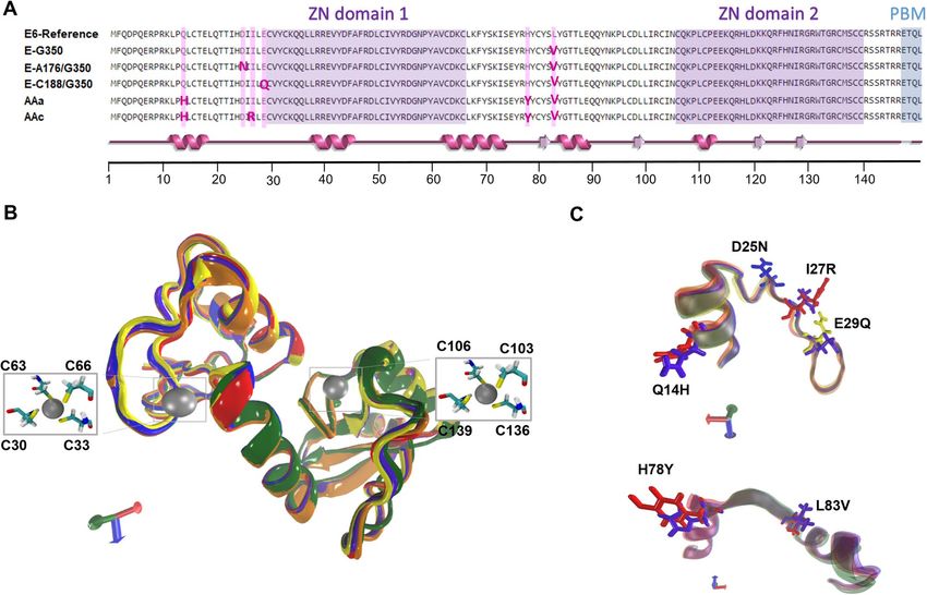
www.nature.com/scientificreports/
Figure 1. Alingment and super position of the 3D structures of the E6-reference and its variants. (A) Multiple
alignment of the sequence of E6 and its variants. Zinc finger domain 1 and 2 are highlighted in purple, PBM
in blue and mutations are highlighted in pink. A super position of the secondary structure of all six proteins is
shown in pink below the alignment. (B) 3D structure of E6-reference: violet, AAa variant: orange, AAc variant:
red, E-G350, green, E-C188/G350: yellow, E-A176/G350: blue. The silver spheres indicate zinc molecules and
the licorice residues correspond to C30, C33, C63, C66, C103, C106, C136 and C139, which make up two zinc
finger domains in the proteins 3D structures. The orientation of the proteins is indicated by the axes, X: red, Y:
green; Z: blue. (C) Visualization of amino acid changes: Q14H, D25N, I27R, H78Y and L83V are in licorice.
2 Å represents accurate models. The RMSD values obtained for the variants were 0.51 Å for E-G350 (green),
0.66 Å for E-C188/G350 (yellow), 0.18 Å for E-A176/G350 (blue), 0.44 Å for AAa (red), and 0.69 Å for AAc
(orange) (Fig. 1B). This mesuments were carried before the MD simulation to ensure that our mutating process
did not damage their 3D strusture. All models were evaluated by Ramachandran plot showing that for all variants
91.9% of the amino acids falled within the favored region and 8.1% in the allowed region, which means a good
stereochemistry for 100% of the residues (Fig. S1).
A close up of the mutated residues in the alignment shows that the side chains of mutants H14 and Y78 were
exposed to the protein’s surface while the amino acids Q14 and H78 side chains of E6-reference were inverted
(Fig. 1C). According to the program HOPE, which analyses the structural and functional effects of point muta-
tions, H14 is bigger than Q14, bigger residues might lead to bumps on the 3D structure of the protein. H14 is
among the observed mutations at this position in other homologous sequences. This sometimes suggests that the
mutant is not damaging for the protein’s structure and function, on the other hand, the residue is located near
a highly conserved position and can gain interactions with target proteins. The Y78 is bigger and more hydro-
phobic than the H78, this can result in loss of hydrogen bonds and disturbance of correct folding (Fig. 1C). The
accessibility of the residues in the mutants could increase the number of interactions with the MAGI-1 m odels46.
Mutations E29Q and D25N remained the same size; therefore, not visible change in 3D structure was observed
(Fig. 1C). A change in residue charge from negative to neutral can cause loss of interactions with other molecules
or residues46. Mutant R27 is bigger than I27, this can be observed in the 3D structure comparison (Fig. 1C). Also,
the change of a neutral to positive residue leads to the possibility of repulsion of ligands or other residues with the
same charge. Moreover, the change of a hydrophobi residue to a neutral one will lead to the loss of hydrophobic
interactions46. The mutation L83V found in all the variants can cause the proteins to lose interactions with other
proteins because, V83 is a smaller residue than L83 (Fig. 1C), loss of interactions with cellular target proteins
could lead to a diminution on the affinity of interactions for the v ariants46.
There are many crystal structures in the RCSB PDB server related to the crystalized MAGI-1; however, none
of these files corresponds to the complete protein structure. Also, there are no reliable software for homology
modelling such large p roteins32. To overcome this drawback, we delimitated our models to domains that have
Scientific Reports | (2022) 12:1898 | https://doi.org/10.1038/s41598-022-05995-1 3
Vol.:(0123456789)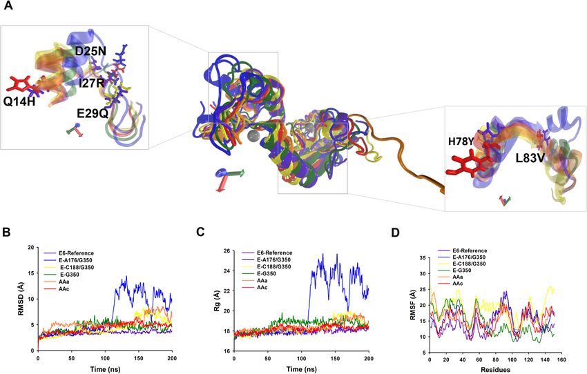
www.nature.com/scientificreports/
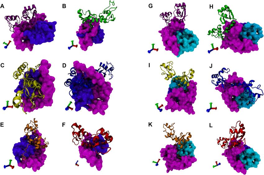
Figure 2. 3D structure of MAGI-1 255 and MAGI-1-329 models. (A) Multiple alignment of the sequence of
model MAGI-1 255 and MAGI-1 329 highlighting domains WW1 and WW2 in soft pink and domain PDZ1 in
soft purple. (B) 3D model visualization of MAGI-1 255 (C) 3D model visualization of MAGI-1 329. The WW1
and the WW2 domains are shown in light pink, the PDZ1 domain is shown in violet, and a highly disordered
region of 76 amino acids in cyan.
been experimentally shown to interact with E 614,25,39. Our first model includes the WW1, WW2, PDZ1 domains
and was denominated MAGI-1 255 and a second model where we added a highly disordered region of 76 amino
acids adjacent to the PDZ1 domain denominated it MAGI-1 329. A sequence alignment of our final models is
shown in (Fig. 2A). This additional region of 76 amino acids in our model 329 was added to compared the inter-
action of the variants with a bigger models of MAGI-1. According data publish by other groups, the interaction
of E6 with MAGI-1 occurs mainly with the PDZ-1 (H472-R554) domain, but different affinity patterns were
observed between adjacent regions closed to the PDZ-1 domaninand E 628–30. Also, computational approaches
have highlighted the role of distal regions of the E6 proteins to form a dynamics networks within PDZ domains,
and a number of studies have characterized these changes in dynamics using NMR, thus providing experimental
evidence for this interaction47–49. Taking into account all the avalible information, we included WW1, WW2
and their interdomains to our models of MAGI-1. Besides these domains we also, added a highly disordered
region of 76 amino acids (G555–T628) to model MAGI-1 329 shown in cyan, this region was reported to have
key role in the interaction of this proteins29. With this we incremented the coverage of MAGI-1 in this model
and predicted that this region increments the affinity of E6 and its variants to MAGI-1.
The 3D homology models of MAGI-1 255 (amino acid 300–554) and of MAGI-1 329 (amino acid 300–628)
were obtained using the crystal structure from human MAGI-1 PDZ1 (PDB ID:2KPK)28 as a template on the
I-TASSER server50 (Fig. 2B,C). The best models were chosen according to the criteria of good alignment with
the template measured by C-Score, TM score, and RMSD values. Model MAGI-1 255 shown in Fig. 2B consists
of six alfa-helix, one 310 helix, eleven beta-sheets and the rest of the residues appeared lightly twisted in random
coils, which are 15.3%, 2.4%, 13.7% and 68.6% respectively of the protein structure. The two WW domains and
the interdomain regions from amino acid G300 to amino acid I471 are shown in magenta, and the PDZ1 domain
from amino acid H472 to amino acid R554 is shown in purple (Fig. 2B). Model MAGI-1 329 consists of seven
alfa-helixes, twelve beta-sheets, and the rest of the residues appeared in loops and coils, which are 15%, 14.1%,
and 70.9%, respectively the protein structure. For this model, the 3D structure from amino acid G300 to R554
are in purple, and the extra region of 76 (G555–T628) amino acids are in cyan (Fig. 2C).
Ramachandran plot for model MAGI-1 255 and MAGI-1 329 exhibited 92.8% and 91.4% respectively of resi-
dues in most favored regions and 7.2% and 8.6% respectively residues are in disallowed regions, which shows a
good stereochemistry for more than 90% of the residues, this makes our models acceptable for more refinement
with MD simulation (Fig. S2).
Scientific Reports | (2022) 12:1898 | https://doi.org/10.1038/s41598-022-05995-1 4
Vol:.(1234567890)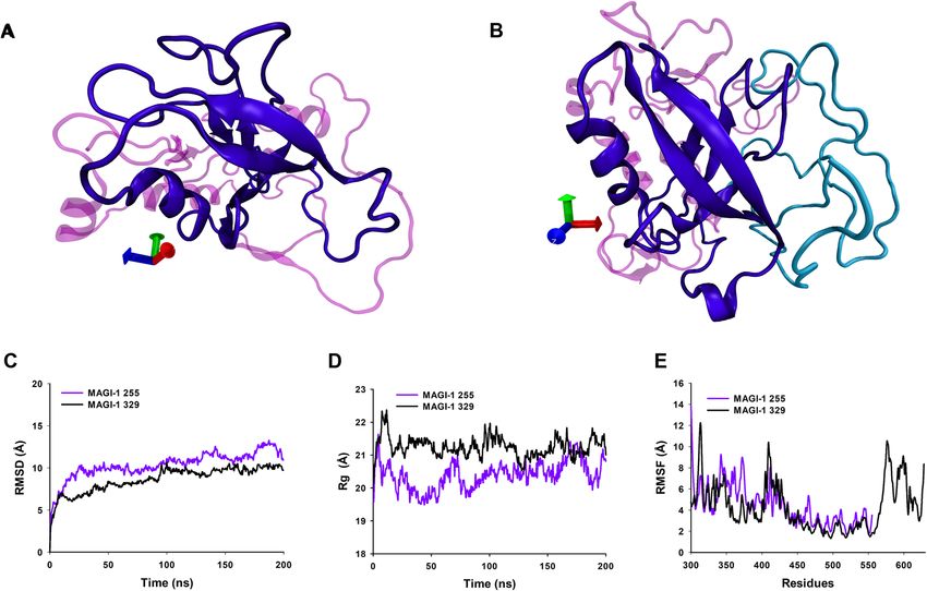
www.nature.com/scientificreports/

                                 Figure 3.  3D structures of the E6-reference and its variants and conformational stability during 200 ns MD
                                 simulation. (A) Super position of average 3D structures of HPV-16 and its variants. Zoom visualization of
                                 amino acid changes: Q14H, D25N, I27R, E29Q, H78Y and L83V. E6-reference: violet, AAa variant: orange,
                                 AAc variant: red, E-G350, green, E-C188/G350: yellow, E-A176/G350: blue. The silver spheres indicate zinc
                                 molecules. The orientation of the proteins is indicated by the axes, X: red, Y: green; Z: blue. (B) RMSD. (C)
                                 Radius of gyration. (D) RMSF.

                                 Molecular dynamics simulation analysis. To examine the change in the protein dynamics and stabil-
                                 ity, the 3D models of HPV-16 E6 and its variants, as well as MAGI-1 255 and MAGI-1 329 were refined by MD
                                 simulation for 200 ns. Trajectories were analyzed by calculating the root mean square deviation of atomic posi-
                                 tions (RMSD), root mean square fluctuation (RMSF), the radius of gyration (Rg), the dPCA analysis and dPCA
                                 based clustering (Fig. 3).
                                      After 200 ns of the MD simulation and using a snapshot of the most populated cluster of E6 and variants, a
                                 structural alignment was done (Fig. 3A). The carboxyl terminus of the E-C188/G350, AAa and AAc proteins
                                 showed a greater difference compared to E6-reference, while the other variants 3D structures remained very
                                 similar compare to E6-reference (Fig. 3A).
                                      The RMSD calculation of the E6-reference (purple) during the 200 ns of MD simulation, reached equilibrium
                                 at 20 ns of trajectory, while the non-European variants AAa (orange) and AAc (red) were equilibrated at 80 ns,
                                 after 150 ns AAa variant loses its equilibrium and recovers it at the end of the DM simulation, probably due to
                                 its mutations and its context (Fig. 3B). Something similar happens to the variant E-C188/G350 (yellow), which
                                 reaches equilibrium at 60 ns, and its equilibrium is disturbed from the 150 ns to the 180 ns. This behaviour is
                                 attributed to mutations in the E29A and L83V positions that directly affect the structure of the protein, causing
                                 disturbs in it’s stability (Fig. 3B). Concerning E-A176/G350 (blue), the equilibrium was reached at the first 20 ns,
                                 but a greater disturbance episode it’s observed from 100 ns to the end of the trajectory it is also thought that the
                                 nature of mutation D25N in the proteins may contribute to this behavior. It was also observed that for E6-ref-
                                 erence, E-G350 (green), and E6-AAc, the RMSD values during the simulation ranged from 2 to 5 Å (Fig. 3B).
                                 While variants AAa, E-A176/G350 and E-C188/G350 were characterized by higher continuous RMSD values
                                 from the 140 ns to the end of the MD simulation (Fig. 3B). The RMSD values of simulated proteins indicated
                                 their stability and particular behavior and provided a suitable basis for further analysis.
                                      The Rg presents different grades of compactness during the simulation evidencing a less compactable grade
                                 at the end of the trajectory, mainly in the variants E-A176/G350, E-C188/G350 and AAa (Fig. 3C in yellow and
                                 orange). Meanwhile, E6-reference and variants E-G350 and AAc maintain compactness during the simulation
                                 (Fig. 3C, green and red). This also confirms that point mutations caused structural destabilizing effects leading
                                 to the loss of protein compactness in the E-A176/G350, E-C188/G350 and AAa variants. Since distance devia-
                                 tions from the starting structure may not necessarily reflect mobility of structural elements, RMSF was used to

Scientific Reports |   (2022) 12:1898 |               https://doi.org/10.1038/s41598-022-05995-1                                                   5

                                                                                                                                              Vol.:(0123456789)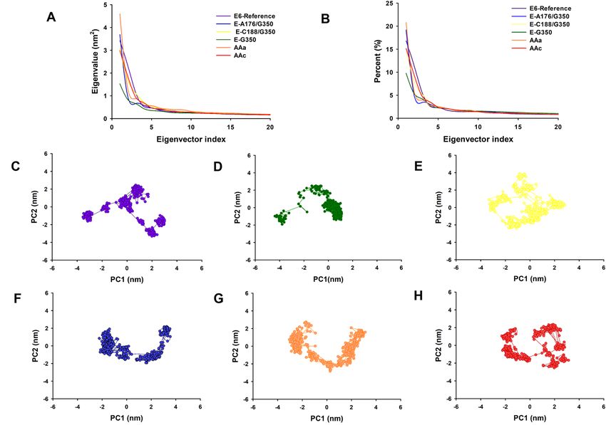
www.nature.com/scientificreports/

                                           Figure 4.  3D structures of MAGI-1 255 and MAGI-1 329 and conformational stability during 200 ns MDs. (A)
                                           Visualization of MAGI-1 255. (B) Visualization of MAGI-1 329. The WW1 and the WW2 domains are shown
                                           in light pink, the PDZ1 domain is shown in violet, and a highly disordered region of 76 amino acids in cyan. (C)
                                           RMSD. (D) Radius of gyration. (E) RMSF.

                                           obtain information on flexibility. According to the data graph in Fig. 3D, there are six maximum fluctuations
                                           peaks areas shared by E6-reference and variants: One at M1 to P5, for E6-reference the fluctuation peak was 16 Å,
                                           and for the variants, the highest peak corresponds to E-C188/G350 with 27 Å, the rest of the variants fluctuate
                                           from 14 to 24 Å of distance being E-G350 the lowest peak, this region is composed by coils and turns with non-
                                           secondary structure (Fig. 3D). The second region at L28–L50 composed of loops and an alpha helix: reached
                                           15 Å for E6-reference and it wasthe highest fluctuation peak. Fluctuation for variants ranges from 20 to 25 Å;
                                           clearly the variants had greatest fluctuation in these residues, where E-C188/G350 had the greates fluctuation
                                           peaks (Fig. 3D). This phenomenon is interesting and it’s attributed to mutation E29Q exclusive of the E-C188/
                                           G350 variant, while the behavior of the other variants is exclusive of their own structural changes caused by
                                           their shared and exclusive mutations. The third peak at C51–L65, in a coil and two beta-sheets, the variants
                                           reached 20–23 Å of fluctuation peaks, while E6-reference’s fluctuation was only 16 Å (Fig. 3D). The fourth region
                                           of fluctuation at C80–L110, was composed of loops, one alpha helix and two beta-sheets and reaches 25 Å for
                                           variant E-A176/G350 and E6-reference. For variants AAa and AAc the fluctuation distance was 20 Å. While the
                                           peak for variant E-G350 was only 14 Å, evidently less flexiblethan the other variants and E6-reference (Fig. 3D).
                                           The fifth fluctuation peak at C111–C140, in two beta sheets, coils and an one alpha helix, for variants AAa,
                                           AAc, E-C188/G350, E-A176/G350 had a fluctuation distance of 20 Å. E6-reference, also, reached a distance of
                                           20 Å. On the other hand, the distance of E-G350 reached 15 Å, and it tends to decrease for the rest of the amino
                                           acids at the carboxyl terminus (Fig. 3D). Finally the residue-based RMSF of the backbone for the E6-reference
                                           displayed less flexible residues than the variants (E-G350, E-C188/G350, E-A176/G350, E6-AAa, and E6-AAc),
                                           at the carboxyl terminus (145–151) composed mainly by loops (Fig. 3D). Interestingly, this region includes the
                                           PBM (ETQV) motif of E6, which is important for this oncoprotein interaction with MAGI-1. Since there is a
                                           higher fluctuation in variants compares to E6-reference, it can be deduced that mutations change the structural
                                           flexibility of the 3D protein structure.
                                               Ramachandran analysis after the MD simulation of these structures shows that more than 98% of the amino
                                           acids of the proteins during the simulation remain in the highly favored regions, which means that the protein’s
                                           conformation are well refined and have native conformations (Fig. S3).
                                               For our two MAGI-1 models we showed a snapshot of the most populated cluster from the dPCA clustering
                                           analysis in (Fig. 4A,B). The PDZ1 domain of our two models have little changes in its 3D structure, but overall
                                           it keeps its main 3D structure (Fig. 4A,B).
                                               The RMSD of MAGI-1 255 (purple) and MAGI-1 329 (black) models during the 200 ns trajectory showed
                                           that both models reached equilibrium before the 100 ns of the simulation and continue stable for the rest of the

          Scientific Reports |   (2022) 12:1898 |              https://doi.org/10.1038/s41598-022-05995-1                                                 6

Vol:.(1234567890)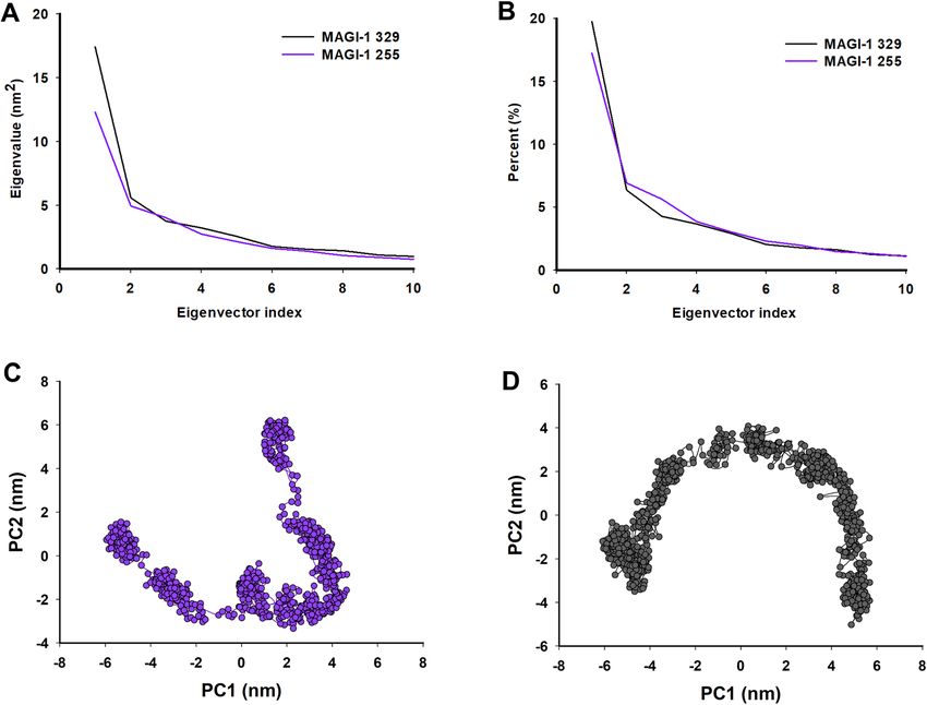
www.nature.com/scientificreports/
Figure 5. Principal component analysis (PCA) of E6-reference and its variants from HPV-16. (A) The
eigenvalues plotted against the corresponding eigenvector indices obtained from the Cα covariance matrix
constructed from the 200 ns MD trajectory. E6-reference: violet, AAa variant: orange, AAc C variant: red,
E-G350, green, E-C188/G350: yellow, E-A176/G350: blue. (B) Percentage of each eigenvector vs. eigenvalues.
E6-reference: violet, AAa variant: orange, AAc C variant: red, E-G350, green, E-C188/G350: yellow, E-A176/
G350: blue. Projection of the motion of the structures of the backbone atoms (PC1 vs PC2) (C) E6-reference.
(D) E-G350. (E) E-C188/G350 (F) E-A176/G350. (G) AAa and (H) AAc.
trajectory. Moreover, MAGI-1 255’s RMSD value after equilibrium ranges from 10 to 13 Å and MAGI-1 329 mod-
el’s RMSD values range from 9 to 10 Å, which means our models are reliable for further investigation (Fig. 4C).
The Rg show that both models maintained a compacted structure throughout the trajectory of MD simulation
(Fig. 4D). We explored the flexibility of the models by measuring Cα. The RMSF values of the models through
trajectory, mainly 3 regions of MAGI-1 255-model showed more flexible areas, those regions correspond to amino
acids G300 to A309 of the WW1 domain, G349 to D379 belong to the WW2 domain and Q399 to H429 that
correspond to an interdomain region between the WW2 and PDZ1 domains of MAGI-1 (Fig. 4E). MAGI-1 329
model showed three regions with more flexibility which include amino acids E304 to I319 of the WW1 domain,
Q399 to V433 corresponding to an inter domain between WW2 and PDZ1 domains and N566 to T628, this
region was the most flexible of the two models. Interestingly, this region corresponds to a highly disordered
region of the whole protein (Fig. 4E)29. The Ramachandran analysis shows the refinement of more than 80% of
the model’s residues (Fig. S4).
Dihedral principal component analysis. dPCA was used to obtain a broader view of dynamic prop-
erties with respect to MD simulation results of E6-reference and its variants, MAGI-1 255 and MAGI-1 329.
The covariance matrix for the first 20 eigenvectors of E6-reference was 11.40 nm2 and 10.09 nm2, 11.14 nm2,
7.12 nm2, 12.23 nm2 and 10.60 nm2 for the variants E-G350, E-A176/G350, E-C188/G350, AAa and AAc, respec-
tively (Fig. 5A). Moreover, the dPCA analysis showed that the first 20 eigenvectors captured 45–57% of the total
protein motions (56.7, 45.5, 52.5, 51.1, 55.3 and 53.8%) for E6-reference, E-G350, E-A176/G350, E-C188/G350,
AAa and AAc respectively (Fig. 5B). Whereas the projections of the first two principal components (PC1 vs PC2)
contributed to 15–28% of the collective motions (28.22, 15.11, 24.20, 22.40, 26.66 and 24.59%) for E6-reference
E-G350, E-A176/G350, E-C188/G350, AAa and AAc respectively (Fig. 5B). There are changes in the motions
of the atoms of the variants E-A176/G350, AAa and AAc compared to E6-reference. Moreover a considerable
change in the motion of the atoms of G350 and E-C188/G350 compared to E6-reference, which suggests that
the properties of the movements described by the first PCs were different in the six protein systems (Fig. 5A,B).
Scientific Reports | (2022) 12:1898 | https://doi.org/10.1038/s41598-022-05995-1 7
Vol.:(0123456789)www.nature.com/scientificreports/
Figure 6. Principal component analysis (PCA) of MAGI-1 255 and MAGI-1 329. (A) First ten eigenvalues
plotted against the corresponding eigenvector indices obtained from the Cα covariance matrix constructed from
the 200 ns MD trajectory. MAGI-1 255 purple and MAGI-1 329 black. (B) Percentage of each eigenvector vs.
eigenvalues. 2D Projection of Principal Component Analysis. Projection of the motion of the protein in phase
space along the first two principal components. (C) MAGI-1 255 and (D) MAGI-1 329.
The projection of the first two eigenvectors (PC2 vs. PC1) for E6-reference, E-G350, E-A176/G350, E-C188/
G350, AAa and AAc (Fig. 5C–H), shows that E6-reference system (Fig. 5C) present different mobility behavior
compared to the variants systems. The variants E-G350, E-C188/G350 and AAc have more restricted motions,
making them the more stable of the six protein systems (Fig. 5D,E,H). The variants E-A176/G350 and AAa were
expanded in their conformational space due to their flexibility (Fig. 5F,G). This points out that the punctual
mutations of residues affect conformation and motion.
With respect to MAGI-1 255 and MAGI-1 329 the matrix value obtained for the for the models were of
17.4 nm2 and 12.3 nm2 and the dPCA analysis showed that the first 10 eigenvectors captured 22.7 and 34.7% of
the proteins total motions (Fig. 6A,B). The projection of the first two eigenvectors (PC2 vs. PC1) for MAGI-1 255
and MAGI-1 329 shows differences in their mobility behaviour (Fig. 6C,D). These results showed a considerable
change in the motion of the atoms of MAGI-1 255 and MAGI-1 329, which means that the missing 76 amino
acids of model 255 restricts its motions, making it more stable. These results support Ramírez et al. observations
about the contribution of the highly disordered 76 amino acid region adjacent to the PDZ-1 domain of MAGI-1
ehavior29.
to its b
Protein–protein docking. Mutations in proteins can affect protein structure and stability, consequently,
these mutations alter the kinetics and thermodynamics of protein–protein interactions (PPI)51. Using ClusPro
blind base docking method, a representative protein structure of the most populated cluster obtained from the
dPCA clustering analysis of the MD simulation refined proteins (E6-reference, E-G350, E-A176/G350, E-C188/
G350, AAa and AAc) were docked against the representative structure of MAGI-1 255 and MAGI-1 329, also,
obtained from dPCA clustering analysis. Docking resulted in 1000 protein conformations of complexes. The top
ten docked complexes from each problem were analyzed for the lowest energy and residues binding between
the two proteins. The best complexes were selected base on a greater number of cluster members and the lowest
energy according to ClusPro guidelines43.
The global free binding energy of the E6-reference against 255 complexes and MAGI-1 329 were calculated
as − 48.14 and − 51.90 kcal/mol respectively, using FiberDock52. These energies were bigger than the energies
obtained from the complexes between the E6 variants and the MAGI-1 models; this means that there is a greater
affinity between MAGI-1 models and E6 variants compare to E6-reference (Table 1). However, the variants that
Scientific Reports | (2022) 12:1898 | https://doi.org/10.1038/s41598-022-05995-1 8
Vol:.(1234567890)www.nature.com/scientificreports/
Complexes No. cluster members’ Binding energy (kcal/mol)
E6R/MAGI-1 255 83 − 48.14
E-G350/MAGI-1 255 126 − 111.14
E-C188/G350/MAGI-1 255 111 − 130.89
E-A176/G350/MAGI-1 255 74 − 191.34
AAa/MAGI-1 255 111 − 138.07
AAc/MAGI-1 255 204 − 120.23
E6R/MAGI-1 329 118 − 54.90
E-G350/MAGI-1 329 88 − 152.50
E-C188/G350/MAGI-1 329 82 − 121.63
E-A176/G350/MAGI-1 329 91 − 148.86
AAa/MAGI-1 329 83 − 148.17
AAc/MAGI-1 329 88 − 166.97
Table 1. Docking binding affinity of E6 HPV-16 and its variants.
presented the lowest binding energy with the MAGI-1 255 model were E-A176/G350, AAa and E-C188/G350
(− 191.34, − 138.07 and − 130.89, respectively). Meanwhile variants AAc, E-G350, and E-A176/G350 and against
MAGI-1 329 showed the lowest energy values (− 166.97, − 152.50 and − 148.86, respectively). We interpreted
this as a gain of interaction affinity between these proteins. In conclusion, the lowest energy docking values was
between MAGI-1 255 and variant E-A176/G350 (Table 1). In addition, there was an increment in the number of
hydrogen bond in the complexes formed by the variants and both models of MAGI-1 compared to the E6-ref-
erence. However the number of salt bridges interactions only increased in the complexes G-350, E-C188/G350
and E-A176/G350 with MAGI-1 255 compared to E6-reference. Concerning the complexes between E6-reference
and its variants with MAGI-1 329 only variants AAa and E-C188/G350 increased their salt bridges interactions
(Table S1). Therefore, we concluded that the variants gain affinity to our two models of MAGI-1.
The protein–protein interfaces of the complexes were analyze using PDBsum generate and are shown in
Fig. 7A–L53. The top docked complex of each variant against MAGI-1 255 and 329 were subjected to PDBsum
to identify the interacting residues. Comparative analysis between the twelve complexes from docking interfaces
of E6-reference and its variants identified a list of different amino acids that were shown to be responsible for
the interaction with the MAGI-255 and MAGI-1 329 (Table S2).
For the complex E6-reference and MAGI-1 255 (Fig. 7A), the interaction occurs mainly through amino acids
of the WW2 domain (Y74, V79, D80, W66, G65, A64, Y78, I76) and from the PDZ1 domain (A234, H231 and
G230) with twenty-four amino acids of E6-reference that included: Y81, R77 and Y76 which are adjacent to H78
a highly mutated amino acid in E6 (Table S2). There are nine hydrogen bonds and three salt bridges (Table S1).
On the other hand, the interaction of E6-reference with MAGI-1 329 was through amino acids corresponding
to WW2 domain and adjacent non-domain regions (Table S2). MAGI-1 329 amino acids C34 to F171 were
responsible for most of the interactions with twenty-three amino acids of E6-reference that included: R147,
R131, H78, R77, Y76, T32 and V31 (Fig. 7G). Some of the E6-reference interacting residues are located closed
to the mutation sites, and its interacting residues are different compared to the variants.
The differences between the interaction of E6-reference with our two models from MAGI-1 is remarkable,
the increase in protein’s coverage results in a gain of interacting residues, an increase of hydrogen bonds, Salt
bridges (Table S1).
For complexes E-G350/MAGI-1 255 (Fig. 7B), E-G350/MAGI-1 329 (Fig. 7H), E-A176/350/MAGI-1 255
(Fig. 7D), E-A176/350/MAGI-1 329 (Fig. 7J), AAc/MAGI-1 255 (Fig. 7E) and AAc/MAGI-1 329 (Fig. 7K)
detailed information about the amino acids involved in the interaction and the type of bonds can be found in
Tables S1 and S2.
We observed that in complex E-C188/G350/MAGI-1 255 (Fig. 7C) the interactions of the PBM motif (E148,
T149, Q150, and 151L) were lost, but, interestedly the interacting residues from complex E-C188/G350/MAGI-1
329 (Fig. 7I) included all the amino acids of the PBM motif of E6 (E148, T149, Q150, and 151L) these residues
interact mainly with amino acids from the WW1 (G1-R33) domain and amino acids from a highly disordered
region of MAGI-1 (G256-T329 in our model) which agrees with the experimental evidence publish by Ramirez
et al.29 (Table S2).
It is important to point out that H78 is mutated to T78 in the Asian American variants this mutation causes
the lost of interaction between this residue and our models of MAGI-1 only for AAa variant, meanwhile, AAc
variant does not exhibit this behaviour. We observed that the interaction of AAa variant with MAGI-1 255 was
not conducted throught its PBM motif (Fig. 7F). Meanwhile, the interaction of this variant with MAGI-1 329
included all the residues from the PBM motif (R147, E148, T149, Q150 and L151) (Fig. 7L).
Regarding the missing interaction of the PBM motif with MAGI-1 255 and 329, we believe that the residues
could be oriented in a way that avoids the interaction or may be block by adjacent residues. According to the
online server HOPE, this could be attributed to changes in size and in charge from the mutations of each v ariant46.
It is important to understand the changes in PPIs caused by these mutations may alters the affinity and stabil-
ity of the interaction of E6 with proteins important for tissue homeostasis. The increase in affinity and stability
of the interaction of E6 with MAGI-1 result in an increase in the degradation of MAGI-1 and as a consequence
in the loss of stability of important cell complexes that maintain cell–cell adherence at the adherents junctions.
Scientific Reports | (2022) 12:1898 | https://doi.org/10.1038/s41598-022-05995-1 9
Vol.:(0123456789)www.nature.com/scientificreports/
Figure 7. Protein–protein docking of E6, its variants with MAGI-1 255 and MAGI-1 329. Protein–protein
docking analysis shows the probable interaction of E6-reference (purple), E-G350 (green), C188/G350 (yellow),
E-A176/G350 (blue), AAa (orange) and AAc (red) with MAGI-1 255 (A–F). Docking between MAGI-1 329
and E6-references and its variants (G–L) E6-reference (purple), E-G350 (green), C188/G350 (yellow), E-A176/
G350 (blue), AAa (orange) and AAc (red). MAGI-1 255 and MAGI-1 329 are represented in quicksurf in color
magenta. The protein–protein docking was performed using the ClusPro 2.0 web server.
Materials and methods
3D protein structures. Multiple alignments of the sequences (Accession number P03126) were performed
using CLUSTAL X 1.8154. The secondary structure of the E6 protein and its variants were predicted using
PSIPRED server55. The crystal structure of HPVt-16 E6 protein was obtained from the Protein Data Bank (RCSB
PDB)56, with the identification number: 4XR8, chain H21. The E6 structure on this PDB contains 151 residues
with four-point mutations in S80C, S97C, S111C, and S140, which were reverted to obtain the E6 reference in
the PyMOL Molecular Graphics System, Version 2.0 Schrödinger, LLC57. After that, all the mutations were car-
ried out in the E6 reference to obtained all the variants of HPV-16. The mutations were done as indicated next,
to obtained E-G350: L83V; E-C188/G350: E29Q and L83V; E-A176/G350: D25N and L83V; AAa: Q14H, H78Y,
and L83V; for AAc: Q14H, I27R, H78Y, and L83V. The obtained proteins were structurally aligned and visualized
using VMD 1.9.358.
To obtained the 3D structure of MAGI-1, a total of 255 and 329 amino acids from the amino acid terminal
region of the protein sequence (300–554 and 300–628) were retrieved from the UniProtKB database59, (accession
number Q96QZ7) and submitted to I-TASSER server as two separate jobs50. First, the 3D structure with 225
residues, which comprises WW1, WW2 and the PDZ1 domains of MAGI-1 was obtained by homology modelling
using the I-TASSER server (https://zhanggroup.org/I-TASSER/), as a template, we selected PDB file: 2KPK28,
which corresponds to the PDZ-1 domain of the MAGI-1. Furthermore, a 3D structure of 329 amino acids of
MAGI-1, which includes the WW1, WW2, PDZ-1 and a 76 amino acidic disordered region of this protein, was
obtained by homology modelling on the I-TASSER server using the same template PDB. All the 3D predicted
structures were evaluated using the Rampage webserver to obtain the Ramachandran plots (http://mordred.
bioc.cam.ac.uk/~rapper/rampage.php).
Molecular dynamics simulation. Parameters for the two Zn2+ ions and eight cysteine-ligand coordina-
tion of E6 were kindly provided by Justin Lemkul from the Virginia Polytechnic Institute and State University,
these parameters included a CYSD patch for the deprotonation of the eight zinc-bound cysteines and a ZN_C
patch to covalently link cysteines to the zinc ions. The correct coordination of the deprotonated cysteines and
the ion zinc using these patches has been demonstrated in previous s tudies60, the CHARMM 36 force field was
Scientific Reports | (2022) 12:1898 | https://doi.org/10.1038/s41598-022-05995-1 10
Vol:.(1234567890)www.nature.com/scientificreports/
employed for the application of the patches using CHARMM software (http://charmm.chemistry.harvard.edu/
charmm_lite.php)61,62. Afterwards, a 200 ns of MD simultion of E6 and its variants were performed using the
NAMD 2.8 (http://www.ks.uiuc.edu/Research/namd/) software p ackage63 with CHARMM36 and CHARMM22
force fields62. For MAGI-1 models we used CHARMM 27 topology and parameter files for proteins. Each system
was placed in a cubic box of TIP3P water with a minimum distance of 10 Å between the solute atoms and the
edge of the b ox64. To neutralize the systems, we added 7568 water molecules, 21 Na+ and 27 Cl− to the E6-ref-
erence. To variant E-G350, we added 7420 water molecules, 21 Na+, and 27 Cl−, to E-C188/G350 variant, 7484
water molecules, 21 Na+, and 28 Cl− were added, to E-A176/G350 variant, 7660 water molecules, 22 Na+ and
29 Cl− were added, to AAa variant 7566 water molecules, 21 Na+ and 27 Cl− were added and to AAc variant,
7554 water molecules, 21 Na+ and 28 Cl− were added. For MAGI-1 255 we added 10,932 water molecules and
18 Sodium, and for MAGI-1 329, 11,456 water molecules and 23 Na were added. Each system was neutralized
to 0.15 mol/L of NaCl and submitted to minimization energy for 10,000 steps of steepest descent minimization
followed by equilibration for 1 ns under constant temperature 310 K and pressure 1 atm (NPT) ensemble with
protein atoms r estraints65,66. MD simulation were run for 200 ns, considering all proteins as soluble.
Trajectory and dPCA analysis. The carma software (https://utopia.duth.gr/~glykos/Qs.html)67, was used
to calculate the root mean square deviation (RMSD) calculates the average deviation in the atomic stability
throughout MD simulation, radius of gyration (Rg) measures the compactness and expansion of the molecules,
and the root means square fluctuation (RMSF) a parameter to explored the flexibility of the protein through MD
simulation, as well as the Principal component analysis (PCA) and dPCA based clustering analysis employing
the last 50 ns of the trajectory. dPCA is a standard tool in statistical mechanics used in order to determine the
correlated motions of the residues to a set of linearly uncorrelated variables called principal components, and it
allows to obtain the large scale collective motions of the atoms on the simulations, which frequently correlates
with the proteins biological function and structural p roperties68. Finally, we obtained the PDB files from the
most populated cluster analysis and performed a protein–protein docking. Molecular graphics were performed
in Sigma plot 12.0. VMD 1.9.3 was used to visualize all the 3D proteins58.
Protein–protein docking. The protein–protein dockings were carried in Cluspro server (https://cluspro.
bu.edu/login.php)69,70, the program has been consistently rated among the best global docking methodologies in
the CAPRI challenge (Critical Assessment of Predicted Interactions)69. For the docking studies, refined models
for most populated cluster from E6-reference or its variants were docked within the MAGI-1 (235 and 329)
homology models, where MAGI-1 models were the receptors and E6, and its variants were used as a ligand.
The conformers with the highest cluster members and the lowest energy calculated in FireDock were taken for
analysis on the PDBSum server52,53. All docking complexes were visualized by VMD 1.9.3 software58.
Conclusions
We proposed an in-silico approach to evidence the differences in the interaction of E6 and five of its natural
variants with two models, cellular protein MAGI-1. According to our results variants, AAa and E-C188/G350
showed less RMSD values, less compactness, a gain of fluctuation regions that are correlated to the increment
of active sites. We attribute this behavior to specific mutations of proteins, and these mutations cause physico-
chemical changes that affect the behavior of proteins. Very marked dynamic changes are observed, particularly
at the amino and carboxyl termini of proteins, where there is a gain in flexibility in the variants compared to
E6-reference. Also, according to the dPCA results a dramatic change of motions behaviour for mutants compared
to E6-reference. These differences in structure and mobility incremented the affinity of variants E-C188/G350
and AAa for our models of MAGI-1. E-C188/G350 increases its affinity for our models by three times, increas-
ing the binding bonds by 50%. A similar pattern is observed among all the variants compared to E6-reference.
Our results suggest that the physicochemical changes that gave rise to thermodynamic changes of the variants
and an increase the affinity for our MAGI-1 models. Here, we were able to represent the possible changes in the
physicochemical properties of E6 proteins and the repercussion in the interaction affinity with MAGI-1. An
experimental validation will be necessary to evaluate the degradation profile of the MAGI-1 protein mediated
by E6-reference and its variants.
Received: 23 October 2021; Accepted: 19 January 2022
References
1. Smith, J. S. et al. Human papillomavirus type distribution in invasive cervical cancer and high-grade cervical lesions: A meta-
analysis update. Int. J. Cancer 121, 621–632. https://doi.org/10.1002/ijc.22527 (2007).
2. Doorbar, J., Egawa, N., Griffin, H., Kranjec, C. & Murakami, I. Human papillomavirus molecular biology and disease association.
Rev. Med. Virol. 25(Suppl 1), 2–23. https://doi.org/10.1002/rmv.1822 (2015).
3. Zehbe, I. et al. Human papillomavirus 16 E6 variants differ in their dysregulation of human keratinocyte differentiation and
apoptosis. Virology 383, 69–77. https://doi.org/10.1016/j.virol.2008.09.036 (2009).
4. CONEVAL. Resultados de pobreza en México 2018 a nivel nacional y por entidades federativas. https://www.coneval.org.mx/
Medicion/Paginas/PobrezaInicio.aspx. (2018).
5. Asadurian, Y. et al. Activities of human papillomavirus 16 E6 natural variants in human keratinocytes. J. Med. Virol. 79, 1751–1760.
https://doi.org/10.1002/jmv.20978 (2007).
6. Niccoli, S., Abraham, S., Richard, C. & Zehbe, I. The Asian–American E6 variant protein of human papillomavirus 16 alone is
sufficient to promote immortalization, transformation, and migration of primary human foreskin keratinocytes. J. Virol. 86,
12384–12396. https://doi.org/10.1128/jvi.01512-12 (2012).
Scientific Reports | (2022) 12:1898 | https://doi.org/10.1038/s41598-022-05995-1 11
Vol.:(0123456789)www.nature.com/scientificreports/
7. Richard, C. et al. The immortalizing and transforming ability of two common human papillomavirus 16 E6 variants with different
prevalences in cervical cancer. Oncogene 29, 3435–3445. https://doi.org/10.1038/onc.2010.93 (2010).
8. Schiffman, M. et al. A population-based prospective study of carcinogenic human papillomavirus variant lineages, viral persistence,
and cervical neoplasia. Cancer Res. 70, 3159–3169. https://doi.org/10.1158/0008-5472.can-09-4179 (2010).
9. Gheit, T. et al. Risks for persistence and progression by human papillomavirus type 16 variant lineages among a population-based
sample of Danish women. Cancer Epidemiol. Biomark. Prev. 20, 1315–1321. https://d oi.o
rg/1 0.1 158/1 055-9 965.e pi-1 0-1 187 (2011).
10. Lichtig, H. et al. HPV16 E6 natural variants exhibit different activities in functional assays relevant to the carcinogenic potential
of E6. Virology 350, 216–227. https://doi.org/10.1016/j.virol.2006.01.038 (2006).
11. Chakrabarti, O. et al. Human papillomavirus type 16 E6 amino acid 83 variants enhance E6-mediated MAPK signaling and dif-
ferentially regulate tumorigenesis by notch signaling and oncogenic Ras. J. Virol. 78, 5934–5945. https://doi.org/10.1128/jvi.78.
11.5934-5945.2004 (2004).
12. Liu, Y., Cherry, J. J., Dineen, J. V., Androphy, E. J. & Baleja, J. D. Determinants of stability for the E6 protein of papillomavirus type
16. J. Mol. Biol. 386, 1123–1137. https://doi.org/10.1016/j.jmb.2009.01.018 (2009).
13. Ganti, K. et al. The human papillomavirus E6 PDZ binding motif: From life cycle to malignancy. Viruses 7, 3530–3551. https://
doi.org/10.3390/v7072785 (2015).
14. Zhang, Y. et al. Structures of a human papillomavirus (HPV) E6 polypeptide bound to MAGUK proteins: Mechanisms of targeting
tumor suppressors by a high-risk HPV oncoprotein. J. Virol. 81, 3618–3626. https://doi.org/10.1128/jvi.02044-06 (2007).
15. Shai, A., Brake, T., Somoza, C. & Lambert, P. F. The human papillomavirus E6 oncogene dysregulates the cell cycle and contributes
to cervical carcinogenesis through two independent activities. Cancer Res. 67, 1626–1635. https://doi.org/10.1158/0008-5472.
can-06-3344 (2007).
16. Pim, D., Thomas, M. & Banks, L. Chimaeric HPV E6 proteins allow dissection of the proteolytic pathways regulating different E6
cellular target proteins. Oncogene 21, 8140–8148. https://doi.org/10.1038/sj.onc.1206026 (2002).
17. Yoshimatsu, Y. et al. Roles of the PDZ-binding motif of HPV 16 E6 protein in oncogenic transformation of human cervical
keratinocytes. Cancer Sci. 108, 1303–1309. https://doi.org/10.1111/cas.13264 (2017).
18. Nominé, Y. et al. A strategy for optimizing the monodispersity of fusion proteins: Application to purification of recombinant HPV
E6 oncoprotein. Protein Eng. 14, 297–305. https://doi.org/10.1093/protein/14.4.297 (2001).
19. García-Alai, M. M., Dantur, K. I., Smal, C., Pietrasanta, L. & de Prat-Gay, G. High-risk HPV E6 oncoproteins assemble into large
oligomers that allow localization of endogenous species in prototypic HPV-transformed cell lines. Biochemistry 46, 341–349.
https://doi.org/10.1021/bi602457q (2007).
20. Illiano, E. et al. Production of functional, stable, unmutated recombinant human papillomavirus E6 oncoprotein: Implications for
HPV-tumor diagnosis and therapy. J. Transl. Med. 14, 224. https://doi.org/10.1186/s12967-016-0978-6 (2016).
21. Martinez-Zapien, D. et al. Structure of the E6/E6AP/p53 complex required for HPV-mediated degradation of p53. Nature 529,
541–545. https://doi.org/10.1038/nature16481 (2016).
22. Charbonnier, S. et al. Defining the minimal interacting regions of the tight junction protein MAGI-1 and HPV16 E6 oncoprotein
for solution structure studies. Protein Expr. Purif. 60, 64–73. https://doi.org/10.1016/j.pep.2008.03.022 (2008).
23. Thomas, M. & Banks, L. In vitro assays of substrate degradation induced by high-risk HPV E6 oncoproteins. Methods Mol. Med.
119, 411–417. https://doi.org/10.1385/1-59259-982-6:411 (2005).
24. Thomas, M., Glaunsinger, B., Pim, D., Javier, R. & Banks, L. HPV E6 and MAGUK protein interactions: Determination of the
molecular basis for specific protein recognition and degradation. Oncogene 20, 5431–5439. https://doi.org/10.1038/sj.onc.12047
19 (2001).
25. Thomas, M. et al. Oncogenic human papillomavirus E6 proteins target the MAGI-2 and MAGI-3 proteins for degradation. Onco-
gene 21, 5088–5096. https://doi.org/10.1038/sj.onc.1205668 (2002).
26. Chen, H. I. & Sudol, M. The WW domain of Yes-associated protein binds a proline-rich ligand that differs from the consensus
established for Src homology 3-binding modules. Proc. Natl. Acad. Sci. USA 92, 7819–7823. https://doi.org/10.1073/pnas.92.17.
7819 (1995).
27. Dobrosotskaya, I., Guy, R. K. & James, G. L. MAGI-1, a membrane-associated guanylate kinase with a unique arrangement of
protein–protein interaction domains. J. Biol. Chem. 272, 31589–31597. https://doi.org/10.1074/jbc.272.50.31589 (1997).
28. Charbonnier, S. et al. The structural and dynamic response of MAGI-1 PDZ1 with noncanonical domain boundaries to the binding
of human papillomavirus E6. J. Mol. Biol. 406, 745–763. https://doi.org/10.1016/j.jmb.2011.01.015 (2011).
29. Ramírez, J. et al. Disorder-to-order transition of MAGI-1 PDZ1 C-terminal extension upon peptide binding: Thermodynamic
and dynamic insights. Biochemistry 54, 1327–1337. https://doi.org/10.1021/bi500845j (2015).
30. Fournane, S. et al. Surface plasmon resonance analysis of the binding of high-risk mucosal HPV E6 oncoproteins to the PDZ1
domain of the tight junction protein MAGI-1. J. Mol. Recognit. 24, 511–523. https://doi.org/10.1002/jmr.1056 (2011).
31. Lee, H. J. & Zheng, J. J. PDZ domains and their binding partners: Structure, specificity, and modification. Cell Commun. Signal 8,
8. https://doi.org/10.1186/1478-811x-8-8 (2010).
32. Schwede, T. Protein modeling: What happened to the “protein structure gap”?. Structure 21, 1531–1540. https://doi.org/10.1016/j.
str.2013.08.007 (2013).
33. Thomas, M., Myers, M. P., Massimi, P., Guarnaccia, C. & Banks, L. Analysis of multiple HPV E6 PDZ interactions defines type-
specific PDZ fingerprints that predict oncogenic potential. PLoS Pathog. 12, e1005766. https://doi.org/10.1371/journal.ppat.10057
66 (2016).
34. Rodríguez-Ruiz, H. A. et al. In silico prediction of structural changes in human papillomavirus type 16 (HPV16) E6 oncoprotein
and its variants. BMC Mol. Cell Biol. 20, 35. https://doi.org/10.1186/s12860-019-0217-0 (2019).
35. Bello-Rios, C. et al. Modeling and molecular dynamics of the 3D structure of the HPV16 E7 protein and its variants. Int. J. Mol.
Sci. https://doi.org/10.3390/ijms22031400 (2021).
36. Grm, H. S. & Banks, L. Degradation of hDlg and MAGIs by human papillomavirus E6 is E6-AP-independent. J. Gen. Virol. 85,
2815–2819. https://doi.org/10.1099/vir.0.80035-0 (2004).
37. Handa, K. et al. E6AP-dependent degradation of DLG4/PSD95 by high-risk human papillomavirus type 18 E6 protein. J. Virol.
81, 1379–1389. https://doi.org/10.1128/jvi.01712-06 (2007).
38. Nakagawa, S. & Huibregtse, J. M. Human scribble (Vartul) is targeted for ubiquitin-mediated degradation by the high-risk papil-
lomavirus E6 proteins and the E6AP ubiquitin-protein ligase. Mol. Cell Biol. 20, 8244–8253 (2000).
39. Glaunsinger, B. A., Lee, S. S., Thomas, M., Banks, L. & Javier, R. Interactions of the PDZ-protein MAGI-1 with adenovirus E4-ORF1
and high-risk papillomavirus E6 oncoproteins. Oncogene 19, 5270–5280. https://doi.org/10.1038/sj.onc.1203906 (2000).
40. Jeong, K. W., Kim, H. Z., Kim, S., Kim, Y. S. & Choe, J. Human papillomavirus type 16 E6 protein interacts with cystic fibrosis
transmembrane regulator-associated ligand and promotes E6-associated protein-mediated ubiquitination and proteasomal deg-
radation. Oncogene 26, 487–499. https://doi.org/10.1038/sj.onc.1209837 (2007).
41. Lee, S. S., Glaunsinger, B., Mantovani, F., Banks, L. & Javier, R. T. Multi-PDZ domain protein MUPP1 is a cellular target for both
adenovirus E4-ORF1 and high-risk papillomavirus type 18 E6 oncoproteins. J. Virol. 74, 9680–9693. https://doi.org/10.1128/jvi.
74.20.9680-9693.2000 (2000).
42. Storrs, C. H. & Silverstein, S. J. PATJ, a tight junction-associated PDZ protein, is a novel degradation target of high-risk human
papillomavirus E6 and the alternatively spliced isoform 18 E6. J. Virol. 81, 4080–4090. https://d oi.o rg/1 0.1 128/j vi.0 2545-0 6 (2007).
Scientific Reports | (2022) 12:1898 | https://doi.org/10.1038/s41598-022-05995-1 12
Vol:.(1234567890)You can also read