National Diagnostic Protocol - Mayetiola destructor Hessian Fly
←
→
Page content transcription
If your browser does not render page correctly, please read the page content below
NDP 41 V1- National Diagnostic Protocol for Mayetiola destructor

      National Diagnostic Protocol
                             Mayetiola destructor
                                Hessian Fly

                                             NDP 41 V1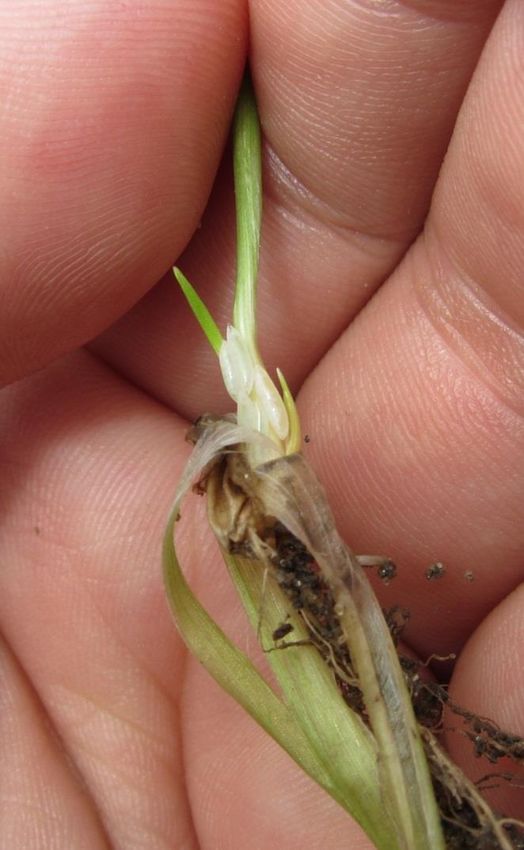
NDP 41 V1 - National Diagnostic Protocol for Mayetiola destructor

© Commonwealth of Australia

Ownership of intellectual property rights
Unless otherwise noted, copyright (and any other intellectual property rights, if any) in this publication is owned by
the Commonwealth of Australia (referred to as the Commonwealth).

Creative Commons licence
All material in this publication is licensed under a Creative Commons Attribution 3.0 Australia Licence, save for content
supplied by third parties, logos and the Commonwealth Coat of Arms.

Creative Commons Attribution 3.0 Australia Licence is a standard form licence agreement that allows you to copy,
distribute, transmit and adapt this publication provided you attribute the work. A summary of the licence terms is
available from http://creativecommons.org/licenses/by/3.0/au/deed.en. The full licence terms are available from
https://creativecommons.org/licenses/by/3.0/au/legalcode.

This publication (and any material sourced from it) should be attributed as: Subcommittee on Plant Health Diagnostics
(2018). National Diagnostic Protocol for Mayetiola destructor – NDP41 V1. (Eds. Subcommittee on Plant Health
Diagnostics) Authors Severtson, D, Szito, A.; Reviewers Nicholas, A, Kehoe, M. ISBN 978-0-6481143-3-8 CC BY 3.0.

Cataloguing data
Subcommittee on Plant Health Diagnostics (2018). National Diagnostic Protocol for Mayetiola destructor – NDP41 V1.
(Eds. Subcommittee on Plant Health Diagnostics) Authors Severtson, D, Szito, A.; Reviewers Nicholas, A, Kehoe, M. ISBN
978-0-6481143-3-8.

ISBN 978-0-6481143-3-8

Internet
Report title is available at: https://www.plantbiosecuritydiagnostics.net.au/resources/#

Department of Agriculture and Water Resources

Street Address 1: 18 Marcus Clarke Street, Canberra City ACT 2601
Street Address 2: 7 London Circuit, Canberra City ACT 2601
Postal Address: GPO Box 858, Canberra City ACT 2601
Switchboard Phone: 02 6272 3933
Web: http://www.agriculture.gov.au

Inquiries regarding the licence and any use of this document should be sent to: copyright@agriculture.gov.au.

The Australian Government acting through the Department of Agriculture and Water Resources has exercised due care
and skill in the preparation and compilation of the information and data in this publication. Notwithstanding, the
Department of Agriculture and Water Resources, its employees and advisers disclaim all liability, including liability for
negligence, for any loss, damage, injury, expense or cost incurred by any person as a result of accessing, using or
relying upon any of the information or data in this publication to the maximum extent permitted by law.

   ii                                                                     Subcommittee on Plant Health Diagnostics
NDP 41 V1 - National Diagnostic Protocol for Mayetiola destructor
Purpose
National Diagnostic Protocols (NDPs) are diagnostic protocols for the unambiguous taxonomic
identification of plant pests. NDPs:
are a verified information resource for plant health diagnosticians
are consistent with ISPM No. 27 – Diagnostic Protocols for Regulated Pests
provide a nationally consistent approach to the identification of plant pests enabling
transparency when comparing diagnostic results between laboratories; and,
are endorsed by regulatory jurisdictions for use (either within their own facilities or when
commissioning from others) in a pest incursion.
Where an International Plant Protection Convention (IPPC) diagnostic protocol exists it should be
used in preference to NDPs although NDPs may contain additional information to aid diagnosis. IPPC
protocols are available on the IPPC website:
https://www.ippc.int/core-activities/standards-setting/ispms
Process
NDPs are facilitated and endorsed by the Subcommittee on Plant Health Diagnostics (SPHD). SPHD
reports to Plant Health Committee and is Australia’s peak technical and policy forum for plant health
diagnostics.
NDPs are developed and endorsed according to Reference Standards developed and maintained by
SPHD. Current Reference Standards are available at
https://www.plantbiosecuritydiagnostics.net.au/initiatives/national-diagnostic-protocols/
NDPs are living documents. They are updated every 5 years or before this time if required (i.e. when
new techniques become available).
Document status
This version of the National Diagnostic Protocol (NDP) for Mayetiola destructor is current as at the
date contained in the version control box below.
PEST STATUS Not present in Australia
PROTOCOL NUMBER NDP 41
VERSION NUMBER V1
PROTOCOL STATUS Endorsed
ISSUE DATE March 2019
REVIEW DATE 2024
ISSUED BY SPHD
The most current version of this document is available from the SPHD website:
https://www.plantbiosecuritydiagnostics.net.au/resources/#
Further information
Inquiries regarding technical matters relating to this project should be sent to:
sphd@agriculture.gov.au
iii Subcommittee on Plant Health Diagnostics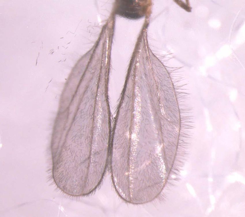
NDP 41 V1- National Diagnostic Protocol for Mayetiola destructor

Contents
1    INTRODUCTION........................................................................................................................................ 2
     1.1         Hosts ................................................................................................................................................................2
2    TAXONOMIC INFORMATION ................................................................................................................ 3
3    DETECTION ................................................................................................................................................ 4
     3.1         In field .............................................................................................................................................................4
     3.2         Plant material ..............................................................................................................................................5
4    IDENTIFICATION...................................................................................................................................... 8
     4.1         Specimen collection and preservation ..............................................................................................8
     4.2         Morphological identification .................................................................................................................9
     4.3         Comparison with similar species ..................................................................................................... 15
     4.4         Molecular identification ....................................................................................................................... 17
5    CONTACTS FOR FURTHER INFORMATION....................................................................................20
6    ACKNOWLEDGEMENTS........................................................................................................................21
7    REFERENCES ............................................................................................................................................22
8    APPENDICES ............................................................................................................................................23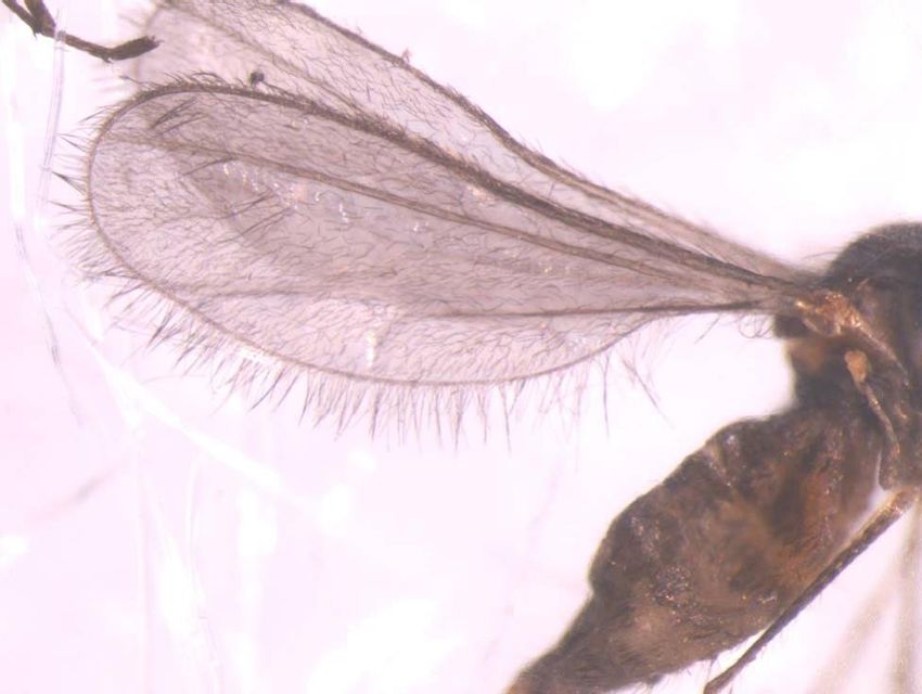
NDP 41 V1 - National Diagnostic Protocol for Mayetiola destructor

1            INTRODUCTION
The Hessian fly (Mayetiola destructor) is a tiny fly (or midge) similar in appearance to a mosquito.

All four life stages (egg, larvae, pupa, and adult) may be found on cereal or grass plants. Elongate,
cylindrical glossy red eggs are laid within leaf veins. Hatched maggots are pale and cylindrical growing
from 0.5 to 4.0 mm long and feed on hidden parts of the plant such as within leaf sheaths. This feeding
damage may cause stunting and death of plants. Larvae pupate approximately three weeks after
hatching from egg. Pupae may be found within leaf sheaths and at the base of plants between stems or
tillers. They are often referred to as the ‘flaxseed’ stage given its close resemblance. An adult winged
midge emerges from the pupa after 6-33 days, with lower temperatures prolonging development. A
full life cycle varies between 20 days to 49 months depending on environmental conditions. Up to six
generations per year have been reported in favourable environments (McColloch 1923).

Hessian fly may be confused with the Barley stem gall midge, Mayetiola hordei, which is also
considered a biosecurity threat to Australia’s cereal industry. It may also be confused with other
midges introduced or native to Australia.

1.1          Hosts
   Primary host: Triticum spp. (wheat)

   Secondary hosts: Agropyron (wheatgrass), Hordeum vulgare (barley), Secale cereale (rye), other
                    grasses

M. destructor has been recorded from some grass genera (Aegilops, Lolium, Elytrigia, Bromus, Elymus
and some species of Agropyron). Elytrigia repens [Elymus repens] is an alternative host in Europe, and
Barnes (1956) suggested that it may have been the original host of Hessian fly. Reproduction on non-
Triticeae grass weeds is negligible.

    2                                                               Subcommittee on Plant Health Diagnostics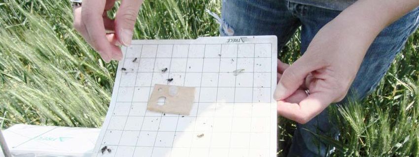
NDP 41 V1 - National Diagnostic Protocol for Mayetiola destructor
2 TAXONOMIC INFORMATION
Taxonomic placement within the order Diptera (flies):
Family: Cecidomyiidae Subfamily: Cecidomyiinae Tribe: Oligotrophini
Genus: Mayetiola
Species: destructor (Say)
Name: Mayetiola destructor (Say) 1817
Synonyms:
Cecidomyia destructor Say 1817
Cecidomyia culmicola Morris 1849
Cecidomyia frumentaria Rondani 1864
Chortomyia secalina (Loew 1858)
Mayetiola secalis Bollow 1950
Phytophaga cerealis Rondani 1843
Phytophaga destructor (Say 1817)
Rhabdophaga elymi Felt, 1909
Rhabdophaga occidentalis Felt, 1908
Rhabdophaga pratensis Felt, 1908
Common name: Hessian fly
3 Subcommittee on Plant Health Diagnostics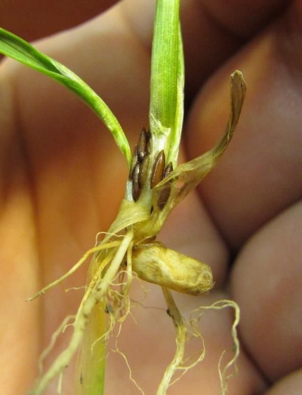
NDP 41 V1 - National Diagnostic Protocol for Mayetiola destructor

3            DETECTION
3.1          In field
Owing to their small size, cryptic nature and short adult lifespan, the Hessian fly may be difficult to
detect in cereal or grass hosts in field situations.

Symptoms of Hessian fly presence may not always be a reliable means of detection, especially where
they may be present in very low numbers or dormant within pupae. However, some symptoms are
important to be aware of in field situations. These include plants that appear greener and stunted.

Patches of darker green plants

Plants infested with Hessian fly, particularly young seedling crops, may appear greener than healthy
uninfested plants (Figure 1) as the larva manipulates the plant to take up more nutrients. Patches of
plants that are darker green relative to surrounding plants should be inspected for Hessian fly larvae.

               Lighter green                                                 Darker green
               uninfested plants                                             infested plants

Figure 1. Wheat plant colour difference with Hessian fly infestation. Kansas State University
glasshouse. Photo: Dustin Severtson, Department of Agriculture and Food Western Australia.

    4                                                               Subcommittee on Plant Health Diagnostics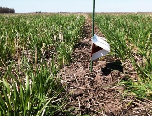
NDP 41 V1 - National Diagnostic Protocol for Mayetiola destructor
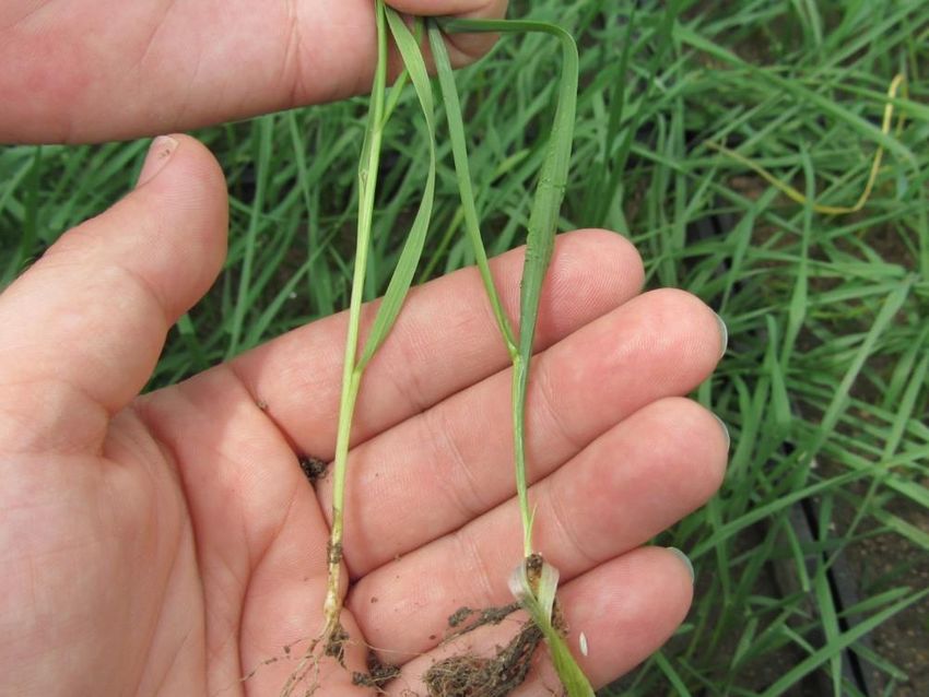
Stunting
Hessian fly infestation in a cereal crop may cause patches of stunted plants where low numbers of
female flies have laid eggs in a very localised area. Larvae hidden within the base of plants do not
move from the area (i.e. move to other plants). If Hessian fly is suspected, collect plants from the
suspect area and adjacent healthy looking plants for comparison of the above and below ground plant
parts. Plant should be dug up carefully keeping the roots intact so as not to displace any larvae or
pupae.
3.2 Plant material
Cereal and grass plant hosts may be inspected for Hessian fly eggs, larvae and pupae. Adult flies may
be captured preferably by pheromone traps (see Appendix) or sweep nets with a fine mesh.
3.2.1 Eggs
The shiny, oblong reddish eggs may be seen where they are laid along the veins of the upper side of
leaves (Figure 2). Severe egg infestation may give the appearance of rust infection. A 10x hand lens
will aid in distinguishing eggs from other material such as disease or soil particles.
Figure 2. Hessian fly egg on the upper surface of a wheat leaf.
5 Subcommittee on Plant Health Diagnostics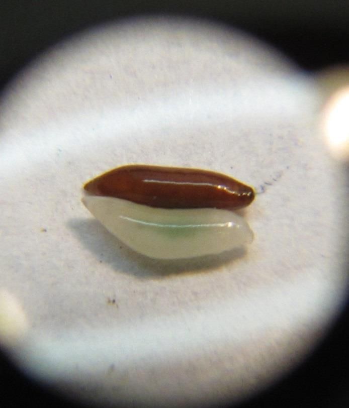
NDP 41 V1 - National Diagnostic Protocol for Mayetiola destructor

        3.2.2       Larvae and pupae
Suspect plant samples may be inspected by peeling the leaf sheaths down to the crown of the wheat
plant where tillers emerge. First instar larvae are very tiny (up to approx. 0.5-1.7 mm long) and can be
found on the crown of the plant using a 10x or 20x magnification hand lens. The larvae colour varies
from almost transparent to opaque and are easily missed when in low numbers (Figure 3).

Second instar larvae are more noticeable, as they are creamy to white and larger ranging in size from
1.7 to 4 mm. The second instar larvae are immobile and remain within the leaf sheaths at the base of
the plant to feed (Figure 4). Third instar larvae are the same, except that they become of the reddish
brown (Figure 5) prior to developing into the dark coloured ‘flaxseed’-looking pupae (2-6 mm) (Figure
6).

 Figure 3. Hessian fly first instar larvae                     Figure 4. Second instar larvae of Hessian fly are
 are very tiny and cryptic. They crawl to                      larger, pale white and immobile at the base of
 the crown of the plant.                                       plants within leaf whorls and between tillers.

  Photos: (Kansas, USA) Dustin Severtson, Department of Agriculture and Food Western
  Australia.

   6                                                                  Subcommittee on Plant Health Diagnostics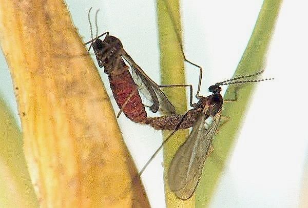
NDP 41 V1 - National Diagnostic Protocol for Mayetiola destructor

 Figure 5. Second (pale) and third (red-                      Figure 6. Pupae of Hessian fly are red
 brown) instars of Hessian fly (10x                           to dark brown at the base of plants
 magnification hand lens). Photo: (Kansas,                    within leaf whorls and between tillers.
 USA) Dustin Severtson, Department of                         Photo: (Kansas, USA) Dustin Severtson,
 Agriculture and Food Western Australia.                      Department of Agriculture and Food
                                                              Western Australia.

   7                                                                 Subcommittee on Plant Health Diagnostics
NDP 41 V1 - National Diagnostic Protocol for Mayetiola destructor
4 IDENTIFICATION
Molecular markers should be used for identification of M. destructor and M. hordei males according to:
Chen, M.S., Wheeler, S., Davis, H., Jeff Whitworth, R., Knutson, A., Giles, K. L., et al. (2014). Molecular
markers for species identification of Hessian fly males caught on sticky pheromone traps. Journal of
Economic Entomology, 107(3), 1110-1117, doi:10.1603/ec13384.
Many specimens (egg, larva, pupa or adult) should be collected for the molecular test to ensure
presence of males.
These methods should be complemented with traditional taxonomic methods based on keys and
descriptions already established for identification of Mayetiola destructor (Say) adults and larvae.
Identification of Mayetiola adults to species level is based principally on microscopic differences in
the male genitalia and other characters, and is best undertaken by expert taxonomists. Most species
have not been well characterised. There are no native or introduced Diptera of the genus Mayetiola
currently in Australia. Given this, specialists familiar with the family Cecidomyiidae are unlikely to
misidentify the species. However, due to the small size of the family, and lack of Mayetiola material
available for comparisons, less skilled identifiers may misidentify the genus. The most obvious initial
identification available is the damage to crops and the location of the larvae in the host plant.
However, correct identification of adult flies will require examination of male genitalia.
4.1 Specimen collection and preservation
Specimens can be killed by standard methods (near boiling water or 70% ethanol), and preserved in
70% ethanol. Specimens for DNA analysis should be collected directly into absolute ethanol
(adults or larvae) and stored at -20C.
4.1.1 Immediate assessment
High power stereo microscopes may be suitable for species diagnostics of adults using morphological
characteristics. Dead adult specimens may be glued to a micropin and observed directly. The dorsal
view of the male terminalia should be observed under 160x magnification.
4.1.2 Dissection and slide mounting
Mounting whole specimens requires clearing in 10% KOH for 3-5 minutes, rinsing specimens with
distilled water 5-6 times, and slide-mounting using a standard method with a mountant such as
Euparal. First instars may not require clearing. Slide mounting of whole specimens may be
unnecessary (except for close observations of female genitalia) when using a high power stereo
microscope.
Adult male genitalia may be dissected under stereo microscope using a scalpel blade. Gently cut
before the last tergite, place directly onto a slide with a drop of Hoyer’s medium (which clears the
specimen) and orient the male genitalia for a dorsal view (Figures 14, 15). Observe under compound
microscope.
8 Subcommittee on Plant Health DiagnosticsNDP 41 V1 - National Diagnostic Protocol for Mayetiola destructor
4.2 Morphological identification
4.2.1 Larvae
Family Cecidomyiidae
Larval body consists of 13 segments
Larvae have head capsule with tentorial arms and mouth apparatus apparent
Subfamily Cecidomyiinae
Larval anal opening slit-like on ventral side of terminal segment
Tribe Oligotrophini
Terminal segment of larvae rounded and with central notch
Eighth abdominal segment of larvae always bearing two dorsal papillae
Genus Mayetiola
Instars of all species reside inside the leaf whorls/sheaths of grasses
The last instar develops into an adult within the penultimate larval cuticle, without
feeding (i.e. “the flaxseed”)
Larvae have either bifid or hastiform (or pointed) breastbone
4.2.2 Adult
Family Cecidomyiidae
Elongate antenna 8–24 segmented.
Antennae ‘simple’ (long, with bead-like segments, often with whorls of hairs)
Slender-bodied; stilt-legged. Ocelli present, or absent.
Eyes asymmetric, nearly or quite connected above the antennae.
The maxillary palps (1–)3–5 segmented; drooping.
Wing venation consists of a few weak veins (Figure 7)
Wing veins reaching the margin (2–)3, or 4(–6).
Wings without a discal cell; without a sub-apical cell; without a closed anal cell.
The costa extending around the entire wing.
Sub-costa absent or only dubiously identifiable.
Wings with the lower calypter much reduced or absent (Watson and Dallwitz, 2003).
4.2.3 Identification of Mayetiola destructor (Say)
Larvae: No definitive species-specific characteristics
The first instar is 0.5-1.7 mm long, dorsoventrally flattened at first, but becoming cylindrical with age.
The second instar is 1.7-4.0 mm long, unevenly cylindrical and with the posterior end variably
tapered. The integument is almost uniformly covered with elongate spicules and the head is directed
ventrally beneath the first thoracic segment. While feeding this instar is white, but when feeding
ceases it turns brown, becomes hard and its shape may be modified by compression, especially when
9 Subcommittee on Plant Health DiagnosticsNDP 41 V1 - National Diagnostic Protocol for Mayetiola destructor
crowded. It becomes a puparium within which the third instar, pupa and adult will develop. The
third instar develops within the second, is not visible, and does not feed. It is dorsoventrally
flattened, becoming cylindrical as the pupal tissues develop. The integument is completely covered
with rounded verrucae, except on the anteroventral areas of the ventral segments, which have
verrucae tipped with anteriorly directed points. A median, ventral, bifid sternal spatula is present on
the prothorax (Gagné and Hatchett, 1989).
Pupae: No definitive species-specific characteristics (Figures 5, 6, 11)
Puparia covered in spiracles
Adult:
Adults 2-4 mm long
Wing venation reduced (Figures 7, 8, 9)
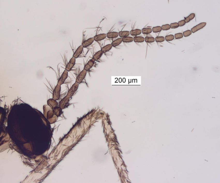
Long antennae with a variable number of flagellomeres (usually 2+15 or 2+16 but ranging
from 2+14 to 2+18) (Figures 9, 10)
Female genitalia has a seventh tergite that flares out anteriorly (Figures 12,13)
Male genitalia has elongate gonostyli and deeply separated hypoproctal lobes (Figures 14, 15)
Figure 7. Hessian fly wings showing hairs and venation pattern. Photo: (Kansas, USA) Dustin
Severtson, Department of Agriculture and Food Western Australia
10 Subcommittee on Plant Health DiagnosticsNDP 41 V1 - National Diagnostic Protocol for Mayetiola destructor Figure 8. Hessian fly wings showing hairs and venation pattern. Photo: (Kansas, USA) Dustin Severtson, Department of Agriculture and Food Western Australia Figure 9. Mating Hessian flies showing wing venation and antennae. Photo: Entomology Dept., University of Nebraska 11 Subcommittee on Plant Health Diagnostics
NDP 41 V1 - National Diagnostic Protocol for Mayetiola destructor

Figure 10. Hessian fly antennae showing whorls of hairs. Photo: (Kansas, USA) Dustin Severtson,
Department of Agriculture and Food Western Australia

Figure 11. Pupae positioned at the base or crown of a wheat plant, no galls present. Photo: K.S. Pike,
WSU (North Carolina Agricultural Extension Service)

   12                                                               Subcommittee on Plant Health Diagnostics
NDP 41 V1 - National Diagnostic Protocol for Mayetiola destructor

Figure 12. Female 7th tergite. Photo: S. Bauer ARS/USAD

Figure 13. Female 7th tergite flares out anteriorly. Photo: Dustin Severtson, Department of Agriculture
and Food Western Australia

   13                                                               Subcommittee on Plant Health Diagnostics
NDP 41 V1 - National Diagnostic Protocol for Mayetiola destructor

                                                                    a
                                                           b

Figure 14. Hessian fly male terminalia cleared and mounted in Hoyer’s medium showing (a) elongate
gonostyli and (b) deeply separated hypoproctal lobes. Photo: Dustin Severtson, Department of
Agriculture and Food Western Australia

Figure 15. Male terminalia of Hessian fly. Note the elongate gonostyli and deeply separated
hypoproctal lobes as in Figure 14. Photo: Pia Scanlon, Department of Agriculture and Food Western
Australia

   14                                                                   Subcommittee on Plant Health Diagnostics
NDP 41 V1 - National Diagnostic Protocol for Mayetiola destructor
4.3 Comparison with similar species
4.3.1 Mayetiola hordei Kieffer 1909
Synonyms: Mayetiola mimeuri (Mesnil, 1934)
Common name: Barley stem gall midge
Hosts: Barley
Comments
This species is not found on wheat (unlike the Hessian fly). Gagne et al. (1991) records the historic
taxonomic confusion between the two species and their structural differences.
NB: This species is also not found in Australia.
External differences specific to M. hordei:
• Larvae induce pea-sized galls in host plant (Figure 16)
• Puparia almost entirely smooth
• Female genitalia has rectangular seventh tergite (Diagram 13 in Figure 17)
• Male genitalia have hypoproctal lobes which are triangular, and short gonostyli (Diagram 14 in
Figure 17).
Figure 16. M. hordei gall in stem of barley plant.
15 Subcommittee on Plant Health DiagnosticsNDP 41 V1 - National Diagnostic Protocol for Mayetiola destructor Figure 17. Hessian fly and barley stem gall midge: Female postabdomens (dorsal) and male termialia (dorsal) of two different species of Mayetiola (Diptera: Cecidomyiidae) in Morocco. (Source: Gagne R., Hatchett J., Lhaloui S., El Boussini M., 1991, Annals of the Entomological Society of America. 84:4, pp. 436-443). (12) female segments 6 to end of M. hordei; (13) same of M. destructor; (14) male terminalia with enlargement of left gonostylus of M. hordei; (15) same of M. destructor 16 Subcommittee on Plant Health Diagnostics
NDP 41 V1 - National Diagnostic Protocol for Mayetiola destructor

4.4          Molecular identification
The PCR protocol for the identification of Hessian fly has been reproduced with permission from a
published method in:

Chen, M.S., Wheeler, S., Davis, H., Jeff Whitworth, R., Knutson, A., Giles, K. L., et al. (2014). Molecular
markers for species identification of Hessian fly males caught on sticky pheromone traps. Journal of
Economic Entomology, 107(3), 1110-1117, doi:10.1603/ec13384.

The testing strategy recommended by Chen et al (2014):

Figure 18. Overall strategy to identify Hessian fly males on a sticky pheromone trap. The numbers
within the circles above or beside a major arrow indicate four major steps in the identification
process: 1) morphology-based preselection to exclude apparent non-midges, 2) Mycetophiloidea-
common marker selection to exclude non-midge insects with similar morphology, 3) selection by
Hessian fly-specific marker 1 to exclude non-Hessian fly midges, and 4) final selection by Hessian fly-
specific marker 2 to reduce misidentification by specific marker 1 due to errors or gene sequence
variation.

        4.4.1       DNA Extraction.
Place individual insects into 1.5-ml Eppendorf tubes with 100 µl STE buffer (10 mM Tris- HCl, 1 mM
EDTA, pH 8.0, 0.1M NaCl) and homogenize with an electric microtube pestle. Incubate in boiling water
for 5 min and centrifuge at 12,000 rpm for 5 min. Transfer the supernatant from each sample into a
new 1.5 ml Eppendorf tube and add 250 µl of -20oC ethanol. Invert the tube several times to mix the
contents. Incubate at -20oC for 14-18 h and then centrifuge at full speed for 20 min at 4oC. Collect the
DNA pellet and discard supernatant (aspirate the supernatant to avoid displacing the DNA pellet).
Reconstitute the pellets with 30µl of ddH20, vortex thoroughly, and store at -20oC for short-term
storage or -80oC for long-term use.

   17                                                               Subcommittee on Plant Health Diagnostics
NDP 41 V1 - National Diagnostic Protocol for Mayetiola destructor
4.4.2 PCR amplification
Amplify according to the following programs:
For the common marker (CM, actin, AF017427) and Hessian-fly specific marker 2 (HFSM2, SSPG31–5,
EV466578)
Steps Temperature (°C) Time Number of cycles
(min)
Denature 94 1
Anneal 55 1 35x
Extension 72 2
For the Hessian-fly specific marker 1 (HFSM1,MDP10, AEGA01028834)
Steps Temperature (°C) Time Number of cycles
(min)
Denature 94 1
Anneal 55 1 45x
Extension 72 2
A negative control without DNA template needs to be included for each PCR run. Optimisation may be
needed due to various PCR mixes available and may differ between laboratories. In the first instance,
test using the above PCR parameters. Marker specificity testing may require optimizing using different
annealing temperatures (35, 40, 45, 50 and 55 °C) with the other PCR conditions remaining
unchanged.
DNA primers
Targeted
Marker Primer sequence pair (5’ – 3’) Bp Reference
gene
CACCAGCCATGTATGTTGCCAT
Common Actin A 148 Chen et al 2014
AAACGGAGGATGGCATGTGGCA
Hessian fly-
specific TGCATTGCTACAACTGAACGA
MDP10 170 Chen et al 2013
marker 1 CAACCGATTGTAGAACAG
(HFSM1)
Hessian fly-
specific AAAGTCATCATTTTAGCTTTGT
SSGP31-5 369 Chen et al 2010
marker 2 TTATGCAGTGGTTGGAGTTGTT
(HFSM2)
18 Subcommittee on Plant Health DiagnosticsNDP 41 V1 - National Diagnostic Protocol for Mayetiola destructor
4.4.3 Interpretation
Bands will differ with the primer sets depending on developmental stages (Figure 19).
Figure 19. PCR amplification of DNA samples extracted from individual Hessian flies from different
developmental stages with different primer sets. CM, HFSM1, and HFSM2 represent primers for the
midge/gnat common marker, Hessian fly-specific marker 1, and Hessian fly-specific marker 2,
respectively (Chen et al 2014).
19 Subcommittee on Plant Health DiagnosticsNDP 41 V1 - National Diagnostic Protocol for Mayetiola destructor
5 CONTACTS FOR FURTHER
INFORMATION
Andras Szito, Department of Primary Industries and Regional Development, Agriculture and Food,
Western Australia.
Email: andras.szito@agric.wa.gov.au.
Dustin Severtson, Department of Primary Industries and Regional Development, Agriculture and Food,
Western Australia.
Email: dsevertson@dpird.wa.gov.au
Dr Ming-shun Chen, Insect Geneticist, US Department of Agriculture
Adjunct Professor, Department of Entomology, Kansas State University.
Email: mchen@ksu.edu
Web: http://www.k-state.edu/hessianfly/
20 Subcommittee on Plant Health DiagnosticsNDP 41 V1 - National Diagnostic Protocol for Mayetiola destructor
6 ACKNOWLEDGEMENTS
This protocol was developed by Andras Szito and Dustin Severtson, (Department of Primary Industries
and Regional Development, Western Australia).
The molecular procedure was reproduced by kind permission of MS Chen. Lucy Tran-Nguyen assisted
with writing the molecular procedure in the required style.
The protocol was reviewed by Adrian Nicholas (NSW DPI) and Monica Kehoe (Department of Primary
Industries and Regional Development, Western Australia)
21 Subcommittee on Plant Health DiagnosticsNDP 41 V1 - National Diagnostic Protocol for Mayetiola destructor
7 REFERENCES
Barnes H.F. (1956). Gall midges of economic importance. Vol. VII: Gall midges of cereal crops. London,
UK: Crosby Lockwood.
Chen, M. S., X. M. Liu, Z. Yang, H. Zhao, R. H. Shukle, J. J. Stuart, and S. Hulbert. (2010). Unconventional
conservation among genes encoding small secreted salivary gland proteins from a gall midge. BMC
Evol. Biol. 10: 296.
Chen, H., Y. C. Zhu, R. J. Whitworth, J. C. Reese, and M. S. Chen. (2013). Serine and cysteine protease-like
genes in the genome of a gall midge and their interactions with host plant genotypes. Insect
Biochem. Mol. Biol. 43: 701-711.
Chen, M.S., Wheeler, S., Davis, H., Jeff Whitworth, R., Knutson, A., Giles, K. L., et al. (2014). Molecular
markers for species identification of hessian fly males caught on sticky pheromone traps. Journal of
Economic Entomology, 107(3), 1110-1117, doi:10.1603/ec13384.
Gagné R.J., Hatchett J.H. (1989). Instars of the Hessian fly (Diptera: Cecidomyiidae). Annals of the
Entomological Society of America, 82, 73-79.
Gagne, R.J., Hatchett J.H., Lhaloui S., El Bouhssini M. (1991). Hessian fly and barley stem gall midge, two
different species of Mayetiols (Diptera: Cecidomyiidae) in Morocco. Annals of the Entomological
Society of America, 84:4, 436-443.
McColloch J.W. (1923). The Hessian fly in Kansas. Kansas Agricultural Experiment Station technical
bulletin, 11.
Szito A., Smith T., Grimm M., Botha J.H. and Poole M.C. (2007). Hessian Fly, Mayetiola destructor (Say)
(Diptera: Cecidomyiidae) Pest Datasheet/Pest Risk review for the cereal Grain Industry.
Department of Agriculture and Food. Government of Western Australia.
Watson, L. and Dallwitz, M.J. (2003 onwards). British insects: the families of Diptera. Version: 1st
January 2012.
22 Subcommittee on Plant Health DiagnosticsNDP 41 V1 - National Diagnostic Protocol for Mayetiola destructor
8 APPENDICES
Pheromone lures for Hessian fly are available from www.phero.net. These are widely used in the U.S.
where Hessian fly is present in wheat producing regions to investigate the fly’s seasonal life cycle in
localised conditions and warn growers of the risk of adult flies being present. The lures are
placed in individual ‘Delta’ sticky traps (available from Trece® Incorporated) which are attached to
a stake towards the ground up to approximately20 cm above the soil for young crops or grasses.
Vertical placement relative to crop height is important as Hessian flies are poor fliers and may not
be able to reach the trap if placed too high (Figure 20). Traps may be placed higher in more advanced
crops or grasses (i.e. heading) for detection of later generations of Hessian fly (Figure 21). Sticky traps
may be placed unfolded in a transparent plastic ( ziplock) bag for transport and later inspection
(Figure 22).
Figure 20. Delta sticky trap with Hessian fly Figure 21. Delta sticky trap with Hessian fly
pheromones. pheromone placed higher in an advanced
wheat crop.
Figure 22. Delta trap showing flies and pheromone.
23 Subcommittee on Plant Health DiagnosticsYou can also read