Noninvasive quantitative assessment of collagen degradation in parchments by polarization-resolved SHG microscopy
←
→
Page content transcription
If your browser does not render page correctly, please read the page content below
SCIENCE ADVANCES | RESEARCH ARTICLE
APPLIED PHYSICS Copyright © 2021
The Authors, some
Noninvasive quantitative assessment of collagen rights reserved;
exclusive licensee
degradation in parchments by polarization-resolved American Association
for the Advancement
SHG microscopy of Science. No claim to
original U.S. Government
Works. Distributed
Margaux Schmeltz1, Laurianne Robinet2, Sylvie Heu-Thao2, Jean-Marc Sintès1, Claire Teulon1, under a Creative
Guillaume Ducourthial1, Pierre Mahou1, Marie-Claire Schanne-Klein1, Gaël Latour1,3* Commons Attribution
License 4.0 (CC BY).
Nondestructive and noninvasive investigation techniques are highly sought-after to establish the degradation
state of historical parchments, which is up to now assessed by thermal techniques that are invasive and destruc-
tive. We show that advanced nonlinear optical (NLO) microscopy enables quantitative in situ mapping of parch-
ment degradation at the micrometer scale. We introduce two parameters that are sensitive to different degradation
stages: the ratio of two-photon excited fluorescence to second harmonic generation (SHG) signals probes severe
degradation, while the anisotropy parameter extracted from polarization-resolved SHG measurements is
sensitive to early degradation. This approach is first validated by comparing NLO quantitative parameters to thermal
measurements on artificially altered contemporary parchments. We then analyze invaluable parchments
Downloaded from http://advances.sciencemag.org/ on August 1, 2021
from the Middle Ages and show that we can map their conservation state and assess the impact of a restoration
process. NLO quantitative microscopy should therefore help to identify parchments most at risk and optimize
restoration methods.
INTRODUCTION At least 215 manuscripts survived the fire but remained in very
Parchment was the main writing support material in the Middle variable states, from almost intact to entirely calcined. Considering
Ages in Western Europe until the growth of paper production in the the fragility of the fragments, a digitization campaign was started
14th to 15th centuries. It is made from untanned animal skin, which in 2006 to make these documents available again for historians (3).
is preserved by liming, scraping, and drying under tension (1). After In highly deformed manuscripts, a restoration treatment was re-
this preparation process, the main remaining constituent is fibrillar quired to flatten the parchment before digitization. Since the parch-
collagen, which arises from the self-assembly of collagen triple-helical ments had suffered from highly damaging conditions, exposure to a
molecules into fibrils and further into fibers. Parchments are very moisture treatment to relax the collagen fibers might cause fur-
sensitive to heat and water that can induce collagen degradation. ther damage. Therefore, it is essential to assess the conservation
The ultimate state of degradation is the formation of gelatin that state of the collagen before such treatment and to monitor its evolu-
corresponds to the unfolding of the collagen triple-helical structure tion in the most degraded parchments.
(denaturation) and the reorganization of the single helices into a Nowadays, the degradation state of skin-based materials in the
network with small triple-helical domains (gelatin) (2). Such an cultural heritage field is commonly assessed by differential scanning
advanced degradation can sometimes be identified macroscopically calorimetry (DSC) or by micro hot table (4–6). These thermal tech-
and qualitatively by a shrinkage, increased transparency, and in- niques measure the temperature, called shrinkage temperature TS,
creased rigidity of the material. As this transformation is irrevers- at which the fibrillar collagen turns into gelatin in a wet sample. The
ible and its primer steps are usually not visible to the eye, the main DSC technique also provides the enthalpy change H related to the
challenge for conservation scientists and restorers is to identify thermal energy required for the denaturation. However, these tech-
these early stages of degradation to avoid gelatin formation follow- niques are invasive, as they require a sampling, and destructive, which
ing exposure to a humid environment. limits the number of analyzed areas and constrains them to the
A vast collection of manuscripts on parchment of major histori- edges or hidden parts of the artifact rather than on the more relevant
cal relevance is housed in the library of Chartres in France. Chartres central part close to the text and illuminations. In addition, the alter-
was one of the most important occidental intellectual centers during ation is often heterogeneous within a single parchment, so a non-
the 11th and 12th centuries, due to its invaluable collection of about invasive technique is needed to repeat the measurements in different
500 medieval manuscripts originating mainly from the famous areas of the artifact to access a complete diagnosis of the manuscript.
Chartres chapter library. On 26 May 1944, during a bombing of the Noninvasive approaches have been recently proposed for parch-
city, a fire irremediably destroyed almost half of the collection. ments, for instance, to identify the animal species (7, 8). Regard-
ing the degradation state, the current analytical techniques show
some limitations. Attenuated total reflection–Fourier transform
infrared (ATR-FTIR) spectroscopy provides information on the
1
Laboratoire d’Optique et Biosciences, Ecole polytechnique, CNRS, INSERM, Institut extent of gelatinization in a parchment (9). However, it requires
Polytechnique de Paris, Palaiseau, France. 2Centre de Recherche sur la Conservation scraping the parchment surface before measurement to avoid
(CRC), Muséum national d’Histoire naturelle, Ministère de la Culture, CNRS, Paris,
France. 3Université Paris-Saclay, Saint-Aubin, France. interferences from surface materials, such as calcium carbon-
*Corresponding author. Email: gael.latour@universite-paris-saclay.fr ate layer or dust. Near-infrared spectroscopy can estimate the
Schmeltz et al., Sci. Adv. 2021; 7 : eabg1090 16 July 2021 1 of 9
SCIENCE ADVANCES | RESEARCH ARTICLE
shrinkage temperature of parchments noninvasively (10), but needs RESULTS
to be calibrated with a large database of reference samples. Synchro- Model parchments
nous fluorescence spectroscopy was suggested for estimating the A set of model parchments D# was obtained by exposure to dry heat
collagen-to-gelatin ratio, but it has not been correlated to quanti- for # days. They were first assessed by DSC measurements of the
tative assessment of collagen degradation such as the shrinkage shrinkage temperature TS and the enthalpy change H, as shown in
temperature (11). Fig. 1 and fig. S1, respectively (see data in table S2). The shrinkage
Some optical techniques provide noninvasive and nondestruc- temperature decreases strongly from D0 (“reference” parchment)
tive three-dimensional (3D) imaging of cultural heritage artifacts. to D16 and then it stabilizes close to room temperature at around
In this respect, the most common technique is optical coherence 32°C. While the shrinkage temperature TS is constant between D16
tomography (OCT) (12). It has been used to study the influence of and D122, the enthalpy change H decreases from 41 to 5 J/g.
inks and pigments on the conservation state of parchments (13, 14), These model parchments were then imaged on both sides,
but it is not well suited to characterize the organization of the fibers namely, the grain and the flesh sides, by NLO microscopy. Image
because of their dense arrangement after the manufacturing pro- stacks were recorded up to a depth of 15 to 100 m, depending on
cess, which homogenizes their optical properties and strongly re- the optical properties of every parchment, to access volume infor-
duces the contrast of OCT images. Alternatively, nonlinear optical mation. No photodamage was observed on the samples throughout
(NLO) microscopy (15, 16), which is widely used for biomedical the imaging. This was assessed by verifying the reproducibility of
studies, is today an emerging technique in cultural heritage scienc- the measurements when the same image was acquired several times
es. It provides micrometer-scale 3D resolution to study pigments, in the same area. Figure 2 shows typical images merging 2PEF in
Downloaded from http://advances.sciencemag.org/ on August 1, 2021
binders and varnishes (17–23), paper and textiles (19, 24, 25), wood red and SHG signals in green. The collagen fibers, revealed by SHG,
used for music instruments (18), and skin-based objects such as are packed and straight, due to the stretching step during manufac-
leathers, parchments, and natural history specimens (26, 27). A key turing. On the grain side, the black (no signal) round structures (white
advantage of this technique is its multimodal capability based on arrowheads in Fig. 2) are attributed to the location of hair folli-
different modes of contrast that are directly linked to the structural cles. On the flesh side, the collagen fiber organization seems more
or chemical nature of the materials. Two-photon excited fluores- homogeneous and dense. In the D0 and D4 parchments, some 2PEF
cence (2PEF) signals are emitted by a wide range of materials signals are collected from structures of few tens of micrometers,
(fluorophores) in historical artifacts with specific absorption and presumably due to residues of keratin, fat, and elastin, as well as
emission fluorescence spectra (18). Second harmonic generation gelatin induced by manufacturing (26). The SHG signals, arising
(SHG) signals are specific for dense and well-aligned structures from fibrillar collagen, prevail on the less degraded parchments,
such as fibrillar collagen (15, 16, 28) and vanish for centrosymmetric from D0 to D16, while from the D40 parchment, the 2PEF signals
materials such as gelatin. Usual SHG imaging is based on circularly seem to dominate with a larger spatial extent. To quantitatively as-
polarized laser excitation, while in polarization-resolved SHG sess the evolution of these signals as a function of heat exposure
(P-SHG) microscopy, the SHG signal is recorded as a function of duration, the ratio of 2PEF to SHG signals I2PEF/ISHG is calculated
the orientation of the linearly polarized laser excitation. This ad- pixel-wise and averaged in depth (see Materials and Methods) for
vanced method provides the main orientation of the collagen fibrils each side of every parchment, as shown in Fig. 3A (table S3). The
and the degree of alignment of the triple helices within and between
fibrils at the submicrometer scale, as demonstrated in various bio-
medical studies (29–37). We have recently shown that SHG micros-
copy provides structural information about the 3D organization of
fibrillar collagen within parchments and other skin-based materials
(26, 27). Notably, this technique discriminates these two extreme
states: well-preserved artifacts characterized by prevailing SHG
signals related to fibrillar collagen and gelatinized ones character-
ized by the loss of SHG signals and the increase of 2PEF (26).
However, basic implementation of NLO imaging does not meet
the need of finely grading intermediate states of degradation, as
thermal methods do.
In this study, we propose two quantitative methods based on
advanced NLO microscopy to quantify these intermediate states of
degradation. The first method is to compute the ratio of 2PEF to
SHG signals in every pixel. The second one is to use P-SHG micros-
copy to compute the collagen alignment at the submicrometer scale.
We first study a set of model parchments made from contemporary
parchments that were artificially altered by heat exposure for in-
creasing durations. The quantitative parameters obtained by these
new methods are compared with the shrinkage temperature and the
enthalpy change measured by DSC to assess their validity. Second, Fig. 1. Shrinkage temperature of model parchments. Shrinkage temperature
these quantitative methods are used to noninvasively estimate the TS obtained by DSC measurements of model parchments artificially altered by dry
degradation state of the heat-damaged medieval parchments from heat exposure during 0 (reference) to 122 days. Three sample measurements were
the Chartres’s library. collected for each model parchment, and the mean value and SD are displayed.
Schmeltz et al., Sci. Adv. 2021; 7 : eabg1090 16 July 2021 2 of 9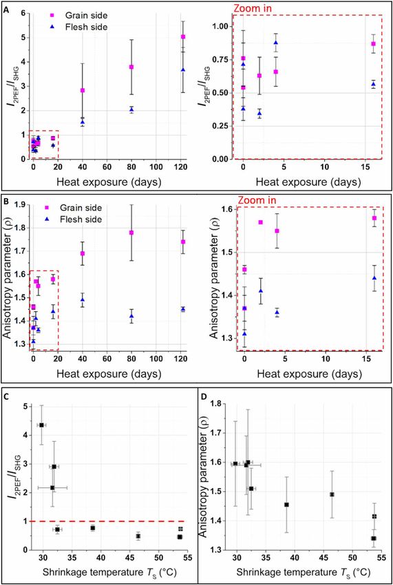
SCIENCE ADVANCES | RESEARCH ARTICLE
Downloaded from http://advances.sciencemag.org/ on August 1, 2021
Fig. 2. NLO imaging of model parchments. NLO imaging on grain and flesh sides of model parchments altered by dry heat exposure during 0, 4, 16, 40, and 122 days.
Images in false colors: SHG signals in green and 2PEF signals in red. On the grain side, hair follicles (black holes) are shown with white arrowheads. Scale bars, 50 m.
graph shows that this ratio does not evolve between 0 and 16 days of Historical parchments
heat exposure and then linearly increases from 16 to 120 days. The medieval parchments from the Chartres’s library suffered from
The model parchments are further analyzed by P-SHG micros- fire and water at the end of the Second World War, as a result of
copy. It provides a measure of the anisotropy parameter that is a bombing and the subsequent action of the firefighters. The most
defined as the square root of the ratio of the two minima of the SHG degraded manuscripts appear as blocks of agglomerated parchment
signal as a function of the incident polarization orientation. This leaves with burned and carbonized areas, without any possibility to
parameter probes the degree of alignment of the triple helices with- separate the folios (or leaves). A small and non-carbonized frag-
in and between fibrils in the focal volume. The expected value from ment of such a block was investigated by NLO microscopy. The col-
previous studies on tissues is around 1.36 for well-aligned type I lected images exhibit a compact structure with flat collagen fibers
fibrillar collagen and increasing values are associated with increas- (Fig. 4). No SHG signals are collected from this sample, except from
ing disorder (32, 34). The evolution of the anisotropy parameter some isolated micrometric particles, likely to be calcium carbonate
versus the heat exposure duration is shown in Fig. 3B (table S3). It (26). The anisotropy parameter cannot be measured in this case be-
increases from D0 to D16 (from 1.45 to 1.58 on the grain side and cause of the absence of SHG signals preventing any P-SHG acquisi-
from 1.3 to 1.45 on the flesh side; see the zoomed-in image in Fig. 3B), tion. Anyway, low SHG signals associated to strong 2PEF signals
and it shows a plateau from D40. This tendency is observed on both confirm that the ultimate state of degradation of collagen has been
sides of the parchments but with values always higher on the grain reached in these parchments even if the morphology of some fibers
side than on the flesh side. is preserved.
To evaluate the relevance of these NLO measurements to probe Three historical parchment leaves originating from a less de-
the degradation state of fibrillar collagen within parchments, the graded manuscript were investigated using quantitative NLO mea-
NLO results are compared to the shrinkage temperature TS and the surements. Results for folios 312 and 297 are presented in Fig. 5 and
enthalpy change H measured by DSC. In contrast to NLO micros- fig. S3. Two areas are investigated on both sides: area #A, in the
copy that collects signals at a selected depth between 0 and 100 m, center, and area #B, on the edge of the folio. The central part ap-
the DSC measurements of collagen degradation are performed over pears less damaged by eye than the edge of the parchment, which is
the entire thickness of the parchment (150 to 300 m). DSC results more rigid, shrunk, and slightly darker. Typical NLO microscopy
are therefore compared to the average of the NLO data collected images from these two areas on both sides are displayed in Fig. 5B. They
from the flesh and grain sides (Fig. 3, C and D). I2PEF/ISHG is con- reveal a lower SHG signal and a higher 2PEF signal in the area #B
stant and remains below 1 as long as TS is above 32°C, but this ratio compared to the area #A, which is confirmed by the computation of
increases with the decrease of the enthalpy change when TS is below the I2PEF/ISHG ratio in all the ROIs imaged within each area (each
32°C (fig. S2). On the contrary, the anisotropy parameter follows point in Fig. 5C corresponds to one ROI and the I2PEF/ISHG ratio is
the same trend as the shrinkage temperature: in the initial stages of always higher in area #B). The anisotropy parameter is also higher
degradation (higher TS), it increases from 1.35 to 1.6. Moreover, for in area #B than in area #A for all the analyzed ROIs (see Fig. 5D). In
the most degraded parchments, the measurements on different re- the central area #A of the folio, I2PEF/ISHG < 1 and ~ 1.4, while in the
gions of interest (ROIs) are strongly dispersed, which suggests that edge, area #B, I2PEF/ISHG varies between 2 and 6 and varies be-
the degradation is heterogeneous within a single parchment. tween 1.45 and 1.6. These results confirm the difference in collagen
Schmeltz et al., Sci. Adv. 2021; 7 : eabg1090 16 July 2021 3 of 9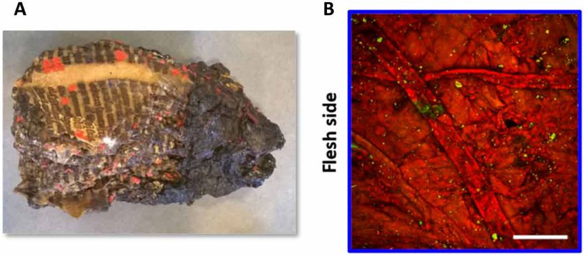
SCIENCE ADVANCES | RESEARCH ARTICLE
Downloaded from http://advances.sciencemag.org/ on August 1, 2021
Fig. 3. Quantitative assessment of model parchments from NLO microscopy. (A) Ratio of 2PEF to SHG signals I2PEF/ISHG and (B) anisotropy parameter on grain and
flesh sides of the model parchments artificially aged by heat for 0, 2, 4, 16, 40, 80, and 122 days (one parchment for each treatment duration, but two reference parch-
ments for D0). Zoom in (on the right): heat exposure for 0 to 16 days. (C) I2PEF/ISHG and (D) mean values from both sides of the parchments versus the shrinkage tempera-
ture TS determined by DSC. The red dashed line in (C) corresponds to I2PEF/ISHG = 1. Error bars correspond to the SD of measurements on several samples (TS) or several
areas and sides (I2PEF/ISHG, ), depending on the technique (see table S1).
Schmeltz et al., Sci. Adv. 2021; 7 : eabg1090 16 July 2021 4 of 9SCIENCE ADVANCES | RESEARCH ARTICLE
contact with water at ambient temperature is sufficient to turn the
remaining fibrillar collagen into gelatin (4). Beyond 16 days of heat
exposure, the shrinkage temperature remains constant but the en-
thalpy change decreases, which may indicate that the amount of pre-
served fibrillar collagen within a sample diminishes and therefore that
the degradation, initially localized on the parchment surface, ex-
tends into the material depth. Together, these measurements clearly
confirm that the collagen degradation increases with heat exposure
duration and that the parameter TS is sensitive to the first steps of
degradation of the fibrillar collagen fragilized by chain cleavage.
Thanks to the correlation established between DSC and NLO
Fig. 4. NLO imaging of a degraded medieval parchment. Investigation of a frag- quantitative parameters on the model parchments, we propose a
ment from a block of agglomerated and strongly degraded parchment leaves from
noninvasive alternative to quantify the collagen degradation state in
the Chartres’s library. (A) Picture of the parchment block (photo credit: M. Schmeltz,
Laboratoire d’Optique et Biosciences). (B) NLO microscopy image on the flesh side
parchments. The SHG signals collected by NLO microscopy in bio-
(SHG signals in green and 2PEF signals in red). Scale bar, 100 m. logical tissues are specific for dense non-centrosymmetric assem-
blies of aligned peptide bonds (15, 16, 28), as in fibrillar collagen,
where the chains are tightly aligned within triple helices, which
conservation that was visually observed between these two areas of are, in turn, tightly aligned within fibrils. In contrast, gelatin (dena-
Downloaded from http://advances.sciencemag.org/ on August 1, 2021
the parchment. Similar results are obtained on folio 297 (see fig. S3). tured collagen) forms a centrosymmetric and low-density network
A parchment leaf (folio 301) originating from the same manu- of single helices, connected by small triple-helical domains, and ac-
script and restored in 2009 was also analyzed to evaluate the influ- cordingly, it exhibits no SHG signal (26). During collagen degrada-
ence of the restoration treatment on the collagen degradation state. tion, SHG signal is thus lost because of the alteration of the collagen
The parchment had been relaxed by cold humidification to be flat- hierarchical structure. Concomitantly, 2PEF intensity increases
tened. Although it improved the visual appearance and the reading until it predominates. The origin of the 2PEF intensity observed in
of the parchment, this water addition may have induced a degrada- degraded collagen is still unknown. Infrared spectroscopy showed
tion of the collagen. The anisotropy parameter values obtained by the onset of a carbonyl band during this transformation, which was
P-SHG measurements in the central part of the restored folio are previously attributed to the formation of gelatin (26). In the present
similar to those obtained in the center of the two untreated folios study, the heat treatment applied to the model parchments induces
from the same manuscript, as shown in Fig. 5 (E and F) and fig. S4. oxidation of the collagen with breakage of the N─C covalent bond
within the main chain and cross-linking of the molecules (6, 38).
However, the preservation of the fibrillar morphology in some of
DISCUSSION the heat-damaged parchments suggests that the increase of the
In the field of cultural heritage, one of the challenges is to develop 2PEF intensity is not specific to gelatin formation, but is rather a
noninvasive and nondestructive approaches that provide a precise general marker of collagen degradation. In the model parchments,
diagnosis related to the conservation state and the type of degrada- the ratio of 2PEF to SHG signals I2PEF/ISHG remains stable below
tion of the historical parchments. The two main chemical degradation 1 in the first stages of the degradation. Then, I2PEF/ISHG increases at
mechanisms taking place in collagen are hydrolysis and oxidation the latest stages of degradation when TS has reached its lowest level,
(1). Both reactions cause chain cleavage with the formation of pep- around 32°C, and the degradation likely progresses in depth, as sup-
tides, and the modification of the collagen side group chemistry, ported by the decrease of the enthalpy change. Our results thus
leading to alteration of hydrogen bonding and other interactions demonstrate unambiguously that I2PEF/ISHG is a marker for the lat-
stabilizing the triple-helical structure and the fibrillar assemblage est stages of degradation. The measurement of I2PEF/ISHG values
(4). Water access is thus facilitated to further break hydrogen bonds above 1 (meaning that 2PEF intensity is predominant over SHG
and unfold the collagen triple helices. As the stabilizing forces signal) in a parchment is therefore a probe of shrinkage tempera-
decrease, the energy required for denaturation of the collagen mol- ture close to ambient conditions. It could thus be used to assess
ecule decreases. This denaturation and the concurrent formation of parchments in advanced stages of degradation, so most at risk.
gelatin correspond to an irreversible degradation. The anisotropy parameter is instead sensitive to the first stages
Currently, collagen stability is assessed by measurement of the of parchment degradation as the DSC shrinkage temperature TS. The
shrinkage temperature from a parchment sample, using destructive anisotropy parameter probes the submicrometer orientation dis-
thermal techniques. In contrast, NLO microscopy is a noninvasive order of collagen triple helices between and within fibrils lying
and nondestructive technique for investigating the conservation within the image plane (32, 34). The lower this parameter, the greater
state of parchments. No variation of the morphology of the collagen the collagen alignment within the focal volume. The increase of the
fibers and no modification of the SHG and 2PEF signals were de- anisotropy parameter is thus attributed to a disorganization of the
tected over time during the image acquisition. It proved the absence fibrils induced by heat exposure. In the early stages of degradation,
of any degradation of the fibrillar collagen due to laser heating this disorganization is not sufficient to reduce the SHG signal, as
during parchment imaging since any damage would have induced a revealed by the absence of evolution of I2PEF/ISHG, but it can be de-
variation of the SHG and 2PEF signals. tected by the P-SHG response, thanks to the high sensitivity of this
In the heat-damaged model samples presented in this study, the polarimetric technique to orientation disorganization (32–34, 37).
shrinkage temperature TS decreases sharply until 16 days of heat expo- The anisotropy parameter is therefore complementary to the I2PEF/
sure, down to its limit, around 30°C. At this state of degradation, ISHG value, as it provides measures of the early stages of the collagen
Schmeltz et al., Sci. Adv. 2021; 7 : eabg1090 16 July 2021 5 of 9SCIENCE ADVANCES | RESEARCH ARTICLE
Downloaded from http://advances.sciencemag.org/ on August 1, 2021
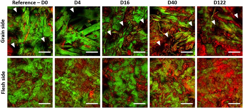
Fig. 5. Investigation of a medieval parchment from the Chartres’s library. (A) Picture of the grain side of folio 312. The measurements were conducted in several ROIs
located in the two areas identified by dotted squares (#A and #B) (photo credit: CNRS–IRHT). (B) NLO microscopy images of ROIs from both areas on grain and flesh sides
of folio 312 (images in false colors: SHG signals in green and 2PEF signals in red). Scale bars, 100 m. (C) Ratio of 2PEF to SHG signals I2PEF/ISHG and (D) anisotropy parame-
ter for various ROIs in area #A and #B of folio 312. (E) I2PEF/ISHG and (F) for the three parchment leaves coming from the same manuscript: two leaves without treatment
over 80 years (folios 312 and 297) and one leaf after restoration by cold humidification in 2009 (folio 301). Error bars correspond to the SD of measurements on several
ROIs (four to six different ROIs in each area, see table S1).
degradation in parchment. This parameter would be particularly treatments are considered to improve their legibility. Most parch-
useful to investigate the impact of a specific environment or conser- ments recovered from the fire display visually heterogeneous degra-
vation treatment on the parchment microstructure. dation states even at the scale of a parchment folio. The study of
Besides the state of alteration, we demonstrate that the two NLO these historical parchments confirms that quantitative NLO is an
quantitative parameters are also sensitive to some important parch- efficient method to assess and compare the conservation state of
ment features. The measurements of the anisotropy parameter collagen in various parchments or within a single leaf, especially in
revealed a difference in the submicrometer-scale fibril distribution central relevant parts near the text and the illuminations. It enables
between the grain and the flesh sides. This difference may be at- the identification of intermediate states of degradation: The edges,
tributed to the collagen organization in skin (26, 27): small and dis- more exposed, are more degraded than the center of the parchment
organized fibers around hair follicles on the grain side (higher folios. Thanks to the correlations established between the I2PEF/ISHG
values) in contrast to greater and well-aligned fibers on the flesh or values and the TS measurements on the model parchments, one
side (lower values). Furthermore, the I2PEF/ISHG is always higher can assess the conservation state of the collagen in these historical
on the grain side compared to the flesh side (Fig. 3A). This is parchment leaves. In the folio edges, I2PEF/ISHG > 1 and values
consistent with the presence of smaller fibrils and hair follicles, as between 1.45 and 1.6 suggest that the shrinkage temperature of the
well as the presence of higher fat content on the grain side that facil- collagen is very low in that area, and probably close to ambient tem-
itates the degradation, in contrast to the denser organization on the perature. In contrast, in the central part, I2PEF/ISHG < 1 and ≈ 1.4
flesh side. In addition, thanks to the high resolution of the tech- suggest that the TS is much higher. In heavily damaged parchments
nique, the I2PEF/ISHG and measurements are sensitive to the het- with low TS, the supply of water even through humidification may
erogeneity of degradation states in the analyzed areas. The be sufficient to turn the collagen into gelatin at ambient tempera-
heterogeneity of the most damaged areas of the parchments is ture. The absence of alteration following the humidification treat-
endorsed by the observation of broader DSC peaks as well as the ment in the central area of the restored parchment thus supports
greater error bars of the I2PEF/ISHG and measurements [20.6% for our inference of a high TS in the central part. Overall, this study of
I2PEF/ISHG (2.9% for ) in the D40 to D122 parchments versus 16.9% the Chartres’s manuscripts on parchment demonstrates that ad-
for I2PEF/ISHG (1.8% for ) in the D0 to D16 parchments]. vanced NLO microscopy can quantitatively and noninvasively as-
The parchment manuscripts that survived the disaster in the sess the degradation state of historical parchments, and in particular
Chartres’s library in 1944 are a precious testimony of the past that before conservation treatment.
aroused the interest of many researchers and historians. This collec- Last, the correlations that were established between DSC and
tion is of great cultural and historical importance, as it not only con- NLO quantitative parameters are based on heat-damaged model
tains records of description of the regional society organization in parchments. In contrast, the historical parchments have been ex-
the Middle Ages but also includes a large number of Carolingian posed to heat (fire) and subsequently water (firefighter response).
manuscripts (8th to 10th centuries), major witnesses to literary Therefore, collagen in these parchments may have undergone oxi-
works from the Antiquity and the high Middle Ages. Considering dation reaction followed by hydrolysis and/or gelatinization. The
the extreme conditions they experienced, the conservation state of these consistent results obtained on the Chartres’s manuscripts thus show that
parchments is an important issue, particularly when conservation NLO quantitative parameters can probe various types of degradation.
Schmeltz et al., Sci. Adv. 2021; 7 : eabg1090 16 July 2021 6 of 9SCIENCE ADVANCES | RESEARCH ARTICLE
Moreover, as the alteration mechanism may affect the evolution of 205, a theological compendium dating from the 13th century [folios
these quantitative parameters, they may be used to reveal some be- 297 (39), 312 (40), and 301 (41)]. The three folios thus underwent
havior specific to the degradation environment. In that perspective, the same degradation conditions, but folios 297 and 312 did not
the morphological observation of the collagen fibers with NLO mi- experience any restoration treatment over 80 years, whereas folio
croscopy may provide additional information related to the origin 301 was treated in 2009 by cold humidification to flatten the
of the degradation. For instance, previous studies based on observa- parchment.
tion under a light microscope (6) have shown that the flat fiber
morphology, also observed by NLO microscopy for the most dam- Differential scanning calorimetry
aged medieval manuscript (Fig. 4), is characteristic of oxidation by A PerkinElmer DSC 8000 calorimeter was used after calibration for
dry heat. This could be assessed quantitatively using texture analy- temperature and heat flow with indium and dodecane standards.
sis or other morphological parameters, as a complement to I2PEF/ Parchment samples of around 1 mg were immersed in deionized
ISHG and . Therefore, the possibility to access quantitative infor- water for 1 hour at low temperature (around 5°C). Wet samples
mation on alteration mechanism in a noninvasive way opens up were then placed in sealed 30-l aluminum capsules (resistant up to
new interesting research perspectives on the degradation of 3 bars pressure) and analyzed by DSC, where they were heated from
parchments. 5° to 120°C at a speed of 10°C/min. The measured thermograms
In conclusion, this study has established advanced NLO micros- displayed an endothermic peak when the collagen from the sample
copy as a nondestructive and noninvasive technique for the quanti- turned into gelatin. The shrinkage temperature TS was measured at
tative diagnosis of collagen degradation in historical parchments. the onset of the peak, determined as the intersection of the peak
Downloaded from http://advances.sciencemag.org/ on August 1, 2021
The methods we have developed can be implemented on a commer- rising slope with the baseline in the thermogram, and the enthalpy
cial NLO setup, provided the few improvements detailed in Materi- change per unit mass H was obtained from the integration of the
als and Methods, so that they could be transferred to heritage surface under the peak (fig. S1A) (5). For each model parchment,
scientists in museums. Measurements of the ratio I2PEF/ISHG could three samples were collected, measured, and lastly averaged (see
help to identify parchments most at risk, close to the irreversible table S2).
collagen denaturation into gelatin. The anisotropy parameter ob-
tained from P-SHG measurements, which probes the earlier stages NLO microscopy
of degradation, could be used for collection follow-up, thus helping A custom-built laser scanning upright microscope was used to per-
heritage professionals to fine-tune conservation conditions, prevent form the NLO measurements (42). The excitation source was a fem-
any degradation onset, and check the collagen preservation during tosecond titanium:sapphire laser (MaiTai, SpectraPhysics) tuned to
a restoration treatment. As a further advantage, these noninvasive 860 nm, delivering 100-fs pulses at an 80-MHz repetition rate, and
NLO measurements could be repeated in different areas of an arti- scanned in the imaging plane using galvanometric mirrors. The
fact to assess heterogeneous progress of degradation or in the most excitation power was 5 to 10 mW at the focus. A shutter was placed
valuable near-text and illumination areas. Last, this new method before the scanning system and opened only while acquiring, to limit
could be applied to other collagen-based materials found in muse- the exposure time and to prevent photodamages. A high–numerical
ums such as leather, natural history specimens, or fluid collections, aperture (NA) air objective (20×, NA 0.80, Plan-Apochromat, Zeiss)
and extended to cellulose-based artifacts, which also exhibit SHG was used. This type of air objective is coverslip-corrected for bio-
signals, such as paper, textiles, and wooden objects. medical studies. To compensate for this coverslip correction and
achieve the best possible resolution, a resin cap with a glass lamella
on its top was mounted on the objective (see fig. S5). With this con-
MATERIALS AND METHODS figuration, the experimental resolution was 0.6 m in the lateral
Model samples direction and 1.85 m in the axial direction near the sample surface
Contemporary “model” parchments were made in 2005 out of calf (43). 2PEF and SHG signals were detected using suitable spectral
skin for the European research project IDAP (Improved Damage filters (FF01-720/SP and FF01-680/SP from Semrock, and GG455
Assessment of Parchment) (38). In this work, we studied the parch- from Schott for the 2PEF signal and FF01-720/SP, FF01-680/SP and
ments subjected to artificial aging by dry heat in an oven at 100°C FF01-427/10 from Semrock for the SHG signal) and photon-counting
for various durations: 0 (“reference” parchment), 2, 4, 16, 40, 80, photomultiplier tubes (P25PC, Sentech). Epidetection was used as
and 122 days. There were two reference parchments and one parch- the only configuration compatible with nontransparent parchments
ment for every other condition, which means eight parchments in or thick books and manuscripts. In all the figures, 2PEF and SHG
total. Each sample is identified by its number of days of aging (for signals are represented in false colors (in red and green, respectively).
instance, the sample D4 corresponds to the parchment that under-
went 4 days of heat exposure). P-SHG microscopy
P-SHG was performed using two motorized achromatic waveplates
Chartres’s medieval manuscripts on parchment inserted at the back pupil of the objective to achieve well-defined
Medieval manuscripts on parchment from the Chartres’s library in tunable linear polarizations, with a residual field ellipticity less than
France were partially destroyed at the end of the Second World War 5% (42) (see fig. S6). 3D data were recorded by axially shifting the
in 1944, exposing its collection to fire and then to water. Following objective mounted on a translation stage with 1-m axial steps, up
this disaster, all recovered parchments were washed in a bath con- to 15 to 100 m depending on the penetration depth of each sample.
taining water and formaldehyde to clean and prevent further micro- Four to six image stacks for each side of each parchment were
bial attack. The studied corpus includes a fragment of an agglomerated recorded, i.e., 67 3D stacks for the model parchments and 34 3D
parchment and three parchment leaves from the same manuscript stacks for the historical parchments (see fig. S7 and table S1).
Schmeltz et al., Sci. Adv. 2021; 7 : eabg1090 16 July 2021 7 of 9SCIENCE ADVANCES | RESEARCH ARTICLE
Image processing and analyses 13. M. Gora, M. Pircher, E. Götzinger, T. Bajraszewski, M. Strlic, J. Kolar, C. Hitzenberger,
Two parameters were extracted pixel-wise from combined 2PEF/P- P. Targowski, Optical coherence tomography for examination of parchment degradation.
Laser Chem. 2006, 68679 (2006).
SHG image stacks. The parameter I2PEF/ISHG was computed pixel by
14. P. Targowski, M. Pronobis-Gajdzis, A. Surmak, M. Iwanicka, E. A. Kaszewska,
pixel as the ratio of 2PEF to SHG signals. The anisotropy parameter M. Sylwestrzak, The application of macro-X-ray fluorescence and optical coherence
was obtained pixel by pixel from the polarimetric diagram ISHG(), tomography for examination of parchment manuscripts. Stud. Conserv. 60, S167–S177
which is the variation of the SHG signal as a function of the incident (2015).
polarization orientation . Details about the theoretical and numer- 15. W. R. Zipfel, R. M. Williams, R. Christie, A. Y. Nikitin, B. T. Hyman, W. W. Webb, Live tissue
intrinsic emission microscopy using multiphoton-excited native fluorescence and second
ical analysis of P-SHG data are provided in the Supplementary harmonic generation. Proc. Natl. Acad. Sci. U.S.A. 100, 7075–7080 (2003).
Materials. Briefly, is defined
_ as the square root of the ratio of the 16. P. J. Campagnola, L. M. Loew, Second-harmonic imaging microscopy for visualizing
I SHG(φ)
= _
two minimal signals:
√
I SHG(φ + )
, where φ is the position of the
first minimum and corresponds to the orientation of the colla-
biomolecular arrays in cells, tissues and organisms. Nat. Biotechnol. 21, 1356–1360 (2003).
17. A. Nevin, D. Comelli, I. Osticioli, G. Filippidis, K. Melessanaki, G. Valentini, R. Cubeddu,
C. Fotakis, Multi-photon excitation fluorescence and third-harmonic generation microscopy
gen fibril (29, 30, 42). When the collagen sample is composed of fi-
measurements combined with confocal Raman microscopy for the analysis of layered
brils aligned within the imaging plane, as in parchments, the samples of varnished oil films. Appl. Phys. A Mater. Sci. Process. 100, 599–606 (2010).
anisotropy parameter has been shown to probe the degree of align- 18. G. Latour, J.-P. Echard, M. Didier, M.-C. Schanne-Klein, In situ 3D characterization
ment within and between these fibrils in the focal volume (32, 34). of historical coatings and wood using multimodal nonlinear optical microscopy.
It increases as the orientation disorder of the fibrils or of the triple Opt. Express 20, 24623–24635 (2012).
19. T. E. Villafana, W. P. Brown, J. K. Delaney, M. Palmer, W. S. Warren, M. C. Fischer,
helices within the fibrils increases at this submicrometer scale. Note
Femtosecond pump-probe microscopy generates virtual cross-sections in historic
that P-SHG imaging also provides the main orientation of fibrils artwork. Proc. Natl. Acad. Sci. U.S.A. 111, 1708–1713 (2014).
Downloaded from http://advances.sciencemag.org/ on August 1, 2021
within the focal volume, but this parameter was not of interest in 20. S. Psilodimitrakopoulos, E. Gavgiotaki, K. Melessanaki, V. Tsafas, G. Filippidis, Polarization
this study. second harmonic generation discriminates between fresh and aged starch-based
A series of thresholding and averaging was then conducted to adhesives used in cultural heritage. Microsc. Microanal. 22, 1072–1083 (2016).
21. H. Liang, M. Mari, C. S. Cheung, S. Kogou, P. Johnson, G. Filippidis, Optical coherence
get a unique value for each parameter per parchment side (see fig. tomography and non-linear microscopy for paintings—A study of the complementary
S7). The mean value and the SD of the parameters were calculated capabilities and laser degradation effects. Opt. Express 25, 19640–19653 (2017).
on each side from z stacks recorded in four to six ROIs for each 22. M. Oujja, S. Psilodimitrakopoulos, E. Carrasco, M. Sanz, A. Philippidis, A. Selimis, P. Pouli,
model parchment and two to six ROIs in the same area for each G. Filippidis, M. Castillejo, Nonlinear imaging microscopy for assessing structural
historical parchment. All the data processing was performed using and photochemical modifications upon laser removal of dammar varnish
on photosensitive substrates. Phys. Chem. Chem. Phys. 19, 22836–22843 (2017).
homemade routines written in Matlab or ImageJ. 23. A. Dal Fovo, M. Oujja, M. Sanz, A. Martínez-Hernández, M. V. Cañamares, M. Castillejo,
R. Fontana, Multianalytical non-invasive characterization of phthalocyanine acrylic paints
SUPPLEMENTARY MATERIALS through spectroscopic and non-linear optical techniques. Spectrochim. Acta Part A 208,
Supplementary material for this article is available at http://advances.sciencemag.org/cgi/ 262–270 (2019).
content/full/7/29/eabg1090/DC1 24. T. E. Villafana, J. K. Delaney, W. S. Warren, M. C. Fischer, High-resolution, three-dimensional
imaging of pigments and support in paper and textiles. J. Cult. Herit. 20, 583–588 (2016).
25. C. Reynaud, M. Thoury, A. Dazzi, G. Latour, M. Scheel, J. Li, A. Thomas, C. Moulhérat,
REFERENCES AND NOTES A. Didier, L. Bertrand, In-place molecular preservation of cellulose in 5,000-year-old
1. M. Kite, R. Thompson, Conservation of Leather and Related Materials (Elsevier Butterworth-
archaeological textiles. Proc. Natl. Acad. Sci. U.S.A. 117, 19670–19676 (2020).
Heinemann, 2006).
26. G. Latour, L. Robinet, A. Dazzi, F. Portier, A. Deniset-Besseau, M.-C. Schanne-Klein,
2. A. Duconseille, T. Astruc, N. Quintana, F. Meersman, V. Sante-Lhoutellier, Gelatin structure
Correlative nonlinear optical microscopy and infrared nanoscopy reveals collagen
and composition linked to hard capsule dissolution: A review. Food Hydrocoll. 43,
degradation in altered parchments. Sci. Rep. 6, 26344 (2016).
360–376 (2015).
27. L. Robinet, S. Thao, M.-C. Schanne-Klein, G. Latour, ICOM-CC 18th Triennal Conference
3. N. Bériou, Rediscovering the Manuscripts from Chartres, website by IRHT-CNRS (2012).
Preprints, J. Bridgland, Ed. (International Council of Museums, 2017).
https://www.manuscrits-de-chartres.fr/en. Accessed 17 June 2021.
28. S. Bancelin, C. Aimé, I. Gusachenko, L. Kowalczuk, G. Latour, T. Coradin, M.-C. Schanne-Klein,
4. G. S. Young, Thermodynamic characterization of skin, hide and similar materials
Determination of collagen fibril size via absolute measurements of second-harmonic
composed of fibrous collagen. Stud. Conserv. 43, 65–79 (1998).
generation signals. Nat. Commun. 5, 4920 (2014).
5. C. Chahine, Changes in hydrothermal stability of leather and parchment
29. P. Stoller, K. M. Reiser, P. M. Celliers, A. M. Rubenchik, Polarization-modulated second
with deterioration: A DSC study. Thermochim. Acta 365, 101–110 (2000).
harmonic generation in collagen. Biophys. J. 86, 3330–3342 (2002).
6. K. Mühlen-Axelsson, R. Larsen, D. V. Sommer, R. Melin, Degradation of collagen
in parchment under the influence of heat-induced oxidation: Preliminary study of changes 30. I. Gusachenko, G. Latour, M.-C. Schanne-Klein, Polarization-resolved second harmonic
at macroscopic, microscopic, and molecular levels. Stud. Conserv. 61, 46–57 (2016). microscopy in anisotropic thick tissues. Opt. Express 18, 19339–19352 (2010).
7. S. Fiddyment, B. Holsinger, C. Ruzzier, A. Devine, A. Binois, U. Albarella, R. Fischer, 31. G. Latour, I. Gusachenko, L. Kowalczuk, I. Lamarre, M.-C. Schanne-Klein, In vivo structural
E. Nichols, A. Curtis, E. Cheese, M. D. Teasdale, C. Checkley-Scott, S. J. Milner, K. M. Rudy, imaging of the cornea by polarization-resolved second harmonic microscopy. Biomed.
E. J. Johnson, J. Vnoucek, M. Garrison, S. McGrory, D. G. Bradley, M. J. Collins, Animal Opt. Express 3, 1–15 (2012).
origin of 13th-century uterine vellum revealed using noninvasive peptide fingerprinting. 32. A. Tuer, M. Akens, S. Krouglov, D. Sandkuijl, B. Wilson, C. Whyne, V. Barzda, Hierarchical
Proc. Natl. Acad. Sci. U.S.A. 112, 15066–15071 (2015). model of fibrillar collagen organization for interpreting the second-order susceptibility
8. A. M. F. Alvarez, J. Bouhy, M. Dieu, C. Charles, O. Deparis, Animal species identification tensors in biological tissue. Biophys. J. 103, 2093–2105 (2012).
in parchments by light. Sci. Rep. 9, 1825 (2019). 33. J. Duboisset, D. Ait Belkacem, M. Roche, H. Rigneault, S. Brasselet, Generic model
9. L. Gonzalez, T. Wess, Use of attenuated total reflection–Fourier transform infrared of the molecular orientational distribution probed by polarization-resolved second-
spectroscopy to measure collagen degradation in historical parchments. Appl. Spectrosc. harmonic generation. Phys. Rev. A 85, 043829 (2012).
62, 1108–1114 (2008). 34. I. Gusachenko, V. Tran, Y. Goulam-Houssen, J.-M. Allain, M.-C. Schanne-Klein,
10. A. Možir, M. Strlic, T. Trafela, I. K. Cigic, J. Kolar, V. Deselnicu, G. de Bruin, On oxidative Polarization-resolved second harmonic generation in tendon upon mechanical
degradation of parchment and its non-destructive characterisation and dating. stretching. Biophys. J. 102, 2220–2229 (2012).
Appl. Phys. A Mater. Sci. Process. 104, 211–217 (2011). 35. A. Golaraei, L. Kontenis, R. Cisek, D. Tokarz, S. J. Done, B. C. Wilson, V. Barzda, Changes of collagen
11. B. Dolgin, V. Bulatov, I. Schechter, Application of synchronous fluorescence to parchment ultrastructure in breast cancer tissue determined by second-harmonic generation double
characterization. Anal. Bioanal. Chem. 395, 2151–2159 (2009). Stokes-Mueller polarimetric microscopy. Biomed. Opt. Express 7, 4054–4068 (2016).
12. P. Targowski, M. Iwanicka, L. Tyminska-Widmer, M. Sylwestrzak, E. A. Kwiatkowska, 36. A. Benoit, G. Latour, M.-C. Schanne-Klein, J.-M. Allain, Simultaneous microstructural
Structural examination of easel paintings with optical coherence tomography. Acc. Chem. and mechanical characterization of human corneas at increasing pressure. J. Mech. Behav.
Res. 43, 826–836 (2010). Biomed. Mater. 60, 93–105 (2016).
Schmeltz et al., Sci. Adv. 2021; 7 : eabg1090 16 July 2021 8 of 9SCIENCE ADVANCES | RESEARCH ARTICLE
37. E. I. Romijn, A. Finnøy, M. B. Lilledahl, Analyzing the feasibility of discriminating between Acknowledgments: We thank C. Merlin and M. Neveu from the Chartres’s library for having
collagen types I and II using polarization-resolved second harmonic generation. facilitated the access and the investigation of the medieval parchments. We also thank
J. Biophotonics 12, e201800090 (2019). D. Gallot and M.-C. Thooris from the Ecole Polytechnique’s library for their support to store the
38. R. Larsen, Improved assessment of parchment (IDAP). Assessment, data collection and historical parchments in appropriate conditions. This work was supported by the Paris Seine
sharing of knowledge. European Commission: Luxembourg (2007). Graduate School Humanities, Creation, Heritage, Investissement d’Avenir ANR-17-
39. BVMM, Bibliothèque virtuelle des manuscripts médiévaux, website by IRHT-CNRS EURE-0021—Foundation for Cultural Heritage Science, ANR-11-EQPX-0029 Morphoscope2,
(2013). https://bvmm.irht.cnrs.fr/consult/consult.php?VUE_ID=106758. Accessed and ANR-10-INBS-04 France. Author contributions: M.S., L.R., M.-C.S.-K., and G.L. designed the
17 June 2021. study. M.S., J.-M.S., P.M., and G.L. optimized the NLO microscope device. M.S. and G.L. acquired
40. BVMM, Bibliothèque virtuelle des manuscripts médiévaux, website by IRHT-CNRS and analyzed the NLO data. M.S., C.T., G.D., and M.-C.S.-K. developed the P-SHG data analysis
(2013). https://bvmm.irht.cnrs.fr/consult/consult.php?VUE_ID=106802. Accessed tools. L.R. and S.H.-T. acquired and analyzed DSC measurements. M.S., L.R., S.H.-T., M.-C.S.-K.,
17 June 2021. and G.L. discussed the results, and M.S., L.R., M.-C.S.-K., and G.L. wrote the paper. All authors
41. BVMM, Bibliothèque virtuelle des manuscripts médiévaux, website by IRHT-CNRS (2013). commented about the manuscript. Competing interests: The authors declare that they have
https://bvmm.irht.cnrs.fr/consult/consult.php?VUE_ID=106771. Accessed 17 June 2021. no competing interests. Data and materials availability: All data needed to evaluate the
42. C. Teulon, I. Gusachenko, G. Latour, M.-C. Schanne-Klein, Theoretical, numerical conclusions in the paper are present in the paper and/or the Supplementary Materials.
and experimental study of geometrical parameters that affect anisotropy measurements Additional data related to this paper may be requested from the authors.
in polarization-resolved SHG microscopy. Opt. Express 23, 9313–9328 (2015).
43. P. Mahou, G. Malkinson, É. Chaudan, T. Gacoin, E. Beaurepaire, W. Supatto, Metrology Submitted 16 December 2020
of multiphoton microscopes using second harmonic generation nanoprobes. Small 13, Accepted 2 June 2021
1701442 (2017). Published 16 July 2021
44. F. Tiaho, G. Recher, D. Rouède, Estimation of helical angles of myosin and collagen by 10.1126/sciadv.abg1090
second harmonic generation imaging microscopy. Opt. Express 15, 12286–12295 (2007).
45. A. E. Tuer, S. Krouglov, N. Prent, R. Cisek, D. Sandkuijl, K. Yasufuku, B. C. Wilson, V. Barzda, Citation: M. Schmeltz, L. Robinet, S. Heu-Thao, J.-M. Sintès, C. Teulon, G. Ducourthial, P. Mahou,
Downloaded from http://advances.sciencemag.org/ on August 1, 2021
Nonlinear optical properties of type I collagen fibers studied by polarization dependent M.-C. Schanne-Klein, G. Latour, Noninvasive quantitative assessment of collagen degradation in
second harmonic generation microscopy. J. Phys. Chem. B 115, 12759–12769 (2011). parchments by polarization-resolved SHG microscopy. Sci. Adv. 7, eabg1090 (2021).
Schmeltz et al., Sci. Adv. 2021; 7 : eabg1090 16 July 2021 9 of 9Noninvasive quantitative assessment of collagen degradation in parchments by
polarization-resolved SHG microscopy
Margaux Schmeltz, Laurianne Robinet, Sylvie Heu-Thao, Jean-Marc Sintès, Claire Teulon, Guillaume Ducourthial, Pierre
Mahou, Marie-Claire Schanne-Klein and Gaël Latour
Sci Adv 7 (29), eabg1090.
DOI: 10.1126/sciadv.abg1090
Downloaded from http://advances.sciencemag.org/ on August 1, 2021
ARTICLE TOOLS http://advances.sciencemag.org/content/7/29/eabg1090
SUPPLEMENTARY http://advances.sciencemag.org/content/suppl/2021/07/12/7.29.eabg1090.DC1
MATERIALS
REFERENCES This article cites 38 articles, 4 of which you can access for free
http://advances.sciencemag.org/content/7/29/eabg1090#BIBL
PERMISSIONS http://www.sciencemag.org/help/reprints-and-permissions
Use of this article is subject to the Terms of Service
Science Advances (ISSN 2375-2548) is published by the American Association for the Advancement of Science, 1200 New
York Avenue NW, Washington, DC 20005. The title Science Advances is a registered trademark of AAAS.
Copyright © 2021 The Authors, some rights reserved; exclusive licensee American Association for the Advancement of
Science. No claim to original U.S. Government Works. Distributed under a Creative Commons Attribution License 4.0 (CC
BY).You can also read