Numerical tests of magnetoreception models assisted with behavioral experiments on American cockroaches
←
→
Page content transcription
If your browser does not render page correctly, please read the page content below
Numerical tests of magnetoreception models
                                                 assisted with behavioral experiments on American
                                                 cockroaches
                                                 Kai Sheng Lee1,* , Rainer Dumke1,2 , and Tomasz Paterek1,3
                                                 1 School   of Physical and Mathematical Sciences, Nanyang Technological University, 637371 Singapore, Singapore
                                                 2 Centre   for Quantum Technologies, National University of Singapore, 117543 Singapore, Singapore
                                                 3 Institute of Theoretical Physics and Astrophysics, Faculty of Mathematics, Physics, and Informatics, University of
arXiv:2010.02072v1 [physics.bio-ph] 5 Oct 2020

                                                 Gdańsk, 80-308 Gdańsk, Poland
                                                 * Correspondence and requests for materials should be addressed to K.S. (email: kaisheng001@e.ntu.edu.sg)

                                                 ABSTRACT

                                                 Many animals display sensitivity to external magnetic field, but only in the simplest organisms the sensing mechanism is
                                                 understood. Here we report on behavioural experiments where American cockroaches (Periplaneta americana) were subjected
                                                 to rotating external magnetic fields with a period of 10 minutes. The insects show increased activity when placed in a rotating
                                                 Earth-strength field, whereas this effect is diminished in ten times stronger rotating field. We analyse established models of
                                                 magnetoreception, the magnetite model and the radical pair model, in light of this adaptation result. Our findings show that the
                                                 magnetite model is excluded and the radical pair model requires strong additional assumptions to be compatible with the data.

                                                 Introduction
                                                 The Earth’s magnetic field has existed for at least 3.5 billion years, the result of an electrically conducting fluid core1 . Most of
                                                 life has evolved in the presence of the Earth’s field and it is unsurprising that organisms have developed adaptations taking
                                                 advantage of it. For instance, magnetotactic bacteria are observed to passively align with magnetic field lines, a phenomena
                                                 coined as magnetotaxis2 . This is achieved via magnetic crystals encased in membranes, and a small enough mass for the
                                                 magnetic torque to steer the organism. For larger animals, an added layer of complexity in translating magnetic information to
                                                 biologically useful neuronal signals is required. We refer to this process of sensing and translating as magnetoreception or
                                                 magnetic sensing (we do not require that it acts as a compass, i.e. provides directional information).
                                                      A plethora of species across the animal phyla, from insects like planthoppers3 or honeybees4–6 , fish like yellowfin tuna7 or
                                                 sockeye salmon8 , migratory birds like homing pigeons9 and mammals like bats10, 11 , have been observed to exhibit the ability
                                                 of magnetoreception. A fuller compilation of known species can be found in12 . However, beyond magnetotactic bacteria, the
                                                 mechanism behind magnetoreception is not known. The usual candidate explanations include the magnetite model13 and the
                                                 radical pair model14–16 . The former involves the presence of ferromagnetic deposits that act like tiny compasses, while the
                                                 latter involves chemical reactions with distinct products that can be modulated by an external magnetic field. It is suggested
                                                 that both methods might be present in a single animal17 . Alternative ideas exist, for example, many experimental findings
                                                 are compatible with the model based on magneto-elastic properties of cells18 . Clearly, more data is required to narrow down
                                                 theoretical possibilities and ultimately localise relevant receptor organs and sensory pathways19 .
                                                      A major practical significance of magnetoreception is not only the ability to sense the Earth field of approximately 0.5 G
                                                 (Gauss) but also sensitivity to time-varying fields with amplitudes in the range 10 − 100 µG, observed in European robins20 and
                                                 American cockroaches21 . This is achieved by biological sensors at room temperature and compact sizes, and once understood
                                                 will lead to robust man-made magnetic sensors.
                                                      Here we report on experiments with American cockroaches (Periplaneta americana) that confirm their magnetoreception
                                                 and show adaptation of the sensory mechanism to the Earth’s magnetic field. We then use these results to put constraints on
                                                 the magnetite and radical pair models. Cockroaches are good candidates for studies on magnetic sensing for several reasons.
                                                 Their genome has been completely sequenced22 , opening the way towards controlled and focused genetic studies related
                                                 to magnetoreception. The size of cockroaches makes them easy to handle and translates to compact table top experiments.
                                                 Cockroaches were shown to be magnetisable, with very long magnetisation decay ranging from about an hour in a living insect
                                                 to about two days in a dead one23 . Finally, cockroaches have already been observed to react to changes in external magnetic
                                                 fields21, 24, 25 , and it is known that the sensing involves the protein cryptochrome26 . All these behavioural experiments have been
Figure 1. Schematic of experimental setup. Cockroaches were placed in Petri dishes and levelled inside the Merritt coil used
to rotate the magnetic north by 60±5◦ clockwise (preserving inclination angle). The coil alternates between being switched on
and off every 5 minutes, from 6 am to 6 pm. A camera records cockroach motion from the top of a covered box prepared to
minimise visual cues from surrounding (bottom left inset). Tracking software recognises the insect (top left inset showing
ellipse fitted to the cockroach) and stores its location and angle in the horizontal plane. Our figure of merit is the activity time
defined as the time a cockroach spends translating or rotating.

conducted in the group of Vacha, and the added value of the present work is an independent confirmation of magnetoreception
in Periplaneta.
    In summary, we video recorded cockroaches in various magnetic fields and utilised tracking software to extract the time
when they were active (rotating or translating). Cockroaches are found to be more active in a periodically rotating Earth-strength
magnetic field. When instead they were faced with a rotating magnetic field of 5 G, ten times the strength of the geomagnetic
field, cockroach activity is comparable to the value observed in control experiments where field rotations were absent. We
simulate leading explanations of magnetoreception and conclude that the magnetite model cannot explain our data, whereas the
radical-pair model with additional assumptions is not excluded, but implausible.

Results
We first describe our methodology, inspired by that of Vacha and extending Refs.21, 24–26 , and then present obtained results.
    The schematic of our setup is depicted in Fig. 1. Experimental procedure and data processing is described in detail in
the Methods section. In essence, the cockroaches were video recorded in an isolated room by a fully automated apparatus.
A camera was attached on top of the box that contained a single cockroach in a Petri dish at a time. Visual cues from the
surrounding were minimised. The box was installed inside the Merritt coil, a configuration of four square loops of wires that
produced the uniform magnetic field across the area accessible to the cockroach.
    Randomised permutations of experimental conditions and cockroach specimen were made, with the constraint that the same
cockroach is not re-used in consecutive experimental runs. Three experimental conditions were studied: one control and two
different classes of tests. In the control runs (N=29), the coils were switched off at all times. In the first class of tests, with
Earth-strength field of 0.5 G (N=29), the coil was arranged such that, when turned on, its field rotates the magnetic north by
60±5◦ clockwise in the horizontal plane. The magnitude and inclination angle of the rotated field was unchanged with respect
to the geomagnetic field. The coil was then periodically switched on and off every five minutes, from 6 am to 6 pm. In the
second class of tests, with the field of 5 G (N=16), a different Merritt coil was used to produce a 60±5◦ rotated field which is
10 times stronger than the Earth’s magnetic field. The same switching profile was used as in the first class of tests.
    From the obtained videos, cockroach position and angle in the horizontal plane was extracted for every frame and used
to compute the activity time, defined as the time the cockroach translates or rotates. The results of all the experiments are
summarised in Fig. 2.

                                                                                                                               2/11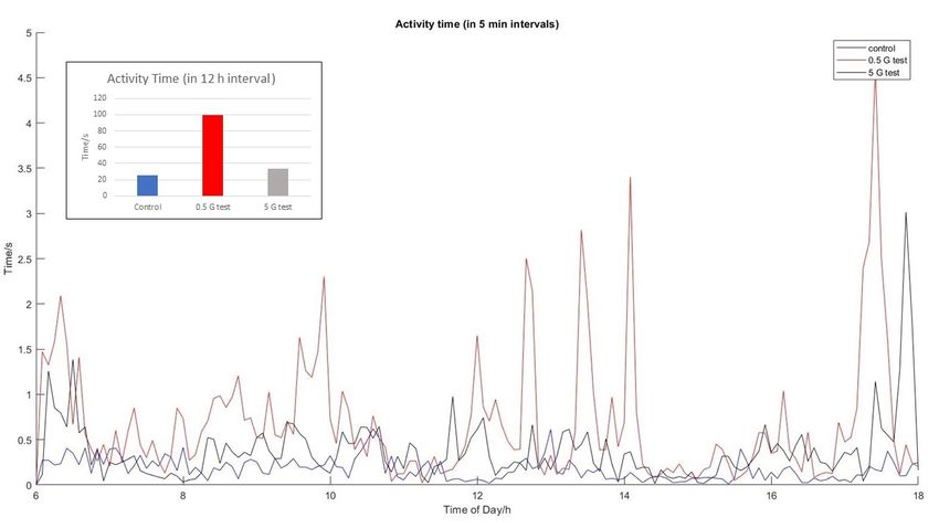
Figure 2. Activity of the American cockroach. The main plot shows the mean activity time, i.e. the time an average cockroach
was translating or rotating. The curves are obtained by connecting points showing the mean activity in the preceding 5 minute
time interval. The inset shows the mean activity accumulated over the time span of the whole experiment (12 hours).

Discussion
The results of the controls show that cockroaches tend to be stationary over the day periods (note that the activity time in Fig. 2
is in seconds). However, the activity is increased when the rotating field has the strength of the geomagnetic field. In a stronger
5 G field this effect is diminished and comparable with the controls. This confirms sensitivity of the American cockroach to the
direction of external magnetic field observed in group of Vacha and indicates that the sensing mechanism is evolutionarily
tuned to the Earth’s field.
     We now discuss two main candidate models of magnetoreception in the light of the obtained results.

Magnetite model
The magnetite model supposes that magnetic materials inside the animal rotate as little compasses in an external magnetic field.
These rotations are transduced to the nervous system and interpreted. The hypothesis gained popularity with the discovery that
certain bacteria are capable of precipitating magnetic grains (either magnetite Fe3 O4 2, 27, 28 or greigite Fe3 S4 29 or both30 ) via
specialised organs. Other animals, including humans, can also mineralise magnetic particles31 . One also verifies plausibility of
this model by noting that magnetic energy of the single domain grain of magnetite with radius 50 nm placed in the Earth’s
magnetic field is almost 10 times larger than the thermal energy at room temperature. Furthermore, the cockroaches were
observed to acquire magnetic moment in the presence of a strong magnetic field, confirming presence of magnetic materials in
their bodies23 . Due to observed long demagnetisation times Kong et al. estimated that a magnetite particle of radius R = 50 nm,
saturation magnetisation Ms = 3 × 10⁵ A / m and mass density ρ = 4.049 × 10³ kg / m3 has to rotate in medium with high
viscosity η = 105 Pa s23 . We now demonstrate that such magnetic grains and their surroundings are disqualified by our data.
    We conducted simulations of a set of 36 spherical magnetic particles with parameters given above. We monitored the
motion in the horizontal plane and initialise the magnetic grains to have magnetisation axes with angular orientations uniformly
distributed between 0 and 2π, i.e. each particle (magnetic moment) is initially rotated by 10 degrees from its neighbours. This
has the macroscopic effect of cancelling out the magnetisation contributions from each individual grain, agreeing with the
experimental observation that cockroaches have no residual magnetisation23 ,
    In the presence of an external magnetic field, the magnetites gradually align toward the field. The rotational motion of the
ith particle is described by Newton’s law:
      I θ̈i = − f θ̇i − µB sin θi ,                                                                                               (1)

                                                                                                                                3/11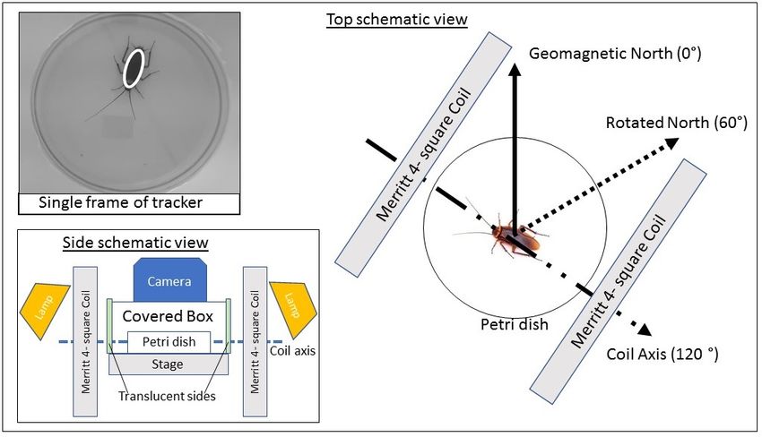
Figure 3. Alignment (normalised magnetisation) in the magnetite model. The dashed line shows the alignment, see Eq. (2),
for a stationary cockroach. The solid colorful lines take into account the actual motion of three typical insects.

where θi is the angle between the external field and the ith magnetic moment, I = 25 ρV R2 is its moment of inertia, and
 f = 8πηR3 the rotational friction coefficient. Note that we have ignored the thermal torque which will additionally misalign
the particles and reduce magnetisation of the whole set. We have simulated the dynamics of the particles for 12 hours changing
the direction of the external field every 5 minutes as in the experiment. Our figure of merit was the “alignment” defined as
normalised total magnetisation:
              n
      M = | ∑ ~mi |/n                                                                                                                (2)
             i=1

where n is the total number of particles and ~mi is the magnetic moment of the ith grain. M is equal to 0 when the individual
moments exactly cancel out and it is given by 1 when all the magnetic moments are parallel.
     The results of our simulations are presented in Fig. 3 and in the Supplementary Information (SI). It turns out that in the
presence of an Earth-strength magnetic field, a highly viscous environment leads to the alignment of only about 40% of all
the magnetites after 12 hours. In the 5 G field, the alignment is complete. This is opposite to experimental findings, where
cockroaches react to rotations of the Earth-strength field but not to rotations in the stronger field. Simulations also show that one
can trade the strength of external field with time. The shape of the alignment curves in 5 G field and in the longer simulations
with 0.5 G field is similar. In SI we show that independently of the strength of the rotating field (0.5 G or 5 G) the particles
(partially) align to the angle of 30 degrees, in between the geomagnetic and rotated fields.
     Additionally, we ask if the motion of the cockroach is an attempt to maintain a certain preferred alignment. For this, let
                                                                     exp
us denote the measured angle of the insect in the jth frame by θ j . We now modify the angle of the magnetic grain at times
                                                                                                                                  exp
t j = j∆t, where ∆t is the time duration between the frames, by adding to them the cockroach motion, i.e. θ j → θ j + θ j .
The simulations show that the motion does not lead to improved alignment. In fact, in the Earth-strength field, the motion
inhibits M which stays below 0.05 level. Note that this holds for both control and test experiments, again in disagreement with
experimental finding that cockroaches behave differently in these two cases.

Radical pair model
The radical-pair mechanism was first conceived in 1969 by Closs, Kaptein and Oosterhoff (CKO model)32, 33 as an explanation of
chemically induced dynamic nuclear polarization, which has ever since been an important technique in NMR spectroscopy34, 35 .
It was later proposed by Schulten et al.14 to be involved in animal magnetoreception.
    The Cryptochrome / Photolyase flavoprotein family are thought to be relevant to light-sensitive biological compasses16 ,
hinting at the radical pair model. We base our discussion on the work by Solov’yov et al.36 who described creation of radical
pairs and their dynamics in Cryptochrome-1 (Cry-1) of the plant Arabidopsis thaliana. While Cry-1 was absent in Periplaneta
americana, Cryptochrome-2 was found to be present and also necessary for magnetic sensitivity26 . The parameters below are
taken from experiments on Photolyase in E. coli (hyperfine axes)37 and Cry-1 in Arabidopsis thaliana (transition rates)38, 39 .
This is justified given that the radical pair mechanism is believed to be contained within the highly conserved FAD molecular
domain40 , common across the Cryptochrome / Photolyase family.
    The radical-pair reaction pathway can be compressed to essential steps given in Fig. 4. The model begins with the creation
of the pair of electrons (each on a different radical molecule) in the state ρ1 , being initially the singlet state. Due to the proximity
of electron-accepting molecules, one of the electrons may hop around, giving rise to a chain of radical pairs ρ1 → ρ2 → ρ3
connected with electron transfer rate ket . This rate has been measured to be on the order of 108 Hz36 . There is also a possibility
of reverse electron transfer, but its rate is estimated to be two orders of magnitude smaller and therefore we ignore it. The whole

                                                                                                                                   4/11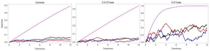
Figure 4. Effective model of the cryptochrome radical pair mechanism. The first radical pair is created in the state ρ1 by an
excitation of the precursor molecule ρ5 . Electron hopping gives rise to other pairs, ρ2 and ρ3 . Their spin dynamics depends on
the external magnetic field and they can recombine to the precursor only if they are in the singlet state. This selective
recombination is represented by the singlet projectors Qs in front of the back-transfer rates, kb , and modifies the yield of the
signalling state ρ4 , perceived by the animal. Variants of the model with solid transitions have been studied previously, see
e.g.36, 41, 42 . We also consider the model with additional dashed arrows, which close the dynamics of the whole system.

system evolves in the external magnetic field ~B and additionally both electrons are coupled to their nearby nuclei, so that the
total Hamiltonian is the sum of the following terms:
                                       ←→      ←→aniso ~
       H j = 2µB~B · ~S j + µB ∑~Ii · ( A iso
                                          i j + A i j ) · S j,                                                               (3)
                              i

where the subscript i = 1, 2 refers to the nuclei, j = 1, 2 to the electrons, and µB is the Bohr magneton. The first term is the
Zeeman interaction of the electron spin ~S j with the external magnetic field. The second terms is the hyperfine interaction with
                                                                                       ←→                                ←
                                                                                                                         →aniso
nuclear spin ~Ii that couples to the electronic spin with diagonal hyperfine tensor A iso  i j and anisotropic tensor A i j . Both of
these have been measured37, 43, 44 and we summarise the values used in our simulations in SI. Due to this interaction, the pair
coherently oscillates between singlet and triplet states and can selectively recombine to the precursor of the radical pairs ρ5 ,
with the estimated rate kb = 107 Hz, only if the pair is in the singlet state. Finally, the last pair in the chain, ρ3 , can decay to the
signalling state ρ4 , with the measured rate kd = 3.3 × 10⁶ Hz. The amount of ρ4 is assumed to be the chemical perceived by the
animal. All the transfers are incoherent processes and are described with the help of the rate equations. The final figure of merit
is the yield of the signalling state Φ being the probability of finding the system in ρ4 after a long time.
     In SI we describe validation of our numerics with analytical estimates and compare our simulations to those of Ref.36 .
Fig. 5 shows Φ as a function of spherical polar and azimuthal angles of the external magnetic field of various magnitudes.
There is a variation of the yield with the direction of the external field for all magnitudes. In particular, for the Earth-strength
field of 0.5 G, the contrast Φmax − Φmin , is around 0.005 and yield is about 0.49, whereas in the 5 G field, we have a contrast of
0.04 and a yield smaller than 0.44. In principle, the result of our experiment could be explained within this model by adding an
assumption that the sensing requires the signalling yield above a threshold value of about 0.45, and that changes in the yield on
the order of one in a thousand are perceivable by the animal.
     The model that was just described begins with the formation of radical pair and ends with the signalling state as a sink.
Many natural processes are closed and we now ask about the possibility of a self-sustaining biocompass that can recycle the
signalling state. For this, we add to the presented radical-pair model two transitions (denoted with dashed magenta lines in
Fig. 4) that close the loop of the process. The transition ρ5 → ρ1 occurs with the rate kex and captures the rate of creation of
the first radical pair, typically as a result of illumination with sunlight. The transition ρ4 → ρ5 occurs with the rate k and
describes conversion of the signalling chemical to the initial precursor molecule. Numerical simulations show that the compass
retains its functionality only if both of these new rates are similar. If k  kex the system accumulates in the precursor state ρ5
and if k  kex the steady state is the signalling state ρ4 independently of the external magnetic field. Fig. 6 shows the results
for exemplary set of kex = 5 × 10⁶ and k = 107 . The variations of the yield are qualitatively similar to those in Fig. 5 but
quantitatively the yield is about ten times smaller and its variations are also correspondingly smaller. This shows that closing
the dynamics has important consequences and makes the radical pair hypothesis rather implausible.

                                                                                                                                   5/11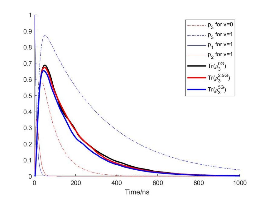
Figure 5. Signalling yield as a function of external magnetic field in the radical pair model with solid arrows in Fig. 4. The
field is parameterised in spherical coordinates ~B = (B0 sin θ cos φ , B0 sin θ sin φ , B0 cos θ ). The hyperfine tensors used in the
simulation were measured using EPR and ENDOR techniques37, 43 . The more complex structures visible for stronger external
fields are the effect of comparable hyperfine interactions (in the range 4 − 16 G) and external fields.

Conclusions
In conclusion, we conducted behavioural experiments on American cockroaches (Periplaneta americana) that confirm their
sensitivity to directional changes of Earth-strength magnetic fields of 0.5 G. The sensitivity is revealed by increased activity of
insects during daytime. Furthermore, the data from experiments in 5 G rotating fields show diminished activity, indicating
adaptation of the sensing mechanism to the Earth’s magnetic field. A similar observation has been made for European robins
exposed to fields 2 times stronger than the Earth’s field45 . We performed numerical analysis of the usual theoretical candidates
of magnetoreception, the magnetite and radical-pair models, in light of the obtained results. Our analyses exclude the magnetite
model with parameters established in previous experiments on cockroaches. They also show that the radical-pair model, with
the parameters presently available from the experiments on Photolyase and Cry-1, can only explain the sensitivity to the

Figure 6. Signalling yield as a function of external magnetic field in the radical pair model with closed dynamics, i.e. with
additional dashed magenta arrows in Fig. 4. The model preserve compass features, but the yield and variations are diminished.

                                                                                                                                6/11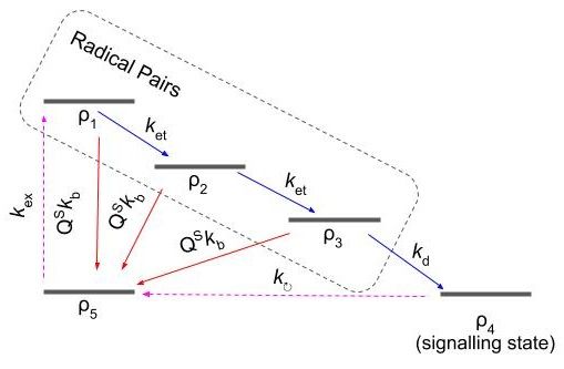
Earth-strength field and its diminishing effect in the stronger field, if we assume that contrast in the chemical yield on the order
of one in a thousand is perceived by the animal, and additionally that this only happens if the yield is above a certain threshold
(attained in 0.5 G but not in 5 G field).

Methods
Experimental procedure
The experiments were conducted in Singapore. Adult female and male cockroaches were kept in separate transparent insectaria
with unlimited water, a diet consisting of cat food pallets and photo-period of 12 light (6am to 6pm) : 12 dark (6pm to 6am)
hours. A day before the experiment, at 6 pm, a single insectarium was placed in a 4◦ C environment in order to immobilise
the insect, which was then moved to a Petri dish (15 cm diameter) with circumference covered with white slip. To minimise
visual cues, the Petri dish was placed in a box with the inside surfaces covered by white paper, except two opposite facing sides
with white translucent films to allow external lights for uniform illumination inside the box. A small aperture was made on the
overhead for the camera configured with a capture rate of 30 frames per second. The box was installed on a stage levelled in the
middle zone (where the generated magnetic field is most uniform) of a Merritt four square coil46 .
    The cockroach was left overnight in an isolated room in order to acclimatise with the new environment. The experiment
automatically begins at 6 am the day after. In the first class of test runs, the magnetic field was alternating between the natural
geomagnetic field in Singapore and the 60 ± 5◦ clockwise-rotated field. This rotation was realised by switching on the Merritt
coil (side dimension 1.2 m), arranged at 120 ± 5◦ from the geomagnetic north. This modifies the horizontal component of the
field while keeping the magnitude and inclination angle of the Earth’s field. In the second class of test experiments we used a
smaller Merritt coil (side dimension 0.3 m) arranged at suitable angle in order to generate the field rotated by 60 ± 5◦ from the
geomagnetic direction (in the horizontal plane) with the strength of 5 G, i.e. about 10 times stronger than the Earth’s magnetic
field. The remaining procedures were the same. The same procedures were also followed in the control runs, except that the
coils were not switched on at all.

Data analysis
The experiments output video recordings of cockroach motion in the Petri dishes. In order to obtain numerical parameters
easily comparable between tests and control runs the videos were processed as follows. The self-written tracking software
identified the cockroach in every frame, fitted an ellipse to it, and stored in a text file timestamp, coordinates of the center of the
ellipse and angle of the main axis. From these values we computed activity time by summing up time intervals between the
frames (33.3 ms) in which the center location changed by more than 3 mm or the angle changed by more than 8 degrees.

Appendix A: Alignment in the magnetite model
Here we show that the magnetite model predicts alignment to the angle between the geomagnetic and rotated fields in both
studied classes of test experiments. The results of the simulations are shown in Fig. 7.

Appendix B: Hyperfine tensors
In our simulations of the radical pair model we used the hyperfine tensors as shown in Tab. 1, which have been measured using
EPR and ENDOR techniques (see compilation in Ref.37 ). Note that these values are different from those used in Ref.36 .

Appendix C: Validation of numerics
We describe here in more detail how we have verified accuracy of our numerics as certain obtained results are different from
those in the literature. In particular, Ref.36 computes the variation in time of the probability that the radical pair ρ3 is in the
singlet / triplet state. Both are found to be at most 0.05 within the first 500 ns of evolution (Fig. 10 of that reference). We now
give analytical estimates of these probabilities which turn out to be an order of magnitude higher and provide the following
intuitive explanation why higher values should be expected. Recall that the electron transfer rate is an order of magnitude faster
than the recombination rates and two orders of magnitude faster than the decay rate to the signalling state. Therefore, all the
dynamics in the chain ρ1 → ρ2 → ρ3 is fast and one expects non-negligible portion of pairs in the state ρ3 .
    In order to place the analytical bounds on the probability that the system is in ρ3 (independently of whether it is the singlet
or triplet state) we note that setting the recombination rate to zero gives the upper bound to the population in ρ3 , whereas
allowing recombination independent of the spin state provides the lower bound on the population. Since in both cases the
system looses spin dependence it is governed by the following set of rate equations, which can be read out from the reaction

                                                                                                                                  7/11
Figure 7. Alignment in the magnetite model. The angular plots show histograms for the orientation angles of the 36 magnetic
particles at different times and different strengths of external magnetic fields as labelled. Initially the particles were distributed
uniformly over the circle. Note that the angle of final alignment is 30◦ . Note also that time for alignment is almost
interchangeable with magnetic field strength, as seen from similarities between the middle and bottom histograms.
scheme in Fig. 4 of the main text:
ṗ1 = (−ket − v kb ) p1 ,
ṗ2 = (−ket − v kb ) p2 + ket p1 ,
ṗ3 = (−kd − v kb ) p3 + ket p2 , (4)
where p j is the population in the jth radical pair and v = 0, 1 turns on and off the possibility of recombination. This set of
equations admits analytical solution:
p1 = exp[(−v kb − ket )t], (5)
p2 = ket t p1 ,
2
ket ket
p3 = 2
[exp ((−v kb − kd )t) − p1 ] − p2 .
(ket − kd ) ket − kd
The corresponding limiting curves (for v = 0 and v = 1) are plotted in Fig. 8 (dashed-dotted lines). The plot confirms that
the populations p1 and p2 quickly decay to zero and shows that the number of radical pairs ρ3 lies well within the obtained
boundaries. Note that the results in Ref.36 are below the analytical lower bound obtained here.
Acknowledgements
We would like to thank Zhang Wei for preparing the first version of the magnetic coils, Anthony Tan and Ng Hong Kuan for
writing and testing the tracking software, Agnieszka Górecka and Herbert Crepaz for assistance in early experiments, and
Dagomir Kaszlikowski for quantum biology spirit. This work is supported by the Singapore Ministry of Education Tier 1 Grant
No. RG 127/14 and the Polish National Agency for Academic Exchange NAWA Project No. PPN/PPO/2018/1/00007/U/00001.
8/11First electron interacts with one nucleus
Aiso
11 [G] Aaniso
11 [G] hyperfine axes
3.93 -4.98 0.4380 0.8655 -0.2432
-4.92 0.8981 -0.4097 0.1595
9.89 -0.0384 0.2883 0.9568
Second electron interacts with two nuclei
Aiso
12 [G] Aaniso
12 [G] hyperfine axes
13.6 0 1 0 0
0 0 1 0
0 0 0 1
Aiso
22 [G] A aniso [G]
22 hyperfine axes
-4 -0.23 -0.984 0.180 0
0.35 0.180 0.984 0
-0.12 0 0 1
Table 1. Hyperfine tensors used in our simulations of the radical pair model.
Figure 8. Population dynamics of the radical pairs. The solid curves are computed with the rates and the hyperfine tensors as
in Ref.36 . The population of the first two radical pairs is transferred to the third pair within 50 ns. The population of the third
pair is plotted in thick lines for various strengths of external magnetic field. All of them lie within the obtained analytical
bounds shown by dashed-dotted lines.
Author contributions statement
All authors researched and wrote this paper.
Additional information
Processed data from tracking software is available on repository at: osf.io/zk9d6.
Competing Interests: The authors declare no competing interests.
References
1. Weiss, N. Dynamos in planets, stars and galaxies. Astron. Geophys. 43, 3.09–3.15 (2002).
2. Blakemore, R. Magnetotactic bacteria. Science 190, 377–379 (1975).
3. Pan, W. et al. Evidence for the presence of biogenic magnetic particles in the nocturnal migratory brown planthopper,
nilaparvata lugens. Sci. Reports 6 (2016).
9/114. Kirschvink, J. L. The horizontal magnetic dance of the honeybee is compatible with a single-domain ferromagnetic
magnetoreceptor. Biosystems 14, 193–203 (1981).
5. Kirschvink, J. L. & Kirschvink, A. K. Is geomagnetic sensitivity real? replication of the walker-bitterman magnetic
conditioning experiment in honey bees. Am. Zool. 31, 169–186 (1991).
6. Kirschvink, J., Padmanabha, S., Boyce, C. & Oglesby, J. Measurement of the threshold sensitivity of honeybees to weak,
extremely low-frequency magnetic fields. J. Exp. Biol. 200, 1363–1368 (1997).
7. Walker, M. M., Kirschvink, J. L., Chang, S.-B. R. & Dizon, A. E. A candidate magnetic sense organ in the yellowfin tuna,
thunnus albacares. Science 224, 751–753 (1984).
8. Mann, S., Sparks, N. H., Walker, M. M. & Kirschvink, J. L. Ultrastructure, morphology and organization of biogenic
magnetite from sockeye salmon, oncorhynchus nerka: implications for magnetoreception. J. Exp. Biol. 140, 35–49 (1988).
9. Tian, L. et al. Testing for the presence of magnetite in the upper-beak skin of homing pigeons. BioMetals 20, 197–203
(2006).
10. Holland, R. A., Kirschvink, J. L., Doak, T. G. & Wikelski, M. Bats use magnetite to detect the earth’s magnetic field.
PLoS ONE 3, e1676 (2008).
11. Tian, L., Lin, W., Zhang, S. & Pan, Y. Bat head contains soft magnetic particles: Evidence from magnetism.
Bioelectromagnetics 31, 499–503 (2010).
12. Wiltschko, R. & Wiltschko, W. Magnetoreception. BioEssays 28, 157–168 (2006).
13. Kirschvink, J. L. & Gould, J. L. Biogenic magnetite as a basis for magnetic field detection in animals. Biosystems 13,
181–201 (1981).
14. Schulten, K., Swenberg, C. E. & Weller, A. A biomagnetic sensory mechanism based on magnetic field modulated coherent
electron spin motion. Zeitschrift für Physikalische Chemie 111, 1–5 (1978).
15. Hore, P. J. & Mouritsen, H. The radical-pair mechanism of magnetoreception. Annu. Rev. Biophys. 45, 299–344 (2016).
16. Ritz, T., Adem, S. & Schulten, K. A model for photoreceptor-based magnetoreception in birds. Biophys. J. 78, 707–718
(2000).
17. Munro, U., Munro, J. A., Phillips, J. B., Wiltschko, R. & Wiltschko, W. Evidence for a magnetite-based navigational
"map" in birds. Naturwissenschaften 84, 26–28 (1997).
18. Krichen, S., Liu, L. & Sharma, P. Biological cell as a soft magnetoelectric material: Elucidating the physical mechanisms
underpinning the detection of magnetic fields by animals. Phys. Rev. E 96, 042404 (2017).
19. Johnsen, S. & Lohmann, K. J. The physics and neurobiology of magnetoreception. Nat. Rev. Neurosci. 6, 703–712 (2005).
20. Thalau, P., Ritz, T., Stapput, K., Wiltschko, R. & Wiltschko, W. Magnetic compass orientation of migratory birds in the
presence of a 1.315 MHz oscillating field. Naturwissenschaften 92, 86–90 (2004).
21. Vacha, M., Puzova, T. & Kvicalova, M. Radio frequency magnetic fields disrupt magnetoreception in american cockroach.
J. Exp. Biol. 212, 3473–3477 (2009).
22. Li, S. et al. The genomic and functional landscapes of developmental plasticity in the american cockroach. Nat. Commun.
9 (2018).
23. Kong, L. J. et al. In-vivo biomagnetic characterisation of the american cockroach. Sci. Reports 8 (2018).
24. Vacha, M. Laboratory behavioural assay of insect magnetoreception: magnetosensitivity of periplaneta americana.
J. Exp. Biol. 209, 3882–3886 (2006).
25. Vacha, M., Kvicalova, M. & Puzova, T. American cockroaches prefer four cardinal geomagnetic positions at rest. Behaviour
147, 425–440 (2010).
26. Bazalova, O. et al. Cryptochrome 2 mediates directional magnetoreception in cockroaches. Proc. Natl. Acad. Sci. 113,
1660–1665 (2016).
27. Frankel, R. B., Blakemore, R. P. & Wolfe, R. S. Magnetite in freshwater magnetotactic bacteria. Science 203, 1355–1356
(1979).
28. Balkwill, D. L., Maratea, D. & Blakemore, R. P. Ultrastructure of a magnetotactic spirillum. J. bacteriology 141 3,
1399–408 (1980).
29. Heywood, B. R., Bazylinski, D. A., Garratt-Reed, A., Mann, S. & Frankel, R. B. Controlled biosynthesis of greigite
(Fe3 S4 ) in magnetotactic bacteria. Naturwissenschaften 77, 536–538 (1990).
10/1130. Bazylinski, D. A. et al. Controlled biomineralization of magnetite (Fe3 O4 ) and greigite (Fe3 S4 ) in a magnetotactic
bacterium. Appl. environmental microbiology 61 9, 3232–9 (1995).
31. Kirschvink, J. L., Kobayashi-Kirschvink, A. & Woodford, B. J. Magnetite biomineralization in the human brain.
Proc. Natl. Acad. Sci. 89, 7683–7687 (1992).
32. Closs, G. L. Mechanism explaining nuclear spin polarizations in radical combination reactions. J. Am. Chem. Soc. 91,
4552–4554 (1969).
33. Kaptein, R. & Oosterhoff, J. Chemically induced dynamic nuclear polarization II. Chem. Phys. Lett. 4, 195–197 (1969).
34. Buchachenko, A. L. Magnetic isotope effect: nuclear spin control of chemical reactions. The J. Phys. Chem. A 105,
9995–10011 (2001).
35. Goez, M. Elucidating organic reaction mechanisms using photo-CIDNP spectroscopy. In
Hyperpolarization Methods in NMR Spectroscopy, 1–32 (Springer Berlin Heidelberg, 2012).
36. Solov’yov, I. A., Chandler, D. E. & Schulten, K. Magnetic field effects in arabidopsis thaliana cryptochrome-1. Biophys. J.
92, 2711–2726 (2007).
37. Cintolesi, F., Ritz, T., Kay, C., Timmel, C. & Hore, P. Anisotropic recombination of an immobilized photoinduced radical
pair in a 50-µt magnetic field: a model avian photomagnetoreceptor. Chem. Phys. 294, 385–399 (2003).
38. Byrdin, M. et al. Intraprotein electron transfer and proton dynamics during photoactivation of DNA photolyase from e.
coli: review and new insights from an “inverse” deuterium isotope effect. Biochimica et Biophys. Acta (BBA) - Bioenerg.
1655, 64–70 (2004).
39. Aubert, C., Vos, M. H., Mathis, P., Eker, . P. M. & Brettel, K. Intraprotein radical transfer during photoactivation of DNA
photolyase. Nature 405, 586–590 (2000).
40. Mei, Q. & Dvornyk, V. Evolutionary history of the photolyase/cryptochrome superfamily in eukaryotes. PLOS ONE 10,
e0135940 (2015).
41. Chia, A. et al. Coherent chemical kinetics as quantum walks. I. reaction operators for radical pairs. Phys. Rev. E 93 (2016).
42. Chia, A., Górecka, A., Kurzyński, P., Paterek, T. & Kaszlikowski, D. Coherent chemical kinetics as quantum walks. II.
radical-pair reactions in Arabidopsis thaliana. Phys. Rev. E 93 (2016).
43. Kay, . W. M. et al. EPR, ENDOR, and TRIPLE resonance spectroscopy on the neutral flavin radical in Escherichia coli
DNA photolyase†. Biochemistry 38, 16740–16748 (1999).
44. Weber, S., Möbius, K., Richter, G. & Kay, C. W. M. The electronic structure of the flavin cofactor in DNA photolyase.
J. Am. Chem. Soc. 123, 3790–3798 (2001).
45. Wiltschko, W., Stapput, K., Thalau, P. & Wiltschko, R. Avian magnetic compass: fast adjustment to intensities outside the
normal functional window. Naturwissenschaften 93, 300–304 (2006).
46. Merritt, R., Purcell, C. & Stroink, G. Uniform magnetic field produced by three, four, and five square coils.
Rev. Sci. Instruments 54, 879–882 (1983).
11/11You can also read