Original Article Abnormality of intestinal cholesterol absorption in ApcMin/+ mice with colon cancer cachexia
←
→
Page content transcription
If your browser does not render page correctly, please read the page content below
Int J Clin Exp Pathol 2019;12(3):759-767
www.ijcep.com /ISSN:1936-2625/IJCEP0090647

Original Article
Abnormality of intestinal cholesterol absorption in
ApcMin/+ mice with colon cancer cachexia
Biao Yu*, Xiao-Huan Peng*, Ling-Yu Wang, An-Bei Wang, Yan-Yan Su, Jia-Huan Chen, Xin-Wei Zhang,
Da-Zhong Zhao, He Wang, Da-Xin Pang, Hong-Sheng Ouyang, Xiao-Chun Tang, Ming-Jun Zhang

Jilin Provincial Key Laboratory of Animal Embryo Engineering, College of Animal Sciences, Jilin University, Changc-
hun, Jilin Province, China. *Equal contributors.
Received December 30, 2018; Accepted January 24, 2019; Epub March 1, 2019; Published March 15, 2019

Abstract: Colorectal cancer syndrome has been one of the greatest concerns in the world, particularly in developed
countries. Several epidemiological studies have shown that dyslipidemia may be associated with the progression
of intestinal cachexia, but there is little research on the function of the small intestine, which is involved in blood
lipid metabolism, in dyslipidemia. In the present study, we aimed to explore the function of intestinal cholesterol
absorption in the ApcMin/+ mouse model using an intestinal lipid absorption test. We found that both triglyceride
(TG) and total cholesterol (TC) uptake were inhibited in the intestine of ApcMin/+ mice with age and the intestinal
peroxisome proliferator-activated receptor α (PPARα) downregulated the processes of β-oxidation, oxidative stress
response, and cholesterol absorption in APC-deficient mice. In addition, reduced expression levels of farnesoid X
receptor (FXR) and apical sodium-dependent bile acid transporter (ASBT) indicated that bile acid metabolism might
be associated with intestinal cholesterol absorption in ApcMin/+ mice. Thus, our data suggested that the intestine
plays an essential role in cholesterol uptake and that bile acid metabolism seems to cause a decrease in intestinal
cholesterol uptake in ApcMin/+ mice.

Keywords: Adenomatous polyposis coli (APC), cholesterol absorption, dyslipidemia, peroxisome proliferator-acti-
vated receptor α (PPARα), bile acid

Introduction                                                    Epithelial cells are an important part of the lipid
                                                                balance in the intestine. Not only can epithelial
Colorectal cancer (CRC) syndrome has been                       cells of the small intestine absorb approximate-
one of the greatest concerns in the world, par-                 ly 95% triglycerides (TGs), but they can also
ticularly in developed countries [1]. A classic                 store TGs within cytosolic lipid droplets (CLDs)
animal model of human familial adenomatous                      [5, 6]. In addition, the absorbed TGs are decom-
polyposis (FAP) is the C57BL/6J-ApcMin/+ mouse,                 posed in the lumen of the gut, resulting in FFAs
and this mouse has a truncated mutation in the                  that are activated, esterified and released into
adenomatous polyposis coli (APC) gene, which                    lymphatic circulation [7, 8]. CLDs play a core
results in multiple intestinal adenoma polyps                   role in the control of enterocyte triglyceride-rich
and is associated with the loss of muscle and                   lipoprotein (TRL) secretion [9].
fat accompanying hyperlipidemia [2, 3]. Hyper-
lipidemia has been reported to be closely asso-                 Previous studies have shown that peroxisome
ciated with the development of intestinal ade-                  proliferator-activated receptor α (PPARα) can
noma polyps in the ApcMin/+ mouse [4]. Previous                 regulate the serum level of TGs in ApcMin/+ mice
studies have shown that a low level of intestinal               through administrating the PPARα ligand bezafi-
lipoprotein lipase (LPL) mRNA expression may                    brate [2]. Interestingly, recent studies have
be involved in dyslipidemia and tumor progres-                  emphasized the importance of PPARα in regu-
sion [2]. However, what we often ignore is the                  lating several important processes such as
role of the small intestine in the process of                   β-oxidation, the oxidative stress response and
blood lipid metabolism.                                         cholesterol absorption in the murine small
Intestinal cholesterol absorption in colorectal cancer

Table 1. Primer pairs used for the qRT-PCR analysis                             care and use of laboratory
Target gene   Primer sequence (5’-3’)     Reverse primer sequence (5’-3’)       animals (NIH Publications
                                                                                No. 8023, revised 1978).
AOX           CCTGTTGGCCTCAATTACTC        GGTCATATGTGGCAGTGGTT
                                                                                In addition, the procedures
ACOT1         GGAGTTGGAGGTGGCCTTCT        CGCAGGTAGTTCACGGCTTC
                                                                                were approved by the In-
ACOT2         GCACGAGCGTCACTTCTTGG        CCGATACTCCAGAAGGCCAC                  stitutional Animal Care and
ACAA2         GGACTTCTCTGCACCGATT         AGAGCCACAGAGCCTGTTGA                  Use Committee of Jilin Un-
GSTK1         AAGCAGTTCTTCCAGGTTCC        CCAGAATGCTCTGATACTCC                  iversity under approved pro-
GSTM3         ATGCCATCCTGCGCTACCT         CCAGGAACTCAGAGTAGAGC                  tocol number 201707025.
GSST          CTGTACCTGGATCTGCTGTC        TAGCCACACTCTCACACAGG
NPC1L1        TGTCCCCGCCTATACAATGG        CCTTGGTGATAGACAGGCTACTG               Intestinal lipid absorption
                                                                                test
CD36          GCAGGTCTATCTACGCTGTG        GGTTGTCTGGATTCTGGAGG
MTTP          GTCAACAGAGAGGCGAGAAG        CTAGCCAAGCCTCTCTTGAG                 At the periods of 8, 14 and
ABCA1         CTCTTCATGACTCTAGCCTGGA      ACACAGACAGGAAGACGAACAC               20 weeks, the mice were
ABCG5         AGAGGGCCTCACATCAACAGA       CTGACGCTGTAGGACACATGC                fasted for 4 hours starting at
ABCG8         AGTGGTCAGTCCAACACTCTG       GAGACCTCCAGGGTATCTTGAA               05:00 prior to undergoing
FXR           GCTTGATGTGCTACAAAAGCTG      CGTGGTGATGGTTGAATGTCC                the intestinal lipid absorption
ASBT          GTACAATGGTGGAGCACAGC        GTGCCTGGATCATTGAACCC                 test. Thirty minutes after
GAPDH         TTGTCTCCTGCGACTTCA          CACCACCCTGTTGCTGTA                   injection, the mice were
                                                                               gavaged with 200 μl of olive
                                                                               oil to assess dietary fat
intestine, which may represent risk factors for            absorption. Blood was sampled via the tail vein
hyperlipidemia [10-12]. However, there are few             at the baseline of 0 h and at 3 and 6 h, and the
studies on the roles of β-oxidation, the oxida-            blood was centrifuged at 2000×g for 10 min-
tive stress response and cholesterol absorp-               utes at 4°C. Fasting plasma total cholesterol
tion in hyperlipidemia; therefore, the exact               (TC) and triglycerides (TGs) were analyzed using
mechanism remains unclear.                                 commercial kits from Biosino (Beijing, China)
                                                           [13].
Thus, whether PPARα signaling regulates intes-
tinal cholesterol absorption in ApcMin/+ mice and          Intestinal permeability assay
the exact mechanism of its action are unclear.
Therefore, we explored the intestinal choles-              FITC-dextran (4 kDa, Sigma, USA) was adminis-
terol absorption ability of ApcMin/+ mice using an         tered by oral gavage (60 mg/100 g body weight,
intestinal lipid absorption test. The aim of this          40 mg/mL) to the fasted mice. After 1 hour,
study was to reveal the potential reason for               blood was collected, stored on ice in the dark
lipid absorption abnormalities in the intestine            and centrifuged 1000×g for 15 minutes at 4°C.
of ApcMin/+ mice.                                          The serum was diluted with the same volume of
                                                           PBS, and the fluorescence intensity was mea-
Materials and methods                                      sured using a fluorescence spectrophotometer
                                                           (λex: 485 nm; λem: 535 nm, Infinite 200 Pro,
Animals                                                    Tecan, Switzerland) [14].
Four- to five-week-old wild-type male C57BL/6J             Gut transit test
(WT, n=20) and mutant male C57BL/6J-ApcMin/+
(ApcMin/+, Min, n=20) mice were purchased from             Overnight-fasted mice were gavaged with 200
the Nanjing Biomedical Research Institute of               μl of Evans blue suspension (5% Evans blue
Nanjing University (Nanjing, China). The mice              and 5% gum arabic in PBS). Afterward, the mice
were provided standard rodent chow and water               had free access to food and water, and the time
ad libitum and housed in cages (≤5 mouse per               until the Evans blue was detected in the feces
cage) that were placed in an SPF animal facility           was recorded [13].
with the laboratory temperature maintained at
22°C and 40-60% humidity with a 12:12 light:               Oil red O staining
dark cycle. All animal welfare and experimental
procedures were performed strictly according               For oil red O staining, the jejunum was isolated
to the National Institutes of Health guide for the         and fixed in 4% neutral-buffered formalin (Carl

760                                                              Int J Clin Exp Pathol 2019;12(3):759-767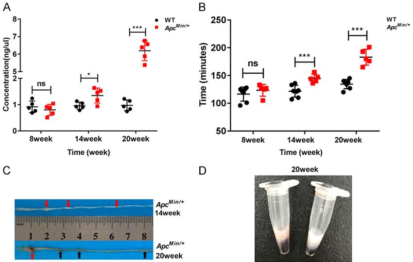
Intestinal cholesterol absorption in colorectal cancer

                                                                                 Figure 2. Intestinal lipid absorp-
                                                                                 tion inhibition occurs in ApcMin/+
                                                                                 mice. A. Study schematic of the
                                                                                 intestinal lipid absorption test
                                                                                 at 8, 14 and 20 weeks of age in
                                                                                 mice. B and C. Plasm triglyceride
                                                                                 (TG) and total cholesterol (TC) lev-
                                                                                 els were determined by tail vein
                                                                                 nick in the ApcMin/+ and WT mice
                                                                                 at 0-, 3- and 6-h time points at 8,
                                                                                 14 and 20 weeks (n≥5). Data are
                                                                                 the mean of three independent
                                                                                 experiments (an average of five
                                                                                 readings was conducted for each
                                                                                 sample), Mean ± SEM. The differ-
                                                                                 ences between the mean values
                                                                                 were assessed by Student’s t-
                                                                                 tests and analyzed using Graph-
                                                                                 Pad Prism software 7.0. *P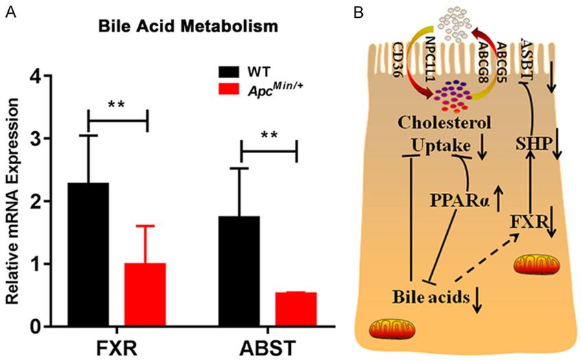
Intestinal cholesterol absorption in colorectal cancer

                                                                               Results

                                                                               The intestines that were exam-
                                                                               ined in our study were taken
                                                                               from ApcMin/+ mice and classi-
                                                                               fied as noncachectic (8 wee-
                                                                               ks of age), precachectic (14
                                                                               weeks of age) and severely
                                                                               cachectic (20 weeks of age).

                                                                               Gut barrier dysfunction in Ap-
                                                                               cMin/+ mice with colon cancer
                                                                               cachexia

                                                                                  To objectively assess intesti-
                                                                                  nal dysfunction in ApcMin/+
                                                                                  mice, the permeability of FITC-
                                                                                  dextran (4 kDa) was investi-
                                                                                  gated. It was remarkable that
                                                                                  the concentration of FITC-
                                                                                  dextran increased by nearly
                                                                                  1.5-fold at 14 weeks of age
                                                                                  (ApcMin/+ mouse group versus
                                                                                  wild-type mouse group: 1.35 ±
Figure 3. Lipid droplets accumulate in the jejunum of ApcMin/+ mice. Oil red O    0.29 ng/μL versus 0.96 ±
staining revealed the accumulation of lipid droplets (red arrow) in the intes-    0.13 ng/μL, P
Intestinal cholesterol absorption in colorectal cancer Figure 4. APC deficiency results in the downregulation of intestinal PPARα target genes. A. LPL and PPARα proteins were measured by western blot with specific antibodies in the intestine of the ApcMin/+ and WT mice (WT: wild type mouse, Min: ApcMin/+ mouse, n=3). B-D. Aox, Acot1, Acot2, Acaa2, Gstk1, Gstm3, Gsst, Npc1l1, CD36, Mttp, Abca1, Abcg5 and Abcg8 were measured by qRT-PCR in the intestine of ApcMin/+ and the WT mice (n=3). Data are expressed as the Mean ± SEM. The differences between the mean values were assessed by Student’s t-tests and analyzed using GraphPad Prism software 7.0. *P
Intestinal cholesterol absorption in colorectal cancer
oxidase (AOX), acyl-CoA thio-
esterase 1 (ACOT1), acyl-CoA
thioesterase 2 (ACOT2) and
acetyl-CoA acyltransferase 2
(ACAA2); oxidative stress re-
sponse (ROS), including gluta-
thione S-transferase kappa 1
(GSTK1), glutathione S-tran-
sferase mu 3 (GSTM3) and
glutathione S-transferase the-
ta (GSST); lipid absorption,
including Niemann-Pick C1
like-1 (NPC1L1), cluster of dif-
ferentiation 36 (CD36), mi-
crosomal triglyceride transfer
protein (MTTP), ATP-binding
cassette subfamily A member
1 (ABCA1), ATP-binding cas-
sette subfamily G member 5
(ABCG5) and ATP-binding cas-
sette subfamily G member 8
(ABCG8). The expression of
Aox, Acot1, Acaa2, Gstm3,
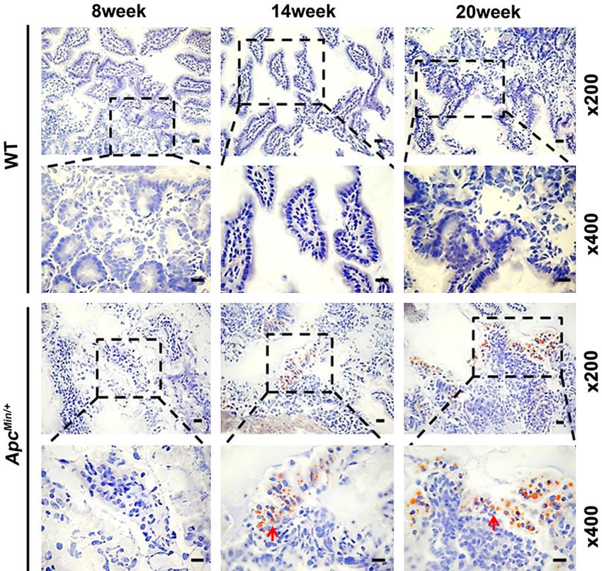
Figure 5. Schematic of fatty acid β-oxidation, oxidative stress response and Gsst, Npc1l1, CD36, Mttp,
cholesterol absorption processes in the enterocytes of mouse. The main Abca1 and Abcg5 was mark-
cholesterol importer NPC1L1 and the cholesterol exporters ABCG5/G8 are edly downregulated in the jeju-
located at the apical membrane of enterocytes and facilitate the uptake of num of the ApcMin/+ mice.
cholesterol across the brush border membrane. ACAT2 esterifies the ab-
sorbed cholesterol, and MTTP transfers triglycerides and cholesteryl esters There was no change in the
to ApoB48 in the smooth ER. The nascent chylomicrons leave the ER, are expression of Acot2, Gstk1
secreted through the Golgi complex to the basolateral side of the entero- and Abcg8 between the two
cyte and reach the venous circulation through lymphatic vessels. In addition groups (Figure 4B-D). The
to the chylomicron pathway, a significant portion of intestinal xanthophylls
are absorbed through an ABCA1/ApoA1 pathway and may be preferentially
results indicated that APC is
delivered to some tissues. The absorption of dietary cholesterol through very important for PPARα acti-
the apical membrane into enterocytes is associated with β-oxidation and/ vation in the jejunum. A sche-
or oxidative stress response. NPC1L1: Niemann-Pick C1 like-1; ABCG5/G8: matic diagram of the fatty acid
ATP-binding cassette transporter G5/G8; CD36: cluster of differentiation 36; β-oxidation, oxidative stress
apoB48: apolipoprotein B48; MTTP: microsomal triglyceride transfer protein;
ACAT2: acyl-coenzyme A cholesterol acyltransferase 2; ER: endoplasmic re- response, and cholesterol ab-
ticulum; Golgi: Golgi apparatus; ABCA1: ATP-binding cassette transporter sorption processes in the
A1; HDL: high-density lipoprotein; AOX: acyl-CoA oxidase; ACOT1/2: acyl-CoA enterocytes of mice is shown
thioesterase 1/2; ACAA2: acetyl-CoA acyltransferase 2; GSTK1: glutathione in Figure 5.
S-transferase kappa 1; GSTM3: glutathione S-transferase mu 3; GSST: glu-
tathione S-transferase theta; OS: oxidative stress; PPARα: peroxisome prolif-
Bile acids affect intestinal
erator-activated receptor α; FXR: farnesoid X receptor; ASBT: apical sodium-
dependent bile acid transporter; SHP: short heterodimer partner. cholesterol absorption in
ApcMin/+ mice
reported, a low level of LPL mRNA expression CD36 plays an essential role in the uptake of
may be associated with dyslipidemia and FFAs and cholesterol from the intestinal lumen
involved in PPARα signaling in the intestine of and is regulated by PPARα [16, 17]. In our study,
ApcMin/+ mice [2]. We repeated the experiment decreased CD36, NPC1L1, ABCG5 and ABCA1
and used a western blot analysis in the jejunum mRNA levels might have been involved in the
of the ApcMin/+ mice (Figure 4A). Moreover, we cholesterol absorption inhibition in ApcMin/+
determined the expression of intestinal PPARα mice. To further determine whether bile acids
target genes: β-oxidation, including acyl-CoA can regulate cholesterol absorption, we ana-
764 Int J Clin Exp Pathol 2019;12(3):759-767Intestinal cholesterol absorption in colorectal cancer
dextran (4 kDa) in the intes-
tine along with a delay in gut
transit with age. Similarly,
research by Puppa et al. sug-
gested that glucose tolerance,
plasma IL-6, TGs, and body
temperature are characteris-
tic of endotoxemia, which is
accompanied with a change in
gut permeability in ApcMin/+
mice [19]. Another study re-
vealed that some inflamma-
tion occurring in intestinal epi-
thelial cells can decrease gut
homeostasis [20]. In fact, in-
Figure 6. Bile acids affected intestinal cholesterol absorption in ApcMin/+ testinal lipid absorption plays
mice. A. FXR and ASBT were measured by qRT-PCR in the intestines of Ap- an essential role in gut lip-
cMin/+ and WT mice (n=3). Data are expressed as the mean ± SEM. The dif- id homeostasis. Our findings
ferences between the mean values were assessed by Student’s t-tests and demonstrated that the lipid
analyzed using GraphPad Prism software 7.0. **PIntestinal cholesterol absorption in colorectal cancer
CD36, NPC1L1, ABCG5 and ABCA1 mRNA [2] Niho N, Takahashi M, Kitamura T, Shoji Y, Itoh
might be involved in cholesterol absorption in M, Noda T, Sugimura T, Wakabayashi K. Con-
the jejunum of ApcMin/+ mouse. comitant suppression of hyperlipidemia and
intestinal polyp formation in apc-deficient mice
Studies have shown that bile acids, as ligands by peroxisome proliferator-activated receptor
for the bile acid receptor farnesoid X receptor ligands. Cancer Res 2003; 63: 6090-6095.
[3] Niho N, Takahashi M, Shoji Y, Takeuchi Y, Mat-
(FXR), alter the transcription of several genes
subara S, Sugimura T, Wakabayashi K. Dose-
that are involved in triglyceride synthesis and dependent suppression of hyperlipidemia and
lipid metabolism [27]. Early clinical studies intestinal polyp formation in Min mice by pio-
have found that PPARα regulates bile acid syn- glitazone, a PPAR gamma ligand. Cancer Sci
thesis, bile acid transport and cholesterol 2003; 94: 960-4.
metabolism pathways [28]. In addition, PPARα [4] Schwarz JM, Linfoot P, Dare D, Aghajanian K.
has been confirmed to be activated in the intes- Hepatic de novo lipogenesis in normoinsulin-
tine of ApcMin/+ mice. Furthermore, our results emic and hyperinsulinemic subjects consum-
indicated that decreased expression of FXR ing high-fat, low-carbohydrate and low-fat,
high-carbohydrate isoenergetic diets. Am J Clin
and ASBT in the jejunum modulates bile acid
Nutr 2003; 77: 43-50.
metabolism in ApcMin/+ mice. Thus, we specu- [5] Whitcomb DC, Lowe ME. Human pancreatic di-
late that the change in cholesterol absorption gestive enzymes. Dig Dis Sci 2007; 52: 1-17.
might be a consequence of a reduced amount [6] Lee B, Zhu J, Wolins NE, Cheng JX, Buhman
of bile acids in the jejunum of ApcMin/+ mice. KK. Differential association of adipophilin and
TIP47 proteins with cytoplasmic lipid droplets
In conclusion, our data suggested that the in mouse enterocytes during dietary fat ab-
intestine plays an essential role in cholesterol sorption. Biochim Biophys Acta 2009; 1791:
uptake and that bile acid metabolism seems to 1173-80.
cause a decrease in intestinal cholesterol [7] Hussain MM, Pan X. Circadian regulators of in-
uptake in ApcMin/+ mice. testinal lipid absorption. J Lipid Res 2015; 56:
761-70.
Acknowledgements [8] Holt PR, Balint JA. Effects of aging on intestinal
lipid absorption. Am J Physiol 1993; 264: G1-
This work was financially supported by the 6.
[9] Bouchoux J, Beilstein F, Pauquai T, Guerrera IC,
National Natural Science Foundation of China
Chateau D, Ly N, Alqub M, Klein C, Chambaz J,
(Grant No. 31472053 and 31572345), Gra- Rousset M, Lacorte JM, Morel E, Demignot S.
duate Innovation Fund of Jilin University (Grant The proteome of cytosolic lipid droplets isolat-
No. 2017094), Program for JLU Science and ed from differentiated caco-2/TC7 enterocytes
Technology Innovative Research Team (JL- reveals cell-specific characteristics. Biol Cell
USTIRT, No. 2017TD-28), and Fundamental 2011; 103: 499-517.
Research Funds for the Central Universities. [10] de Vogel-van den Bosch HM, Bünger M, de
Groot PJ, Bosch-Vermeulen H, Hooiveld GJ,
Disclosure of conflict of interest Müller M. PPARalpha-mediated effects of di-
etary lipids on intestinal barrier gene expres-
None. sion. BMC Genomics 2008; 9: 231.
[11] van den Bosch HM, Bünger M, de Groot PJ, van
Address correspondence to: Ming-Jun Zhang, Jilin der Meijde J, Hooiveld GJ, Müller M. Gene ex-
Provincial Key Laboratory of Animal Embryo pression of transporters and phase I/II meta-
Engineering, College of Animal Sciences, Jilin bolic enzymes in murine small intestine during
University, 5333 Xi’an Road, Lvyuan District, fasting. BMC Genomics 2007; 8: 267.
Changchun 130062, Jilin Province, China. Tel: (86) [12] Bünger M, van den Bosch HM, van der Meijde
431-87836122; Fax: (86) 431-86758018; E-mail: J, Kersten S, Hooiveld GJ, Müller M. Genome-
mjzhang@jlu.edu.cn wide analysis of PPARalpha activation in mu-
rine small intestine. Physiol Genomics 2007;
References 30: 192-204.
[13] Obrowsky S, Chandak PG, Patankar JV, Povo-
[1] Takahashi H, Hosono K, Endo H, Nakajima A. den S, Schlager S, Kershaw EE, Bogner-Strauss
Colon epithelial proliferation and carcinogene- JG, Hoefler G, Levak-Frank S, Kratky D. Adipose
sis in diet-induced obesity. J Gastroenterol triglyceride lipase is a TG hydrolase of the
Hepatol 2013; 28 Suppl 4: 41-7. small intestine and regulates intestinal PPA-
766 Int J Clin Exp Pathol 2019;12(3):759-767Intestinal cholesterol absorption in colorectal cancer
Ralpha signaling. J Lipid Res 2013; 54: 425- [22] Zhou X, Cao L, Jiang C, Xie Y, Cheng X, Krausz
35. KW, Qi Y, Sun L, Shah YM, Gonzalez FJ, Wang
[14] Yang R, Han X, Uchiyama T, Watkins SK, Yagu- G, Hao H. PPARalpha-UGT axis activation re-
chi A, Delude RL, Fink MP. IL-6 is essential for presses intestinal FXR-FGF15 feedback signal-
development of gut barrier dysfunction after ling and exacerbates experimental colitis. Nat
hemorrhagic shock and resuscitation in mice. Commun 2014; 5: 4573.
Am J Physiol Gastrointest Liver Physiol 2003; [23] Howles PN, Carter CP, Hui DY. Dietary free and
285: G621-G629. esterified cholesterol absorption in cholesterol
[15] Davis MR, Arner E, Duffy CR, De Sousa PA, esterase (bile salt-stimulated lipase) gene-tar-
Dahlman I, Arner P, Summers KM. Expression geted mice. J Biol Chem 1996; 271: 7196-202.
of FBN1 during adipogenesis: relevance to the [24] Febbraio M, Guy E, Coburn C, Knapp FF Jr,
lipodystrophy phenotype in marfan syndrome Beets AL, Abumrad NA, Silverstein RL. The im-
and related conditions. Mol Genet Metab pact of overexpression and deficiency of fatty
2016; 119: 174-85. acid translocase (FAT)/CD36. Mol Cell Bio-
[16] Uchida A, Slipchenko MN, Cheng JX, Buhman chem 2002; 239: 193-7.
KK. Fenofibrate, a peroxisome proliferator-acti- [25] Yamanashi Y, Takada T, Yoshikado T, Shoda J,
vated receptor alpha agonist, alters triglycer- Suzuki H. NPC2 regulates biliary cholesterol
ide metabolism in enterocytes of mice. Bio- secretion via stimulation of ABCG5/G8-medi-
chim Biophys Acta 2011; 1811: 170-6. ated cholesterol transport. Gastroenterology
[17] Nassir F, Wilson B, Han X, Gross RW, Abumrad 2011; 140: 1664-1674.
NA. CD36 is important for fatty acid and cho- [26] Colin S, Briand O, Touche V, Wouters K, Baron
lesterol uptake by the proximal but not distal M, Pattou F, Hanf R, Tailleux A, Chinetti G,
intestine. J Biol Chem 2007; 282: 19493-501. Staels B, Lestavel S. Activation of intestinal
[18] Yen CL, Nelson DW, Yen MI. Intestinal triacylg- peroxisome proliferator-activated receptor-al-
lycerol synthesis in fat absorption and system- pha increases high-density lipoprotein produc-
ic energy metabolism. J Lipid Res 2015; 56: tion. Eur Heart J 2013; 34: 2566-74.
489-501. [27] Staels B, Handelsman Y, Fonseca V. Bile acid
[19] Puppa MJ, White JP, Sato S, Cairns M, Baynes sequestrants for lipid and glucose control. Curr
JW, Carson JA. Gut barrier dysfunction in the Diab Rep 2010; 10: 70-7.
Apc(Min/+) mouse model of colon cancer ca- [28] Li T, Chiang JY. Regulation of bile acid and cho-
chexia. Biochim Biophys Acta 2011; 1812: lesterol metabolism by PPARs. PPAR Res
1601-6. 2009; 2009: 501739.
[20] Okumura R, Takeda K. Roles of intestinal epi-
thelial cells in the maintenance of gut homeo-
stasis. Exp Mol Med 2017; 49: e338.
[21] Seyer A, Cantiello M, Bertrand-Michel J,
Roques V, Nauze M, Bézirard V, Collet X,
Touboul D, Brunelle A, Coméra C. Lipidomic
and spatio-temporal imaging of fat by mass
spectrometry in mice duodenum during lipid
digestion. PLoS One 2013; 8: e58224.
767 Int J Clin Exp Pathol 2019;12(3):759-767You can also read