Polyad Presentation Inga feuillei (Mimosaceae-Ingeae): Anther Opening and - Zobodat
←
→
Page content transcription
If your browser does not render page correctly, please read the page content below
©Verlag Ferdinand Berger & Söhne Ges.m.b.H., Horn, Austria, download unter www.biologiezentrum.at

        Phyton (Horn, Austria)     Vol. 46      Fasc. 1      141-158       18. 12. 2006

      Inga feuillei (Mimosaceae-Ingeae): Anther Opening and
                        Polyad Presentation
                                               By
                 Herwig TEPPNER*) and Edith STABENTHEINER**)
                                        With 50 Figures
                                 Received September 26, 2006

          Key w o r d s : Inga feuillei, Ingeae, Leguminosae, Mimosaceae, Mimosoideae. -
     Anther, anther opening, anther septation, anthesis, filament isthmus, floral ecology,
     morphology, pollen presentation, polyads. - Environmental scanning electron micro-
     scopy.

                                             Summary
          TEPPNER H. & STABENTHEINER E. 2006. Inga feuillei (Mimosaceae-Ingeae): Anther
     opening and polyad presentation. - Phyton (Horn, Austria) 46 (1): 141 - 158, with 50
     figures. - English with German summary.
          The development of anthers in the opening flowers of Inga feuillei DC. (Mimo-
     saceae-Ingeae) was investigated with the help of stereo microscopy and environ-
     mental scanning electron microscopy (ESEM) and documented with photos. The two
     thecae sit far apart on a massive connective and have transverse septa by par-
     enchymatous tissue. The filament inserts in an ellipsoid attachment zone on the dor-
     sal side of the anther with a narrow isthmus, its apex dehydrates and the cells col-
     lapse during the anther opening. The latter starts with a slit along the whole sto-
     mium. The theca valves strongly fold basally inwards, the adjacent (central) parts of
     the valves become flat and port the polyads; the margin of the valves shrivels and
     bend back. So, finally, four flat bulges with two polyads each are exposed in a rubber
     stamp-like manner. The polyads have two different faces and are covered with a thin
     layer of pollenkitt. The opening process of the anthers lasts for 1/2 - 1V2 hours and
     occurs in the afternoon. The flowers are usually open for one night, the preceding

          *) Pens. Univ.-Prof. Dr. Herwig TEPPNER, Institute of Plant Sciences, Division of
     Systematics and Geobotany, Karl-Franzens University Graz, Holteigasse 6, A-8010
     Graz, Austria, Europe; e-mail: herwig.teppner@uni-graz.at
          **) Ass.-Prof. Dr. Edith STABENTHEINER, Institute of Plant Sciences, Division of
     Plant Physiology, Karl-Franzens University Graz, Schubertstrasse 51, A-8010 Graz,
     Austria, Europe; e-mail: edith.stabentheiner@uni-graz.at
©Verlag Ferdinand Berger & Söhne Ges.m.b.H., Horn, Austria, download unter www.biologiezentrum.at
        142

        afternoon and the following morning. Sphingids and noctuids are most probably the
        normal pollinators.

                                       Zusammenfassung
             TEPPNER H. & STABENTHEINER E. 2006. Inga feuillei (Mimosaceae-Ingeae): An-
        theren-Öffnung und Polyaden-Präsentation. - Phyton (Horn, Austria) 46 (1): 141 -
        158, mit 50 Abbildungen. - Englisch mit deutscher Zusammenfassung.
             Die Entwicklung der Antheren in den sich öffnenden Blüten von Inga feuillei
        DC. (Mimosaceae-Ingeae) wurde mit Hilfe von Stereolupe und REM verfolgt und
        durch Photos dokumentiert. Die beiden Theken sitzen weit getrennt auf einem mas-
        siven Konnektiv und sind durch Parenchym quer septiert. In einer ovalen Anhef-
        tungszone am Rücken der Anthere sitzt das Filament mit einem schmalen Isthmus
        an, dessen Apex während des Antheren-Öffnens austrocknet und kollabiert. Der
        Öffnungsprozeß beginnt mit einem Spalt entlang des ganzen Stomiums. Die Theka-
        Valven falten sich an ihrer Basis stark einwärts, die angrenzenden (zentralen) Teile
        der Valven werden flach und tragen die Polyaden. Der Rand der Valven schrumpft
        und krümmt sich zurück. So sind schließlich vier oben ebene Wülste mit je zwei
        Polyaden emporgehoben und bilden insgesamt eine stempeiförmige Struktur. Die
        Polyaden haben zwei verschieden gestaltete Flächen und sind von einer dünnen
        Schicht Pollenkitt überzogen. Die Blüten sind meist für eine Nacht, den vorherge-
        henden Nachmittag und den folgenden Vormittag offen. Schwärmer und Eulen
        dürften die normalen Besucher sein.

                                        1. I n t r o d u c t i o n
            In Ingae and as well as in Mimosaceae as a whole, anther opening and
        pollen presentation have become features of interest in characterization of
        taxa. HERNANDEZ 1986 and GUINET & HERNANDEZ 1989 used such char-
        acteristics for the delimitation of the new genus Zapoteca from Calliandra.
        PRENNER & TEPPNER 2005 described Calliandra angustifolia. HUGHES 1997
        (among other things) discusses open anthers in Leucaena (Mimoseae) and
        KENRICK & KNOX 1979 those of Acacia (Acacieae).
            According to LEWIS & Rio ACRE 2005 the Inga alliance (10 genera) is a
        core group of Ingeae. Thus it would be of interest to know its anthers in
        more detail and to provide a basis for comparison with other genera,
        especially Calliandra. The short description and the low magnification
        image in TEPPNER 1998 are not sufficient for this purpose. According to the
        monograph of PENNINGTON 1997 Inga comprises c. 300 species (a number of
        imperfectly known taxa included). A discussion of the age of speciation
        within Inga was initiated by BERMINGHAM & DICK 2001 and RICHARDSON &
        al. 2001.
             Inga feuillei DC. (a variety with fruits hairy at the dorsal and ventral
        dilatation), is being cultivated at the botanic garden since 1979. Five trees
        were planted in the division for Andean plants in the entrance hall of the
        new greenhouse complex of the Institut für Pflanzenwissenschaften in
        Graz in 1995. The trees have now a DBH (diameter at breast height) of 10 -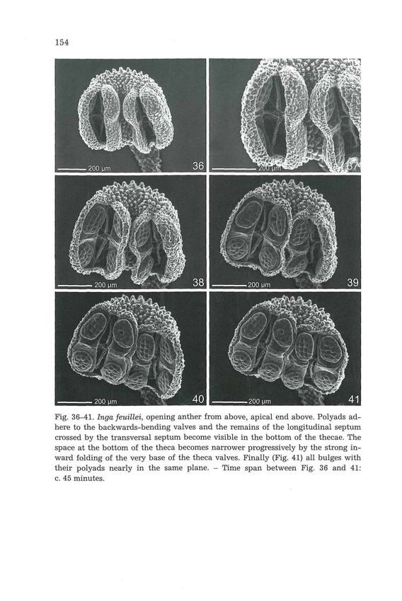
©Verlag Ferdinand Berger & Söhne Ges.m.b.H., Horn, Austria, download unter www.biologiezentrum.at

                                                                                      143
    20 cm. I. feuillei has been described and reviewed in a previous paper
    (TEPPNER 1998).

                               2. M a t e r i a l a n d M e t h o d s
         Seeds were purchased in Lima, Peru, on June 12, 1979, H. TEPPNER 79/428 &
    K. KEPLINGER, and were sown few days later in the cool greenhouse in Graz. The
    treelets were c. 3 m high, when planted 1995 in the new greenhouse of the Institute of
    Plant Sciences of the University in Graz (Austria, Europe).
         Anthesis was observed on intact inflorescences directly on the trees. In addition,
    cut inflorescences were watered and investigated in the laboratory using a stereo-
    microscope (Wild M 3B) and the environmental scanning electron microscope
    (ESEM). Flower buds of cut inflorescences also open on the second and third day
    approx. at normal time, thus one can assume that on the first day the timing of an-
    thesis is the same as on the intact trees. This is also in accordance with the experi-
    ence on the trees.
         For an overview of the internal structure of the anther, longitudinal and trans-
    versal manual cuts in water were used.
         Time scale is indicated in CET (Central European normal time).
         For ESEM investigations fresh anthers were mounted on aluminum stubs using
    C-impregnated double sided tape and investigated without any further preparation
    using a Philips XL 30 ESEM scanning electron microscope (FEI), using the following
    conditions: 0.8-0.9 torr chamber pressure, 9 mm working distance, 20 kv acceleration
    voltage, LFGSED (large field gaseous secondary electron detector).
         For the observation of the opening process, fully ripe but still closed anthers
    from opening flowers were inserted into the chamber of the ESEM; after a few mi-
    nutes opening of the anthers began.

                                         3. R e s u l t s
                                        3.1. Anthesis
         Flowers within a spike open, beginning from the base, acropetally
    (Fig. 1). Anthesis of all flowers of a day is nearly synchronous. The start of
    anthesis, the opening of the valvate corolla lobes, takes place between
    10:00 - 13:00 hours. The hank of anthers and coiled filaments appears
    (Fig. 5 and 6). The longitudinal axis of the anther is + parallel to the longi-
    tudinal axis of the (apical part of the) filament, which lies in a flat longi-
    tudinal depression on the dorsal side of the anther (Fig. 6, compare Fig. 12
    and 13). The filaments extend progressively and the anthers slowly start to
    turn in a position perpendicular to the filaments. At c. 16:00 usually, the
    opening of the anthers begins (only few single anthers open earlier, from
    15:00). The filaments are coiled to nearly straight and greenish in the distal
    part, surpassing the tips of the corolla by c. 1.0-1.5 cm. Till 17:00 opening
    has progressed, in a part of anthers polyads are exposed, the filaments are
    more or less straight but still greenish and not of full length; the position of
    anthers is more or less oblique. When the filaments become straight, be-
    tween 17:00 and 18:00, the nectar secretion starts but remain low in the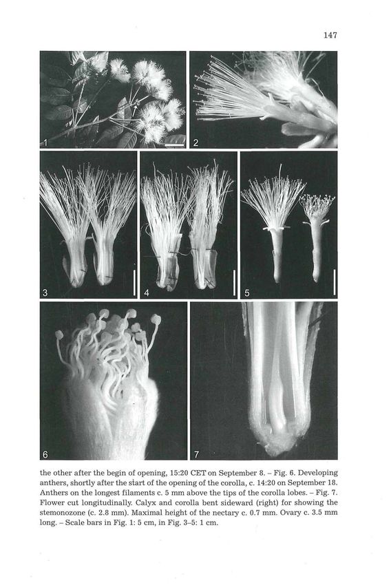
©Verlag Ferdinand Berger & Söhne Ges.m.b.H., Horn, Austria, download unter www.biologiezentrum.at

          144
          first 2-3 hours (up to 1-3 mm high, one-sided between ovary and fila-
          ments). At 18:00 nearly all anthers are open and the polyads exposed,
          nevertheless, not all anthers are in the final horizontal position and the
          filaments are still a little greenish at their tips. Thus, the full presentation
          of the polyads and the beginning of nectar secretion take place before the
          duskfall. In full darkness, at c. 19:00-20:00, the opening process seems to
          be finished: the filaments are extended to their final length (surpassing the
          corolla tips for 2.0-2.5 cm), are white and the anthers are perpendicular to
          the filaments (Fig. 2 and 42). The process from the first splitting of the
          corolla lobes up to the presentation of the polyads lasts for 3-4 hours, the
          anther opening itself usually for c. 1/2 to 1 or l1/2 hours. The time span
          needed in the ESEM lies in the same dimension. Anthesis is accompanied
          by a weak but distinct and characteristic odour (night and day, with vary-
          ing intensity) which may be described as perfume-like when fresh and a
          little strong when fading. The anthers persist on the withered and dry fi-
          laments.
                Anthesis of a single flower lasts for one night in Peru. The same is true
          for our greenhouse in c. June to August (day length 14-16 hours), where
          wilting of the filaments takes place between 9:00-11:00 of the next morn-
          ing. In September (day length 12-13 hours) fading of the filaments begins
          at c. 20:00-21:00 on the day after the opening day and they are distinctly
          withered at 23:00-24:00. Under cooler conditions in November opening of
          the flowers occurs later, anthers open just in the night (after 21:00) and
          withering takes place in the morning of the third day.

                              3.2. Some Notes on Flower Morphology
               Calyx length measures c. 10-13 mm, the corolla c. (13-)20-24 mm, the
          fully developed filaments c. 40-45 mm (Fig. 2 and 3); their basal part is
          fused into a tube shorter than the corolla, c. 12-17 mm long (Fig. 3 and 7),
          and forming a stemonozone of c. 2.0-4.0 mm (Fig. 7). The numbers of sta-
          mens varies between (75-)100-120 per flower (Fig. 2 and 3).
               The thecae and the locules, respectively, are partitioned by a trans-
          versal septum of parenchymatous tissue. In the centre of the locule the
          septum is thin, 1-2-layered, towards the periphery it dilates to 5-7 layers
          (compare ENDRESS & STUMPF 1991:            253, DNYANSAGAR 1954        and   ENGLER
          1876: 275-291 for other Mimosaceae). So an anther consists of two thecae,
          four locules and eight locule halves with one polyad each (diagram and
          discussion of terminology in PRENNER & TEPPNER 2005: 270, 272, 279-280).
          As well known for the longitudinal septum (e.g., KEIJZER 1987: 489, 490,
          492), this transversal septum is also dissolved during late developmental
          stages in the flower bud. The remains of the peripheral cells of the trans-
          versal septum appear on the valves as bulges (e.g., Fig. 35, 41, 45) dilated
          like a gusset towards the valve margin (Fig. 33). In the depth of the locules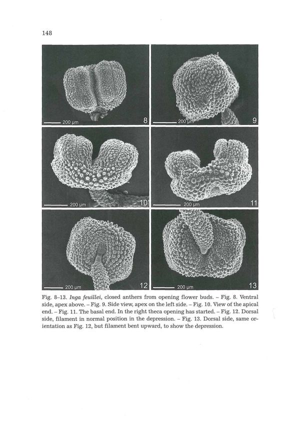
©Verlag Ferdinand Berger & Söhne Ges.m.b.H., Horn, Austria, download unter www.biologiezentrum.at

                                                                                    145
   the 'cross' of the remains of the longitudinal and the transversal septum
   can be seen (e.g., Fig. 37 and 47). Here the cellular structure of the septa
   can be clearly recognized.
        The nectary is a circular, shallowly ten-lobed bulge, c. 0.6-1.0 mm
   high, adnate for its whole length to the stemonozone (Fig. 7). The inner
   diameter near the base of the nectar room measures c. 2.0 mm. The nectar
   fills the whole tube, but also flowers without nectar may occur spor-
   adically, in spite of the presence of a nectary. Nectar access is very narrow,
   the entrance is completely closed by filaments and style, thus access is
   possible between the filaments only. It is relatively easy to put a needle (up
   to 1.0 mm in diameter) between the filaments to the nectar but not thicker
   objects, e.g., the tip of a small forceps. The gynoeceum is l(often 2-, rarely
   3)-carpellate (Fig. 3, 7 and 4). Male flowers with a small carpellodium
   (c. 2.0 mm long) may occur along with the hermaphrodite ones.

               3.3. Anthers, Filament Attachment Zone and Isthmus
         The closed anthers (Fig. 8-13) measure c. 0.5 mm in length x 0.7 mm
   in width. A massive cushion-like connective holds the two thecae. The
   apical end of the anther (connective) is a little rounded, the basal one a
   little emarginated. The epidermal cells form shortly conical, ribbed pa-
   pillae. The distinct stomium passes along the whole length, over the
   shoulders of the anthers up to the connective (Fig. 8 and 10).
         The construction around the filament attachment seems to be very
   complicated. The filament is diminished at its apex and forms a narrow
   isthmus (Fig. 12-15). This is responsible for the versatility of the anthers.
   Its epidermis consists of c. twenty rows of shortly cylindrical cells (Fig. 15).
   The attachment zone lies in a depression on the dorsal side of the anther, is
   elliptical and divided by the small filament insertion point unequally. The
   larger apical part, is covered by shortly papillate cells with smooth or
   partially striate cuticle. The very small basally directed area on the other
   side of the filament shows small cells but otherwise similar to the other
   epidermal cells (Fig. 14 and 15). During the maturation of the anthers in
   the afternoon, the isthmus- and attachment zone-cells change dramati-
   cally: they collapse, apparently by dehydration. The collapsing in the fila-
   ment isthmus begins in the epidermis directly at the anther (Fig. 16 and
   17), progresses successively (Fig. 18-22) and finally, in the evening, the
   whole apical half of the isthmus is collapsed (Fig. 23). The cells of the at-
   tachment zone, especially of the larger apical field can collapse more (Fig.
   19-21) or less (Fig. 22-23). If the turgescence of these cells should be re-
   sponsible for the longitudinal, erect original position of the anthers, then
   the loss of turgor may be the cause for the bending of the anther in the
   position perpendicular to the filament.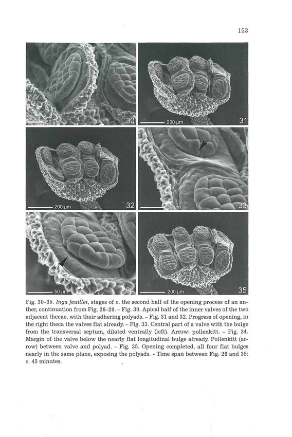
©Verlag Ferdinand Berger & Söhne Ges.m.b.H., Horn, Austria, download unter www.biologiezentrum.at

         146
                          3.4. Anther Opening and Polyad Presentation
              The opening process is presented here for three anthers in different
         positions: The view of the basal end of the anther (Fig. 11, 24 and 25), of
         the apical end from a little above (Fig. 26-35) and of the inner (ventral)
         side from above (Fig. 36-41). It begins with the appearance of a slit
         through the stomium (Fig. 11), practically simultaneously along the whole
         length. The bending back of the theca walls follows (Fig. 24, 26 and 36).
         The theca valves are sharply folded inward at their very base (e.g., Fig. 25,
         27, 31 and 37). The sides, a little arched by the volume of the polyads, bend
         back, and this zone which bears the polyads becomes more and more flat
         (e.g., Fig. 27-35, 38-41). Finally the ends of this zone are a little bent down
         (e.g., Fig. 35, 41 and 42). The margin of the theca valves crumples further
         back and shrivels (e.g., Fig. 34, 35 and 42-46). So the polyads appear ele-
         vated. Practically, a longitudinal bulge with a flat upper side is formed
         inward in each valve and the four bulges of an anther lie in one plane,
         approximately. The end sectors of the bulges bear the polyads and, as a
         whole, the anther forms a rubber stamp-like structure at the end of the
         opening process (Fig. 42).
              If the thecae of an anther do not open synchronously, the inner walls
         can overlap (Fig. 25 and 31) and eventually a polyad can be wedged in.
              The remains of the transversal septum form a distinct bulge on the
         valves (e.g., Fig. 33). In the open anther the 'cross' of this transversal bulge
         with the remains of the longitudinal septum can be seen (e.g., Fig. 35, 36-
         41 and 47). No tapetal membrane nor other sporopollenin structures were
         observed in the locules around the polyads.

                                              3.5. Polyads
              The roundish to elliptic polyads are 32-celled. The inner (= upper or
         exposed) side of the polyad (e.g., Fig., 36-46) and the outer (= lower or ad-
         herent) side (Fig. 48 and 49) show distinct differences: on the upper side
         the pollen grains are arched evenly, whereas on the lower side the bulge is
         flattened (Fig. 50). Furthermore the upper side of the polyad (contiguous to
         the longitudinal septum of the developing anther) is more or less in a single
         plane, whereas the lower side is arched (Fig. 50), what can be seen through
         the theca wall already (e.g., Fig. 8, 26 and 37). The polyads are covered by a
         thin layer of pollenkitt which can be seen in the optical section of the

         Fig. 1-7. Inga feuillei. Fig. 1. A twig with axillary inflorescences (spikes), in part
         crowded at the end. Arrow: end of the shoot axis. - Fig. 2. One flower in anthesis
         (length c. 4.3 cm) and two withered ones. - Fig. 3. Two flowers in late anthesis with
         G 1, split longitudinally. - Fig. 4. Two flowers at the day after anthesis, one with G 2,
         the other (right) with G 3, split longitudinally. - Fig. 5. One flower in late anthesis,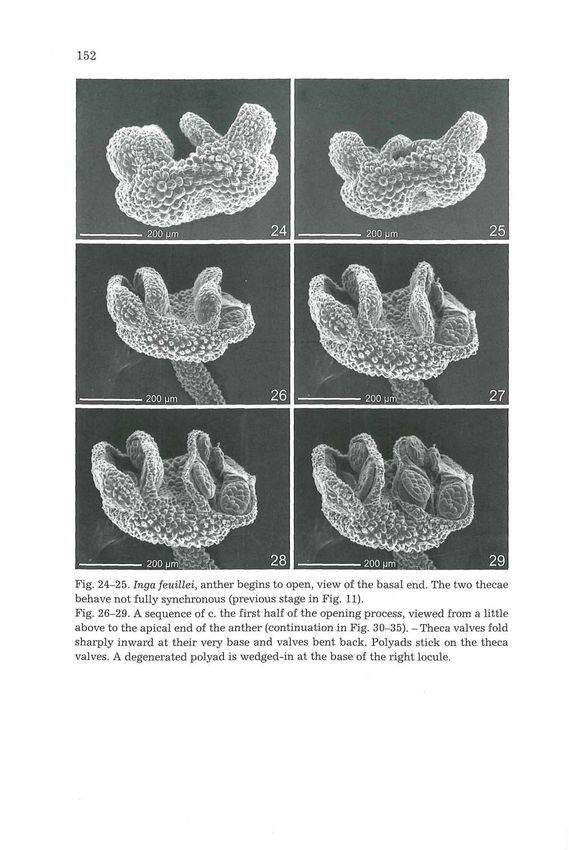
©Verlag Ferdinand Berger & Söhne Ges.m.b.H., Horn, Austria, download unter www.biologiezentrum.at

                                                                                        147

    the other after the begin of opening, 15:20 CET on September 8. - Fig. 6. Developing
    anthers, shortly after the start of the opening of the corolla, c. 14:20 on September 18.
    Anthers on the longest filaments c. 5 mm above the tips of the corolla lobes. - Fig. 7.
    Flower cut longitudinally. Calyx and corolla bent sideward (right) for showing the
    stemonozone (c. 2.8 mm). Maximal height of the nectary c. 0.7 mm. Ovary c. 3.5 mm
    long. - Scale bars in Fig. 1: 5 cm, in Fig. 3-5: 1 cm.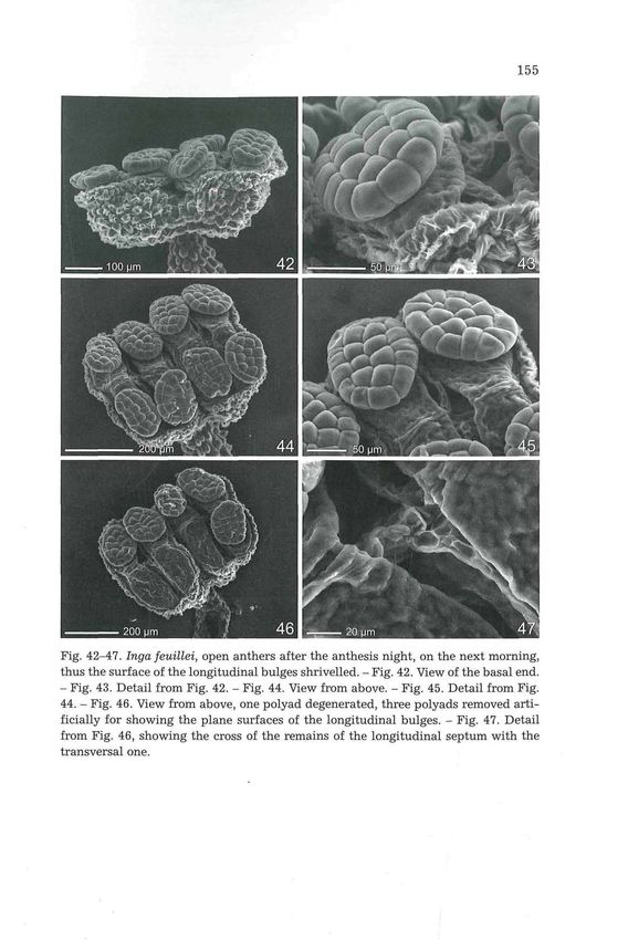
©Verlag Ferdinand Berger & Söhne Ges.m.b.H., Horn, Austria, download unter www.biologiezentrum.at

         148

         Fig. 8-13. Inga feuülei, closed anthers from opening flower buds. - Fig. 8. Ventral
         side, apex above. - Fig. 9. Side view, apex on the left side. - Fig. 10. View of the apical
         end. - Fig. 11. The basal end. In the right theca opening has started. - Fig. 12. Dorsal
         side, filament in normal position in the depression. - Fig. 13. Dorsal side, same or-
         ientation as Fig. 12, but filament bent upward, to show the depression.
©Verlag Ferdinand Berger & Söhne Ges.m.b.H., Horn, Austria, download unter www.biologiezentrum.at
149
polyad in the light microscope. Sometimes a small amount can be re-
cognized in the ESEM (Fig. 30, 33 and 34). The tip of a needle usually is not
sufficient to remove a polyad by adhesion from the anther, but with the
side of a needle the plane of contact is of appropriate size. On a finger-tip
or on a slide or cover slip the polyads adhere very well.
4. D i s c u s s i o n
4.1. Morphology
Inga stamens show characteristic features which are common in Mi-
mosaceae: long and coiled filaments in the bud stage enclosed in the cor-
olla and extending during anthesis; glabrous filaments; narrow isthmus at
the apex of the filaments (TUCKER 1996: 238); papillate epidermis. The tip
of the anther (connective) has no effiguration as in many Mimosaceae, but
at least the tip is rounded. The dehiscence suture extends over both
shoulders of the thecae (ENDRESS & STUMPF 1991: 253).
From the few figures of open anthers in the literature (e.g., Acacia
ENDRESS 1996: 282, Zapoteca GUINET & HERNANDEZ 1989: 9, Calliandra
PRENNER & TEPPNER 2005: 279) it seems, that a longitudinal bulge formed
from the theca valve by inward folding, should be common in Mimosaceae
(and other angiosperms). The peculiar feature in Inga is the exposed part
of this bulge, which becomes nearly plane, so that the polyads can usually
adhere with a large part of their face. A somewhat similar situation seems
to occur in Zapoteca, but the figures in GUINET & HERNANDEZ 1989: 9 and
HERNANDEZ 1990: 228 give the impression, that the longitudinal bulge is
not flat and the polyads adhere with a smaller part of their face only.
Longitudinal and transversal septum are largely dissolved before opening;
this phenomenon, in general terms, is discussed in KEIJZER 1987: 489-492.
The Ingeae-stamens most similar to Inga feuillei figured with details
in the literature are Albizia julib7'issin and Archidendron vaillantii (END-
RESS & STUMPF 1991: 250, 252), Par archidendron pruinosum and Callian-
dra houstoniana (TUCKER 1996: 242). Isthmus details are not discernible in
these figures, so comparisions are not possible. For Gasteria verrucosa
(Asphodelaceae) dehydration of the filament tip is mentioned in KEIJZER
1987: 490. SCHMID 1976 discusses the importance of changes in the filament
structure for interruption of water transport as one possible support of
desiccation of anthers. Such changes might be caused in the vessels by the
very quick elongation of the filaments. We do not know, if the collapsing of
the cells at the isthmus apex is also important in this connection. Some
Mimosaceae abscise and shed the anthers, Inga and others do not. There-
fore, abscission and shedding of anthers is not a necessary or logical con-
sequence of a narrow isthmus.
From the few species, in which the nectary is better documented, the
nectary adnated to the stemonozone in I. vera WILLD. subsp. affinis (DC.)©Verlag Ferdinand Berger & Söhne Ges.m.b.H., Horn, Austria, download unter www.biologiezentrum.at
150
Fig. 14-19. Inga feuülei, filament attachment zone and isthmus. - Fig. 14. Fresh an-
ther soon after the start of the opening of the corolla. Filament bent sideward to show
the whole attachment zone. Apical part left and the small basal part right. - Fig. 15.
Detail from Fig. 14. - Fig. 16. Detail from Fig. 12. The filament in the original posi-
tion in the basal depression. On the isthmus the first cells collapsed. - Fig. 17. Detail
from Fig. 16. - Fig. 18. Open anther at c. 19:30, filament bent sideward. Dehydration
of the isthmus progressed, attachment zone cells also collapsing. - Fig. 19. Detail
from Fig. 18.©Verlag Ferdinand Berger & Söhne Ges.m.b.H., Horn, Austria, download unter www.biologiezentrum.at
151
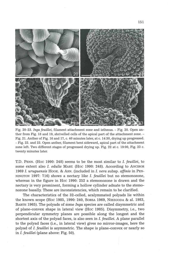
Fig. 20-23. Inga feuülei, filament attachment zone and isthmus. - Fig. 20. Open an-
ther from Fig. 18 and 19, shrivelled cells of the apical part of the attachment zone. -
Fig. 21. Anther of Fig. 16 and 17, c. 40 minutes later, at c. 14:30, drying up progressed.
- Fig. 22. and 23. Open anther, filament bent sideward, apical part of the attachment
zone left. Two different stages of progressed drying up. Fig. 22 at c. 19:00, Fig. 23 c.
twenty minutes later.
T.D. PENN. (HOC 1990: 248) seems to be the most similar to I. feuillei, to
some extent also I. edulis MAET. (HOC 1990: 248). According to ANCIBOR
1969 I. uruguensis HOOK. & ARN. (included in I. vera subsp. affinis in PEN-
NINGTON 1997: 716) shows a nectary like I. feuillei but no stemonozone,
whereas in the figure in Hoc 1990: 252 a stemonozone is drawn and the
nectary is very prominent, forming a hollow cylinder adnate to the stemo-
nozone basally. These are inconsistencies, which remain to be clarified.
The characteristics of the 32-celled, acalymmated polyads lie within
the known scope (Hoc 1985, 1990: 240, SORSA 1969, NIEZGODA & al. 1983,
BARTH 1965). The polyads of some Inga species are called disymmetric and
of plane-convex shape in lateral view (Hoc 1985). Disymmetry, i.e., two
perpendicular symmetry planes are possible along the longest and the
shortest axis of the polyad faces, is also seen in I. feuillei. A plane parallel
to the polyad faces (i.e., in lateral view) gives no mirror-images, here the
polyad of I. feuillei is asymmetric. The shape is plane-convex or nearly so
in I. feuillei (plane above: Fig. 50).©Verlag Ferdinand Berger & Söhne Ges.m.b.H., Horn, Austria, download unter www.biologiezentrum.at
152
Fig. 24-25. Inga feuillei, anther begins to open, view of the basal end. The two thecae
behave not fully synchronous (previous stage in Fig. 11).
Fig. 26-29. A sequence of c. the first half of the opening process, viewed from a little
above to the apical end of the anther (continuation in Fig. 30-35). - Theca valves fold
sharply inward at their very base and valves bent back. Polyads stick on the theca
valves. A degenerated polyad is wedged-in at the base of the right locule.©Verlag Ferdinand Berger & Söhne Ges.m.b.H., Horn, Austria, download unter www.biologiezentrum.at
153
Fig. 30-35. Inga feuillei, stages of c. the second half of the opening process of an an-
ther, continuation from Fig. 26-29. - Fig. 30. Apical half of the inner valves of the two
adjacent thecae, with their adhering polyads. - Fig. 31 and 32. Progress of opening, in
the right theca the valves flat already. - Fig. 33. Central part of a valve with the bulge
from the transversal septum, dilated ventrally (left). Arrow: pollenkitt. - Fig. 34.
Margin of the valve below the nearly flat longitudinal bulge already. Pollenkitt (ar-
row) between valve and polyad. - Fig. 35. Opening completed, all four flat bulges
nearly in the same plane, exposing the polyads. - Time span between Fig. 26 and 35:
c. 45 minutes.©Verlag Ferdinand Berger & Söhne Ges.m.b.H., Horn, Austria, download unter www.biologiezentrum.at
154
LI
Fig. 36-41. Inga feuillei, opening anther from above, apical end above. Polyads ad-
here to the backwards-bending valves and the remains of the longitudinal septum
crossed by the transversal septum become visible in the bottom of the thecae. The
space at the bottom of the theca becomes narrower progressively by the strong in-
ward folding of the very base of the theca valves. Finally (Fig. 41) all bulges with
their polyads nearly in the same plane. - Time span between Fig. 36 and 41:
c. 45 minutes.©Verlag Ferdinand Berger & Söhne Ges.m.b.H., Horn, Austria, download unter www.biologiezentrum.at
155
Fig. 42-47. Inga feuillei, open anthers after the anthesis night, on the next morning,
thus the surface of the longitudinal bulges shrivelled. - Fig. 42. View of the basal end.
- Fig. 43. Detail from Fig. 42. - Fig. 44. View from above. - Fig. 45. Detail from Fig.
44. - Fig. 46. View from above, one polyad degenerated, three polyads removed arti-
ficially for showing the plane surfaces of the longitudinal bulges. - Fig. 47. Detail
from Fig. 46, showing the cross of the remains of the longitudinal septum with the
transversal one.©Verlag Ferdinand Berger & Söhne Ges.m.b.H., Horn, Austria, download unter www.biologiezentrum.at
Fig. 48-49. Inga feuillei, under side (= outer or adherent side) of the polyads. - Fig. 48.
All eight polyads of one anther „stamped" onto the viscid tape of the stub. One
polyad a little shifted during the preparation. - Fig. 49. Detail of Fig. 48.
Fig. 50. A polyad in lateral view, differences in the arching of the surface of the pollen
grains between upper and lower side.
4.2. Flower Ecology
Phenology of flowering with respect to seasonality as well as to flower
opening and duration is highly variable in the genus. KOPTUR 1983a,b ob-
served in seven species of Inga in Costa Rica. She reports nocturnal or day
and night anthesis, opening in the morning, the afternoon or continuously
etc. and a duration of flowers between 5 and 21 hours; nectar production
starts when filaments are fully extended; in I. feuillei this was a little ear-
lier. The nectar composition of eight species is discussed in KOPTUR 1994
(freshly secreted nectar is sucrose-dominated).
A wide range of pollinators is possible in Inga species depending,
among others, on the time of anthesis. Nocturnal flowers may be visited by
sphingids, noctuids and bats, whereas, when daytime is also involved,
hummingbirds, butterflies, bees etc. may be visitors as well (e.g., KOPTUR
1983a,b, 1984: 1133-1134, SALAS 1974, VOGEL 1968: 579-581, HEITHAUS &
al. 1975, HABER & al. 1981). In I. feuillei the size of the flowers, the mainly
nocturnal nectar production and the narrow access to the nectar makes©Verlag Ferdinand Berger & Söhne Ges.m.b.H., Horn, Austria, download unter www.biologiezentrum.at
157
sphingids and noctuids as pollinators probable, which were observed as to
be effective pollinators in the greenhouse (TEPPNER 1998: 41-42). In our
greenhouse we had colonies of bumblebees (Bombus t e r r e s t r i s , B.
h a e m a t u r u s ) who collected the floral and extrafloral nectar and caused
some accidental pollinations. To what extent other visitors of I. feuillei are
involved in the natural environment in the afternoon and in the morning,
is not known.
5. A c k n o w l e d g e m e n t s
Our sincere thanks to Mag. Dr. U. BROSCH for procuring literature, which was
not available at the institute. We would like to thank also Mag. Dr. W. OBERMAYER for
scanning and layout of Fig. 1-7 and to Mag. P. HARVEY for the careful check of the
language. Last but not least many thanks of H. T. goes to his wife, Erika, who typed
with patience the manuscript; without her support this paper would not have been
possible.
6. R e f e r e n c e s
ANZIBOR E. 1969. Los nectarios florales en Leguminosas-Mimosöideas. - Darwiniana
15(1-2): 128-142.
BARTH O. M. 1965. Feinstruktur des Sporoderms einiger brasilianischer Mimosoiden-
Polyaden. - Pollen et Spores 7(3): 429-441.
BERMINGHAM E. & DICK C. 2001. The Inga - newcomer or museum antiquity ? - Sci-
ence 293:2214-2216.
DNYANSAGAE V. R. 1954. Embryological studies in the Leguminosae. VI. Inflorescence,
sporogenesis, and gametophytes of Dichrostachys cinerea W. & A. and Parkia
biglandulosa W. & A. - Lloydia 17(4): 263-74.
ENDRESS P. K. 1996. Diversity and evolutionary biology of tropical flowers. - Cam-
bridge tropical biology series. - Cambridge University Press.
— & STUMPF S. 1991. The diversity of stamen structures in 'Lower' Rosidae (Ro-
sales, Fabales, Proteales, Sapindales). -Bot. J. linn. Soc. 107: 217-293.
ENGLER A. 1876. Beiträge zur Kenntniss der Antherenbildung der Metaspermen. -
Jahrb. wiss. Bot. 10: 275-316, plates XX-XXIV.
GRIMES J. 1998. [Review of] T.D. PENNINGTON, illustrations by Rosemary WISE. The
genus Inga - Botany. - Kew Bull. 53(1): 252-253.
GUINET P. & HERNANDEZ H. M. 1989. Pollen characters in the genera Zapoteca and
Calliandra (Leguminosae, Mimosoideae). Their systematic and phylogenetic
relevance. - Pollen et Spores 31(1-2): 5-22.
HABER W.A., FRANKIE G. W, BAKER H. G., BAKER I. & KOPTUR S. 1981. Ants like flower
nectar. - Biotropica 13(3): 211-214.
HEITHAUS E. R., FLEMING T.H. & OPLER P. A. 1975. Foraging patterns and resource
utilization in seven species of bats in a seasonal tropical forest. - Ecology
56(4): 841-854.
HERNANDEZ H. M. 1986. Zapoteca: A new genus of neotropical Mimosoideae. - Ann.
Missouri bot. Garden 73: 755-763.
— 1990. A new subgenus and a new species of Zapoteca (Leguminosae). - Sys-
tematic Botany 15(2): 226-230.©Verlag Ferdinand Berger & Söhne Ges.m.b.H., Horn, Austria, download unter www.biologiezentrum.at
158
Hoc P. S. 1985. Inga MILL, in Argentina: Palynological study. - Bull, internat. Group
Study Mimosoideae 13: 70-86.
— 1990. Las especies Argentinas de Inga (Leguminosae, Mimosoideae). - Dar-
winiana 30(1-4): 237-258.
HUGHES C. E. 1997. Variation in anther and pollen morphology in Leucaena BENTH.
(Leguminosae-Mimosoideae). -Bot. J. linn. Soc. 123: 177-196.
KEIJZER C. J. 1987. The processes of anther dehiscence and pollen dispersal. I. The
opening mechanism of longitudinally dehiscing anthers. - New Phytologist
105(3): 487-498.
KENRICK J. & KNOX R.B. 1979. Pollen development and cytochemistry in some Aus-
tralian species of Acacia. - Austral. J. Bot. 27: 413-427.
KOPTUR S. 1983a. Flowering phenology and floral biology of Inga (Fabaceae: Mimo-
soideae). - Syst. Bot. 8(4): 354-368.
— 1983b. Inga. - In: JANZEN D.H. (ed.), Costa Rican natural history, p. 259-261. -
Chicago and London.
— 1984. Outcrossing and pollinator limitation of fruit set: Breeding systems of
neotropical Inga trees (Fabaceae: Mimosoideae). -Evolution 38(5): 1130-1143.
— 1994. Floral and extrafloral nectars of Costa Rican Inga trees: A comparison of
their constituents and composition. - Biotropica 26(3): 276-284.
LEWIS G. P. & Rio ACRE L. 2005. Tribe Ingeae. - In: LEWIS G., SCHRIRE B., MACKINDER
B. & LOCK M. (eds.), Legumes of the world, p. 192-213. - The Royal Botanic
Gardens, Kew.
NIEZGODA C. J., FEUER S. M. & NEVLING L. I. 1983. Pollen ultrastructure of the tribe
Ingeae (Mimosoideae: Leguminosae). - Amer. J. Bot. 70(5): 650-667.
PENNINGTON T. D. 1997. The genus Inga. Botany. - The Royal Botanic Gardens, Kew.
[Before use read GRIMES 1998].
PRENNER G. & TEPPNER H. 2005. Anther development, pollen presentation and pollen
adhesive of parenchymatous origin in Calliandra angustifolia (Leguminosae-
Mimosoideae-Ingeae). - Phyton (Horn, Austria) 45(2): 267-286.
RICHARDSON J. E., PENNINGTON R. T., PENNINGTON T. D. & HOLLINGSWORTH P. M. 2001.
Rapid diversification of a species-rich genus of neotropical rain forest trees. -
Science 293: 2242-2245.
SALAS S. 1974. Analisis del sistema de polinizacion de Inga vera ssp. spuria. - Thesis,
Universidad de Costa Rica. (Citation after KOPTUR 1983b).
SCHMID R. 1976. Filament histology and anther dehiscence. - Bot. J. linn. Soc. 73:
303-315.
SORSA P. 1969. Pollen morphological studies on the Mimosaceae. - Ann. bot. fenn. 6:
1-34.
TEPPNER H. 1998. Inga feuillei DC. (Mimosaceae-Ingeae). - Samentauschverzeichnis
1998 (Bot. Garten Inst. Bot. Univ. Graz) p. 37-45.
TUCKER S.C. 1996. Stamen structure and development in legumes, with emphasis on
poricidal stamens of caesalpinioid tribe Cassieae. - In: D'ARCY W. G. &
KEATING R.C. (eds.), The anther. Form, function and phylogeny. - Cambridge
University Press.
VOGEL S. 1968. Chiropterophilie in der neotropischen Flora. Neue Mitteilungen I. -
Flora, Abt. B, 157: 562-602.You can also read