Potential roles of mesenchymal stem cells and their exosomes in the treatment of COVID-19
←
→
Page content transcription
If your browser does not render page correctly, please read the page content below
[Frontiers in Bioscience-Landmark, 26(10), 948-961, DOI:10.52586/4999]                                       https://www.fbscience.com

Review

Potential roles of mesenchymal stem cells and their
exosomes in the treatment of COVID-19
Xiaoyun Cheng1 , Mao Jiang2 , Lingzhi Long2 , Jie Meng2, *
1
 Department of Pulmonary and Critical Care Medicine, Xiangya Hospital of Central South University, 410008
Changsha, Hunan, China, 2 Department of Pulmonary and Critical Care Medicine, The Third Xiangya Hospital of Central
South University, 410008 Changsha, Hunan, China

TABLE OF CONTENTS

1. Abstract
2. Introduction
3. MSCs regulate immune cells in COVID-19
         3.1 MSCs interfere with the differentiation, maturation, and function of antigen-presenting cells (APCs) in COVID-19
         3.2 MSCs regulate the polarization of macrophages in COVID-19: from M1 to M2
         3.3 MSCs regulate lymphocyte subsets and apoptosis in COVID-19
4. MSCs improve ARDS in COVID-19
5. MSCs promote lung regeneration and reverse PF in COVID-19
6. MSC-exosomes for the treatment of COVID-19
         6.1 Comparison of MSC-exosomes and MSCs
         6.2 Unique advantages of MSC-exosomes over MSCs for COVID-19 treatment
7. Clinical trials of MSCs and MSC-exosome therapy for COVID-19
8. Conclusions
9. Author contributions
10. Ethics approval and consent to participate
11. Acknowledgment
12. Funding
13. Conflict of interest
14. References

1. Abstract                                                              strategies were as follows: ((Mesenchymal stem cells)
                                                                         OR (MSCs)) AND (COVID-19). Results: MSCs regulate
          Background:       Corona Virus Disease 2019                    the immune system to prevent cytokine release syndrome
(COVID-19) is an acute respiratory infectious disease                    (CRS) and to promote endogenous repair by releasing
caused by severe respiratory syndrome coronavirus 2                      various paracrine factors and exosomes. Conclusions:
(SARS-CoV-2).        The primary pathogenesis is over-                   MSC therapy is thus a promising candidate for COVID-19.
activation of the immune system. SARS-CoV-2 continues
to mutate and spread rapidly and no effective treatment
options are yet available. Mesenchymal stem cells (MSCs)                 2. Introduction
are known to induce anti-inflammatory macrophages,
regulatory T cells and dendritic cells. There are a rapidly                       According to real-time WHO network data, the
increasing number of clinical investigations of cell-based               worldwide number of confirmed COVID-19 cases to April
therapy approaches for COVID-19.             Objective: To               22, 2021 was 143,488,236 and 3,055,587 deaths, posing an
summarize the pathogenic mechanism of SARS-CoV-2,                        unprecedented threat to the global economy and to human
and systematically formulated the immunomodulation                       health [1]. The International Committee on the Taxonomy
of COVID-19 by MSCs and their exosomes, as well as                       of Viruses named the COVID-19 pathogen as SARS-CoV-
research progress. Method: Searching PubMed, clinical-                   2. This virus gains phagocytic entry into AT2 via inter-
trials.gov and Chictr.cn for eligible studies to be published            action with angiotensin-converting enzyme 2 (ACE2) (See
or registered by May 2021. Main keywords and search                      Fig. 1). It increases Angiopoietin-2 (Ang-2) levels, lead-

Submitted: 6 June 2021 Revised: 17 August 2021 Accepted: 1 September 2021 Published: 30 October 2021
This is an open access article under the CC BY 4.0 license (https://creativecommons.org/licenses/by/4.0/).
© 2021 The Author(s). Published by BRI.
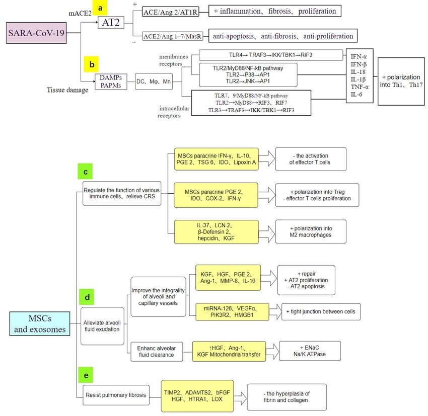
949

Fig. 1. The pathological processes of COVID-19 (blue arrows) and multiple therapeutic mechanisms of MSCs and their exosomes in COVID-19
(green arrows). (a) SARS-CoV-2 gains entry into AT2s via ACE2. (b) Immune cells recognize SARS-CoV-2 via TLRs. (c) T cells are polarized into
pro-inflammatory phenotypes. (d) Excessive activation of M1 causes CRS. (e) Inflammation and oxidative damage cause lung fibrosis and remodeling.
MSCs reduce fibrosis by various paracrine factors. (f) MSCs force T cells to polarize into Tregs. (g) MSCs and MSC-exosomes increase the number of
anti-inflammatory M2 macrophages. (h) MSCs restore microvascular permeability. (i) MSCs activate the Na+/K+ pump to remove lung fluid and reduce
pulmonary edema. Abbreviations: ECM, extracellular matrix; PGE 2, Prostaglandin E 2; COX, Cyclooxygenase; HGF, hepatocyte growth factor; KGF,
keratinocyte growth factor.

ing to long-term and intense activation of pro-inflammatory                rapid and sharp rise in the level of cytokines. These include
Ras-related pathways. A high concentration of Ang-2 in                     C-X-C Motif Chemokine Ligand 10 (CXCL10), monocyte
the lung interstitium promotes cell apoptosis, releases pro-               chemo-attractant protein-1 (MCP-1/CCL2), macrophage
inflammatory cytokines and triggers the inflammatory re-                   inflammatory protein-1 (MIP-1), platelet-derived growth
sponse, thereby causing immune-induced tissue damage                       factor (PDGF), tumor necrosis factor-α (TNF-α) and vas-
and increased vascular permeability [2, 3]. In patients with               cular endothelial growth factor (VEGF) [4]. This mecha-
severe disease, the development of CRS is an abnormal sys-                 nism results in an imbalance between tissue damage and
temic inflammatory response that manifests clinically as a                 repair, leading to respiratory failure. Patients may eventu-
950

                          Table 1. The potential roles and mechanisms of MSC-exosomes in various diseases.
 Functions                                   Involved genes/factors/pathways                           Diseases                    References
 (+) Immunomodulatory activities (+) re- miR-21, miR-24, miR-124, miR-145, miR-122, KGF, COVID-19                                   [17–22]
 pair (+) angiogenesis (–) fibrosis          VEGF, HGF, VE-cadherin, Occludin-1, Claudin-1, PCNA,
                                             Cyclin D1, IDO, Wnt/β-catenin signalling, TGF-β1/Smad 2
 (–) EMT                                     miR-182-5p, miR-23a-3p (-) NF-κB, Hedgehog                LPS induced lung injury        [23]
 (–) Inflammation, proliferation             miR-34a, miR-122, miR-124, miR-127                        IPAH                           [24]
 (–) Leukocyte infiltration                  ICAM-1                                                    ARDS                           [25]
 Anti–viral                                  COX-2, PGE2                                               Influenza                      [26]
 (–) Airway hyperresponsiveness            miR-146a-5p                                             Allergic airway inflammation       [27]
 (–) Autophagy, apoptosis                  miR-125b, Bcl-xL, Bcl2, and BIRC8 (-) Caspase1, Caspase Myocardial infarction, acute     [28, 29]
                                           8, lymphotoxin α                                        kidney injury.
 (–) Fibrosis                              (-) Col-I, Col-III, TGF-β1/Smad                         Liver fibrosis                   [30, 31]
 (+) M2 macrophage polarization            miR-1260b (-) Wnt5a/RANKL                               Periodontitis, Tendon healing    [32, 33]
 (+) Treg cell induction, T–cell apoptosis FASL/FAS                                                Acute colitis                      [34]
 (–) Dendritic cell maturation               a blockade in G0/G1 phase of the cell cycle               LPS intervention               [35]
 (+) Autophagy                               AMPK/ /mTOR, Akt/mTOR                                     Myocardial I/R                 [36]
 (+) indicates stimulation. (–) indicates inhibition.

ally die from multiple organ failure. Disruption of matrix                     vates the intracellular receptors TLR3/7 and membrane re-
metalloproteinases (MMPs) during the inflammatory stage                        ceptors TLR2/4. TLR3 in dendritic cells (DCs) specifically
causes complex damage to the alveolar epithelium and to                        recognize dsRNA, an intermediate product of viral repli-
the pulmonary vascular endothelium [5]. Moreover, per-                         cation, thereby activating nuclear factor kappa-B (NF-κB)
sistent stimulation of epithelial cells results in senescence-                 and interferons (IFNs). TLR2 and TLR4 located on the sur-
related phenotypes. Consequently, abnormal interactions                        face of macrophages and DCs can activate interferon reg-
between fibroblasts and epithelial cells generate irreversible                 ulatory factors (IRFs) and NF-κB, resulting in the produc-
damage and fibrosis [6].                                                       tion of different cytokines and chemical activators. TLR2
          Although the efficacy of vaccines in preventing                      can mediate DCs to express interleukin-8 (IL-8) and IL-23
COVID-19 ranges from 50% to 95%, an increasing num-                            (Fig. 2b), while TLR4 mainly mediates DCs to produce IL-
ber of COVID-19 cases still require effective treatment op-                    12 and IFN-γ-induced protein 10 (IP-10). IP-10 in turn
tions [7, 8]. There is currently no standard drug therapy for                  stimulates T cells to secrete IFN and promotes the differ-
COVID-19. Antiviral, anti-malarial and anti-inflammatory                       entiation of T helper-type (Th) cells into Th1 cells (Fig. 1c)
drugs are unable to repair or regenerate lung tissues, espe-                   [24].
cially in patients with severe complications such as acute                               MSCs can interfere with the antigen-presenting
respiratory distress syndrome (ARDS) [9–11]. Since the                         function, differentiation and maturation of DCs by
outbreak of COVID-19, a number of studies have been car-                       paracrine IFN-γ, indoleamine 2,3-dioxygenase (IDO),
ried out on MSCs and MSC-exosome therapy for this dis-                         transforming growth factor-β (TGF-β), IL-10 and
ease. MSCs have good safety for the treatment of COVID-                        prostaglandin E2 (PGE2) [37]. Consequently, this reduces
19 and show particular clinical efficacy in shortening the                     the activation of DCs and their pro-inflammatory secretions
course of disease, alleviating lung injury and reducing the                    [38, 39]. Leng et al. [40] observed a significant increase
level of inflammatory factors [6, 12, 13]. This is expected                    in the number of CD14+CD11c+CD11mid low-activity
to provide a new approach for the treatment of severe and                      phenotypic DCs on day 6 after MSC transplantation. This
critical COVID-19. MSC-exosomes also show promise as                           prevented the excess proliferation of T cells in COVID-19
a cell-free substitute for COVID-19 [14–16] (Table 1, Ref.                     patients. The interaction between MSCs and DCs also
[17–36]). The potential mechanisms of action of MSCs and                       leads to an indirect conversion of pro-inflammatory Th1 to
MSC-exosome therapy are shown in Figs. 1,2.                                    anti-inflammatory Th2 immunity [41].
                                                                               3.2 MSCs regulate the polarization of macrophages in
                                                                               COVID-19: from M1 to M2
3. MSCs regulate immune cells in COVID-19
                                                                                         Pro-inflammatory macrophages were reportedly
3.1 MSCs interfere with the differentiation,                                   more abundant in the bronchoalveolar lavage fluid from se-
maturation, and function of antigen-presenting cells
(APCs) in COVID-19                                                             vere compared to mild COVID-19 cases [42]. Zhang et
                                                                               al. [43] proposed that CRS in severe COVID-19 is mainly
        Once SARS-CoV-2 infects the human lung epithe-                         a virus-triggered macrophage activation syndrome. Viral
lium and is internalized (Fig. 1a), SARS-CoV-2 RNA acti-                       RNA stimulates macrophages to produce various soluble
951 Fig. 2. MSCs act on COVID-19 through multiple mechanisms and specific secretory products (light yellow squares). (a) SARS-CoV-2 causes immune-induced tissue damage via pro-inflammatory Ras-related pathways. (b) TLRs of immune cells recognize SARS-CoV-2, leading to CRS. (c) MSCs have powerful anti-inflammatory and immunomodulatory functions. (d) MSCs repair microvascular permeability and alleviate pulmonary edema. (e) MSCs secrete substances to inhibit the hyperplasia of fibrin and collagen, thereby alleviating PF. + indicates stimulation. – indicates inhibition. factors via the activation of TLRs. These factors include refractory respiratory failure in COVID-19 and it has been IFN-γ, IP10, MCP1, MIP10, granulocyte colony stimu- reported they are almost entirely depleted in severely in- lating factor (G-SCF), IL-2, IL-6, IL-7 and TNF-α. In fected patients [44]. particular, IL-6 and TNF-α cause macrophages to differ- MSCs regulate macrophage polarization to limit entiate into M1 (Fig. 1d), thus causing an imbalance in inflammation and to promote tissue healing following in- M1/M2. In addition to direct stimulation by viral RNA, jury. Anti-inflammatory soluble factors and two main in- ATP is released by the dead cells as DAMPs and binds to hibitory molecules secreted or expressed by MSCs trigger the P2X7 receptor (P2X7R). This in turn activates NOD- the immune system’s inhibitory response. MSCs are stim- like receptor protein 3 (NLRP3) inflammasomes and in- ulated to produce IL-10, thyrotropin-releasing hormone-6 creases macrophage-derived IL-1β and IL-18 [37]. The (TRH-6), human leukocyte antigen (HLA), human growth loss of alveolar macrophages is a major underlying cause of factor (HGF), heme oxygenase-1 (HO-1) [45], superoxide
952

dismutase (SOD), cyclooxygenase-2 (COX-2), PGE2 and             with severe COVID-19. Moreover, the mean CD3+, CD4+
IDO [46]. Meanwhile, MSCs are induced by IFN-γ to up-           and CD8+ lymphocyte counts increased by 46% (p < 0.05),
regulate the expression of other inhibitory molecules, in-      45% (p < 0.05) and 46% (p < 0.001), respectively.
cluding PD-1/PDL-1 and FAS-FASL [47–49]. These sol-                       The mechanism of action of MSCs in reversing
uble factors and molecules inhibit pro-inflammatory path-       lymphocytopenia and reducing inflammatory mediators in
ways such as the NF-κB signaling pathway and also polar-        COVID-19 is mainly attributed to the more than 30 solu-
ize M1 macrophages into the anti-inflammatory M2 phe-           ble paracrine factors such as PEG2, IDO and COX-2 [61].
notype [50, 51]. In this way, MSCs reduce the production        These have been shown to inhibit the proliferation of CD4+
of inflammatory cytokines by macrophages in COVID-19            Th1 and Th17 cells as well as CD8+T cells, and to in-
[52].                                                           duce Foxp3+ Treg differentiation (Fig. 1f). They also in-
                                                                directly inhibit excessive T cell proliferation by interact-
3.3 MSCs regulate lymphocyte subsets and apoptosis in
COVID-19                                                        ing with APCs and other immune cells. IL-10 is a critical
                                                                negative regulator of T cell responses and directly inhibits
          Flow cytometry analysis has shown that the num-       the ability of T cells to produce pro-inflammatory media-
ber of CD4+ and CD8+T cells in the peripheral blood of          tors. IL-10 also reduces the antigen presenting capacities
COVID-19 patients was significantly reduced, with the de-       and co-stimulation of macrophages and DCs, thereby de-
gree of reduction being related to the severity of COVID-       creasing T cell-derived IL-6 and TNF-α, which is one of
19 [53]. This phenomenon may be related to the re-              the essential mechanisms by which MSCs alleviate inflam-
cruitment of T cells from peripheral blood to lung tis-         mation [62, 63]. This was demonstrated in a recent clinical
sue, and to the apoptosis of T cells induced by the virus       report by Meng et al. [64] which showed that patients who
[54]. COVID-19 patients present with lymphocyte defi-           received MSCs had lower IL-6 levels than those who re-
ciency and over-activation of T cells. These effector T cells   ceived placebo [65].
are stimulated by pro-inflammatory mediators produced by                  Through their expression of PD-L1 and FasL,
DCs, macrophages and neutrophils [55, 56]. A significant        MSCs can inhibit abnormally activated Th1 cells, thus
rise in HLA-DR+CD38+ cell levels can manifest in the            inhibiting IL-γ release from Th1. This prevents fur-
over-activation of T cells. The proportion of highly pro-       ther macrophage activation in a vicious loop and restores
inflammatory CCR4+CCR6+ Th17 cells amongst CD4+ T               Th1/Th2 balance. Long-term FasL interaction can induce
cells then increases [57]. High expression of IL-17A in         apoptosis of cytotoxic T cells [66].
Th17 induces the migration of inflammatory white blood                    At the same time, MSCs release TGF-β which pro-
cells, leading to inflammatory infiltration and destruction     motes the proliferation of CD4(+) CD25(+) FoxP3(+) Treg,
of lung tissue. Additionally, the major histocompatibility      CD3(+) CD8(+) CD28(–) T-suppressor cells (Ts), and IL-
complex 1 (MHC-1) of infected cells presents viral anti-        10-producing B cells [67]. MSCs also up-regulate IDO and
gens, thus activating CTLs to produce high levels of cyto-      PGE2, which have synergistic inhibitory effects on NK and
toxic granules such as perforin and granzymes. This implies     Th17 cells. MSCs inhibit B cells through cell cycle stagna-
that over-activation of T cells and the elevated cytotoxic-     tion in the G0/G1 phase rather than by inducing apoptosis.
ity of CD8+T cells leads to an excessive immune response.       With regard to cellular immunity, MSCs reduce the produc-
T cell-derived cytokines and chemokines such as TNF-α,          tion of immunoglobin M (IgM), IgG, and IgA, and down-
IFN-γ, IL-2, IL-12, CCL2, IL-18, CCL9, CXCL10, IL-6             regulate the expression of CXCR4, CXCR5 and CXCR7 in
and IL-17 are released in large quantities and damage the       B cells, thereby altering their chemotactic properties [68].
lung tissue [58]. When the T cell count falls to its lowest
level, the concentrations of serum IL10, IL2, IL4, TNF-α
and IFN-γ reach their peak on days 4–6. Moreover, the
                                                                4. MSCs improve ARDS in COVID-19
levels of IL-6, IL-7, G-CSF, IP-10, monocyte chemotactic                 ACE2 is down-regulated following the entry of
protein-1 (MCP-1) and MIP1a increase significantly, thus        SARS-CoV-2 into alveolar epithelial cells, resulting in
causing CRS [59].                                               an imbalance of ACE/Ang II/AT1R and ACE2/Ang 1-
          Leng et al. [40] reported that on day 4 after MSC     7/MASR. Elevated Ang-2 levels then lead to cell apoptosis
transplantation, the absolute lymphocyte count increased        and trigger inflammatory responses, giving rise to immune-
to 0.58 × 10⁹ /L and lymphocytopenia improved signifi-          induced tissue damage and increased vascular permeability
cantly. On days 3 to 6 after transplantation, the level of      [69–71] (Fig. 2b).
TNF-α decreased whereas that of IL-10 increased. Similar                 In COVID-19 patients the average time from
reversals were reported in another study [60]. T cell counts    symptom onset to dyspnea is 5 days, the average hospital
were also analyzed in a non-randomized, open-label cohort       stay is 7 days, and the average time for onset of ARDS
study of COVID-19 patients [18]. This indicated that MSC-       is 8 to 9 days [72]. By day 8 to 14 of disease onset, the
exosome therapy significantly improved the absolute neu-        overexpression of cytokines such as IL-2, IL-6, IL-7, IP10,
trophil count by a mean of 32% [p value < 0.001] in patients    MCP1, MIP1A and TNFα causes activation of lympho-
953
cytes and macrophages, leading to an excessive inflamma- alleviating PF [89]. MSCs can also reverse PF by over-
tory response [73]. The integrity of alveolar walls and pul- expressing MMP-P1 and decreasing collagen-1 (COL-I)
monary capillaries are destroyed, resulting in edema that production during TGF-β1-induced fibrosis. In conclu-
impairs oxygen exchange and respiration and inevitably de- sion, MSCs can promote angiogenesis and the regenera-
velops into ARDS [74, 75]. tion of alveolar epithelial cells, prevent the apoptosis of en-
MSCs and MSC-exosomes can effectively allevi- dothelial cells, reduce the levels of TGF-β, TNF-α, COL-I,
ate COVID-19-induced ARDS in a dose-dependent manner COL-III, Hydroxyproline and serum ceruloplasmin, inhibit
by increasing alveolar fluid clearance and by improving air- myofibroblast growth, and thereby alleviate or reverse PF
way and hemodynamic parameters [76]. MSC-exosomes (Fig. 2e).
have been used as intravenous infusion therapy for ALI Human embryonic stem cells (hESCs) derived
and pulmonary fibrosis (PF) [23, 77, 78]. An earlier study from immune and stromal regulatory cells (IMRCs) have
showed the exosomes release keratinocyte growth factor been used to treat lung injury and fibrosis in vivo. IMRCs
(KGF) and Lipoxin A4 which act to prevent long-term lung have superior efficacy to FDA-approved pirfenidone [14]
damage caused by COVID-19 and to promote tissue re- and show excellent efficacy and safety in both mice and
pair by activating Na+/K+ pumps [79] (Fig. 2d). Impor- monkeys [90].
tantly, MSCs have been shown to restore epithelial protein
permeability, stabilize endothelial fluid leakage, and main-
tain alveolar-capillary barrier function by secreting Ang-1
6. MSC-exosomes for the treatment of
[80–82]. In addition, MSCs can inhibit cellular signaling COVID-19
pathways mediated by TLRs or PRRs, as well as reduc- MSC-exosomes are able to transfer cargoes such
ing local immune cell recruitment (Fig. 2c). miRNA-126, as mRNA, miRNA, proteins, lipids and even mitochon-
VEGF-α, phosphoinositide-3-kinase regulatory subunit 2 dria to target cells and tissues, resulting in changes to gene
(PIK3R2) and high mobility group box chromosomal pro- expression and in the behavior of target cells. Hence,
tein 1 (HMGB1) can each restore the vascular endothelial MSC-exosomes could have a therapeutic role in COVID-
cadherin-catenin (VE-Cadherin) complex and reduce en- 19 [91, 92]. Preclinical studies have confirmed that MSC-
dothelial barrier permeability to relieve ARDS. exosomes are able to serve as acellular alternatives [78].
6.1 Comparison of MSC-exosomes and MSCs
5. MSCs promote lung regeneration and 6.1.1 Can MSC-exosomes effectively replace MSCs?
reverse PF in COVID-19
A growing number of studies have established
PF is a refractory lung disease that develops due to that the healing, nutritional, immunoregulation, and anti-
persistent alveolar injury, repeated destruction, repair, re- inflammatory effects of administered MSCs are due to the
construction, and excessive deposition of extracellular ma- exosomes they release. These effects of MSCs have been
trix (ECM) [83]. Current studies have determined that only observed in vitro after the addition of MSC-exosomes [37].
1% of AT2 cells can regenerate following SARS-CoV-2- MSCs were cleared from the circulation within 24 hours,
induced lung injury [84, 85]. but MSC-exosomes were detected in lung parenchymal
High expression of IL-17A by Th17 in COVID- cells and macrophages just 1 hour after injection and re-
19 can induce the migration of inflammatory leukocytes, mained there for up to 7 days [93]. The efficacy and safety
leading to inflammatory infiltration and the destruction of of a single intravenous injection of MSC-exosomes were
lung tissue [86]. High levels of TNF-α induce the recruit- recently assessed in 24 COVID-19 patients who presented
ment of immune cells and reduce antioxidant molecules with moderate to severe ARDS. The clinical symptoms,
in parenchymal and endothelial cells, causing lung fibro- oxygenation, serum markers of acute inflammation, neu-
sis and remodeling (Fig. 1e). MSCs improve angiogene- trophil and lymphocyte counts all improved in patients who
sis mainly through paracrine release of pro-angiogenic/anti- received MSC-exosomes, with no side effects reported [18].
apoptotic agents such as Ang, IL-3, MMP-1 and VEGF The immunomodulatory effects of MSC-
[87]. They also secrete ECM regulators such as fibrob- exosomes have also been attributed to their anti-
last growth factor, HGF and MMPs to regenerate damaged inflammatory cargo, such as IDO, HLA-G, PD-L1,
tissues [88]. Moreover, MSCs express or secrete ADAM, galectin-1, IL-10, TGF-β1 and HGF. These factors inhibit
metallopeptidase with thrombospondin Type 1 Motif 2 IL-1, IL-6, NK cells, effector and cytotoxic T cells, while
(ADAMTS2), basic fibroblast growth factor (bFGF), col- activating M2 macrophages and Treg to further suppress
lagen 15A1 (COL15A1), COL16A2, COL18A1, HGF, the over-activated immune system [94, 95]. Importantly,
high temperature requirement A1 (HTRA1), lipoxyge- MSC-exosomes also transfer miRNAs that play a role in
nase (LOX), and tissue inhibitor of metalloproteinases 2 COVID-19, including miRNA-145, miRNA-126, miRNA-
(TIMP2). These regulate the expression of collagen, fi- 199a, miRNA-221, miRNA-27 and Let-7f [88, 96, 97]. In
bronectin and elastin fibrilaments in lung tissue, thereby coronavirus pneumonia and influenza, mi-RNAs carried by954
MSC-exosomes inactivate cytoplasmic mRNA encoding exosomes can be loaded with miRNAs either by direct
proteins and change nuclear DNA through methylation insertion of the nucleic acids, or by collecting the exo-
[26]. Thus by changing the expression of cell receptors and somes from genetically-modified MSCs [106]. For exam-
directly preventing the entry of RNA viruses. In addition, ple, miR-32, the first miRNA found to target viral RNA,
MSCs have comparable immunoregulatory activity. binds to retrovirus PFV-1 transcripts to reduce viral replica-
MSC-exosome-transferred miRNAs cause APCs tion [107], while miR-146a has been shown to specifically
to produce fewer Ag/MHC molecules on their surface, thus inhibit COX-2 in lung epithelial cells. miR-375 inhibits the
resulting in reduced activation of effector T cells. miR- trans-differentiation of myofibroblasts and their synthesis
NAs carried by MSC-exosomes also mediate the function of collagen by blocking P38 [108].
of macrophages, NK cells, T cells and B cells to inhibit in- MSC-exosomes are thus a novel intervention tool
fection [98]. for COVID-19 treatment that can successfully deliver ex-
6.1.2 MSC-exosomes are safer than MSCs for COVID-19 ogenous miRNAs to exert antiviral function. When com-
treatment bined with antiviral drugs such as Remdesivir, MSC-
exosomes can therefore serve as an effective drug delivery
In the context of COVID-19, MSCs are known system [109].
to aggregate in the peripheral microvasculature and ex-
acerbate vascular clots, causing central or peripheral vas- 6.2.3 Potential of MSC-exosomes for vaccine development
cular dysfunction. This is probably because MSCs ex- Spike protein is one of the structural proteins of
press procoagulant tissue factor (TF/CD142) on their sur- SARS-CoV-2 that facilitates viral entry into the host cells.
face [99, 100]. Therefore, spike protein is a good target for the develop-
The small size and low immunogenic effect of ment of anti-SARS coronavirus vaccines. Seraphin et al.
MSC-exosomes allows them to pass through small blood showed that MSC-exosome-based vaccines containing the
capillaries without triggering a blood clot [101]. Because of SARS-CoV-2 spike protein could induce high levels of neu-
their strong ability for self-replication and differentiation, tralizing antibodies [17, 110–112].
the carcinogenicity of MSCs is also another clinical chal-
lenge. MSC-exosomes cannot replicate and hence there is
no risk of endogenous tumor formation [102]. 7. Clinical trials of MSCs and MSC-exosome
6.2 Unique advantages of MSC-exosomes over MSCs therapy for COVID-19
for COVID-19 treatment
Current treatment trials for COVID-19 include
6.2.1 Advantages of MSC-exosomes for COVID-19: corticosteroids, PD-1/PD-L1 checkpoint inhibitors, cy-
practical considerations tokine absorption devices, convalescent plasma [113] and
The challenges surrounding the use of MSCs for anti-malarial and antiviral drugs [114]. Definite clinical
COVID-19 that still need to be overcome include their benefits from these treatments have yet to be established
immuno-compatibility, stability, heterogeneity, differenti- and their safety and efficacy still need to be validated
ation and migration. The low homing rate of MSCs is through Phase II and III clinical trials.
also the focus of current research. Although Xiao et al. However, clinical trials have shown that MSC
raised the possibility that CD90 binding to the specific inte- therapy and its derivatives are promising candidates for
grins b3 and b5 could to some extent promote MSC homing COVID-19 with known safety and efficacy. The United
[103], MSC-exosomes have an important advantage in their States FDA has approved MSCs for severe COVID-19 pa-
homing ability. Due to their nanosized dimension, intra- tients as compassionate use and progress has been made
venously injected MSC-exosomes accumulate in COVID- in this field. A study from Spain involving 13 COVID-19
19-damaged organs through blood circulation [104]. MSC- patients requiring mechanical ventilation reported that no
exosomes from allogenic sources can also be used immedi- treatment-related adverse events (TRAEs) were observed
ately after thawing and without washing. In addition, MSC- [115]. After the first intervention with MSCs, clinical im-
exosomes are easier to use routinely in hospitals compared provements were observed in 9 patients (70%) after a me-
to MSCs. Finally, the cost of using MSC-exosomes is much dian follow-up of 16 days. Seven patients were extubated
lower than that of MSCs. and discharged, while 4 patients continued intubation (2
with improved ventilation and radiological parameters, and
6.2.2 MSC-exosome as a drug and miRNA delivery
2 with stable conditions). The research team compared the
system for COVID-19
clinical progress and mortality rates of their study cohort
Designing miRNAs that specifically bind to the with similar cases in the intensive care unit (ICU). The mor-
SARS-CoV-2 genome could allow disruption of SARS- tality rate of patients who received MSC therapy dropped
CoV-2 without any side effects on human gene expression from 70–85% to 15% (2/13). Only 2 patients died during
[105]. Thus, MSC-exosomes that carry miRNAs may be the study, one from massive gastrointestinal bleeding unre-
a promising new approach to COVID-19 therapy. MSC- lated to the MSC treatment, and the other from secondary955
Table 2. Completed clinical trials of MSCs and MSC-exosomes for the treatment of COVID-19.
NCT Number Phase Interventions Outcome measures Enrollment Allocation
Change of clinical symptoms as respiratory Randomized
distress or need for oxygen support
NCT04713878 NA Biological: MSCs 21
Change of cytokine storm parameters Parallel Assignment
Change of pulmonary functions Open Labe
Change of clinical symptoms Primary Purpose: Treatment
Biological: UC-MSCs Change in lesion proportion (%) of full lung Randomized
volume from baseline to day 28
NCT04288102 2 100
Biological: Saline containing 1% Change in ground-glass lesion proportion Parallel Assignment
Human serum albumin (%) of full lung volume
Time to clinical improvement in 28 days Masking: Quadruple
Oxygenation index Primary Purpose: Treatment
Biological: PrimePro Survival Rates Single Group Assignment
NCT04573270 1 Other: Placebo Contraction Rates 40 Masking: Triple
Primary Purpose: Treatment
Biological: UC-MSCs + Heparin Incidence of pre-specified infusion associ- Randomized
along with best supportive care. ated adverse events
Other: Vehicle + Heparin along Incidence of Severe Adverse Events Parallel Assignment
NCT04355728 1/2 24
with best supportive care Survival rate after 90 days post first infusion Masking: Triple
Ventilator-Free Days Primary Purpose: Treatment
Change in Oxygenation Index (OI)
C-Reactive Protein levels
Single Group Assignment
NCT04522986 1 Biological: MSCs Safety: Adverse Event 6
Open Labe Primary Purpose: Treatment
Drug: allogeneic MSCs Incidence of TEAE in Treatment group Randomized
Other: Placebo Survival rate Parallel Assignment
Duration of hospitalization Masking: Quadruple
NCT04535856 1 9
Clinical improvement Ordinal scale Primary Purpose: Treatment
Clinical improvement Oxygenation index
Inflammation markers change
Adverse reaction (AE) and severe adverse re- Single Group Assignment
action (SAE)
Biological: MSCs-derived Time to clinical improvement (TTIC) Open Labe Primary Purpose: Treatment
NCT04276987 1 24
exosomes Number of patients weaning from mechani-
cal ventilation
Duration (days) of ICU monitoring
Duration (days) of vasoactive agents usage
Rate of mortality
Procedure: Therapeutic Plasma Survival Non-Randomized
exchange
NCT04492501 NA Biological: Convalescent Plasma Duration of hospitalization 600 Factorial Assignment
Drug: Tocilizumab Time to resolution of cytokine release storm Masking: Open Label
Drug: Remdesivir Time of viral clearance Primary Purpose: Treatment
Biological: MSCs Complications
Drug: EXO 1 inhalation Adverse Events Randomized
Drug: EXO 2 inhalation Time to Clinical Recovery (TTCR) Parallel Assignment
NCT04491240 1/2 30
Drug: Placebo inhalation SpO2 Concentration Masking: Double
LDH Primary Purpose: Treatment
NA, Not Applicable.956
Table 3. Clinical trials of MSCs and MSC-exosomes for Covid-19 registered in Chictr.cn
ChiCTR number Biological Interventions Phase Enrollment Registration
date
ChiCTR2000031430 umbilical cord MSCs + Routine treatment 2 200 2020/2/5
Routine treatment
ChiCTR2000030835 umbilical cord High dose group: routine treatment + MSCs (2 × 10⁶ /kg/time) NA 20 2020/3/15
Low dose group: routine treatment + MSCs (1 × 10⁶ /kg/time)
ChiCTR2000030866 umbilical cord MSCs based on conventional treatments NA 30 2020/3/16
ChiCTR2000030261 exosomes Aerosol inhalation of exosomes NA 13 2020/2/26
Blank 13
ChiCTR2000030088 Wharton’s Jelly Iv injection of Wharton’s Jelly MSCs (1 × 10⁶ /kg) NA 40 2020/2/22
saline
ChiCTR2000030020 MSCs MSCs therapy NA 20 2020/2/20
ChiCTR2000029580 Ruxolitinib combined Ruxolitinib combined with MSCs NA 70 2020/2/5
with MSCs Routine treatment
ChiCTR2000029990 MSCs MSCs therapy 1–2 60 2020/2/18
saline 60
ChiCTR2000030116 MSCs MSCs in dose 1 NA 8 2020/2/1
MSCs in dose 2 8
ChiCTR2000030138 UC-MSCs UC-MSCs NA 30 2020/2/24
Routine treatment + placebo 30
ChiCTR2000030173 UC-MSCs UC-MSCs NA 30 2020/2/17
Routine treatment 30
ChiCTR2000030484 HUMSCs HUMSCs: intravenous infusion, 5 × 10⁷ cells/time, once/week, NA 30 2020/2/2
twice/course
HUMSCs: intravenous infusion, 5 × 10⁷ cells/time, 1 time/week, 2 30
times/course, a total of 2 courses; Exosomes: intravenous administra-
tion, 180 mg/time, 1 time/day, 7 days/course, 2 courses in total
The same amount of placebo (stem cell solvent) 30
ChiCTR2000030944 NK cells and MSCs NK cells and MSCs + Routine treatment 1 10 2020/9/1
Routine treatment + placebo 10
ChiCTR2000031319 Human dental pulp Human dental pulp stem cells + Routine treatment 1 10 2020/4/1
stem cells Routine treatment + placebo 10
ChiCTR2000031430 MSCs MSCs + Routine treatment 2 100 2020/3/20
Routine treatment 100
ChiCTR2000031494 MSCs MSCs + Routine treatment 1 18 2020/2/1
Routine treatment 18
NA, Not Applicable.
fungal pneumonia caused by Candida spp. We searched to placebo, UC-MSCs reduced the volume of lung lesions
for “COVID-19”, AND “exosome” OR “extracellular vesi- (median difference: –13.31%, 95% CI: –29.14%, 2.13%, p
cles” OR “mesenchymal stem cells” up to April 22, 2021. = 0.080).
Clinicaltrial.gov had 83 registered trials for the clinical use
of MSCs, MSC-exosomes or MSC-exosome. Of these, 38 Compared with MSCs, MSC-exosomes have the
are ongoing and are recruiting patients. Nine trials had been ability to transmit and exchange intracellular chemical in-
completed to that date (Table 2). Using similar methodol- formation. However, MSC-exosomes have received less at-
ogy, 16 registered clinical trials of MSCs for the treatment tention than MSCs in COVID-19 research to date. At April
of COVID-19 were found in the Chinese Clinical Trial Reg- 22, 2021, only two clinical trials using MSC-exosomes
ister (Chictr).cn (Table 3). One clinical trial enrolled 101 to treat COVID-19 had been completed (NCT04276987
patients with severe COVID-19 lung injury. Patients re- and NCT04491240; see Table 2). Preliminary results of
ceived human umbilical cord MSCs (HU-MSCs) (4 × 10⁷ NCT04491240 released on 21 October 2020 showed that
cells per infusion) on days 0, 3 and 6 [116]. In this phase compared to placebo, the clinical recovery time, C-reaction
1 trial (NCT 04252118), the researchers demonstrated that protein (CRP) and layered double hydroxide (LDH) lev-
intravenous HU-MSCs are safe and well tolerated in pa- els were lower for 10 consecutive days after inhalation of
tients with moderate and severe COVID-19. Compared a solution containing 0.5–2 × 10¹⁰ nanoparticles (MSC-
exosomes) twice daily. These effects may have been me-957
diated by the contents released from the MSC-exosomes, 10. Ethics approval and consent to participate
which included for example miRNA-126, miRNA-290,
miRNA-21, miRNA-30b-3p, let-7, miRNA-200, miRNA- Not applicable.
145, miRNA-27a-3p, Syndecan-1, HGF and Ang-1 [117–
119]. Clearly, the application of MSC-exosomes instead of 11. Acknowledgment
MSC therapy offers significant advantages [120], includ-
ing more manageable dosing, easier storage, more readily Thanks to all the peer reviewers for their opinions
available sources, better stability and lower immunogenic- and suggestions.
ity [121–123]. Moreover, its noninvasive administration
via inhalation avoids the side effects and pain that are com- 12. Funding
monly associated with parenteral therapy.
This work was supported by The National Natu-
For these reasons, MSC-exosomes are a highly
ral Science Foundation of China (NSFC) [grant numbers
promising, cell-free therapy for COVID-19 [124, 125]. The
82070070].
U.S. Food and Drug Administration has in fact allowed the
expanded use of MSC-exosome preparations for the treat-
ment of COVID-19 [126]. These include aerosol inhalation 13. Conflict of interest
of MSC-exosomes, targeted drug delivery based on MSC-
exosomes, and the development of MSC-exosome-based The authors declare no conflict of interest.
vaccines [127, 128]. However, a phase 3 trial is needed to
further evaluate the effects of MSC-exosomes on mortality 14. References
and long-term lung dysfunction from COVID-19.
[1] Yadav P, Vats R, Bano A, Bhardwaj R. Mesenchymal stem cell
immunomodulation and regeneration therapeutics as an amelio-
rative approach for COVID-19 pandemics. Life Sciences. 2020;
8. Conclusions 263: 118588.
[2] Fang Y, Liu H, Huang H, Li H, Saqi A, Qiang L, et al. Dis-
tinct stem/progenitor cells proliferate to regenerate the trachea,
COVID-19 treatment is currently very challeng- intrapulmonary airways and alveoli in COVID-19 patients. Cell
ing, especially because of its complications and sequelae. Research. 2020; 30: 705–707.
Intravenous MSC administration or inhalation of MSC- [3] Hernandez JJ, Beaty DE, Fruhwirth LL, Lopes Chaves AP, Ri-
exosomes can improve the overall prognosis for COVID-19 ordan NH. Dodging COVID-19 infection: low expression and
localization of ACE2 and TMPRSS2 in multiple donor-derived
by a variety of mechanisms: (1) through their immune reg- lines of human umbilical cord-derived mesenchymal stem cells.
ulation, (2) by promoting tissue repair and regeneration, (3) Journal of Translational Medicine. 2021; 19: 149.
through their anti-fibrosis effect, and (4) by resuming nor- [4] Liu S, Peng D, Qiu H, Yang K, Fu Z, Zou L. Mesenchymal stem
mal vascular permeability. All these mechanisms can inter- cells as a potential therapy for COVID-19. Stem Cell Research
& Therapy. 2020; 11: 169.
act to strengthen lung repair and to protect the organs from [5] Manolio TA, Boerwinkle E, O’Donnell CJ, Wilson AF. Genet-
damage caused by the excessive immune response. De- ics of Ultrasonographic Carotid Atherosclerosis. Arteriosclero-
spite the readily available sources, high proliferation rate, sis, Thrombosis, and Vascular Biology. 2004; 24: 1567–1577.
minimally invasive or noninvasive administration, and no [6] Khoury M, Cuenca J, Cruz FF, Figueroa FE, Rocco PRM, Weiss
DJ. Current status of cell-based therapies for respiratory virus
ethical concerns, several challenges remain to be addressed infections: applicability to COVID-19. European Respiratory
with MSC and MSC-exosomes therapy. In particular, the Journal. 2020; 55: 2000858.
dosing and timing of MSC and MSC-exosome therapy re- [7] Teo SP. Review of COVID-19 mRNA Vaccines: BNT162b2 and
quire careful consideration, since improper use may aggra- mRNA-1273. Journal of Pharmacy Practice. 2021. (in press)
[8] Wang X, Xu W, Hu G, Xia S, Sun Z, Liu Z, et al. Retraction
vate immunosuppression and lead to an unfavorable prog- Note to: SARS-CoV-2 infects T lymphocytes through its spike
nosis for COVID-19. protein-mediated membrane fusion. Cellular & Molecular Im-
munology. 2020; 17: 894–894.
[9] Zhang J, Xie B, Hashimoto K. Current status of potential thera-
peutic candidates for the COVID-19 crisis. Brain, Behavior, and
9. Author contributions Immunity. 2020; 87: 59–73.
[10] Abraham A, Krasnodembskaya A. Mesenchymal stem cell-
JM contributed to the conception of the study derived extracellular vesicles for the treatment of acute respi-
ratory distress syndrome. Stem Cells Translational Medicine.
and led to the submission. XC performed the tables and 2020; 9: 28–38.
wrote the manuscript; LL helped perform the figures with [11] Zhang L, Hei F. Mesenchymal Stem Cell-derived Exosomes: are
constructive discussions; MJ contributed significantly to they another Therapeutic Method for Extracorporeal Membrane
manuscript preparation; All authors approved the final ver- Oxygenation-supported Acute Respiratory Distress Syndrome?
American Journal of Respiratory and Critical Care Medicine.
sion. 2020; 202: 1602–1603.958
[12] Li Z, Niu S, Guo B, Gao T, Wang L, Wang Y, et al. Stem cell Derived from Human Umbilical Cord Mesenchymal Stem Cells
therapy for COVID-19, ARDS and pulmonary fibrosis. Cell Pro- Alleviate Liver Fibrosis. Stem Cells and Development. 2013;
liferation. 2020; 53: e12939. 22: 845–854.
[13] Ji H, Liu C, Zhao R. Stem cell therapy for COVID-19 and other [31] Lou G, Yang Y, Liu F, Ye B, Chen Z, Zheng M, et al. MiR-122
respiratory diseases: Global trends of clinical trials. World Jour- modification enhances the therapeutic efficacy of adipose tissue-
nal of Stem Cells. 2020; 12: 471–480. derived mesenchymal stem cells against liver fibrosis. Journal of
[14] Wang J, Zou W, Liu J. Mesenchymal stem cells in the treat- Cellular and Molecular Medicine. 2017; 21: 2963–2973.
ment of COVID-19-progress and challenges. Chinese Journal of [32] Hogan BLM, Barkauskas CE, Chapman HA, Epstein JA, Jain R,
Biotechnology. 2020; 36: 1970–1978. Hsia CCW, et al. Repair and regeneration of the respiratory sys-
[15] Aheget H, Tristán-Manzano M, Mazini L, Cortijo-Gutierrez M, tem: complexity, plasticity, and mechanisms of lung stem cell
Galindo-Moreno P, Herrera C, et al. Exosome: A New Player function. Cell Stem Cell. 2014; 15: 123–138.
in Translational Nanomedicine. Journal of Clinical Medicine. [33] Chamberlain CS, Clements AEB, Kink JA, Choi U, Baer
2020; 9: 2380. GS, Halanski MA, et al. Extracellular Vesicle-Educated
[16] Muthu S, Bapat A, Jain R, Jeyaraman N, Jeyaraman M. Exoso- Macrophages Promote Early Achilles Tendon Healing. Stem
mal therapy-a new frontier in regenerative medicine. Stem cell Cells. 2019; 37: 652–662.
Investigation. 2021; 8: 7. [34] Heidari N, Abbasi-Kenarsari H, Namaki S, Baghaei K, Zali MR,
[17] Hassanpour M, Rezaie J, Nouri M, Panahi Y. The role of extra- Ghaffari Khaligh S, et al. Adipose-derived mesenchymal stem
cellular vesicles in COVID-19 virus infection. Infection, Genet- cell-secreted exosome alleviates dextran sulfate sodium-induced
ics and Evolution. 2020; 85: 104422. acute colitis by Treg cell induction and inflammatory cytokine
[18] Sengupta V, Sengupta S, Lazo A, Woods P, Nolan A, Bremer reduction. Journal of Cellular Physiology. 2021; 236: 5906–
N. Exosomes Derived from Bone Marrow Mesenchymal Stem 5920.
Cells as Treatment for Severe COVID-19. Stem Cells and De- [35] Shahir M, Mahmoud Hashemi S, Asadirad A, Varahram M,
velopment. 2020; 29: 747–754. Kazempour-Dizaji M, Folkerts G, et al. Effect of mesenchymal
[19] Akbari A, Rezaie J. Potential therapeutic application of mes- stem cell-derived exosomes on the induction of mouse tolero-
enchymal stem cell-derived exosomes in SARS-CoV-2 pneumo- genic dendritic cells. Journal of Cellular Physiology. 2020; 235:
nia. Stem Cell Research & Therapy. 2020; 11: 356. 7043–7055.
[20] Alzahrani FA, Saadeldin IM, Ahmad A, Kumar D, Azhar EI, [36] Liu L, Jin X, Hu C, Li R, Zhou Z, Shen C. Exosomes De-
Siddiqui AJ, et al. The Potential Use of Mesenchymal Stem Cells rived from Mesenchymal Stem Cells Rescue Myocardial Is-
and their Derived Exosomes as Immunomodulatory Agents chaemia/Reperfusion Injury by Inducing Cardiomyocyte Au-
for COVID-19 Patients. Stem Cells International. 2020; 2020: tophagy via AMPK and Akt Pathways. Cellular Physiology and
8835986. Biochemistry. 2017; 43: 52–68.
[21] Raghav A, Khan ZA, Upadhayay VK, Tripathi P, Gautam KA, [37] Jayaramayya K, Mahalaxmi I, Subramaniam MD, Raj N, Dayem
Mishra BK, et al. Mesenchymal Stem Cell-Derived Exosomes AA, Lim KM, et al. Immunomodulatory effect of mesenchy-
Exhibit Promising Potential for Treating SARS-CoV-2-Infected mal stem cells and mesenchymal stem-cell-derived exosomes
Patients. Cells. 2021; 10: 587. for COVID-19 treatment. BMB Reports. 2020; 53: 400–412.
[22] Chouw A, Milanda T, Sartika CR, Kirana MN, Halim D, Faried [38] Lu Z, Chang W, Meng S, Xu X, Xie J, Guo F, et al. Mesenchymal
A. Potency of Mesenchymal Stem Cell and Its Secretome in stem cells induce dendritic cell immune tolerance via paracrine
Treating COVID-19. Regenerative Engineering and Transla- hepatocyte growth factor to alleviate acute lung injury. Stem Cell
tional Medicine. 2021. (in press) Research & Therapy. 2019; 10: 372.
[23] Xiao K, He W, Guan W, Hou F, Yan P, Xu J, et al. Mesenchymal [39] Reis M, Mavin E, Nicholson L, Green K, Dickinson AM, Wang
stem cells reverse EMT process through blocking the activation X. Mesenchymal Stromal Cell-Derived Extracellular Vesicles
of NF-κB and Hedgehog pathways in LPS-induced acute lung Attenuate Dendritic Cell Maturation and Function. Frontiers in
injury. Cell Death & Disease. 2020; 11: 863. Immunology. 2018; 9: 2538.
[24] Aliotta JM, Pereira M, Wen S, Dooner MS, Del Tatto M, Papa E, [40] Leng Z, Zhu R, Hou W, Feng Y, Yang Y, Han Q, et al. Trans-
et al. Exosomes induce and reverse monocrotaline-induced pul- plantation of ACE2(-) Mesenchymal Stem Cells Improves the
monary hypertension in mice. Cardiovascular Research. 2016; Outcome of Patients with COVID-19 Pneumonia. Aging and
110: 319–330. Disease. 2020; 11: 216.
[25] Qiao Q, Liu X, Yang T, Cui K, Kong L, Yang C, et al. [41] Luz-Crawford P, Kurte M, Bravo-Alegría J, Contreras R, Nova-
Nanomedicine for acute respiratory distress syndrome: the latest Lamperti E, Tejedor G, et al. Mesenchymal stem cells gener-
application, targeting strategy, and rational design. Acta Phar- ate a CD4+CD25+Foxp3+ regulatory T cell population during
maceutica Sinica B. 2021. (in press) the differentiation process of Th1 and Th17 cells. Stem Cell Re-
[26] Khatri M, Richardson LA, Meulia T. Mesenchymal stem cell- search & Therapy. 2013; 4: 65.
derived extracellular vesicles attenuate influenza virus-induced [42] Liao M, Liu Y, Yuan J, Wen Y, Xu G, Zhao J, et al. Single-
acute lung injury in a pig model. Stem Cell Research & Therapy. cell landscape of bronchoalveolar immune cells in patients with
2018; 9: 17. COVID-19. Nature Medicine. 2020; 26: 842–844.
[27] Fang S, Zhang H, Wang C, He B, Liu X, Meng X, et al. Small [43] Zhang X, Tan Y, Ling Y, Lu G, Liu F, Yi Z, et al. Viral and
extracellular vesicles derived from human mesenchymal stromal host factors related to the clinical outcome of COVID-19. Na-
cells prevent group 2 innate lymphoid cell-dominant allergic air- ture. 2020; 583: 437–440.
way inflammation through delivery of miR-146a-5p. Journal of [44] Brody AR, Salazar KD, Lankford SM. Mesenchymal stem cells
Extracellular Vesicles. 2020; 9: 1723260. modulate lung injury. Proceedings of the American Thoracic So-
[28] Xiao C, Wang K, Xu Y, Hu H, Zhang N, Wang Y, et al. Trans- ciety. 2010; 7: 130–133.
planted Mesenchymal Stem Cells Reduce Autophagic Flux in [45] Chen X, Wu S, Tang L, Ma L, Wang F, Feng H, et al. Mes-
Infarcted Hearts via the Exosomal Transfer of miR-125b. Circu- enchymal stem cells overexpressing heme oxygenase-1 amelio-
lation Research. 2018; 123: 564–578. rate lipopolysaccharide-induced acute lung injury in rats. Jour-
[29] Bruno S, Grange C, Collino F, Deregibus MC, Cantaluppi V, nal of Cellular Physiology. 2019; 234: 7301–7319.
Biancone L, et al. Microvesicles derived from mesenchymal [46] Li X, Michaeloudes C, Zhang Y, Wiegman CH, Adcock IM,
stem cells enhance survival in a lethal model of acute kidney Lian Q, et al. Mesenchymal stem cells alleviate oxidative stress-
injury. PLoS ONE. 2012; 7: e33115. induced mitochondrial dysfunction in the airways. Journal of Al-
[30] Li T, Yan Y, Wang B, Qian H, Zhang X, Shen L, et al. Exosomes lergy and Clinical Immunology. 2018; 141: 1634–1645.e5.959
[47] Gao F, Chiu SM, Motan DAL, Zhang Z, Chen L, Ji H, et al. tion/reactivation opportunity. Journal of Biomolecular Structure
Mesenchymal stem cells and immunomodulation: current status and Dynamics. 2021; 39: 5831–5842.
and future prospects. Cell Death & Disease. 2016; 7: e2062. [66] Basil MC, Katzen J, Engler AE, Guo M, Herriges MJ, Kathiriya
[48] Bernardo ME, Fibbe WE. Mesenchymal stromal cells: sensors JJ, et al. The Cellular and Physiological Basis for Lung Repair
and switchers of inflammation. Cell Stem Cell. 2013; 13: 392– and Regeneration: Past, Present, and Future. Cell Stem Cell.
402. 2020; 26: 482–502.
[49] Prockop DJ. Concise review: two negative feedback loops place [67] Jerkic M, Masterson C, Ormesher L, Gagnon S, Goyal S, Rabani
mesenchymal stem/stromal cells at the center of early regulators R, et al. Overexpression of IL-10 Enhances the Efficacy of Hu-
of inflammation. Stem Cells. 2013; 31: 2042–2046. man Umbilical-Cord-Derived Mesenchymal Stromal Cells in E.
[50] Matthay MA. Therapeutic potential of mesenchymal stromal coli Pneumosepsis. Journal of Clinical Medicine. 2019; 8: 847.
cells for acute respiratory distress syndrome. Annals of the [68] Xiong J, Chen L, Zhang L, Bao L, Shi Y. Mesenchymal Stromal
American Thoracic Society. 2015; 12: S54–S57. Cell-Based Therapy: a Promising Approach for Severe COVID-
[51] Matthay MA, Goolaerts A, Howard JP, Lee JW. Mesenchymal 19. Cell Transplantation. 2021; 30: 096368972199545.
stem cells for acute lung injury: preclinical evidence. Critical [69] Song J, Zhang C, Fan X, Meng F, Xu Z, Xia P, et al. Immuno-
Care Medicine. 2010; 38: S569–S573. logical and inflammatory profiles in mild and severe cases of
[52] Taghavi-Farahabadi M, Mahmoudi M, Soudi S, Hashemi SM. COVID-19. Nature Communications. 2020; 11: 3410.
Hypothesis for the management and treatment of the COVID- [70] Xu Z, Shi L, Wang Y, Zhang J, Huang L, Zhang C, et al. Patho-
19-induced acute respiratory distress syndrome and lung injury logical findings of COVID-19 associated with acute respiratory
using mesenchymal stem cell-derived exosomes. Medical Hy- distress syndrome. The Lancet Respiratory Medicine. 2020; 8:
potheses. 2020; 144: 109865. 420–422.
[53] Tsuchiya A, Takeuchi S, Iwasawa T, Kumagai M, Sato T, [71] Szekely L, Bozoky B, Bendek M, Ostad M, Lavignasse P, Haag
Motegi S, et al. Therapeutic potential of mesenchymal stem cells L, et al. Pulmonary stromal expansion and intra-alveolar coag-
and their exosomes in severe novel coronavirus disease 2019 ulation are primary causes of COVID-19 death. Heliyon. 2021;
(COVID-19) cases. Inflammation and Regeneration. 2020; 40: 7: e07134.
14. [72] Bernheim A, Mei X, Huang M, Yang Y, Fayad ZA, Zhang N,
[54] Liu J, Li S, Liu J, Liang B, Wang X, Wang H, et al. Longitu- et al. Chest CT Findings in Coronavirus Disease-19 (COVID-
dinal characteristics of lymphocyte responses and cytokine pro- 19): Relationship to Duration of Infection. Radiology. 2020;
files in the peripheral blood of SARS-CoV-2 infected patients. 295: 200463.
EBioMedicine. 2020; 55: 102763. [73] Giamarellos-Bourboulis EJ, Netea MG, Rovina N, Akinosoglou
[55] Coelho A, Alvites RD, Branquinho MV, Guerreiro SG, Maurício K, Antoniadou A, Antonakos N, et al. Complex Immune Dys-
AC. Mesenchymal Stem Cells (MSCs) as a Potential Therapeu- regulation in COVID-19 Patients with Severe Respiratory Fail-
tic Strategy in COVID-19 Patients: Literature Research. Fron- ure. Cell Host & Microbe. 2020; 27: 992–1000.e3.
tiers in Cell and Developmental Biology. 2020; 8: 602647. [74] Dutra Silva J, Su Y, Calfee CS, Delucchi KL, Weiss D, McAuley
[56] Song N, Wakimoto H, Rossignoli F, Bhere D, Ciccocioppo R, DF, et al. Mesenchymal stromal cell extracellular vesicles rescue
Chen KS, et al. Mesenchymal stem cell immunomodulation: In mitochondrial dysfunction and improve barrier integrity in clin-
pursuit of controlling COVID-19 related cytokine storm. Stem ically relevant models of ARDS. European Respiratory Journal.
Cells. 2021; 39: 707–722. 2021; 58: 2002978.
[57] Wang W, Lei W, Jiang L, Gao S, Hu S, Zhao Z, et al. Therapeutic [75] Silva JD, Krasnodembskaya AD. Investigation of the MSC
mechanisms of mesenchymal stem cells in acute respiratory dis- Paracrine Effects on Alveolar-Capillary Barrier Integrity in the
tress syndrome reveal potentials for Covid-19 treatment. Journal in Vitro Models of ARDS. Methods in Molecular Biology. 2021;
of Translational Medicine. 2021; 19: 198. 97: 63–81.
[58] Mahat RK, Panda S, Rathore V, Swain S, Yadav L, Sah SP. The [76] Ragni E, Banfi F, Barilani M, Cherubini A, Parazzi V, Larghi
dynamics of inflammatory markers in coronavirus disease-2019 P, et al. Extracellular Vesicle-Shuttled mRNA in Mesenchymal
(COVID-19) patients: a systematic review and meta-analysis. Stem Cell Communication. Stem Cells. 2017; 35: 1093–1105.
Clinical Epidemiology and Global Health. 2021; 11: 100727. [77] Muraca M, Pessina A, Pozzobon M, Dominici M, Galderisi U,
[59] Li D, Wang C, Chi C, Wang Y, Zhao J, Fang J, et al. Bone Lazzari L, et al. Mesenchymal stromal cells and their secreted
Marrow Mesenchymal Stem Cells Inhibit Lipopolysaccharide- extracellular vesicles as therapeutic tools for COVID-19 pneu-
Induced Inflammatory Reactions in Macrophages and Endothe- monia? Journal of Controlled Release. 2020; 325: 135–140.
lial Cells. Mediators of Inflammation. 2016; 2016: 2631439. [78] Harrell CR, Jovicic N, Djonov V, Arsenijevic N, Volarevic V.
[60] Basiri A, Pazhouhnia Z, Beheshtizadeh N, Hoseinpour M, Sag- Mesenchymal Stem Cell-Derived Exosomes and Other Extracel-
hazadeh A, Rezaei N. Regenerative Medicine in COVID-19 lular Vesicles as New Remedies in the Therapy of Inflammatory
Treatment: Real Opportunities and Range of Promises. Stem Diseases. Cells. 2019; 8: 1605.
Cell Reviews and Reports. 2021; 17: 163–175. [79] Zhu Y, Feng X, Abbott J, Fang X, Hao Q, Monsel A, et al. Hu-
[61] Sang L, Guo X, Shi J, Hou S, Fan H, Lv Q. Characteristics and man mesenchymal stem cell microvesicles for treatment of Es-
Developments in Mesenchymal Stem Cell Therapy for COVID- cherichia coli endotoxin-induced acute lung injury in mice. Stem
19: an Update. Stem Cells International. 2021; 2021: 1–16. Cells. 2014; 32: 116–125.
[62] Grégoire C, Lechanteur C, Briquet A, Baudoux É, Baron F, [80] Gorman E, Millar J, McAuley D, O’Kane C. Mesenchymal stro-
Louis E, et al. Review article: mesenchymal stromal cell therapy mal cells for acute respiratory distress syndrome (ARDS), sep-
for inflammatory bowel diseases. Alimentary Pharmacology & sis, and COVID-19 infection: optimizing the therapeutic poten-
Therapeutics. 2017; 45: 205–221. tial. Expert Review of Respiratory Medicine. 2021; 15: 301–
[63] Mangalmurti N, Hunter CA. Cytokine Storms: Understanding 324.
COVID-19. Immunity. 2020; 53: 19–25. [81] Mei SHJ, McCarter SD, Deng Y, Parker CH, Liles WC, Stew-
[64] Meng F, Xu R, Wang S, Xu Z, Zhang C, Li Y, et al. Human um- art DJ. Prevention of LPS-induced acute lung injury in mice by
bilical cord-derived mesenchymal stem cell therapy in patients mesenchymal stem cells overexpressing angiopoietin 1. PLoS
with COVID-19: a phase 1 clinical trial. Signal Transduction Medicine. 2007; 4: e269.
and Targeted Therapy. 2020; 5: 172. [82] Xu J, Qu J, Cao L, Sai Y, Chen C, He L, et al. Mesenchymal stem
[65] Elrashdy F, Aljaddawi AA, Redwan EM, Uversky VN. On cell-based angiopoietin-1 gene therapy for acute lung injury in-
the potential role of exosomes in the COVID-19 reinfec- duced by lipopolysaccharide in mice. The Journal of Pathology.
2008; 214: 472–481.960
[83] Magro C, Mulvey JJ, Berlin D, Nuovo G, Salvatore S, Harp J, et cells (MSC) and convalescent plasma must consider exosome in-
al. Complement associated microvascular injury and thrombosis volvement: do the exosomes in convalescent plasma antagonize
in the pathogenesis of severe COVID-19 infection: a report of the weak immune antibodies? Journal of Extracellular Vesicles.
five cases. Translational Research. 2020; 220: 1–13. 2020; 10: e12004
[84] Sinclair K, Yerkovich ST, Chambers DC. Mesenchymal stem [102] Cai Y, Li J, Jia C, He Y, Deng C. Therapeutic applications of
cells and the lung. Respirology. 2013; 18: 397–411. adipose cell-free derivatives: a review. Stem Cell Research &
[85] Sinclair KA, Yerkovich ST, Chen T, McQualter JL, Hopkins Therapy. 2020; 11: 312.
PM, Wells CA, et al. Mesenchymal Stromal Cells are Readily [103] Xiao K, Hou F, Huang X, Li B, Qian ZR, Xie L. Mesenchymal
Recoverable from Lung Tissue, but not the Alveolar Space, in stem cells: current clinical progress in ARDS and COVID-19.
Healthy Humans. Stem Cells. 2016; 34: 2548–2558. Stem Cell Research & Therapy. 2020; 11: 305.
[86] De Biasi S, Meschiari M, Gibellini L, Bellinazzi C, Borella R, [104] Harrell CR, Jovicic BP, Djonov V, Volarevic V. Therapeu-
Fidanza L, et al. Marked T cell activation, senescence, exhaus- tic Potential of Mesenchymal Stem Cells and their Secretome
tion and skewing towards TH17 in patients with COVID-19 in the Treatment of SARS-CoV-2-Induced Acute Respiratory
pneumonia. Nature Communications. 2020; 11: 3434. Distress Syndrome. Analytical Cellular Pathology. 2020; 2020:
[87] Du H, Jiang L, Geng W, Li J, Zhang R, Dang J, et al. Growth 1939768.
Factor-Reinforced ECM Fabricated from Chemically Hypoxic [105] Ivashchenko AA, Dmitriev KA, Vostokova NV, Azarova VN,
MSC Sheet with Improved in Vivo Wound Repair Activity. Blinow AA, Egorova AN, et al. AVIFAVIR for Treatment of
BioMed Research International. 2017; 2017: 2578017. Patients with Moderate COVID-19: Interim Results of a Phase
[88] Rocha JLM, de Oliveira WCF, Noronha NC, dos Santos NCD, II/III Multicenter Randomized Clinical Trial. Clinical Infectious
Covas DT, Picanço-Castro V, et al. Mesenchymal Stromal Cells Diseases. 2021; 73: 531–534.
in Viral Infections: Implications for COVID-19. Stem Cell Re- [106] Jamalkhah M, Asaadi Y, Azangou-Khyavy M, Khanali J,
views and Reports. 2021; 17: 71–93. Soleimani M, Kiani J, et al. MSC-derived exosomes carrying
[89] Shukla L, Yuan Y, Shayan R, Greening DW, Karnezis T. Fat a cocktail of exogenous interfering RNAs an unprecedented
Therapeutics: The Clinical Capacity of Adipose-Derived Stem therapy in era of COVID-19 outbreak. Journal of Translational
Cells and Exosomes for Human Disease and Tissue Regenera- Medicine. 2021; 19: 164.
tion. Frontiers in Pharmacology. 2020; 11: 158. [107] Elahi FM, Farwell DG, Nolta JA, Anderson JD. Pre-
[90] Wu J, Song D, Li Z, Guo B, Xiao Y, Liu W, et al. Immunity-and- clinical translation of exosomes derived from mesenchymal
matrix-regulatory cells derived from human embryonic stem stem/stromal cells. Stem Cells. 2020; 38: 15–21.
cells safely and effectively treat mouse lung injury and fibrosis. [108] de Jong B, Barros ER, Hoenderop JGJ, Rigalli JP. Recent Ad-
Cell Research. 2020; 30: 794–809. vances in Extracellular Vesicles as Drug Delivery Systems and
[91] Paliwal S, Chaudhuri R, Agrawal A, Mohanty S. Regenerative Their Potential in Precision Medicine. Pharmaceutics. 2020; 12:
abilities of mesenchymal stem cells through mitochondrial trans- 1006.
fer. Journal of Biomedical Science. 2018; 25: 31. [109] O’Driscoll L. Extracellular vesicles from mesenchymal stem
[92] Qin H, Zhao A. Mesenchymal stem cell therapy for acute respi- cells as a Covid-19 treatment. Drug Discovery Today. 2020; 25:
ratory distress syndrome: from basic to clinics. Protein & Cell. 1124–1125.
2020; 11: 707–722. [110] Kuate S, Cinatl J, Doerr HW, Uberla K. Exosomal vaccines
[93] Varderidou-Minasian S, Lorenowicz MJ. Mesenchymal stro- containing the S protein of the SARS coronavirus induce high
mal/stem cell-derived extracellular vesicles in tissue repair: levels of neutralizing antibodies. Virology. 2007;362(1):26-37.
challenges and opportunities. Theranostics. 2020; 10: 5979– [111] Jafari D, Shajari S, Jafari R, Mardi N, Gomari H, Ganji F, et al.
5997. Designer Exosomes: a New Platform for Biotechnology Thera-
[94] Al-Khawaga S, Abdelalim EM. Potential application of mes- peutics. BioDrugs. 2020; 34: 567–586.
enchymal stem cells and their exosomes in lung injury: an [112] Rezakhani L, Kelishadrokhi AF, Soleimanizadeh A, Rahmati
emerging therapeutic option for COVID-19 patients. Stem Cell S. Mesenchymal stem cell (MSC)-derived exosomes as a cell-
Research & Therapy. 2020; 11: 437. free therapy for patients Infected with COVID-19: Real oppor-
[95] Allan D, Tieu A, Lalu M, Burger D. Mesenchymal stromal cell- tunities and range of promises. Chemistry and Physics of Lipids.
derived extracellular vesicles for regenerative therapy and im- 2021; 234: 105009.
mune modulation: Progress and challenges toward clinical ap- [113] Yoon HA, Bartash R, Gendlina I, Rivera J, Nakouzi A, Bortz
plication. STEM CELLS Translational Medicine. 2020; 9: 39– RH, 3rd, et al. Treatment of severe COVID-19 with convalescent
46. plasma in Bronx, NYC. JCI insight. 2021. (in press)
[96] Basiri A, Mansouri F, Azari A, Ranjbarvan P, Zarein F, Heidari [114] Beigel JH, Tomashek KM, Dodd LE, Mehta AK, Zingman BS,
A, et al. Stem Cell Therapy Potency in Personalizing Severe Kalil AC, et al. Remdesivir for the Treatment of Covid-19 - Final
COVID-19 Treatment. Stem Cell Reviews and Reports. 2021; Report. New England Journal of Medicine. 2020; 383: 1813–
17: 193–213. 1826.
[97] Wang J, Huang R, Xu Q, Zheng G, Qiu G, Ge M, et al. Mes- [115] Sánchez-Guijo F, García-Arranz M, López-Parra M, Monedero
enchymal Stem Cell-Derived Extracellular Vesicles Alleviate P, Mata-Martínez C, Santos A, et al. Adipose-derived mes-
Acute Lung Injury Via Transfer of miR-27a-3p. Critical Care enchymal stromal cells for the treatment of patients with se-
Medicine. 2020; 48: e599–e610. vere SARS-CoV-2 pneumonia requiring mechanical ventilation.
[98] Lucas C, Wong P, Klein J, Castro TBR, Silva J, Sundaram M, a proof of concept study. EClinicalMedicine. 2020; 25: 100454.
et al. Longitudinal analyses reveal immunological misfiring in [116] Shi L, Huang H, Lu X, Yan X, Jiang X, Xu R, et al. Effect of
severe COVID-19. Nature. 2020; 584: 463–469. human umbilical cord-derived mesenchymal stem cells on lung
[99] Moll G, Drzeniek N, Kamhieh-Milz J, Geissler S, Volk HD, damage in severe COVID-19 patients: a randomized, double-
Reinke P. MSC Therapies for COVID-19: Importance of Pa- blind, placebo-controlled phase 2 trial. Signal Transduction and
tient Coagulopathy, Thromboprophylaxis, Cell Product Quality Targeted Therapy. 2021; 6: 58.
and Mode of Delivery for Treatment Safety and Efficacy. Fron- [117] Xu N, He D, Shao Y, Qu Y, Ye K, Memet O, et al. Lung-derived
tiers in Immunology. 2020; 11: 1091. exosomes in phosgene-induced acute lung injury regulate the
[100] Riphagen S, Gomez X, Gonzalez-Martinez C, Wilkinson N, functions of mesenchymal stem cells partially via miR-28-5p.
Theocharis P. Hyperinflammatory shock in children during Biomedicine & Pharmacotherapy. 2020; 121: 109603.
COVID-19 pandemic. The Lancet. 2020; 395: 1607–1608. [118] Gardin C, Ferroni L, Chachques JC, Zavan B. Could Mes-
[101] Askenase PW. COVID-19 therapy with mesenchymal stromal enchymal Stem Cell-Derived Exosomes Be a Therapeutic Op-You can also read