Review-Pencil Graphite Electrodes as Platform for Enzyme and Enzyme-Like Protein Immobilization for Electrochemical Detection - IOPscience
←
→
Page content transcription
If your browser does not render page correctly, please read the page content below
Journal of the Electrochemical
Society

OPEN ACCESS

Review—Pencil Graphite Electrodes as Platform for Enzyme and
Enzyme-Like Protein Immobilization for Electrochemical Detection
To cite this article: Akash Nathani et al 2020 J. Electrochem. Soc. 167 037520

View the article online for updates and enhancements.

                               This content was downloaded from IP address 46.4.80.155 on 01/12/2020 at 08:03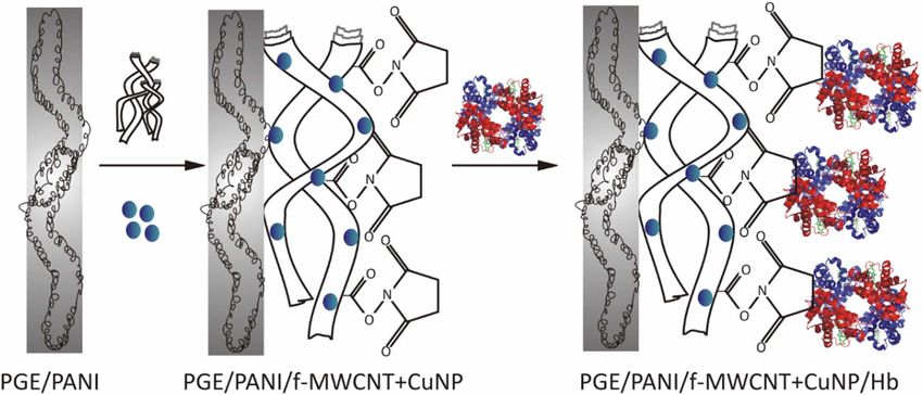
Journal of The Electrochemical Society, 2020 167 037520

                                  Review—Pencil Graphite Electrodes as Platform for Enzyme and
                                  Enzyme-Like Protein Immobilization for Electrochemical
                                  Detection
                                                            =,z                              =
                                  Akash Nathani,                  Nandimalla Vishnu,             and Chandra S. Sharma
                                  Creative & Advanced Research Based On Nanomaterials (CARBON) Laboratory, Department of Chemical
                                  Engineering, Indian Institute of Technology Hyderabad, Kandi, 502285 Telangana, India

                   Carbon-based electrodes are being used widely nowadays for biosensor applications, primarily owing to their good electrical conduc-
                   tivity and ease of functionalization. At the same time, the increasing demand for the low cost, disposable and the ease of availability
                   for the do-it-yourself assemblies have provided an opportunity to look beyond conventional carbon materials for electrochemical
                   analysis. In recent time, the pencil lead, entitled as the pencil graphite has been used as an electrode for the enzyme-based elec-
                   trochemical biosensors. The review highlights the various aspects involved in using pencil graphite electrode (PGE) as a working
                   electrode. This includes the various pretreatment strategies used, which is the first step toward the effective surface functionalization,
                   followed by strategies used for the immobilization of the functional nanomaterials and the enzymes and finally, the integration of
                   the PGE with different types of sensor assemblies. A comprehensive discussion on the latest development in this area also suggests
                   future perspectives based on PGE to develop low-cost point-of-care diagnostics.
                   © The Author(s) 2019. Published by ECS. This is an open access article distributed under the terms of the Creative Commons
                   Attribution 4.0 License (CC BY, http://creativecommons.org/licenses/by/4.0/), which permits unrestricted reuse of the work in any
                   medium, provided the original work is properly cited. [DOI: 10.1149/2.0202003JES]

                   Manuscript submitted September 18, 2019; revised manuscript received November 5, 2019. Published December 11, 2019. This
                   paper is part of the JES Focus Issue on Sensor Reviews.

     In recent times, pencil graphite electrodes (PGE) are emerging                        the SPCEs are single-use and disposable type electrodes, and its sur-
as an alternative for the conventional carbon and other metal elec-                        face cannot be renewed easily.16 In addition, the overall cost of PGE
trodes. The prime composition of PGE is graphite, which is a most                          from a commercially available pencil lead is less than the SPCE.11
stable form of carbon available in mineral ores like coal.1 Over carbon                        Among the hetero-structured graphite, pencil lead is one of the
and other conventional metal electrodes, PGE, as a disposable elec-                        low-cost and abundantly available materials used for various purposes
trode gained much consideration among the electrochemical groups                           in day-to-day life. Note that, as the name suggests, pencil lead does
for its abundance at ultra-low-cost with various interesting proper-                       not hold a metallic lead in it and it is an example for an intercalated
ties like; mechanical rigidity, chemical inertness, low background                         compound containing a mixture of clay and other particles in a con-
current, wide potential window, analyte adsorption, ease of minia-                         ducting graphite material.17 However, depending on the hardness and
turization, and modification.2,3 Note that sp2 hybridized carbons of                       blackness, pencils are categorised from 9H to 9B, here H stands for the
graphite are devoted to the good adsorption and higher conductivity.                       hardness and B indicates the blackness.17,18 For instance, 6B contains
Thus, graphite composite films on the conventional electrodes are used                     85% graphite, 10% clay and 5% wax.19,20 EDX of the same displayed
for the voltammetric determination of genotoxic nitro compounds,4                          82.9% of C, 6.5% of Si, 6.0% of O, 2.2% of Fe and 2.4% of Al21
organic compounds,5 antibiotics6 and nucleic acids.7 Interestingly,                        whereas 2B consists of 79% graphite and 21% clay22 with elemental
PGEs can facilitate the renewable surface easily unlike, the strin-                        composition of ((% w/w) O (8.5), P (0.1), Al (2.5), Si (3.0), K (0.18),
gent polishing procedures required for the conventional electrode like                     Mg (0.05), Fe (0.6), C (85.0) and Na (0.18)) thus the main composition
glassy carbon electrode (GCE), and yields better reproducibility for                       being aluminum silicate and HB consists of 68% graphite, 26% clay
the analyses.2,8,9                                                                         and 5% wax.23 However, there is a variation in the composition of a
     Based on the interesting properties of PGEs and thrust, our group                     particular grade by different manufactures. Although it seems obvi-
member has already exploited the pre-anodized PGE (PGE∗ ) as surface                       ous that the high graphite content serve as a better electrode due to
renewable electrochemical sensor for (i) the detection of total pheno-                     high electrical conductivity which is true in certain cases,24 the choice
lic preservatives in commercially available pharmaceutical insulin9 (ii)                   for the appropriate grade mostly depends upon the specific interaction
separation-free sensing of dihydroxy benzene isomers in tea dust10 and                     between the analyte and the graphite/clay composite. For instance,
(iii) simultaneous sensing of hypoxanthine (Hx), xanthine (X) and uric                     in the enzymatic sensing of hydrogen peroxide, 6H displayed a su-
acid (UA) in fish samples for quality monitoring.11 Before our attempt,                    perior performance compared to HB, 2B, 1H, 2H, 3H, 4H and 5H
several researchers successfully used PGE for the electrochemical ap-                      by delivering a good reversible redox current.25 In other cases, where
plications and proved that PGEs are the better choice over the other                       the sensor configuration involves electrodes drawn on the cellulose
conventional carbon electrodes. For instance, Wang et al. observed that                    paper, the ease of delivering sufficient material to the paper dictates
PGE yielded better electrochemical responses over the GCE for strip-                       the choice of the electrode.26 On the other hand, several researchers
ping based detection of nucleic acids.12 Similarly, J. K. Kariuki in 2012                  were using the chemically modified pencil leads as PGE based sen-
explored and compared the physical and electrochemical characteris-                        sors for various electrochemical and biological applications. For in-
tics of PGE with GCE.1 He studied the electron transfer rate of PGE                        stance, Prussian Blue modified PGEs for H2 O2 sensing.27 Likewise,
and GCE with redox systems (Eg. [Fe(CN)6 ]3-/4− , [Ru(NH3 )6 ]3+/2+ )                      1-napthylamine polymerised PGE for pH studies in non-invasive body
and found that PGE provided well defined and promising voltam-                             fluids,28 MoS2 grown PGE for guanine and adenine electrochemical
metric peaks with comparable electron rate transfer to GCE. On the                         detection29 and FeS2 grown PGE as an in-expensive tool for detection
other hand, a screen-printed carbon electrode (SPCE) may act as an                         of UA in human urine samples.30
alternative to low-cost PGE. For instance, SPCE based electrochem-                             Due to the increasing trend on modified PGE based electrochemical
ical biosensors are used to evaluate the damage of nucleic acids,13                        sensors, Akanda et al.,22 clearly discussed the trends in the fabrication
antioxidant activity of beer, coffee and tea,14 and to determine Metho-                    of metal/metal oxide/metal complex nanostructure, carbon nanostruc-
carbamol and Paracetamol simultaneously.15 Unfortunately, most of                          ture and polymer modified PGE as chemical or bio-sensors. In ad-
                                                                                           dition, Torrinha et al.,31 discussed the biosensors based on enzyme-
                                                                                           modified PGE. However, their discussion limited to research findings
  =
       These authors contributed equally to this work.                                     of the nanomaterial and enzyme-modified PGE’s. In this regard, we
   z
       E-mail: ch14m16p000001@iith.ac.in                                                   are motivated to discuss the biosensors not limited to development
Journal of The Electrochemical Society, 2020 167 037520

Figure 1. Some of the enzymes and enzyme-like proteins used for immobilization on PGE (Source: Wikipedia).

of biosensors based on enzyme only but also on research trend in              GOx in phosphate buffer solution (PBS) in the refrigerator and prior
the construction of enzyme-like proteins like haemoglobin (Hb) on             to use, all PVP/GOx modified electrodes were first rinsed with PBS.
PGEs. Starting from 1997, enzymes like glucose oxidase (GOx), al-             Then in an aerated pH 7 solution, at + 0.18 V vs SCE, the glucose
kaline phosphatase (ALP), xanthine oxidase (XOD), cholesterol oxi-            current response was measured for 1–14 mM glucose concentrations
dase (ChOx), horseradish peroxidase (HRP), urease, alcohol dehydro-           which yielded a linear relationship with a 0.0325 A M−1 sensitivity
genase (ADH), laccase, glucose oxidase (GlOX), Uricase, Ascorbic              and 0.5 mM as detection limit (LOD). Although the reusability of the
oxidase, Lipase, Glycerol Kinase (GK) & Glycerol -3-phosphate Ox-             enzyme electrode retained over a week, the developed sensor suffers
idase and proteins like Hb (Figure 1) are used for the modification           from some common interferents like lactic acid, ascorbic acid (AA)
of chemically modified PGE or bare PGEs and employed as electro-              and 4-aceto amino phenol.
chemical biosensors.                                                              In another case, Cheng et al.44 fabricated the glucose sensor by
    Electrochemical biosensors are one of the biosensors where they           using a hydroxyl methyl ferrocene mediator and HB-PGE modified
transform the information of the target biochemical to a readable sig-        with the carbon paste (CP) and GOx linked nano-Au (AuNP) par-
nal in the current or voltage. These electrochemical biosensors are           ticles (i.e., HB-PGE/CP-AuNP/GOx). The HB-PGE/CP-AuNP/GOx
majorly useful in a wide range of applications like the development           preparation procedure includes five-steps, in step-I, PGE surface was
of point of care devices, assessing the quality of food and monitoring        coated with a 0.8 cm layer of CP and dried for 10 min at 120°C. In
the environment.31–37 Fabrication of an electrochemical biosensor in-         step-II, AuNP were electrodeposited on the surface of CP by electro
cludes the two-step procedure, i.e., development of a biocompatible           reduction. In step-III, HB-PGE/CP-AuNP was dipped in L-cysteine
platform and immobilization of the desired enzyme. Immobilization             (CySH) solution for 1 hr at 25°C to create the covalent bonds between
of the desired enzyme on a biocompatible platform and retaining its ac-       the sulphydryl groups of CySH and AuNP. Further, the electrode was
tivity during the analyses are crucial in the construction of a successful    washed gently with distilled water to avoid the loosely bounded CySH.
electrochemical biosensor. Although enzymes are complex and expen-            To create the effective bonds between the electrode and GOx, electrode
sive, immobilization process yields various advantages like reusabil-         was placed in 40 mM solution of N, N’-dicyclohexylcarbodiimide
ity, improved stability and reduction in the cost of operation.38–43          (DCHCDI) chloroform solution for 1 hour at 40°C in step IV. Fur-
Thus based on the enzyme used for modification of PGEs, the                   ther, the electrode was dipped for 24 h in GOx in step-V. During the
current review article is sub-divided into five main categories, (i)          process, an amide bond was created between the carboxyl group and
GOx, (ii) ALP, (iii) Hb, (iv) XOD and (v) other enzyme-modified               the amino group of CySH and enzyme, respectively. Prior to use, HB-
PGEs.                                                                         PGE/CP-AuNP/GOx sensor was preserved at 4°C in pH 7 PBS. At
                                                                              0.33 V, the HB-PGE/CP-AuNP/GOx with a mediator showed an oxi-
    GOx modified PGEs.—Among various enzymes, GOx is used for                 dation peak corresponding to glucose. For 45-days, calibration curve
monitoring glucose. In the development of an electrochemical biosen-          by addition of glucose exhibited good linearity at 0–33.41 mM with 5
sor for glucose, GOx will be immobilized on the platform constructed          × 10−3 A M−1 and 22.3 μM as sensitivity and LOD respectively. The
by nanomaterial, and it easily catalyses glucose with better sensitivity      HB-PGE/CP-AuNP/GOX was usable till 228 days and showed 5–8%
and improved selectivity. In 1997, Zahir et al.22 were first to fabricate     interference to mannose, galactose and xylitol and 0–1% to cellobiose,
the mediator-less 2B-PGE based glucose sensor. In this work, 0.003 M          xylose, and arabinose. The developed HB-PGE/CP-AuNP/GOx elec-
of 4- vinyl pyridine (4VP) was polymerized to poly (4- vinyl pyridine)        trode was used for the analyses of glucose produced by the hydrolysis
(PVP) on 2B- pencil rod under the constant potential of +0.40 V vs            of Cinnamomum caphora tree branch fiber and validated with HPLC.
SCE in pH 3 conditions. After successful polymerization, the optimal          In his next attempt, Cheng et al.45 used HB-PGE/CP-AuNP/GOx for
electrode was immobilized with GOx (i.e., by three different tech-            the flow injection analysis (FIA) of glucose. In this work, the HB-
niques as (i) 5 μL of GOx solution applied for 5 min (ii) the disc            PGE/CP-AuNP/GOx prepared by following the aforementioned five
was immersed in a GOx solution and stirred for 0.5 hr at room tem-            steps and with a new step. This new step was prior to step-IV and
perature, dried and placed at room temperature, and (iii) 0.2% GOx            in this step the CySH bonds containing electrode was placed in re-
solution was placed in cell before the 4-vinyl pyridine (4-VP) poly-          dox mediator, 20 mM ferrocene carboxaldehyde (FcAld; dissolved
merisation at pH 6.4. Each PVP/GOx electrodes were placed still in            in EtOH/HCl, 99.5/0.5, v/v) for 1 hr at 75°C and rinsed further with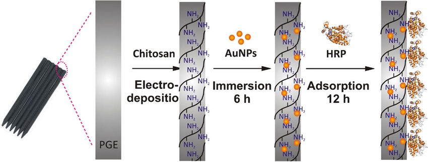
Journal of The Electrochemical Society, 2020 167 037520

Figure 2. Schematics on the preparation of PGE/GO/ZnO/CuO2 /GOx and its reaction to glucose oxidation.47

distilled water. Thus, the enzyme immobilized electrode was devel-             was polished by a glossy paper sheet and then pre-anodized by ap-
oped by chemical bonds between the redox mediator, FcAld and GOx               plying +1.8 V oxidation potential in 1 M NaOH solution for 10 s
modified HB-PGE/CP-AuNP. The new FIA electrode showed the lin-                 (2B-PGE∗ , ∗ = pre-anodized). Then 2 μL of GO solution (0.5 mg/mL)
ear response in a concentration range of 0–39.0 mM with 2.21 × 10⁻³            was drop-casted on the 2B-PGE∗ and kept at room temperature to
A M−1 sensitivity and 7.8 μM LOD. The FIA electrode can be used                dry. 2 μL of GOx solution (5 mg mL−1 in pH 7 PBS) was drop-cast
for more than 50 days and displayed 5–7% interference to mannose               onto dried GO and was kept at 15 ± 3°C for 5 min. For the two
and galactose. The developed electrode was successfully tested for             fabricated electrodes, the enzyme immobilization with simultaneous
the glucose detection in immobilized enzyme hydrolysate of waste               reduction of GO was done by two electrochemical methods. In the
bamboo chopsticks.                                                             first method, 6 continuous cyclic voltammetry (CV) cycles in 0 to
    Similarly, Dervisevic et al.46 developed a glucose sensor by immo-         −1.5 V potential range at a scan rate of 50 mV s−1 was employed, and
bilizing GOx on a poly(glycidyl methacrylate-co-vinylferrocene) (i.e.,         in the second method, the −1.5 V potential was applied constantly
poly(GMA-co-VFc)) polymer casted PGE (i.e., PGE/ poly(GMA-                     for 30 s. Finally, the as-prepared modified electrodes are identified
co-VFc)-GOx). In the fabrication of electrode, poly(GMA-co-VFc)                as 2B-PGE∗ /rGO/GOx. The developed 2B-PGE∗ /rGO/GOx electrode
was drop-casted on the electrode surface of PGE and left to                    showed a pair of redox peaks at −0.521 V corresponding to the en-
air dry. After drying, PGE/poly(GMA-co-VFc) was dipped in 3-                   zyme, and by amperometric method, 2B-PGE∗ /rGO/GOx displayed a
aminophenylboronic acid and in flavin adenine dinucleotide (FAD)               1.77 × 10⁻³ A M−1 sensitivity and 0.61 μM LOD at 0.04 to 0.6 mM
to create the linkages between the polymer and FAD. Further, the de-           glucose concentrations in pH 7 PBS.
veloped electrode was placed still for 24 h in the enzyme solution to              In yet another study, Vijayaraj et al. used rGO and GOx with the
let the reconstitution on the FAD monolayer. Using amperometric i-t            pretreated Type-B PGE for sensitive detection of glucose in PBS (pH
technique, current response of PGE/poly(GMA-co-VFc)-GOx to glu-                7).49 Here, the pretreatment was carried out by running 5 cycles of
cose was measured at an applied potential of 0.3 V in pH 7.5 PBS               CV in 0.1M H3 PO4 solution at the scan rate of 50 mV s−1 . Prior to
and found linearity in 1–16 mM range with a LOD 2.7 μM (S/N =                  the pretreatment, the curved surface was covered with Teflon, and the
3). Further, stability measurements of the PGE/poly(GMA-co-VFc)-               end was polished smooth using weighting paper. The rGO-GOx com-
GOX revealed that the electrode retained 90% of its original activity          posite was synthesized on the surface of PGE∗ in a single step. First,
during first 13 additions and displayed 65% in last 6 spikes. Prior to         the GO was synthesized using Hummers and Offeman method. Later,
the next measurement, the PGE/poly(GMA-co-VFc)-GOx electrode                   the PGE∗ was immersed in the mixture of GO (1 mg mL−1 in DI) and
should be stored at 4°C (0.1 M; pH 7.0 PBS) for 5 min. Then the stor-          GOx (10 mg mL−1 in 0.1 M PBS, pH 7.0) dispersions for 4h followed
age studies displayed a stable response for the first week and reduced         by the electrochemical treatment of 10 cycles CV from −1.3 to 0.2 V
to 60% in the next 6 days.                                                     in N2 saturated PBS (pH 5) at 50mVs−1 to form rGO–GOx/PGE∗ .
    In the next phase, Y. Mortazavi et al.47 started using graphene            Here, the reduction of GO to rGO and the covalent bonding of GOx
oxide (GO) and reduced GO (rGO) for the fabrication of GOx                     to GO was confirmed using fourier-transform infrared spectroscopy
based glucose sensors. In the first case, PGE was modified by a GO             (FT-IR) and X-ray photoelectron spectroscopy (XPS). The formation
and ZnO/Cu2 O (PGE/GO/ZnO/CuO2 ). GOx was immobilized on the                   of oxygen functionalities during pretreatment and reduction of GO was
PGE/GO/ZnO/CuO2 using the electrostatic interaction of positively              systematically investigated by the comparative voltammetry studies of
charged PGE/GO/ZnO/CuO2 and negatively charged enzyme. The                     GO–GOx/PGE∗ and GO–GOx/PGE while the immobilization of GOx
PGE/GO/ZnO/CuO2 /GOx preparation includes drop-casting of GO                   by comparing the CV’s of PGE, PGE∗ , GO/PGE∗ and GO-GOx/PGE∗
suspension on the cleaned surface of PGE (PGE/GO), and by a two-               in PBS. The greater reduction in the reduction peak current for GO–
step, electrochemical process, ZnO/Cu2 O compounds were prepared               GOx/PGE∗ as compared to GO-GOx/PGE over the successive cycles
on PGE/GO. At first, ZnO was formed by applying a potential of                 indicated the presence of a greater number of oxygen functionalities
−1.4 V for 200 s and by further by applying −0.7 V for 150 s,                  in case of GO–GOx/PGE∗ . It was observed that the reduction current
Cu2 O was formed on PGE/GO to yield PGE/GO/ZnO/CuO2 electrode.                 increases in the order PGE< PGE∗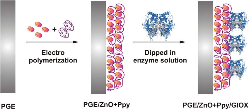
Journal of The Electrochemical Society, 2020 167 037520

   Reaction 1:                                                              linear response was obtained within 0.1–8 mM range and the LOD
                                                                            was 0.05 and 0.1 mM for the manual measurement and automated
               Glucose + O2 → gluconicacid + H2 O2                [R1a]     measurement respectively.
                                      −
                      O2 + 4H + 4e → 2H2 O                        [R1b]
                                                                                ALP modified PGEs.—In E. coli, ALP of molecular weight around
    Further, Sağlam et al.50 also immobilized GOx on quantum dot           94000 kDa is dimeric and a periplasmic protein. However, it is enzy-
(QD) (ZnS-CdS) modified PGE, crosslinked with chitosan (CT) for             matically inactive in cytoplasm. Most of the immunoassays use ALP
PGE∗ /ZnS-CdS/CT/GOx fabrication and for the electrochemical de-            as a label in the analyses. On the other hand, detection by electrochem-
tection of glucose through FIA method. For this PGE was activated at        ical methods achieved wide attention due to its precise measurement of
an applied potential +1.4 V for 60 s in pH 6 PBS. By electrochemical        current in turbid and coloured samples. In 2005, Ozsoz et al.52 were the
precipitation, quantum dots (ZnS-CdS) were deposited on the sur-            first to report on the hybridization detection of enzyme labelled DNA
face of activated PGE (PGE∗ ). The PGE∗ was placed for 10 min in a          by covalently immobilization technique by α-naphthyl phosphate (α-
mixture solution of 15 mM CdCl2 , 8 mM Na2 S2 O3 , 8 mM ethylene-           NAP) signal. In this work, on PGE surface was immobilized with ALP
diaminetetraacetic acid (EDTA) and 0.05 mM mercapto acetic acid             using the biotin-extravidin (Ex) and electrochemically assayed using
(MAA) (to avoid the coagulation of quantum dots) containing pH              a substrate, α-NAP, for the detection of hybridization. Using coupling
6 PBS and applied −1 V potential for 1000 s at 30°C. Further, the           agents, N-(dimethylamino) propyl- N‘-ethylcarbodiimide hydrochlo-
PGE∗ /CdS exposed to similar conditions with 15 mM ZnCl2 . The ob-          ride (EDC) and N-hydroxysulfosuccinimide (NHS) probes were co-
tained PGE∗ /CdS-ZnS was placed for 1 hr in GOX solution mixed with         valently attached on the surface of PGE. For hybridization, the PGE
CT and dried at 4°C in the refrigerator. Using FIA, dried PGE∗ /ZnS-        attached with probes are placed in a solution containing the oligonu-
CdS/CT/GOx was used as an electrochemical detector for glucose at           cleotides. ALP labelled extravidin (Ex-ALP) bind to hybrid and α-
flow rate = 1.3 mL min−1 , transmission tubing length = 10 cm, in-          NAP was added. Consequently, the reaction will occur between the
jection volume = 100 μL and constant applied potential = −500 mV            ALP and α-NAP. Post hybridization, the current response obtained by
vs Ag/AgCl. Under optimal conditions, the glucose sensor showed a           the reduction of α-NAP, i.e., 1-naphtol was measured by DPV (Fig-
linear response in the 0.01–1 mM range with LOD = 3 mM.                     ure 3).
    Teanphonkrang et al. developed a prototype for the auto-                    After DNA hybridization, Ozsoz et al.53 used a similar technique
mated robotic amperometric quantification of glucose in 24-well             to detect the mir21 in breast cancer cells (i.e., micro RNA). To con-
microplates.51 The device consisted of a PGE working electrode with         struct a sensor, c-probes were attached covalently on 0.5 mm HB-
a computer-controlled stepper motor to move the three-electrode as-         PGE using, EDC and NHS, coupling agents. For hybridization, the
sembly sequentially between the samples. The computer-controlled            HB-PGE attached with c-probes is placed in a solution containing the
micropositioner allowed the stepper motor to move the assembly in           target probes. ALP-Ex binds to the target due to the interaction be-
all three directions, x,y & z, i.e. right-left, backwards-forward and up-   tween biotin-avidin and enzyme transforms electro-inactive α-NAP
down respectively and the 2.5 ml vials of the 24-well plastic microtiter    (substrate) to electro-active 1-naphtol (product). Post hybridization,
served as the electrochemical cells. The working electrode consisted of     the current response obtained by electroactive 1-naphtol was quanti-
HB type PGE with 0.5 mm diameter and 3 cm length, and heat shrink           fied by DPV at +0.23V. Similar to the above work, Erdem et al.54 and
tubing was used to cover the length of the electrode leaving 2 mm of        co-authors of the above work, developed protocol for the electrochem-
the length to interact with the electrolyte. A systematic study of the      ical detection of specific Polymerase chain reaction (PCR)-amplified
pretreatment was carried out considering the untreated and the treat-       DNA fragments by the introduction of biotin tags into the DNA ampli-
ment using continuous and pulsed-potential driven electro-oxidation         cons during the PCR run in the presence of a biotinylated nucleoside
in 1 M KNO3 solution. The efficiency of the pretreatment was assessed       triphosphate. For electrochemical detection, PGE with biotinylated
by the ion exchange technique. The H+ ions from the carboxyl groups         DNA at the surface using the streptavidin-ALP conjugate, are used.
formed during pretreatment were exchanged with Ag+ ions by im-              The ALP converts the indicator α-NAP which is electrochemically
mersing the PGE∗ in 1 M AgNO3 in 0.1 M KNO3 for 1 min. Later the            inactive to an electroactive 1-naphthol. The amount of 1-naphthol was
efficiency of the ion exchange and hence the pretreatment was exam-         quantified by linear sweep voltammetry (LSV).
ined by DPV in 1 M KNO3 . The DPV reduction of bonded Ag was                    In another study for the detection of miRNA-21, Mandli et al. used
significantly higher in the case of pulsed treatment as compared to the     a sandwich hybridization technique using PGE working electrode to
continuous and untreated electrode, confirming the high efficiency of       overcome the need for modification of the target thus making the tech-
the pulsed treatment. Later the enzyme immobilization was carried out       nique more suitable for the real samples.55 In this study, the authors
using 1-Ethyl-3-(3-dimethylaminopropyl)carbodiimide (EDC) – N-              used HB-type pencil lead with 0.5mm diameter and 1.5 cm length of
hydroxysuccinimide (NHS) chemistry for both the types of pretreated         which 1 cm was immersed into the electrolyte. Here the pretreatment
electrodes and also the untreated electrode. A comparative sensing          was carried out by applying 1.4 V for 60 s in 0.5 M acetate buffer
study was carried out using chronoamperometry for the reduction of          (pH 4.8). The strategy involved functionalization of PGE∗ with gold
H2 O2 formed due to enzymatic reaction at 600 mV by spiking the             nanoparticles (NP) followed by the immobilization of the thiol termi-
supporting electrolyte with 1mM glucose solution at regular intervals.      nated capture probe (SH-p1). Later the nonspecific sites of the AuNP
The H2 O2 reduction current was significantly higher in the case of         were covered using 6-mercapto-1-hexanol (MCH) which also helped
pulse treated compared to the continuous treated and the untreated          in the orientation of the probe. After the hybridization of the miRNA-
electrode. After confirming pulsed treatment as the better technique,       21 (T) with the capture probe, a biotinylated complementary probe
the time of treatment was optimized by varying the pretreatment for 2,      (B-P2) was hybridization with the target to form a sandwich struc-
5, 15 and 30 min. It was observed that the H2 O2 reduction current in-      ture. Finally, streptavidin-conjugated ALP was immobilized on the
creased with the increase in the pretreatment period and got saturated      B-P2/T/MCH/SH-P1/AuNPs/PGE∗ by specific interaction with biotin
at 15 min of pretreatment although the improvement in the slop and the      to complete the sensor assembly B-P2/MCH/T/SH-P1/AuNPs/PGE∗ .
width of the linear region was optimal at 5min of pretreatment, so the      The hybridization of the target was investigated with 1 mg mL−1 α-
same was used for the later studies. Later the automated stability and      NAP in diethanolamine (DEA) buffer wherein, in the presence of
calibration testing were carried out in the 24-well microplates where       the ALP the electro-inactive α-NAP was converted to electroactive
the alternate wells were filled with buffer and glucose solution of dif-    1-naphtol that was sensed by voltammetry. A systematic study of each
ferent concentrations. The buffer solutions were mainly used for the        processing step was evaluated to get the optimal sensor performance.
baseline correction and cleaning of the electrode. The signal stability     These include the numbers of deposition cycles for the synthesis of
was within 5% of the mean value after the continuous operation for          Au nanoparticles where the PGE∗ was cycled between 0.9 to −0.3 V
6.5 h. The assembly offered the evaluation of 4 and 20 samples a per        at a scan rate of 50 mV s−1 in 0.1 M KNO3 containing 4mM HAuCl4
plate run in standard addition and calibration mode, respectively. The      followed by potential cycling between 0.2 to 1.6 V at 50 mV s−1 in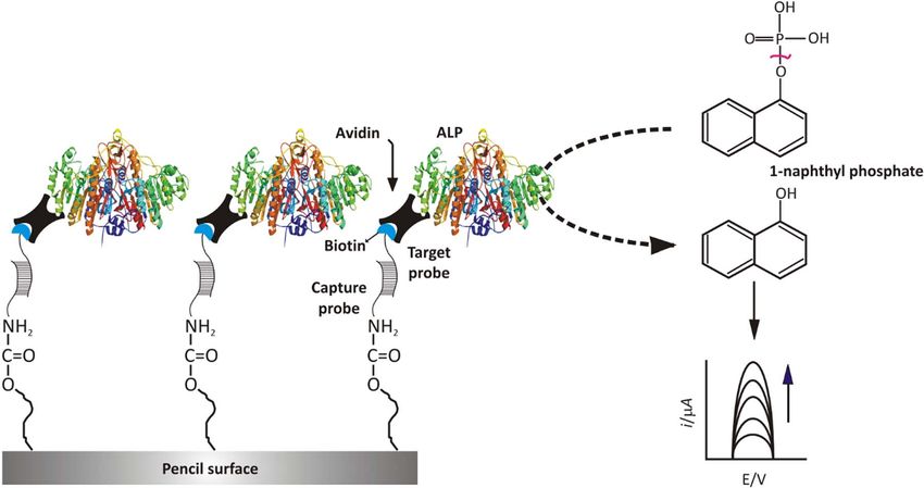
Journal of The Electrochemical Society, 2020 167 037520
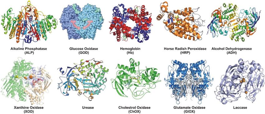
Figure 3. Schematic representation on the sensor preparation for the detection of mir21 in breast cancer cells.52
0.5 M H2 SO4 . The maximum CV peak current which could be ob- peroxidase and display enzyme-like activity. The interactions among
tained was tested using 5mM [Fe(CN)6 ]3−/4− in 0.1 M PBS (7.4) from the subunits may lead to allosteric properties of the protein. Each sub-
−0.2 to 0.8 V at 100mV s−1 . It was found that the maximum peak unit consists of a chain of a protein associated with a non-protein heme
current was obtained for 5 cycles of deposition and 6 cycles of acid group. Four iron-heme groups are liable for the electroactivity of Hb
treatment. Also, the effect of SH-P1 and SA-ALP concentration was displaying the reversibility of Fe(III) to Fe(II).56 Based on its elec-
evaluated, and the optimal response was obtained for 1.1 μM and troactivity, Batra et al.57 reported an acrylamide sensor. The central
12.2 U mL−1 , respectively. With the optimal parameters, the linear idea behind the sensor was an interaction between the acrylamide and
range was found to be 200 pM to 388 nM and the limit of detection Hb which leads to an adduct which is responsible for the change in
as 100 pM. Finally, the selectivity of the sensor was demonstrated electroactivity of Hb. The increase in the concentration of acrylamide-
in the presence of the non-complimentary sequence miRNA-125a, Hb adduct displayed the decrement in the peak current response. This
and the reproducibility was demonstrated by fabricating four identical is considered as an analytical signal for detection of acrylamide. In this
T-P2/MCH/SH-P1/AuNPs/PGE∗ and tested at different target concen- work, an electrochemical biosensor for acrylamide was developed by
trations where the minimum relative standard deviation obtained was immobilization of Hb on a composite formed by conducting polymer
2.1% and maximum as 9.9%. like polyaniline (PANI), carboxylated multiwalled carbon nanotube
(c-MWCNT) and copper nanoparticle (PANI/c-MWCNT/CuNP).
Hb modified PGEs.—Hb with a molecular weight of 64,500 is an Prior to the immobilization, the composite was electro-deposited
association of four globular subunits of proteins bonded noncovalently on PGE to develop PGE/PANI/c-MWCNT/CuNP and the elec-
to each other. Though Hb may not play a crucial role as an electron trode developed in this work is PGE/PANI/c-MWCNT/CuNP/Hb
carrier in biological systems, heme proteins of Hb act analogous to (Figure 4).
Figure 4. Schematic representation on the preparation of PGE/PANI/CuNP/cMWCNT/Hb.57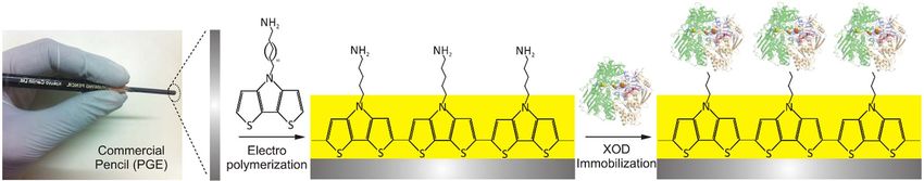
Journal of The Electrochemical Society, 2020 167 037520

    To fabricate the sensor, aniline was electropolymerised to PANI          to 2H+ + O2 + 2e− at 0.5 V. At pH 7.4 and 35°C room temper-
on the surface cleaned PGE by continuous 20 cycles at a potential            ature, PGE/Au-FeNP/CT/XOD exhibited optimum current response
window −0.1 to 0.2 V. c-MWCNT was prepared by ultrasonication                to X at 0.1-300 μM with 1.169 A M−1 cm−2 sensitivity and 0.1 μM
of MWCNTs in 3:1 ratio of H2 SO4 and HNO3 acid for 3–4 hrs. The              (S/N = 3) as LOD. The PGE/Au-FeNP/CT/XOD was later extended
obtained black coloured c-MWCNT was mixed with EDC and NHS.                  for the analysis of X in meat samples. The stability and reusability
This c-MWCNT solution is added with CuNP suspension, and the                 of PGE/Au-FeNP/CT/XOD displayed a 75% retention of the sensor
mixture was electrodeposited on the surface of PGE/PANI by 20 con-           activity after its usage of 100 times in 100 days when stored at 4°C.
tinuous cycles at −0.1 to 0.2 V. As an indication of PGE/PANI/c-             In another report, Devi et al.62 covalently immobilized XOD (from
MWCNT/CuNP formation, the surface of PGE become green. For the               buttermilk) onto boronic acid-functionalized Au-Fe NPs electrode-
immobilization of Hb, the surface of PGE/PANI/c-MWCNT/CuNP                   posited on PGE via the boro-ester linkages, i.e., the bond between the
electrode was dipped in Hb solution for overnight at room temper-            –NH2 groups of enzyme and free hydroxyl groups of boronic acid.
ature. Then PGE/PANI/c-MWCNT/CuNP/Hb was gently rinsed for                   In this work, PGE/Au-FeNP/XOD used boronic acid-activated Au-
3–4 times in pH 5 acetate buffer and preserved at 4°C until its fur-         FeNPs (12 h stirring of 10 g 4-mercaptophenylboronic acid and 6 g
ther use. The developed sensor showed the optimal response in pH             Au-FeNPs in 100 mL ethanol yielded a residue which was further
5 acetate buffer at 35°C operated at 20 mV s−1 . Under optimal DPV           washed in diethyl ether to remove 4-mercaptophenylboronic acid from
conditions, PGE/PANI/CuNP/cMWCNT/Hb exhibited 72.5 × 10³ A                   the obtained residue and vacuum dried at 40°C) and these Au-FeNPs
M−1 cm−2 sensitivity and 0.2 nM LOD with a linear range of 5 nM              were deposited on pretreated PGE in a 22mL solution containing 5 mM
to 75 mM. Further, the sensor was used for 120 times in a span of            of [Fe(CN)6 ]3−/4− and 50 mg of Au-FeNP (by 20 continuous CV cycles
100 days and stored at 4°C.                                                  between −0.6 V to +0.6 V at v 50 mV s−1 ). The resulting PGE/Au-
    In another case, Majidi et al. reported Hb modified electrodes for       FeNP modified electrode was rinsed gently in distilled water to avoid
the electrochemical detection of nitrite and H2 O2 .58,59 In the first re-   the loosely bound material and set aside in dry atmosphere at 4°C.
port, Hb modified H-PGE (H-PGE/Hb) was used for the electrochemi-            Enzyme was then immobilized as per their previous literature to yield
cal reduction of nitrite. In this work, Majidi et al.58 prepared H-PGE/Hb    PGE/Au-FeNP/XOD. Then at pH 7.2 and 30°C, PGE/Au-FeNP/XOD
where the PGE was tightly covered with a Teflon band leaving the top         showed a linear response to 0.05-150 μM of Hx with a LOD = 0.05
end for soldering the copper wire for electrical contact and the bot-        μM (S/N = 3). Further, PGE/Au-FeNP/XOD was tested for Hx levels
tom end was polished and used for the Hb immobilization by simple            in various meat samples. The PGE/Au-FeNP/XOD optimal response
drop-cast of Hb solution prepared by adding glycerol and Hb in pH            dropped by 50% after its usage over 100 days by placing it at 4°C.
7.4 PBS. The electrode was gently rinsed in pH 7.4 PBS and placed                In another report, an amperometric X biosensor was fabricated
at 4°C until its further use. DPV was carried out to determine the           by immobilization of XOD on an electrochemically polymerized con-
nitrite by H-PGE/Hb. The peaks corresponding the reduction of ni-            ducting polymer film (i.e., 10-[4H-dithieno (3, 2-b: 2 , 3 -d) pyrrole-4-
trite were found linear with the concentration of nitrite in a range         yl] decane-1-amine) coated PGE (i.e., PGE/DTP-NH2 /XOD).63 The
of 10–220 μM with a LOD of 5 μM. Further, the H-PGE/Hb elec-                 PGE/DTP-NH2 /XOD was prepared as per the following procedure
trode was tested for nitrite in the spinach and tap water. In another        (Figure 5), electropolymerization of DTP-NH2 on a cleaned and pre-
report, Majidi et al.59 prepared H-PGE/Hb as per their previous re-          treated PGE was carried out by applying CV potential in the range of
port with introducing a new step. Ormosil, prepared by 5 min stir-           −1.5 to 2.5 V vs Ag/AgCl and at v = 100 mV s−1 (PGE/DTP-NH2 ).
ring of methyltrimethoxysilane (MTMOS) and methanol and 0.1 M                Then, the PGE/DTP-NH2 was immersed in 2.5% GA for 3 hrs. To
HCL in 6:3:1 ratio, was drop-casted on the H-PGE/Hb to prevent               obtain PGE/DTP-NH2 /XOD, immobilization of XOD was performed
the loss of Hb. The dried H-PGE/Hb was stored at 4°C for overnight           by immersing PGE/DTP-NH2 in 2 U mL−1 XOD-ammonium sulfate
and used for the reduction of nitrite and H2 O2 . Under optimal DPV          suspension for 48 h on an orbital shaker set at 150 rpm at 4°C. The
conditions, H-PGE/Hb showed a linear response to H2 O2 and nitrite           fabricated PGE/DTP-NH2 /XOD electrode shown an optimal amper-
concentration ranging from 5–240 and 10–240 μM with 3 and 5 μM               ometric response at 30°C and at 0.5 V vs Ag/AgCl applied potential.
as LOD respectively. Further, the developed PGE/Hb electrode was             The current response increased linearly with increase in the X con-
used for the analyses of nitrite in tap water and H2 O2 in mother’s milk     centration ranging from 0.3 to 25 μM and showed the sensitivity and
samples.                                                                     detection limit of 0.124 A M−1 and 0.074 μM respectively. In the
                                                                             end, the applicability of PGE/DTP-NH2 /XOD electrode was verified
    XOD modified PGEs.—XOD is a molybdopterin-containing flavo-              to measure the X concentration in chicken meat samples.
protein that catalyses hypoxanthine (Hx) to xanthine (X) and X to uric
acid (UA) by oxidation with molecular oxygen while reducing H2 O.60             Other enzyme-modified PGEs.—Apart from GOx, ALP, XOD and
In 2013, Devi et al.61,62 immobilized XOD on PGE and used for X              Hb, there are some more enzymes used for the immobilization on
detection in the first report and Hx detection in another report. In the     the PGE surface. Other enzymes include ChOx, HRP, urease, ADH,
first report,61 XOD was covalently immobilized on the surface of a           lacasse, GlOx, uricase, lactate dehydrogenase, lipase, glycerol ki-
modified PGE by CT and Au-FeNP electrodeposition (i.e., PGE/Au-              nase and glycerol 3-phosphate oxidase (GPO), glucose dehydrogenase
FeNP/CT/XOD). Prior to the modification, the surface of the PGE              (GDH) and ascorbate oxidase.
wad cleaned using piranha solution [a mixture of H2 SO4 and H2 O2               Chauhan et al. immobilized ChOx (from Streptomycin sp.) on HB-
in 3:1] and followed by distilled water. The surface of PGE was pol-         PGE (15 mm diameter and 20 mm long) and used for amperometric
ished using a slurry of alumina. On the PGE surface, Au-FeNP/CT was          determination of serum cholesterol.64 To fabricate the HB-PGE/ChOx
electrodeposited by continuous electrochemical cycling of PGE in a           (Figure 6), wooden part of a pencil was removed completely and one
mixture of electrolyte containing the K3 [Fe(CN)6 ]/K4 [Fe(CN)6 ] in 1:1     end was dipped into 60% HCl for 24 hr followed by dipping in 70%
and Au-FeNP/CT solution between −0.37 V and 0.6 V. The PGE/Au-               HNO3 for 24 hrs. Then the cleaned electrode was placed for 8 hrs at
FeNP/CT electrode was dried at room temperature after gentle rinse           4°C in 0.2% enzyme solution (dissolved in pH 7.4 PBS). The elec-
in distilled water. On the dried PGE/Au-FeNP/CT, XOD was immo-               trode was then washed and used as HB-PGE/ChOx. The linear current
bilized via glutaraldehyde (GA) coupling. The GA groups formed on            response was observed at a cholesterol concentration ranging from
PGE/Au-FeNP/CT help the immobilization of XOD by simple dipping              1.29-10.33 mM, and LOD was 0.09 mM. The HB-PGE/ChOx elec-
for overnight at 4°C to obtain PGE/Au-FeNP/CT/XOD. Prior to use,             trode was used for 200 times for a span of 25 days, under 4°C storage
the PGE/Au-FeNP/CT/XOD was rinsed thoroughly in pH 7.4 PBS to                conditions.
avoid the excess and unbound enzyme. The XOD sensor was tested                  In another case, Teepoo et al. fabricated a renewable 6H-PGE
by adding X solution in pH 7.4 PBS, and the current response was             by forming a AuNP and HRP multilayer using layer-by-layer as-
measured at 0.5 V. The current response was due to the electrochemi-         sembly (Figure. 7).25 Prior to the fabrication, the 6H-PGE was
cal reactions between the XOD and X to yield H2 O2 and it’s splitting        pre-anodized by subjecting 1.8 V for 5 min in pH 4.8 acetate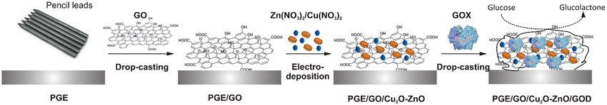
Journal of The Electrochemical Society, 2020 167 037520

Figure 5. Preparation of PGE/DTP-NH2 /XOD electrode.63

buffer containing NaCl. The pre-anodized 6H-PGE∗ was immersed             GA solution was dropped and allowed to dry. Then electrode was
in 0.5% w/v CT solution and subjected to 1.5 V for 5 min (6H-             placed in 5 mg mL−1 urease solution for 90 min. Then the PGE/f-
PGE∗ /CT). Then the 6H-PGE∗ /CT was dipped in AuNP solution for           MWCNT-PANI/urease electrode was dried and placed in pH 7.2 PBS
6 h (i.e., 6H-PGE∗ /CT/AuNP) and then placed in HRP for 12 h (6H-         and stored at 4°C until its further use. At constant potential (0.3 V
PGE∗ /CT/AuNP/HRP), which were repeated to obtain more num-               vs Ag/AgCl) and in pH 7.2 PBS, amperometric current responses
ber of layers. The developed 6H-PGE∗ /CT/AuNP/HRP showed a lin-           of PGE/f-MWCNT-PANI/urease displayed linearity for 0.07-10 mM
ear current response between 0.01 and 1.5 mM range of H2 O2 and           and showed 12 × 10−3 A M−1 and 0.04 mM sensitivity and LOD re-
displayed 0.15 A M−1 cm−2 , 0.002 mM sensitivity and LOD re-              spectively. But the PGE/f-MWCNT-PANI/urease retained only 50%
spectively and placed in pH 7 PBS at 4°C until further use. The           of original response after 15 days. In another case, Kashyap et al.
6H-PGE∗ /CT/AuNP/HRP was further tested for H2 O2 content in dis-         fabricated the cost-effective bio-electrodes by immobilizing Laccase
infector and hair dye samples.                                            on PGE@PANI/MWCNT (i.e., PGE@PANI/MWCNT/Laccase).67
    Next, Zhu et al. fabricated pyrocatechol violet electro-deposited     The electrode was fabricated by electro-polymerization of aniline
on single walled carbon nanotubes (SWCNT)-modified PGE (i.e.,             on a pre-treated PGE in the potential range of −0.2-1 V at v =
PGE/SWCNT/PCV) for electrochemical oxidation of dihydronicoti-            50 mV s−1 (i.e., PGE@PANI). The PGE@PANI was placed in
namide adenine dinucleotide (NADH) at 0.2 V vs saturated calomel          1 mg mL−1 MWCNT+EDC/NHS solution for 2 h. The bio-cathode
electrode (SCE).65 The PGE/SWCNT/PCV electrode displayed a                (PGE@PANI/MWCNT/Laccase) was developed by simple dipping
linear response to 1.3-280 μM of NADH and displayed 0.15 A                of PGE@PANI/MWCNT into 3 mg mL−1 enzyme solution for 24 h
M−1 cm−2 and 1.3 μM as sensitivity and LOD, respectively. The             under room temperature. Finally, the PGE@PANI/MWCNT/Laccase
PGE/SWCNT/PCV electrode was further fabricated as an ethanol              sensor displayed > 75% of the initial activity over the measurement
biosensor by immobilizing ADH through GA and bovine serum albu-           period.
min (BSA) coupling agents (i.e., PGE/SWCNT/PCV/ADH). The fea-                 In continuation, Batra et al. developed l-Glutamate biosensor by
sibility of PGE/SWCNT/PCV/ADH was tested for ethanol in 5 mM              immobilization of GlOx on a polymer-modified PGE (Figure 8).68
NAD+ containing pH 7.5 PBS. At 0.2 V vs Ag/AgCl, amperomet-               To prepare the polymer-modified PGE, the surface of electrode was
ric current response of PGE/SWCNT/PCV/ADH toward ethanol in               cleaned and placed in pyrrole (py) & ZnO containing KCl and per-
the presence of NAD+ displayed linearity for 9.3-320 μM ethanol           formed electropolymerisation in −0.25 to 0.8 V potential window for
and shown a sensitivity of 0.002 A M−1 cm−2 . The fabricated              20 continuous cycles. The resulted PGE/Ppy-ZnO was immersed in
PGE/SWCNT/PCV/ADH was further used to detect the ethanol con-             GlOx solution (5 U mL−1 ) for overnight at room temperature for en-
tent in liquor samples.                                                   zyme immobilization. Thus formed PGE/Ppy-ZnO/GlOx was rinsed
    Later, Meibodi et al.66 polymerized aniline on functionalized mul-    with pH 7.5 PBS and stored at 4°C. Further, the developed l-glutamate
tiwalled carbon nanotube (f-MWCNT) modified PGE and used to               showed optimal response at 0.1 M Tris–HCl buffer of pH 8.5 and
immobilize the urease via physical adsorption and electrochemical         30°C. The PGE/Ppy-ZnO/GlOx displayed 0.1 nM as LOD and was
entrapment technique and further used for the amperometric determi-       extended to detect l-glutamate in Chinese soups. Even after storage
nation of urea. To prepare urease biosensor, the PGE was prepared         at 4°C, biosensor displayed a 30% loss in the activity over 100 uses
using a pencil graphite cylinder and modelled epoxy resin. On pre-        in a span of 90 days. Similarly, Tsai and Wen developed a biosen-
pared PGE, suspension (sono-dispersion of 2 mg f-MWCNT+0.2 M              sor for UA,69 a final product of purine metabolism, using uricase and
aniline+0.8 M perchloric acid for 2 h, into 30 mL of solution) was        HRP. Here, UA gets converted to H2 O2 using uricase, and HRP fur-
drop-casted and subjected to electrochemical treatment for 30 cy-         ther detects the generated H2 O2 . Using the GA coupling technique,
cles between 0.1 V and −1 V at v = 40 mV s−1 . After this, 0.5%           the uricase and HRP are co-immobilized on the HB-PGE. Under op-

Figure 6. Preparation procedure of HB-PGE/ChOX for serum cholesterol.64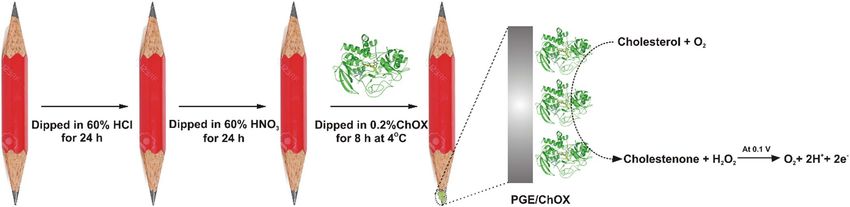
Journal of The Electrochemical Society, 2020 167 037520

Figure 7. Preparation of HRP based sensor on chitosan covered and AuNPs electrodeposited PGE surface.25

timal amperometric conditions, using the ferrocene monocarboxylic              LOD 0.1 μM. The sensor performance was also evaluated for the real
acid indicator, the electrode displayed linearity up to 0.12 mM with           samples including blood samples from healthy males and females and
sensitivity and LOD of 2.6 × 10⁻³ A M−1 and 0.6 μM. The developed              for the persons diagnosed with lactate acidosis as well as for milk,
UA sensor was tested for UA in blood serum.                                    cheese, curd, yogurt, white wine, red wine and beer. There was no
    In another case, Batra et al. developed a lactate dehydrogenase            effect on the sensor response due to interferents such ascorbic acid,
based sensor for the detection of lactic acid.70 Here the sensing prin-        glutamic acid, citric acid and glucose and 75% of the initial activity
ciple was based on the redox signal of NADH in the presence of the             was maintained after the regular usage for 60 days when stored at 4°C.
enzyme and the lactate. In this work, authors have used graphene oxide             For the detection of triglycerides (TG), Narwal & Pundir used
nanoparticles decorated PGE to immobilize the enzyme. Firstly, the             multiple enzymes co-immobilized on the surface of PGE∗ .71 In this
graphene oxide nanoparticles were synthesized by a modified Hum-               study, authors have used the enzymes lipase, glycerol kinases (GK)
mer’s method where HB type pencil rod was grounded in a mortar                 and glycerol 3-phosphate oxidase (GPO) for the detection of TG tri-
pestle. Later the graphite powder was mixed with the NaNO3 and                 olein. Here the enzyme lipase hydrolyzes the TG to glycerol and free
KMnO4 and was allowed to react with H2 SO4 and H2 O2 in succession             fatty acids. The glycerol thus formed is phosphorylated to glycerol-
followed by washing with H2 O2 , HCl and deionized (DI) water until            3-phosphate in the presence of adenosine triphosphate (ATP) by
the neutral pH was attained by the supernatant. The graphene oxide             GK which gets further oxidized to dihydroxyacetone phosphate and
nanoparticles thus obtained were dispersed in the 0.1 M KOH solu-              H2 O2 by the action of GPO. The CV peak current obtained by the
tion by continuous stirring and heating at 92°C. Later the nanoparticles       electro-oxidation of H2 O2 is then measured as the sensor response.
were electrodeposited on the PGE (6B, 2mm diameter) by potential               To prevent the denaturation of the enzyme and maintain their activ-
cycling in the dispersion from −0.15 to 2V at 20 mV s−1 for 20 cy-             ity and stability, the enzymes were first agglomerated to their re-
cles. Prior to electrodeposition, the PGE was polished by 0.05 μm              spective nanoparticle by glutaraldehyde crosslinking. The enzyme
alumina slurry on a polishing cloth followed by thorough washing               NP’s were separately synthesized by desolvation method where ab-
with ethanol and DI water. The enzyme was then immobilized using               solute ethanol was added to enzyme solutions dropwise followed by
EDC-NHS chemistry to complete the senor assembly. Various sensing              crosslinking using a glutaraldehyde solution. The Enzyme NP thus
parameters such as solution pH, temperature and time of incubation             formed were surface-functionalized using cystamine to retain a pos-
for enzyme immobilization were optimized to get the optimal sensor             itive charge on the NP surface and were characterized using scan-
performance. Later the sensor performance was evaluated by cyclic              ning electron microscope (SEM), transmission electron microscopy
voltammetry in 0.1 M sodium phosphate buffer (pH 7.3) from −0.15               (TEM) and UV-vis spectroscopy. The PGE with 2 mm diameter and
to 0.7 V in the presence of 6.6 mM NAD+ and varying concentration              2.5 cm length was pretreated by potential cycling from −1.1 to 0 V in
of lactic acid. The linear range was found to be from 5 to 50 mM with          0.2 M H2 SO4 . Later PGE∗ was immersed in the suspension of Lipase

Figure 8. Preparation of glutamate sensor by immobilization of GlOx on ZnO and polypyrrole coated PGE surface.68
Journal of The Electrochemical Society, 2020 167 037520
NPs/GKNPs/GPONPs (1:1:1) for the immobilization to take place via of a hundredfold of glutamic acid and saccharose and a thousandfold
electrostatic interaction between the negatively charged surface and of galactose higher than glucose. However, a significant increase in the
the positively charged –NH2 of cysteamine dihydrochloride. Later the oxidation peak current was observed in the equimolar concentration
CV studies were carried out for Lipase NPs/GKNPs/GPONPs/PGE∗ of AA, UA, dopamine and CySH mainly due to overlapping of their
using 0.1 M PBS containing 6.6mM ATP, 10mM MgCl2 and 1mM oxidation peaks with that of NADH.
Triton X100 at 20mV s−1 scan rate and the same was compared with The antioxidants present in orange juice are well known to protect
Lipase/GK/GPO/PGE∗ . It was found that the oxidation current ob- biological targets from the reactive oxygen, nitrogen and hydroxyl
tained for NP’s immobilized electrode was much higher as compared species.73,74 To distinguish the contribution of the antioxidant capac-
to the native enzyme immobilized electrode. The linear range was ity of AA and phenols and to quantify the content of AA, Barberis et
found to be 0.1 mM – 45 mM with LOD as 0.1 nM. Finally, the study al. developed a PGE and ascorbate oxidase based telemetric sensor.75
was carried out in the sera of real blood samples collected from 20 The authors used a strategy where the oxidation current obtained with
apparently healthy adult males and females and also from the 20 sub- (biosensor-BS) and without (sensor -S) presence of the enzyme was
jects diagnosed with hypertriglyceridemia in various age groups. The evaluated simultaneously by using two working electrodes. The differ-
study with real samples was also carried out with a standard enzymic ence in the current obtained at a particular voltage was used to estimate
colourimetric kit, and a good correlation coefficient was obtained be- the selectivity index and content of AA and the antioxidant capacity
tween the two methods. Interferants such as urea, uric acid, ascorbic in the juices. The sensor/biosensor assembly involved four 2H-Type
acid, glutamic acid and citric acid did not show any significant effect PGE (length- 30 mm, diameter- 300 μm) consisting of one pseudo ref-
on the response of the sensor, and it was found the electrode lost 20% erence, one auxiliary and two working electrodes. The working elec-
of the initial response after regular usage of 240 days when stored at trodes were initially coated with the epoxy resin and later polished
4°C. with alumina wheel attached to a high-speed drill to get a disc-shaped
In another study based on the ZnS-CdS quantum dot (QD), Ertek active area. The biosensor electrode surface was initially coated with
et al. developed QD modified PGE for the development of photo- polyethyleneimine (PEI) by a dip evaporation method, which acts as
electrochemical glucose sensor based on the enzyme glucose dehy- an enzyme stabilizer. Later the PEI coated PGE was further coated
drogenase and the redox couple NAD+ /NADH using FIA system.72 with 25 μL of 10% bovine serum albumin (BSA) solution containing
Initially, the PGE was activated by 1.4 V for 60 s in pH 7 PBS. Later 25 U of ascorbate oxidase. The PEI and enzyme coating steps were
the electrode surface was immobilized sequentially with ZnS and CdS alternately repeated 10 times, followed by a final coating layer of
by electrochemical precipitation method in the presence of mercapto polyurethane (PU) which acts as an enzyme immobilizer. The sensor
acetic acid (MAA) as described by Sağlam et al. When the QD mod- electrode was prepared in a similar fashion except for the use enzyme.
ified electrode is irradiated with the light source, the photoexcitation All the calculations were carried out based on the assumptions that 1)
leads to the formation of electron-hole pair in the conduction-valance The total oxidation current recorded by the sensor in more than the
band. The transfer of electrons between the electrode material and biosensor in the presence of AA as some amount of AA gets oxidized
the holes of valance band of QD with further transfer of electrons by the enzyme before it reaches the PGE surface. 2) The total current
between the conduction band of QD and the electroactive molecule recorded by the sensor and biosensor is the same in the absence of
leads to the generation of anodic/ cathodic photocurrent. Addition- the AA. The LOD and limit of quantification were calculated as 0.26
ally, the hybrid QD provide better charge separation and high quan- and 0.77 μM respectively for the biosensor. The biosensor displayed
tum yield compared to individual QD. The CV and impedance stud- good operational stability for 30 measurements, after which the cur-
ies of bare PGE∗ , CdS/PGE∗ and MAA-ZnS-CdS/PGE∗ in 10.0 mM rent response decreased to 70% of the initial value. To further enchase
[Fe(CN)6 ]3−/4− containing 0.10 M KCl showed that the redox CV peak the sensitivity and lower the LOD, Barberis et al. immobilized differ-
current decreases and the charge transfer resistance increases with suc- ent carbon allotropes including SWCNT, MWCNT, fullerene C60 and
cessive immobilization of QDs indicating the semiconducting nature fullerene C70 on the PGE as they are known to have high electron affin-
of the QD. To demonstrate the photocatalytic effect of the QD, CV ity, high specific surface area and display good adsorption capacity for
studies of bare PGE∗ was compared with QD immobilized PGE∗ with organic molecules.76 The biosensor-sensor assembly was carried out
and without illumination in the presence of 2 mM NADH in PBS (pH as described above except prior to PEI coating, the PGE was coated
7.0). It was observed that while there was a very little increment in the with fullerene (FC) or CNT’s by dip evaporation in their dimethyl-
peak current for the bare PGE∗ , the QD immobilized PGE∗ showed formamide (DMF) dispersion containing 60 mg mL−1 fullerene or
a significant increase in the oxidation current under irradiation with 10 mg mL−1 CNT with 50 tip cycles, besides some preliminary stud-
250 W halogen lamp. To evaluate the biosensing performance, the ies were carried out to choose the best solvent and the concentration
CV’s of GDH/ PGE∗ and GDH/MAA-ZnS-CdS/PGE∗ were recorded for FC/CNT dispersion. Later the CV’s of S-FC60 (sensor-FC60 ), B-
in absence and presence of illumination in 0.1 M PBS (pH 7.0) con- FC60 (Biosensor-FC60 ), S-FC70 , B-FC70 , S-SWCNT, B-SWCNT, S-
taining 0.1 M KCl and 10 mM NAD+ with 40mM glucose at 20 mV MWCNT and B-MWCNT were compared for the baseline current
s−1 . It was observed that the redox current obtained was the largest and in the presence of 1mM AA. It was observed that for B-FC60 and
for GDH/MAA-ZnS-CdS/PGE∗ with illumination demonstrating the B-FC70 the CV’s in AA solution completely overlapped with that of
photocatalytic performance of QD for glucose sensing application. the baseline while that for B-SWCNT and B-MWCNT, the current
Later, various operating parameters such as transmission tube length, was higher than the baseline. This was mainly due to the complete
sample volume, flow rate and the applied potential were optimized at shielding of AA by oxidation before it can reach the sensor surface
10 cm, 100 μL, 0.6 ml min−1 and 800 mV vs Ag/AgCl respectively to in the case of FC while an incomplete shielding effect in the case of
get the optimal sensor performance in FIA configuration. For the cal- CNTs. It is hypothesized that this is mainly due to higher enzyme
ibration studies, varying concentration glucose was injected with two loading onto the FCs when compared to CNTs due to their high sur-
injections of each concentration through a 100 μL sample loop. The face/ volume ratio. The hypothesis was confirmed by measuring the
LOD obtained without (amperometric) and with (photoamperometric) CVs in the AA solution and at different concentrations of O2 . It was
illumination were 0.09 mM and 0.05 mM respectively, and a two-fold observed that for 0% O2 , CV currents for sensors were statistically
increase in the sensitivity was observed for the photoamperometric same as that of biosensors and with the increase in the O2 levels the
detection as compared to amperometric detection of glucose. The pre- current for biosensors decreased while that of the sensors remained
cision studies carried out by repetitive injection of 7 pulses of 0.5 mM the same. This was mainly due to the enzymatic oxidation of some
glucose solution gave an RSD of 3.5% and 4.5% for amperometric quota of AA before it reached the sensor surface. At 21% O2 , there
and photoamperometric detection, respectively. Finally, the interfer- was 100% decrease in the current for FC based biosensors while it
ence studies were carried out in the presence of AA, glutamic acid, was 85.5% and 76.4% decrease for B-SWCNT and B-MWCNT re-
UA, saccharose, dopamine, CySH and galactose. It was observed that spectively. Thus at a given AA concentration, the enzymatic activ-
there was no significate change in the oxidation current in the presence ity depends upon the O2 concentration while at a given AA and O2You can also read