Serum carotenoids and Pediatric Metabolic Index predict insulin sensitivity in Mexican American children
←
→
Page content transcription
If your browser does not render page correctly, please read the page content below
www.nature.com/scientificreports
OPEN Serum carotenoids and Pediatric
Metabolic Index predict insulin
sensitivity in Mexican American
children
Srinivas Mummidi1,11*, Vidya S. Farook1,11, Lavanya Reddivari2,11, Joselin Hernandez‑Ruiz3,
Alvaro Diaz‑Badillo1, Sharon P. Fowler4, Roy G. Resendez1, Feroz Akhtar1,
Donna M. Lehman5, Christopher P. Jenkinson1, Rector Arya1, Jane L. Lynch6, Jose A. Canas7,
Ralph A. DeFronzo5, Daniel E. Hale8, John Blangero1, Juan Carlos Lopez‑Alvarenga1,
Ravindranath Duggirala1 & Jairam K. P. Vanamala9,10*
High concentrations of carotenoids are protective against cardiometabolic risk traits (CMTs) in
adults and children. We recently showed in non-diabetic Mexican American (MA) children that serum
α-carotene and β-carotene are inversely correlated with obesity measures and triglycerides and
positively with HDL cholesterol and that they were under strong genetic influences. Additionally, we
previously described a Pediatric Metabolic Index (PMI) that helps in the identification of children who
are at risk for cardiometabolic diseases. Here, we quantified serum lycopene and β-cryptoxanthin
concentrations in approximately 580 children from MA families using an ultraperformance liquid
chromatography-photodiode array and determined their heritabilities and correlations with CMTs.
Using response surface methodology (RSM), we determined two-way interactions of carotenoids and
PMI on Matsuda insulin sensitivity index (ISI). The concentrations of lycopene and β-cryptoxanthin
were highly heritable [h2 = 0.98, P = 7 × 10–18 and h2 = 0.58, P = 1 × 10⁻⁷]. We found significant (P ≤ 0.05)
negative phenotypic correlations between β-cryptoxanthin and five CMTs: body mass index
(− 0.22), waist circumference (− 0.25), triglycerides (− 0.18), fat mass (− 0.23), fasting glucose
(− 0.09), and positive correlations with HDL cholesterol (0.29). In contrast, lycopene only showed
a significant negative correlation with fasting glucose (− 0.08) and a positive correlation with HDL
cholesterol (0.18). Importantly, we found that common genetic influences significantly contributed
to the observed phenotypic correlations. RSM showed that increased serum concentrations of
α- and β-carotenoids rather than that of β-cryptoxanthin or lycopene had maximal effects on ISI.
In summary, our findings suggest that the serum carotenoids are under strong additive genetic
influences and may have differential effects on susceptibility to CMTs in children.
Obesity or overweight affects one in three children in the United States1 and is associated with other comorbid
conditions such as insulin resistance, hypertension, non-alcoholic fatty liver disease, obstructive sleep apnea,
dyslipidemia, and psycho-behavioral problems2–9. A major concern is that about 70% of these children are at
risk of developing adult obesity that predisposes them to develop several chronic diseases, including metabolic
1
South Texas Diabetes and Obesity Institute, Department of Human Genetics, School of Medicine, University of
Texas Rio Grande Valley, Edinburg, TX, USA. 2Department of Food Science, Purdue University, West Lafayette, IN,
USA. 3Clinical Pharmacology Unit, Hospital General de México Dr. Eduardo Liceaga, Mexico City, Mexico. 4School
of Public Health, University of Texas Health Houston, Houston, TX, USA. 5Department of Medicine, School of
Medicine, University of Texas Health San Antonio, San Antonio, TX, USA. 6Department of Pediatrics, School of
Medicine, University of Texas Health San Antonio, San Antonio, TX, USA. 7Johns Hopkins All Children’s Hospital,
St. Petersburg, FL 33701, USA. 8Department of Pediatrics, Penn State Health Milton S. Hershey Medical
Center, Hershey, PA, USA. 9Department of Food Science, Pennsylvania State University, University Park, PA,
USA. 10Department of Plant Science, Pennsylvania State University, University Park, PA, USA. 11These authors
contributed equally: Srinivas Mummidi, Vidya S. Farook and Lavanya Reddivari. *email: srinivas.mummidi@
utrgv.edu; jairam.vanamala@gmail.com
Scientific Reports | (2021) 11:871 | https://doi.org/10.1038/s41598-020-79387-8 1
Vol.:(0123456789)www.nature.com/scientificreports/
syndrome (MS)10,11. Growing evidence suggests that the accumulation of adipose tissue resulting from a positive
energy balance leads to a low-grade pro-inflammatory state and oxidative stress12,13. There are marked dispari-
ties in adult and childhood obesity between various ethnic groups with increased prevalence in Hispanics and
African Americans14, which are most likely due to environmental and genetic factors and their i nteractions15,16.
Fruits and vegetables contain phytonutrients such as carotenoids, which are considered anti-obesogenic due
to their anti-inflammatory and anti-oxidant properties17,18. Carotenoid functionality is most likely mediated
through modulation of inflammation-related gene transcription and signal transduction pathways and scav-
enging reactive oxygen s pecies17,19–23. While the U.S. Department of Health and Human Services recommends
daily fruit and vegetable consumption24, ~ 60% of children aged 1 to 18 years do not meet the suggested levels of
intake of 1–2 cups of fruits and 1–3 cups of vegetables25 and often low socioeconomic status compromises the
quality of their d iet26. Although Hispanic children and adolescents may have higher fruit and vegetable intake
compared with African–American and white youth27, their relative serum carotenoid concentrations could be
variable28, suggesting that additional factors should be taken into consideration for understanding metabolic
dysfunction in this high-risk population.
Six carotenoids (α-carotene, β-carotene, β-cryptoxanthin, lycopene, lutein, and zeaxanthin) constitute > 95%
of the circulating carotenoids in the human body29,30. Chemically, carotenoids are lipophilic polyisoprenoid
compounds that are either hydrocarbons (α-carotene, β-carotene, lycopene) or the oxygenated xanthophylls
(β-cryptoxanthin, lutein, zeaxanthin)30. Among these, α-carotene, β-carotene and β-cryptoxanthin, are con-
sidered to be provitamin A carotenoids. As their name implies, they can be further metabolized to retinol and
related compounds that function as Vitamin A, which is involved in growth, development, and visual f unction31.
Lycopene, lutein, and zeaxanthin are non-vitamin A carotenoids, of which the latter two are intimately involved
in blue light filtering in the macular region of the retina32. Lycopene is the most efficient singlet oxygen quencher
among the carotenoids mentioned a bove33.
Humans cannot synthesize carotenoids and rely on the dietary intake of fruits and vegetables for their supply.
There is substantial inter-individual variability in serum and tissue carotenoid levels. Several factors, includ-
ing age, gender, body weight, physical activity, alcohol consumption, drug use, smoking status, and infectious
diseases, may determine this inter-individual v ariability34. Also, several candidate-association and genome-
wide association studies have identified genetic variants that could potentially influence carotenoid levels and
bioavailability by altering their absorption, cleavage, transport, metabolism, and tissue i ncorporation35–38. The
genetic basis for inter-individual variation in carotenoids in Mexican Americans is poorly understood. Our
recent data showed that serum levels of α-carotene and β-carotene are under strong genetic additive influences
in Mexican American children who were at high risk for overweight (53%), obesity (34%), prediabetes (13%),
and metabolic syndrome (19%)39,40 and that they have phenotypic correlations with several cardiometabolic risk
traits (CMTs)39. Our recent studies also demonstrated that genetic factors might influence positive phenotypic
correlations between β-carotene and HDL cholesterol and negative phenotypic correlation between β-carotene
and waist circumference and body mass index39. In the current study, our primary objective was to examine
the genetic basis of lycopene and β-cryptoxanthin variability among these children and their correlation with
CMTs. Importantly, we assessed the interactions between the four carotenoids and a Pediatric Metabolic Index
(PMI) which, incorporates adiposity and lipid measures in predicting insulin sensitivity using the response
surface methodology.
Materials and methods
Subjects. The current study is a family-based cross-sectional study in Mexican American children to deter-
mine the heritability of two different carotenoids, β-cryptoxanthin and lycopene, and their association with
CMTs. The participants of this study were Mexican American children and adolescents (N = 673, age 6–17 years
old), who were recruited as part of the San Antonio Family Assessment of Metabolic Risk Indicators (SAFARI)
study40. These children were from 401 MA nuclear families/sibships, which were embedded within extended
families that we recruited previously as part of three well established genetic epidemiological studies in San
Antonio, TX, and vicinity40. Many of the children belonged to predominantly low-income extended families.
Each nuclear family/sibship had ~ 2 children (range 1–5 children), and the children from all the families gener-
ated a total of 3664 relative pairs. As described previously, this study aimed to assess CMTs and their genetic and
environmental bases in MA children in San Antonio, Texas. The present study excluded three children who had
type 2 diabetes; thus, it involved 670 non-diabetic children. The Institutional Review Board of the University
of Texas Health, San Antonio, Texas, reviewed and approved all the research protocols. For each child, written
informed consent was obtained from one or both parents, and for children seven years or older, a signed assent
was obtained. All studies were performed in accordance with the relevant guidelines and regulations.
Phenotype data. Extensive information such as family history, socio-demographic characteristics, and
environmental data were collected using questionnaires at the clinic or home interviews as described in detail
previously40. All physical, clinical, and laboratory assessments were performed at the Children’s Center of the
Texas Diabetes Institute (TDI), and some laboratory assessments using serum samples were carried out at the
Texas Biomedical Research Institute, San Antonio, TX, the USA. as previously r eported40. Aliquots of biospeci-
mens (e.g., fasting serum samples) collected at the same clinic visits of the children were prepared and promptly
frozen at − 70 ºC for future use. The following ten obesity-related quantitative traits were used for estimating the
phenotypic, genetic, and environmental correlations with serum carotenoid levels: waist circumference, body
mass index (BMI), fat mass assessed by dual X-ray absorptiometry (DXA), blood pressure [systolic (SBP) and
diastolic blood pressure (DBP)], fasting plasma glucose, fasting serum specific insulin, the homeostasis model
of assessment-insulin resistance (HOMA-IR), HDL cholesterol (HDL-C), and triglycerides. As described pre-
Scientific Reports | (2021) 11:871 | https://doi.org/10.1038/s41598-020-79387-8 2
Vol:.(1234567890)www.nature.com/scientificreports/
viously, serum carotenoids were measured with the available serum samples using Waters’ ultra-performance
liquid chromatography-photodiode array (UPLC-PDA)39. The carotenoid standards were obtained from Sigma-
Aldrich (Saint Louis, MO, USA). The α- and β-carotenoid data were previously reported39, and measurements
of lycopene and β-cryptoxanthin were obtained for this study (N = ~ 580). Both dietary intake of carotenoid
levels as assessed by the self-reported Block Kids Food Frequency Questionnaire (FFQ) and serum carotenoid
data were available for a subset of our SAFARI children (N = ~ 440). The obtained FFQ information was based
on 78 questions regarding the consumption of food/beverage items (frequency and amounts consumed) in the
previous week41,42. The Block FFQs were analyzed by NutritionQuest (Berkeley, CA). Adjustments were made to
carotenoid concentrations using age, sex, ln BMI, total energy intake (kcals), and total cholesterol as covariates
to assess correlations between dietary intake of carotenoid and serum carotenoid levels43,44.
Statistical analyses. Variance components analysis. The genetic basis of lycopene and β-cryptoxanthin
were determined using a variance-components approach (VCA) as implemented in the computer program
SOLAR45. Briefly, in a simple model, variances or covariances between relatives as a function of the genetic
relationships can be specified, and the proportion of phenotypic variance that is attributed to (additive) genetic
influences (i.e., heritability, h2) is estimated from the components of variance. A likelihood ratio test was used to
test whether the heritability of a given carotenoid was significant (P ≤ 0.05). The lycopene and β-cryptoxanthin
values were inverse normalized for the genetic analyses, and all analyses accounted for covariate effects (e.g., age
and sex). We reanalyzed the data to determine the heritability estimate of a given carotenoid after accounting for
the dietary intake of the same carotenoid as an additional covariate using a sub-set of our data.
Bivariate genetic analysis. Bivariate genetic analysis was used to determine the phenotypic, genetic, and envi-
ronmental correlations between the carotenoids and the ten CMTs. In this approach, a given phenotypic cor-
relation (ρP) between a pair of phenotypes (e.g., lycopene and BMI) is partitioned into additive genetic (ρG) and
environmental (ρE) correlations. The phenotypic correlation (ρP) between a pair of traits is given by:
ρP = h21 h22 ρG + e12 e22 ρE
where ρP = the phenotypic correlation; ρG = the additive genetic correlation; ρE = the random environmental cor-
relation; h21 = the heritability of trait 1; h22 = the heritability of trait 2; e12 = equal to 1 − h21; e22 = equal to 1 − h22. The
significance (P ≤ 0.05) of the phenotypic, additive genetic, and random environmental correlation was determined
using likelihood ratio tests. The additive genetic correlation (ρG) is a measure of the shared genetic basis of the
two traits (i.e., pleiotropy).
Pediatric Metabolic Index (PMI). PMI was based on two different adiposity-related components obtained from
396 Mexican c hildren46. It was shown to be correlated well with HOMA-IR, Matsuda ISI, and hepatic enzymes
and could be used to identify children who are at increased risk for cardiometabolic diseases. The first compo-
nent of PMI was based on the distribution of adipose tissue, and the second component was based on adipose
dysfunction. For the second component, median values from the normal-weight children in the population
served as the cut-offs with considerations for age (10 years) and sex. PMI index for SAFARI data was calculated
as follows:
♀ < 10 years:
WC TG 1.32
(BMI z score) + × ×
36.13 + (2.3 ∗ Age) 0.88 HDL − C
♀ > 10 years:
WC TG 1.34
(BMI z score) + × ×
36.13 + (2.3 ∗ Age) 1.04 HDL − C
♂ < 10 years:
WC TG 1.38
(BMI z score) + × ×
44.08 + (1.48 ∗ Age) 0.77 HDL − C
♂ > 10 years:
WC TG 1.30
(BMI z score) + × ×
44.08 + (1.48 ∗ Age) 1.06 HDL − C
The abbreviations in the above equations are defined as follows: TG = fasting triglycerides (mmol/L); HDL-C:
HDL cholesterol (mmol/L); WC = waist circumference (cm); and BMI = Body Mass Index expressed as Z-score).
Age is in years.
Response surface methodology (RSM). The response surface analysis with quadratic effects was used to deter-
mine two-way interactions of serum carotenoids and PMI on Matsuda insulin sensitivity index (ISI), a better
measure of IR compared to HOMA-IR40. Statistical analysis was performed using STATISTICA (version 7).
Scientific Reports | (2021) 11:871 | https://doi.org/10.1038/s41598-020-79387-8 3
Vol.:(0123456789)www.nature.com/scientificreports/
Variable N Mean ± SD or% h2 ± SE P-value References
Girls 670 49.3 – – –
Age (years) 670 11.5 ± 3.5 – – –
Overweight 670 52.7 – – –
Obese 670 33.6 – – –
Pre-diabetes 630 13.2 – – –
MSa 625 18.7 – – –
Acanthosis nigricans 661 33.1 – – –
BMI (kg/m2) 670 22.7 ± 6.5 0.75 ± 0.11 1.1 × 10⁻¹¹ Fowler et al.40
Waist circumference (mm) 664 764.5 ± 179.7 0.63 ± 0.12 3.0 × 10⁻⁸ Fowler et al.40
Fat mass (kg) [DXA] 634 16.0 ± 11.1 0.69 ± 0.12 1.8 × 10⁻⁹ Fowler et al.40
Fasting glucose (mg/dl) 630 89.5 ± 7.5 0.39 ± 0.11 6.3 × 10⁻⁵ Fowler et al.40
Fasting insulin (µIU/ml) 626 13.6 ± 9.4 0.55 ± 0.11 2.0 × 10⁻⁷ Fowler et al.40
HOMA-IR 622 2.0 ± 1.3 0.60 ± 0.11 1.8 × 10⁻⁸ Fowler et al.40
–8
HDL cholesterol (mg/dl) 623 45.8 ± 10.9 0.64 ± 0.12 2.9 × 10 Fowler et al.40
–13
Triglycerides (mg/dl) 623 74.9 ± 39.8 0.77 ± 0.11 8.8 × 10 Fowler et al.40
–10
SBP (mmHg) 670 104.1 ± 9.7 0.66 ± 0.11 1.0 × 10 Fowler et al.40
–10
DBP (mmHg) 670 63.2 ± 7.0 0.64 ± 0.11 9.7 × 10 Fowler et al.40
b,c −11
α-carotene (µmol/L) 565 0.60 ± 0.97 0.81 ± 0.12 6.7 × 10 Farook et al.39
b,c −15
β-carotene (µmol/L) 572 0.48 ± 0.59 0.90 ± 0.11 3.6 × 10 Farook et al.39
b,c –18
Lycopene (µmol/L) 590 0.41 ± 0.58 0.98 ± 0.10 7 × 10 This study
β-cryptoxanthin (µmol/L)b,c 593 0.51 ± 0.42 0.58 ± 0.12 1 × 10⁻⁷ This study
Table 1. Characteristics of the 670 non-diabetic SAFARI children and heritability estimates for selected
cardiometabolic risk traits and carotenoids. BMI, body mass index; DXA, dual X-ray absorptiometry,
HOMA-IR. the homeostasis model of assessment-insulin resistance; HDL cholesterol, high-density lipoprotein
cholesterol; SBP, systolic blood pressure; DBP, diastolic blood pressure. a MS = metabolic syndrome as defined in
Fowler et al.40. b Traits were inverse-normalized for the genetic analyses. c Traits were adjusted for the significant
covariate effects of age and sex terms for the genetic analyses.
Results
Serum carotenoid levels are highly heritable. A total of 670 non-diabetic children were included in
the study after excluding three children with T2D. The serum levels of lycopene and β-cryptoxanthin were highly
heritable (lycopene, h 2 = 0.98, P = 7.3 × 10⁻¹⁸; β-cryptoxanthin, h2 = 0.58, P = 1 × 10⁻⁷). We repeated the heritabil-
ity analysis of a given trait after adjusting for dietary intake of the same carotenoid using a subset of our data
based on the availability of the dietary information. Prior to the reanalysis of the data, we found low but signifi-
cant correlations between dietary intake of carotenoids and serum carotenoid levels as follows: β-cryptoxanthin
(ρ ± S.E., P-value): 0.14 ± 0.05, 0.003 and lycopene: 0.17 ± 0.05, 0.0004. However, our reanalyzed data revealed
minimal changes in our heritability estimates, in turn suggesting that the original heritability estimates of
β-cryptoxanthin and lycopene are largely related to post-absorption-derived concentrations of carotenoids. We
previously published the heritabilities of the CMTs as well as the serum α-carotene and β-carotene levels from
SAFARI children39,40 and are included in Table 1 to aid in data description and additional analyses and inter-
pretation.
Serum carotenoid levels show different patterns of correlations with CMTs. The results of phe-
notypic (ρP), genetic (ρG), and environmental (ρE) correlations between lycopene and β-cryptoxanthin and the
CMTs are shown in Fig. 1 and Tables 2, 3. For comparative purposes, we also included our previously published
data on α-carotene (Fig. 1A) and β-carotene (Fig. 1B). Both β-cryptoxanthin and lycopene showed a significant
positive correlation with HDL-C. However, for the other traits, the two carotenoids showed distinct phenotypic
and genetic correlations. As shown in Table 2, a significant negative phenotypic correlation (ρP) was found
between lycopene and fasting glucose (ρP = − 0.08; for example, with each S.D. increment of lycopene, the fast-
ing glucose decreased by 5 mg/dL). In contrast, the ρP between lycopene and HDL-C was positive (ρP = 0.18;
for example, for each S.D. increment of lycopene, HDL-C increased by 2.1 mg/dL) and significant. Likewise, as
shown in Table 3, β-cryptoxanthin exhibited a significant positive ρP of 0.29 with HDL-C. However, it exhibited
significant negative ρPs with waist circumference, BMI, triglycerides, and fat mass, which ranged from − 0.25
(waist circumference) to − 0.18 (triglycerides). When the ρPs were partitioned into ρGs and ρEs, only ρGs between
β-cryptoxanthin and waist circumference (− 0.34), B.M.I. (− 0.30), triglycerides (− 0.44), and HDL-C (0.62), but
not ρEs with the same traits, were statistically significant. In the case of lycopene, only the ρG with HDL-C was
significant. When corrected for multiple testing using the Benjamini–Hochberg p rocedure47, only the pheno-
typic correlation with HDL-C remained significant for lycopene (i.e., Benjamini–Hochberg procedure-based
False Discovery Rate (FDR) adjusted P-value < 0.02 corresponds to a nominal P-value < 0.05). In the case of
β-cryptoxanthin, the phenotypic correlations with waist circumference, BMI, HDL-C, triglycerides, fat mass,
Scientific Reports | (2021) 11:871 | https://doi.org/10.1038/s41598-020-79387-8 4
Vol:.(1234567890)www.nature.com/scientificreports/
Figure 1. Genetic correlation coefficients (ρG) from α-carotene (A), β-carotene (B), β-cryptoxanthin (C),
and lycopene (D) and their correlation with CMTs. The solid dots show the estimated rho coefficient; bars
correspond to 95% confidence intervals, and the dotted vertical line corresponds to the absence of correlation
(ρG = 0). Both carotenoids show a positive correlation with HDL-C. Several obesity-related traits showed a
negative correlation with β-carotene and β-cryptoxanthin. If the FDR correction is applied, the significant
P-value threshold is < 0.02 (corresponding to a nominal P-value < 0.05). For simplicity, the data for SBP and DBP
are not shown.
Trait paira ρP (95% CI) P-value ρG (95% CI) P-value ρE (95% CI) P-value
Waist circumference − 0.05 (− 0.128, 0.028) 0.24 − 0.06 (− 0.295, 0.175) 0.61 − 0.02 (− 1,1) 0.97
Body Mass Indexb − 0.04 (− 0.138, 0.058) 0.35 − 0.06 (− 0.276, 0.156) 0.61 0.06 (− 1,1) 0.93
HDL cholesterol 0.18 (0.102, 0.258) 5.69 × 10–5* 0.24 (0.005, 0.475) 0.05 − 0.08 (− 1, 0.998) 0.87
b
Triglycerides − 0.03 (− 0.128, 0.068) 0.51 − 0.1 (− 0.316, 0.116) 0.39 0.47 (− 1,1) 0.46
Fat massb − 0.03 (− 0.128, 0.068) 0.52 − 0.01 (− 0.245, 0.225) 0.91 − 0.14 (− 1,1) 0.82
Systolic blood pressure − 0.03 (− 0.108, 0.048) 0.53 − 0.04 (− 0.275, 0.195) 0.70 0.066 (− 0.9728,1) 0.90
Diastolic blood pressure − 0.03 (− 0.108, 0.048) 0.54 − 0.02 (− 0.255, 0.215) 0.89 − 0.03 (− 1,1) 0.58
Fasting glucoseb − 0.08 (− 0.158, − 0.002) 0.04 − 0.25 (− 0.544, 0.044) 0.09 0.33 (− 0.67,1) 0.38
Fasting insulinb 0.01 (− 0.068, 0.088) 0.78 0.17 (− 0.065, 0.405) 0.18 − 0.72 (− 1, 0.828) 0.12
HOMA-IRc 0.02 (− 0.058, 0.098) 0.70 0.13 (− 0.105, 0.365) 0.27 − 0.59 (− 1, 0.821) 0.24
Table 2. Phenotypic (ρP), genetic (ρG) and environmental (ρE) correlations between lycopene and the
cardiometabolic risk traits. HDL cholesterol, high-density lipoprotein cholesterol, HOMA-IR the homeostasis
model of assessment-insulin resistance. Asterisks indicate the p-values that remain significant after applying
FDR correction. Bold values denote statistical significance at P-value ≤ 0.05. a All traits were adjusted for
the covariate effects of age and sex terms. b Data were log-transformed. c Trait was transformed using inverse
normal transformation.
and systolic blood pressure and genotypic correlations with HDL-C and triglycerides remained significant after
FDR correction. For explanation, a positive genetic correlation between two traits indicates that the same genetic
factors increase (or decrease) their levels. In contrast, a negative correlation between the two traits indicates that
the same genetic factors increase the levels of one trait and decrease the levels of the other trait.
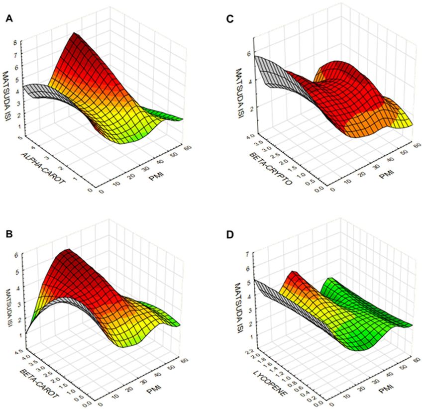
Differential interactions of individual serum carotenoid levels with insulin sensitivity. We used
RSM to determine two-way interactions of serum carotenoids and PMI on Matsuda ISI. RSM generates a surface
Scientific Reports | (2021) 11:871 | https://doi.org/10.1038/s41598-020-79387-8 5
Vol.:(0123456789)www.nature.com/scientificreports/
Trait paira ρP (95% CI) P-value ρG (95% CI) P-value ρE (95% CI) P-value
− 0.25 (− 0.328, − − 0.34 (− 0.595, −
Waist circumference 2.93 × 10 *
–9
0.02 − 0.1 (− 0.551, 0.351) 0.67
0.172) 0.085)
− 0.22 (− 0.2984, −
Body Mass Indexb 2.08 × 10⁻⁷* − 0.3 (− 0.555, − 0.045) 0.03 − 0.06 (− 0.589, 0.469) 0.82
0.142)
HDL cholesterol 0.29 (0.212, 0.368) 1.32 × 10 *
–12
0.62 (0.346, 0.894) 5.75 × 10 *
–5
− 0.19 (− 0.641, 0.261) 0.35
− 0.18 (− 0.258, − − 0.44 (− 0.714, −
Triglyceridesb 5.30 × 10–5* 0.002* 0.36 (− 0.189, 0.909) 0.14
0.102) 0.166)
− 0.23 (− 0.308, − − 0.27 (− 0.525, −
Fat massb 1.7 × 10–7* 0.054 − 0.16 (− 0.611, 0.291) 0.51
0.152) 0.015)
Systolic blood pressure − 0.07 (− 0.148, 0.008) 0.01* − 0.13 (− 0.424, 0.164) 0.35 0.03 (− 0.362, 0.422) 0.87
Diastolic blood pres-
− 0.07 (− 0.148, 0.008) 0.10 − 0.18 (− 0.474, 0.114) 0.23 0.1 (− 0.292, 0.492) 0.60
sure
− 0.09 (− 0.168, −
Fasting glucoseb 0.04 − 0.3 (− 0.692, 0.092) 0.12 0.09 (− 0.204, 0.384) 0.54
0.0116)
− 0.56 (− 0.932, −
Fasting insulinb − 0.07 (− 0.148, 0.008) 0.10 0.27 (− 0.024, 0.564) 0.15 0.002
0.188)
− 0.52 (− 0.932, −
HOMA-IRc − 0.07 (− 0.148, 0.008) 0.11 0.21 (− 0.084, 0.504) 0.16 0.009
0.108)
Table 3. Phenotypic (ρP), genetic (ρG) and environmental (ρE) correlations between β-cryptoxanthin and the
cardiometabolic risk traits. HDL cholesterol, high-density lipoprotein cholesterol, HOMA-IR the homeostasis
model of assessment-insulin resistance. Asterisks indicate the p-values that remain significant after applying
FDR correction. Bold values denote statistical significance at P-value ≤ 0.05. a All traits were adjusted for
the covariate effects of age and sex terms. b Data were log-transformed. c Trait was transformed using inverse
normal transformation.
fitted three-dimensional plot using a distance-weighted least-squares procedure while minimizing the variance
of estimators by a polynomial regression model weighted for the inverse of the variances. The plots shown in
Fig. 2 represent the interactions between serum carotenoid concentrations with PMI while showing the extent
of insulin sensitivity measured by Matsuda ISI. Interestingly, the resulting function is nonlinear, and a maximum
response was obtained with increasing serum α- and β- carotenoids (Fig. 2A,B), but not with β-cryptoxanthin
or lycopene (Fig. 2C,D). However, there is a significant decrease in Matsuda ISI with lower levels of α- and
β-carotenoids. PMI values between 2 and 4 showed the maximum variability associated with carotenoids.
Discussion
Our family-based study demonstrates that genetic factors play a crucial role in determining the serum carot-
enoid concentrations in MA children. Similarly, several other studies reported a range of heritabilities for
carotenoids39,48–50, given that the heritability is a population-specific parameter. For example, high heritability
(r2 = 0.98) has been reported for relative peak macular pigment density in monozygotic t wins51 and moderate
heritability (h2 = 30.5%) for serum retinol levels in a French family study49. A recent study in Older Order Amish
adults estimated the heritability of serum lycopene to be 0.38 ± 0.1248. While it can be argued that shared environ-
mental influences could have resulted in high heritabilities observed in our study, we believe that these estimates
are plausible as most of the children are distributed across large families. These findings are similar to those we
found for the α-carotene and β-carotene in the same group of c hildren39. The serum levels of β-cryptoxanthin
and lycopene consistently showed a positive correlation with HDL-C levels while there was a marked differ-
ence in their effects on other CMTs. We previously observed a similar correlation between serum α-carotene
and β-carotene and HDL-C39. These results are in agreement with the concept that plasma lipoproteins as the
primary transporters of carotenoids52.
Given that diet is the primary source of serum carotenoids, based on our available data, we assessed the extent
to which the observed high heritabilities are related to the dietary intake of carotenoids in our study. Despite
the limitations of the FFQ derived estimates of dietary markers, including carotenoids, we found low but highly
significant correlations between dietary and serum carotenoids after making appropriate covariate adjustments.
Here, it is worth noting that there is no consensus on the nature of the relationship between dietary intake
and blood (either serum or plasma) carotenoid concentrations43,44,53–55. Also, such relationships may differ by
individual carotenoids43. Moreover, blood concentrations of carotenoids may be able to better predict disease
risk when compared to dietary intake56. However, our findings of minimal changes in heritability estimates of
carotenoids after adjustments for dietary carotenoids suggest that the observed high heritabilities are mainly
related to post-absorption-derived concentrations of carotenoids. This conclusion may not be that surprising
since many other factors, aside from diet, related to carotenoid uptake, distribution, metabolism, and excretion,
ody34. Also, it should be noted that carotenoids have long and variable half-lives
contribute to their levels in the b
and follow first-order depletion k inetics57.
In this study, β-cryptoxanthin showed a negative correlation with several CMTs, including waist circumfer-
ence, BMI, triglycerides, and fat mass, but lycopene failed to show any correlation with any of the examined
CMTs. Despite strong evidence for the possible role of lycopene in the suppression of adipogenesis, there are
conflicting reports on the correlation between lycopene and BMI and related traits28,58. Notwithstanding the
Scientific Reports | (2021) 11:871 | https://doi.org/10.1038/s41598-020-79387-8 6
Vol:.(1234567890)www.nature.com/scientificreports/
Figure 2. Response surface contour plots showing the relationship of α-carotene (A), β-carotene (B),
β-cryptoxanthin (C), and lycopene (D) and PMI with Matsuda ISI. The contour plots are colored to aid in better
visualization of the graphics.
observation that BMI could influence the association between metabolic syndrome and serum lycopene levels,
the associations between lycopene and the metabolic syndrome were only significant for normal-weight and
participants who are overweight and not for individuals with obesity in the adult National Health and Nutrition
Examination Survey (NHANES) data59. A previous study reported higher serum concentrations of α-tocopherol,
α-carotene, and trans-β-carotene in Mexican American children were associated with reduced childhood
overweight and o besity60. While similar associations were found in the SAFARI children with α-carotene and
39
β-carotene , our current study further extends these observations to include β-cryptoxanthin suggesting an
overall beneficial effect of serum levels of these three carotenoids.
In a systemic review of multiple studies conducted on anti-oxidant vitamins, Asplund showed that lower
cardiovascular risk is associated with high intake of β-carotene (OR = 0.88; 95%CI 0.77–1.01) or high serum/
plasma levels of β-carotene (OR = 0.46; 95%CI 0.37–0.58) whereas such effect is not seen in randomized con-
trolled trials with β-carotene supplements (OR = 1.02; 95%CI 0.96–1.08)61. Additional studies also have shown
that serum carotenoids have anti-oxidant activity, and low levels of serum carotenoids and other anti-oxidants
are associated with insulin resistance, T2D, and metabolic syndrome62–70. Kinetic analysis of plasma carotenoid
concentrations shows extreme variability in absorption curves and plasma clearance rates71. While the mecha-
nism of intracellular translocation of carotenoids is not completely understood, recent studies have shown that in
addition to passive diffusion, transporters such as SR-BI may play a role in their absorption72. Nevertheless, the
overall carotenoid bioaccessibility is considered to be poor and variable among individuals, and such variability
may contribute to the carotenoid “low responder” and “high responder” phenotypes72. Additional factors such
as dietary fiber content and composition of gut microbiota may also alter carotenoid absorption, bio-activation,
Scientific Reports | (2021) 11:871 | https://doi.org/10.1038/s41598-020-79387-8 7
Vol.:(0123456789)www.nature.com/scientificreports/
and circulating plasma concentrations. How consumption of carotenoids can regulate gut immune responses
and microbiota is a subject of active ongoing research73.
We initially developed PMI as an omnibus clinical measure that included adiposity, HDL-C, and triglyceride
concentration to assess the relative metabolic health of children in a population after adjusting for age and sex.
Remarkably, we found that serum α- and β- carotenoid concentrations were inversely associated with PMI in
the SAFARI children using the RSM approach. However, their protective effects were modified by underlying
metabolic conditions and were diminished in the presence of metabolic impairment (PMI > 4). Some previous
studies showed both dietary and plasma β-carotene, but not lycopene and β-cryptoxanthin, may positively
associate with insulin sensitivity. Sluijs et al. reported that diets high in β-carotene were protective against T2D
[Hazard Ratio quartile 4 versus quartile 1 (HRQ4): 0.78 (95%CI 0.64, 0.95), P-linear trend 0.01]67. In contrast,
they found that the effect of serum α-carotene on T2D was marginal [HRQ4 of 0.85 (95%CI 0.70,1.03), and
P-linear trend 0.05], whereas other carotenoids did not show any association. In a study conducted on non-
diabetic adult subjects with obesity, plasma β-carotene concentrations were positively associated with insulin
sensitivity, as assessed by HOMA-IR, whereas such associations were not detected with lycopene and lutein/
zeaxanthin58. These studies, along with our present study, highlight the fact that various carotenoids may have
non-overlapping and differential associations with insulin sensitivity in children, as previously reported in adults.
However, the protective associations of the α- and β-carotenoids were not detectable in the presence of metabolic
dysfunction. This concept needs further exploration, given our previous finding that fruit and vegetable juice
concentrate supplementation could lead to increased serum β-carotene levels and improved insulin resistance
in overweight prepubertal boys74.
A balanced diet should incorporate a variety of fruits and vegetables as sources of different carotenoids.
Orange colored vegetables such as carrots and green vegetables such as broccoli and spinach are rich sources
of α-carotene and β-carotene75,76. Red-colored vegetables such as tomatoes are lycopene-rich, and orange-
colored fruits such as mandarins are rich in β-cryptoxanthin75,76. Our perplexing finding that the serum levels
of β-cryptoxanthin, but not lycopene, being protective against many CMTs in the SAFARI children has prompted
us to examine this issue further. Unlike many nutrients, lycopene’s bioavailability is increased by cooking, and
processing and tomato-based products like ketchup and sauces are a common dietary source of lycopene77.
However, limited data exists on the relative health benefits of consuming whole tomatoes versus processed
tomato products and lycopene supplements. Thus rigorous, well-controlled studies are needed to understand
the differential effects of carotenoids in CMTs.
Reinforcing our results that indicate high heritability of serum carotenoid levels, genome-wide association
analyses (GWAS) as well as candidate gene association studies (CGAS) have identified several genetic vari-
ants that could potentially contribute to inter-individual variability in carotenoid levels in the b ody36. These
genetic variants map to various stages in carotenoid metabolism, including absorption, conversion, fasting,
and post-prandial blood concentrations, and tissue levels. Also, gene variants that alter lipid absorption and
metabolism can influence circulating carotenoid levels78. For example, genetic variants in BCO1, ABCA1, APOB,
and LPL, have been implicated in fasting blood β-carotene concentrations, whereas genetic variants in RBP4 in
fasting blood retinol concentrations (reviewed by Borel and D esmarchelier79). Similarly, serum lycopene and
78,80
β-cryptoxanthin levels may also be genetically determined . Such genetic variation could potentially lead to
differences in carotenoid metabolism in global populations. However, irrespective of such genetic variability,
consumption of a wide variety of fruits and vegetables need to be encouraged, given the broad protective effects
of carotenoids on human health.
Conclusions
While dietary intake of fruits, vegetables, or supplements is critical for serum carotenoid levels, they are also
determined by genetic influences that may alter their absorption, transport, tissue concentration, and utiliza-
tion of the carotenoids. Other modifying factors may include the differences in gut microbiota, socioeconomic
strata, other metabolic conditions, and their interactions. Importantly, our findings suggest a connection between
concentrations of certain serum carotenoids and insulin sensitivity patterns; increased serum concentrations of
α- and β- carotenoids were shown to have maximal effects on insulin sensitivity. In summary, our study reveals
that the serum carotenoids are under strong additive genetic influences; and, may have differential effects on
susceptibility to childhood obesity and its related CMTs in children and adolescents.
Received: 20 March 2020; Accepted: 1 December 2020
References
1. Ogden CL, C. M., Fryar CD, Flegal KM. in N.C.H.S. data brief, no 219 (ed National Center for Health Statistics) (Hyattsville,
Maryland, 2015).
2. Estrada, E. et al. Children’s Hospital Association consensus statements for comorbidities of childhood obesity. Child Obes. 10,
304–317. https://doi.org/10.1089/chi.2013.0120 (2014).
3. Gungor, N. K. Overweight and obesity in children and adolescents. J. Clin. Res. Pediatr. Endocrinol. 6, 129–143. https://doi.
org/10.4274/Jcrpe.1471 (2014).
4. Herouvi, D., Karanasios, E., Karayianni, C. & Karavanaki, K. Cardiovascular disease in childhood: The role of obesity. Eur. J. Pediatr.
172, 721–732. https://doi.org/10.1007/s00431-013-1932-8 (2013).
5. Poyrazoglu, S., Bas, F. & Darendeliler, F. Metabolic syndrome in young people. Curr. Opin. Endocrinol. Diabetes Obes. 21, 56–63.
https://doi.org/10.1097/01.med.0000436414.90240.2c (2014).
6. Wirix, A. J., Kaspers, P. J., Nauta, J., Chinapaw, M. J. & Kist-van Holthe, J. E. Pathophysiology of hypertension in obese children:
A systematic review. Obes. Rev. 16, 831–842. https://doi.org/10.1111/obr.12305 (2015).
Scientific Reports | (2021) 11:871 | https://doi.org/10.1038/s41598-020-79387-8 8
Vol:.(1234567890)www.nature.com/scientificreports/
7. Anderson, E. L. et al. The prevalence of non-alcoholic fatty liver disease in children and adolescents: A systematic review and
meta-analysis. PLoS ONE 10, e0140908. https://doi.org/10.1371/journal.pone.0140908 (2015).
8. Mofid, M. Obstructive sleep apnea: The sleeping giant of the childhood obesity epidemic. JAAPA 27, 27–30. https://doi.
org/10.1097/01.J.A.A.0000453860.16582.9c (2014).
9. Pulgaron, E. R. & Delamater, A. M. Obesity and type 2 diabetes in children: Epidemiology and treatment. Curr. Diab. Rep. 14, 508.
https://doi.org/10.1007/s11892-014-0508-y (2014).
10. Biro, F. M. & Wien, M. Childhood obesity and adult morbidities. Am. J. Clin. Nutr. 91, 1499S-1505S. https://doi.org/10.3945/
ajcn.2010.28701B (2010).
11. Singh, A. S., Mulder, C., Twisk, J. W., van Mechelen, W. & Chinapaw, M. J. Tracking of childhood overweight into adulthood: A
systematic review of the literature. Obes. Rev. 9, 474–488. https://doi.org/10.1111/j.1467-789X.2008.00475.x (2008).
12. Bondia-Pons, I., Ryan, L. & Martinez, J. A. Oxidative stress and inflammation interactions in human obesity. J. Physiol. Biochem.
68, 701–711. https://doi.org/10.1007/s13105-012-0154-2 (2012).
13. Karalis, K. P. et al. Mechanisms of obesity and related pathology: Linking immune responses to metabolic stress. FEBS J. 276,
5747–5754. https://doi.org/10.1111/j.1742-4658.2009.07304.x (2009).
14. Falkner, B. & Cossrow, N. D. Prevalence of metabolic syndrome and obesity-associated hypertension in the racial ethnic minorities
of the United States. Curr. Hypertens. Rep. 16, 449. https://doi.org/10.1007/s11906-014-0449-5 (2014).
15. Aguilera, C. M., Olza, J. & Gil, A. Genetic susceptibility to obesity and metabolic syndrome in childhood. Nutr. Hosp. 28(Suppl
5), 44–55. https://doi.org/10.3305/nh.2013.28.sup5.6917 (2013).
16. Hill, J. O., Wyatt, H. R. & Peters, J. C. Energy balance and obesity. Circulation 126, 126–132. https: //doi.org/10.1161/CIRCUL ATIO
NAHA.111.087213 (2012).
17. Bonet, M. L., Canas, J. A., Ribot, J. & Palou, A. Carotenoids and their conversion products in the control of adipocyte function,
adiposity and obesity. Arch. Biochem. Biophys. 572, 112–125. https://doi.org/10.1016/j.abb.2015.02.022 (2015).
18. Bonet, M. L., Canas, J. A., Ribot, J. & Palou, A. Carotenoids in adipose tissue biology and obesity. Subcell Biochem. 79, 377–414.
https://doi.org/10.1007/978-3-319-39126-7_15 (2016).
19. Edge, R., McGarvey, D. J. & Truscott, T. G. The carotenoids as anti-oxidants—A review. J. Photochem. Photobiol. B 41, 189–200
(1997).
20. Fiedor, J. & Burda, K. Potential role of carotenoids as anti-oxidants in human health and disease. Nutrients 6, 466–488. https://doi.
org/10.3390/nu6020466 (2014).
21. Kim, J. H. et al. The non-provitamin A carotenoid, lutein, inhibits NF-kappaB-dependent gene expression through redox-based
regulation of the phosphatidylinositol 3-kinase/P.T.E.N./Akt and NF-kappaB-inducing kinase pathways: Role of H(2)O(2) in
NF-kappaB activation. Free Radic Biol Med 45, 885–896. https://doi.org/10.1016/j.freeradbiomed.2008.06.019 (2008).
22. Galano, A., Vargas, R. & Martinez, A. Carotenoids can act as anti-oxidants by oxidizing the superoxide radical anion. Phys. Chem.
Chem. Phys. 12, 193–200. https://doi.org/10.1039/b917636e (2010).
23. Gordon, M. H. Significance of dietary anti-oxidants for health. Int. J. Mol. Sci. 13, 173–179. https://doi.org/10.3390/ijms130101
73 (2012).
24. U.S. Department of Health and Human Services. 2015–2020 dietary guidelines for Americans. 8th ed. https://health.gov/dietarygui
delines/2015/guidelines/ (U.S. Department of Health and Human Services; U.S. Department of Agriculture, Washington, DC,
2015).
25. Herrick, K. A., Rossen, L. M., Nielsen, S. J., Branum, A. M. & Ogden, C. L. Fruit consumption by youth in the United States.
Pediatrics 136, 664–671. https://doi.org/10.1542/peds.2015-1709 (2015).
26. Lorson, B. A., Melgar-Quinonez, H. R. & Taylor, C. A. Correlates of fruit and vegetable intakes in U.S. children. J. Am. Diet Assoc.
109, 474–478. https://doi.org/10.1016/j.jada.2008.11.022 (2009).
27. Di Noia, J. & Byrd-Bredbenner, C. Determinants of fruit and vegetable intake in low-income children and adolescents. Nutr. Rev.
72, 575–590. https://doi.org/10.1111/nure.12126 (2014).
28. Ford, E. S., Gillespie, C., Ballew, C., Sowell, A. & Mannino, D. M. Serum carotenoid concentrations in U.S. children and adolescents.
Am. J. Clin. Nutr. 76, 818–827 (2002).
29. Britton, G., Liaaen-Jensen, S. & Pfander, H. Carotenoids Handbook (Birkhäuser Verlag, Basel, 2004).
30. Milani, A., Basirnejad, M., Shahbazi, S. & Bolhassani, A. Carotenoids: Biochemistry, pharmacology and treatment. Br. J. Pharmacol.
174, 1290–1324. https://doi.org/10.1111/bph.13625 (2017).
31. Stahl, W. & Sies, H. Bioactivity and protective effects of natural carotenoids. Biochim. Biophys. Acta 1740, 101–107. https://doi.
org/10.1016/j.bbadis.2004.12.006 (2005).
32. Mares, J. Lutein and zeaxanthin isomers in eye health and disease. Annu. Rev. Nutr. 36, 571–602. https://doi.org/10.1146/annur
ev-nutr-071715-051110 (2016).
33. Di Mascio, P., Kaiser, S. & Sies, H. Lycopene as the most efficient biological carotenoid singlet oxygen quencher. Arch. Biochem.
Biophys. 274, 532–538 (1989).
34. Bohn, T. et al. Host-related factors explaining interindividual variability of carotenoid bioavailability and tissue concentrations in
humans. Mol. Nutr. Food Res. https://doi.org/10.1002/mnfr.201600685 (2017).
35. Beydoun, M. A., Nalls, M. A., Canas, J. A., Evans, M. K. & Zonderman, A. B. Gene polymorphisms and gene scores linked to low
serum carotenoid status and their associations with metabolic disturbance and depressive symptoms in African–American adults.
Br. J. Nutr. 112, 992–1003. https://doi.org/10.1017/S0007114514001706 (2014).
36. Borel, P. Genetic variations involved in interindividual variability in carotenoid status. Mol. Nutr. Food Res. 56, 228–240. https://
doi.org/10.1002/mnfr.201100322 (2012).
37. Borel, P., Desmarchelier, C., Nowicki, M. & Bott, R. A combination of single-nucleotide polymorphisms is associated with inter-
individual variability in dietary beta-carotene bioavailability in healthy men. J. Nutr. 145, 1740–1747. https://doi.org/10.3945/
jn.115.212837 (2015).
38. Borel, P. et al. Interindividual variability of lutein bioavailability in healthy men: Characterization, genetic variants involved, and
relation with fasting plasma lutein concentration. Am. J. Clin. Nutr. 100, 168–175. https://doi.org/10.3945/ajcn.114.085720 (2014).
39. Farook, V. S. et al. Genetics of serum carotenoid concentrations and their correlation with obesity-related traits in Mexican
American children. Am. J. Clin. Nutr. https://doi.org/10.3945/ajcn.116.144006 (2017).
40. Fowler, S. P. et al. Genetic epidemiology of cardiometabolic risk factors and their clustering patterns in Mexican American children
and adolescents: The SAFARI Study. Hum. Genet. 132, 1059–1071. https://doi.org/10.1007/s00439-013-1315-2 (2013).
41. Smith, C. & Fila, S. Comparison of the kid’s block food frequency questionnaire to the 24-hour recall in urban native American
youth. Am. J. Hum. Biol. 18, 706–709. https://doi.org/10.1002/ajhb.20475 (2006).
42. Cullen, K. W., Watson, K. & Zakeri, I. Relative reliability and validity of the Block Kids Questionnaire among youth aged 10 to 17
years. J. Am. Diet Assoc. 108, 862–866. https://doi.org/10.1016/j.jada.2008.02.015 (2008).
43. Talegawkar, S. A. et al. Carotenoid intakes, assessed by food-frequency questionnaires (FFQs), are associated with serum carotenoid
concentrations in the Jackson Heart Study: Validation of the Jackson Heart Study Delta N.I.R.I. Adult FFQs. Public Health Nutr.
11, 989–997. https://doi.org/10.1017/S1368980007001310 (2008).
44. Prasad, M. et al. Carotenoid intake and serum concentration in young Finnish children and their relation with fruit and vegetable
consumption. Nutrients https://doi.org/10.3390/nu10101533 (2018).
Scientific Reports | (2021) 11:871 | https://doi.org/10.1038/s41598-020-79387-8 9
Vol.:(0123456789)www.nature.com/scientificreports/
45. Almasy, L. & Blangero, J. Multipoint quantitative-trait linkage analysis in general pedigrees. Am. J. Hum. Genet. 62, 1198–1211.
https://doi.org/10.1086/301844 (1998).
46. Hernandez, M. J. G. et al. Pediatric visceral adiposity index adaptation correlates with HOMA-IR, Matsuda, and transaminases.
Endocr. Pract. 24, 294–301. https://doi.org/10.4158/EP-2017-0086 (2018).
47. Bejamini, Y. & Hochberg, Y. Controlling the false discovery rate: A practical and powerful approach to multiple testing. J. R. Stat.
Soc. SerB 57, 289–300 (1995).
48. D’Adamo, C. R. et al. A common variant in the SETD7 gene predicts serum lycopene concentrations. Nutrients 8, 82. https://doi.
org/10.3390/nu8020082 (2016).
49. Gueguen, S. et al. Genetic and environmental contributions to serum retinol and alpha-tocopherol concentrations: The Stanislas
Family Study. Am. J. Clin. Nutr. 81, 1034–1044 (2005).
50. Tremblay, B. L., Guenard, F., Lamarche, B., Perusse, L. & Vohl, M. C. Genetic and common environmental contributions to familial
resemblances in plasma carotenoid concentrations in healthy families. Nutrients https://doi.org/10.3390/nu10081002 (2018).
51. Hogg, R. E. et al. Heritability of the spatial distribution and peak density of macular pigment: A classical twin study. Eye 26,
1217–1225. https://doi.org/10.1038/eye.2012.98 (2012).
52. Clevidence, B. A. & Bieri, J. G. Association of carotenoids with human plasma lipoproteins. Methods Enzymol. 214, 33–46 (1993).
53. van Kappel, A. L. et al. Serum carotenoids as biomarkers of fruit and vegetable consumption in the New York Women’s Health
Study. Public Health Nutr. 4, 829–835. https://doi.org/10.1079/phn2000115 (2001).
54. Brady, W. E., Mares-Perlman, J. A., Bowen, P. & Stacewicz-Sapuntzakis, M. Human serum carotenoid concentrations are related
to physiologic and lifestyle factors. J. Nutr. 126, 129–137. https://doi.org/10.1093/jn/126.1.129 (1996).
55. Morgan, E. H., Graham, M. L., Marshall, G. A., Hanson, K. L. & Seguin-Fowler, R. A. Serum carotenoids are strongly associated
with dermal carotenoids but not self-reported fruit and vegetable intake among overweight and obese women. Int. J. Behav. Nutr.
Phys. Act. 16, 104. https://doi.org/10.1186/s12966-019-0869-3 (2019).
56. Aune, D. et al. Dietary compared with blood concentrations of carotenoids and breast cancer risk: A systematic review and meta-
analysis of prospective studies. Am. J. Clin. Nutr. 96, 356–373. https://doi.org/10.3945/ajcn.112.034165 (2012).
57. Burri, B. J., Neidlinger, T. R. & Clifford, A. J. Serum carotenoid depletion follows first-order kinetics in healthy adult women fed
naturally low carotenoid diets. J. Nutr. 131, 2096–2100. https://doi.org/10.1093/jn/131.8.2096 (2001).
58. Ben Amara, N. et al. Independent positive association of plasma beta-carotene concentrations with adiponectin among non-
diabetic obese subjects. Eur. J. Nutr. 54, 447–454. https://doi.org/10.1007/s00394-014-0728-6 (2015).
59. Han, G. M., Soliman, G. A., Meza, J. L., Islam, K. M. & Watanabe-Galloway, S. The influence of BMI on the association between
serum lycopene and the metabolic syndrome. Br. J. Nutr. 115, 1292–1300. https://doi.org/10.1017/S0007114516000179 (2016).
60. Gunanti, I. R., Marks, G. C., Al-Mamun, A. & Long, K. Z. Low serum concentrations of carotenoids and vitamin E are associated
with high adiposity in Mexican–American children. J. Nutr. 144, 489–495. https://doi.org/10.3945/jn.113.183137 (2014).
61. Asplund, K. Antioxidant vitamins in the prevention of cardiovascular disease: A systematic review. J. Intern. Med. 251, 372–392
(2002).
62. Cicero, A. F. G. & Colletti, A. Effects of carotenoids on health: Are all the same? results from clinical trials. Curr. Pharm. Des. 23,
2422–2427. https://doi.org/10.2174/1381612823666170207095459 (2017).
63. Goncalves, A. & Amiot, M. J. Fat-soluble micronutrients and metabolic syndrome. Curr. Opin. Clin. Nutr. Metab. Care 20, 492–497.
https://doi.org/10.1097/MCO.0000000000000412 (2017).
64. Henriksen, E. J., Diamond-Stanic, M. K. & Marchionne, E. M. Oxidative stress and the etiology of insulin resistance and type 2
diabetes. Free Radic. Biol. Med. 51, 993–999. https://doi.org/10.1016/j.freeradbiomed.2010.12.005 (2011).
65. Kaulmann, A. & Bohn, T. Carotenoids, inflammation, and oxidative stress–implications of cellular signaling pathways and relation
to chronic disease prevention. Nutr. Res. 34, 907–929. https://doi.org/10.1016/j.nutres.2014.07.010 (2014).
66. Ribeiro, D., Freitas, M., Silva, A. M. S., Carvalho, F. & Fernandes, E. Antioxidant and pro-oxidant activities of carotenoids and
their oxidation products. Food Chem. Toxicol. 120, 681–699. https://doi.org/10.1016/j.fct.2018.07.060 (2018).
67. Sluijs, I. et al. Dietary intake of carotenoids and risk of type 2 diabetes. Nutr. Metab. Cardiovasc. Dis. 25, 376–381. https://doi.
org/10.1016/j.numecd.2014.12.008 (2015).
68. Beydoun, M. A. et al. Carotenoids, vitamin A, and their association with the metabolic syndrome: A systematic review and meta-
analysis. Nutr. Rev. 77, 32–45. https://doi.org/10.1093/nutrit/nuy044 (2019).
69. Coyne, T. et al. Diabetes mellitus and serum carotenoids: Findings of a population-based study in Queensland, Australia. Am. J.
Clin. Nutr. 82, 685–693 (2005).
70. Sugiura, M., Nakamura, M., Ogawa, K., Ikoma, Y. & Yano, M. High-serum carotenoids associated with lower risk for developing
type 2 diabetes among Japanese subjects: Mikkabi cohort study. BMJ Open Diabetes Res. Care 3, e000147. https://doi.org/10.1136/
bmjdrc-2015-000147 (2015).
71. Olson, J. A. Absorption, transport, and metabolism of carotenoids in humans. Pure Appl. Chem. 66, 1011–1016 (1994).
72. Reboul, E. Mechanisms of carotenoid intestinal absorption: Where do we stand?. Nutrients https://doi.org/10.3390/nu11040838
(2019).
73. Lyu, Y., Wu, L., Wang, F., Shen, X. & Lin, D. Carotenoid supplementation and retinoic acid in immunoglobulin A regulation of
the gut microbiota dysbiosis. Exp. Biol. Med. 243, 613–620. https://doi.org/10.1177/1535370218763760 (2018).
74. Canas, J. A. et al. Insulin resistance and adiposity in relation to serum beta-carotene levels. J. Pediatr. 161(58–64), e51-52. https://
doi.org/10.1016/j.jpeds.2012.01.030 (2012).
75. Maiani, G. et al. Carotenoids: Actual knowledge on food sources, intakes, stability and bioavailability and their protective role in
humans. Mol. Nutr. Food Res. 53(Suppl 2), S194-218. https://doi.org/10.1002/mnfr.200800053 (2009).
76. Jaswir, I., Noviendri, D., Hasrini, R. F. & Octavianti, F. Carotenoids: Sources, medicinal properties and their application in food
and nutraceutical industry. J. Med. Plants Res. 5, 7119–7131. https://doi.org/10.5897/JMPRx11.011 (2011).
77. Clinton, S. K. Lycopene: Chemistry, biology, and implications for human health and disease. Nutr. Rev. 56, 35–51. https://doi.
org/10.1111/j.1753-4887.1998.tb01691.x (1998).
78. Borel, P. et al. Human plasma levels of vitamin E and carotenoids are associated with genetic polymorphisms in genes involved in
lipid metabolism. J Nutr 137, 2653–2659. https://doi.org/10.1093/jn/137.12.2653 (2007).
79. Borel, P. & Desmarchelier, C. Genetic variations associated with vitamin A status and vitamin A bioavailability. Nutrients https://
doi.org/10.3390/nu9030246 (2017).
80. Moran, N. E., Erdman, J. W. & Clinton, S. K. Complex interactions between dietary and genetic factors impact lycopene metabolism
and distribution. Arch. Biochem. Biophys. 539, 171–180. https://doi.org/10.1016/j.abb.2013.06.017 (2013).
Acknowledgements
This study was supported by grants from the National Institute of Health (R01 HD049051, R01 AI119131,
HD049051-5S1 (A.R.R.A.), HD041111, DK053889, DK047482, P01 HL45522 and MH59490), Veterans Admin-
istration Epidemiologic Grant, Voelcker Foundation, and the National Research Initiative Grant 2009-55200-
05197 from the U.S.D.A. National Institute for Food and Agriculture. We thank Dr. William Rogers, Dr. Rolando
Lozano, Dr. Richard Granato, Margaret Fragoso, David Rupert, Rhonda Lyons, Tanya Prado, and Ram Prasad
Scientific Reports | (2021) 11:871 | https://doi.org/10.1038/s41598-020-79387-8 10
Vol:.(1234567890)You can also read