Exclusively heteronuclear NMR experiments for the investigation of intrinsically disordered proteins: focusing on proline residues - Magnetic ...
←
→
Page content transcription
If your browser does not render page correctly, please read the page content below
Magn. Reson., 2, 511–522, 2021                                                                                Open Access
https://doi.org/10.5194/mr-2-511-2021
© Author(s) 2021. This work is distributed under
the Creative Commons Attribution 4.0 License.

              Exclusively heteronuclear NMR experiments for the
               investigation of intrinsically disordered proteins:
                         focusing on proline residues
                                Isabella C. Felli1 , Wolfgang Bermel2 , and Roberta Pierattelli1
              1 CERM    and Department of Chemistry “Ugo Schiff”, University of Florence, Via Luigi Sacconi 6,
                                            50019 Sesto Fiorentino, Florence, Italy
                            2 Bruker BioSpin GmbH, Silberstreifen 4, 76287 Rheinstetten, Germany

         Correspondence: Isabella C. Felli (felli@cerm.unifi.it) and Roberta Pierattelli (roberta.pierattelli@unifi.it)

                                     Received: 27 March 2021 – Discussion started: 12 April 2021
                                Revised: 20 May 2021 – Accepted: 2 June 2021 – Published: 1 July 2021

       Abstract. NMR represents a key spectroscopic technique that contributes to the emerging field of highly flexi-
       ble, intrinsically disordered proteins (IDPs) or protein regions (IDRs) that lack a stable three-dimensional struc-
       ture. A set of exclusively heteronuclear NMR experiments tailored for proline residues, highly abundant in
       IDPs/IDRs, are presented here. They provide a valuable complement to the widely used approach based on amide
       proton detection, filling the gap introduced by the lack of amide protons in proline residues within polypeptide
       chains. The novel experiments have very interesting properties for the investigations of IDPs/IDRs of increasing
       complexity.

1   Introduction                                                     due to the open conformations that, when approaching phys-
                                                                     iological pH and temperature, broaden amide proton reso-
Invisible in X-ray studies of protein crystals, intrinsically dis-   nances beyond detection. While several elegant experiments
ordered regions (IDRs) of complex proteins have for a long           were proposed to exploit exchange processes with the sol-
time been considered to be passive linkers connecting func-          vent (Kurzbach et al., 2017; Olsen et al., 2020; Szekely et
tional globular domains and are, thus, often ignored in struc-       al., 2018; Thakur et al., 2013), in general initial NMR in-
tural biology studies. However, in many cases they comprise          vestigations of IDPs/IDRs are carried out in conditions in
a significant fraction of the primary sequence of a protein,         which these critical points are mitigated. Exchange broad-
and for this reason, they are expected to have a role in pro-        ening strongly depends on pH and temperature; conditions
tein function (Van Der Lee et al., 2014). The characterization       can be optimized to recover most of the amide proton res-
of highly flexible regions of large proteins and entire pro-         onances enabling the acquisition of amide-proton-detected
teins characterized by the lack of a 3D structure, now gen-          triple resonance experiments needed for sequence-specific
erally referred to as intrinsically disordered proteins (IDPs),      assignment of the resonances. However, in particular for pro-
lies well behind that of their folded counterparts and is nowa-      teins that are largely exposed to the solvent, it may be inter-
days pursued by an increasingly large number of studies to           esting to study their near-physiological pH and temperature
fill this knowledge gap. NMR plays a strategic role in this          conditions (Gil et al., 2013). In this context, 13 C direct detec-
context since it constitutes the major, if not the unique, spec-     tion NMR developed into a valuable alternative.
troscopic technique to achieve atomic resolution information            Although the intrinsic sensitivity of 13 C is lower with re-
on their structural and dynamic properties. However, intrin-         spect to that of 1 H, 13 C nuclear spins are characterized by
sic disorder and high flexibility have very relevant effects         a large chemical shift dispersion (Dyson and Wright, 2001)
for NMR investigations, such as reduction in chemical shift          and, when coupled to 15 N nuclei, provide a well-defined
dispersion and efficient exchange processes with the solvent         fingerprint of a polypeptide (Bermel et al., 2006a; Hsu et

Published by Copernicus Publications on behalf of the Groupement AMPERE.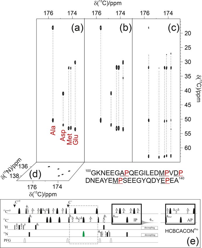
512                                                                 I. C. Felli et al.: IDPs by NMR: focusing on proline residues

al., 2009; Lopez et al., 2016; Schiavina et al., 2019). These        2     Materials and methods
features were exploited to design a suite of 3D experiments
based on carbonyl-carbon direct detection for sequential as-         Isotopically labelled α-synuclein (13 C and 15 N) was ex-
signment and to measure NMR observables (Felli and Pier-             pressed and purified, as previously described (Huang et
attelli, 2014). These experiments, starting from 1 H polariza-       al., 2005). The NMR sample has 0.6 mM protein concen-
tion, exploit only heteronuclear chemical shifts in the indirect     tration in a 20 mM phosphate buffer at pH 6.5 and 100 mM
dimensions to maximize chemical shift dispersion (exclu-             NaCl in H2 O with 5 % D2 O for the lock signal.
sively heteronuclear experiments) and can be used to study              Isotopically labelled CBP-ID4 (13 C and 15 N) was
IDPs/IDRs – also in conditions in which amide proton reso-           expressed and purified as previously described (Piai
nances are too broad to be detected. In addition, they reveal        et al., 2016). The NMR sample has 0.9 mM pro-
information about proline residues that lack the amide proton        tein concentration in a water buffer containing 20 mM
when part of the polypeptide chains and cannot be detected           tris(hydroxymethyl)aminomethane (TRIS) and 50 mM KCl,
in 2D 1 H−15 N correlation experiments (2D HN), even if pH           at pH 6.9, with 5 % D2 O added for the lock signal.
and temperature conditions are optimized to reduce exchange             NMR experiments were acquired at 288 K (for α-
broadening.                                                          synuclein) and at 283 K (for CBP-ID4) with a 16.4 T Bruker
   Proline residues are abundant in IDPs/IDRs and often oc-          AVANCE NEO spectrometer operating at 700.06 MHz 1 H,
cur in proline-rich sequences with repetitive units (Theillet et     176.05 MHz 13 C, and 70.97 MHz 15 N frequencies, equipped
al., 2014). Initial bioinformatics studies on the relative abun-     with a 5 mm cryogenically cooled probe head optimized for
                                                                     13 C direct detection (TXO). Radio frequency (RF) pulses
dance of each amino acid in regions of the protein that could
not be observed in X-ray diffraction studies led to the clas-        and carrier frequencies typically employed for the investiga-
sification of prolines as “disorder promoting” amino acids           tion of intrinsically disordered proteins were used, except for
(Dunker et al., 2008). Nevertheless, proline, the only imino         the modifications introduced to zoom into the proline 15 N
acid, features a closed ring in its side chain, which confers lo-    region. Carrier frequencies were set to 4.7 ppm (parts per
cal rigidity compared to all other amino acids (Williamson,          million; 1 H), 176.4 (13 C0 ), 53.9 (13 Cα ), and 44.9 (13 Cali ).
1994), as also exploited in Förster Resonance Energy Trans-          The 15 N carrier was set to 137 ppm in the centre of 15 N
fer (FRET) studies in which proline residues are used as rigid       resonances of proline residues. Hard pulses were used for
                                                                     1 H. Band-selective 13 C pulses used were Q5 and Q3 (Ems-
spacers to measure distances (Schuler et al., 2005). These
observations clearly show the importance of experimental             ley and Bodenhausen, 1990) of 300 and 231 µs for 90 and
atomic resolution information on the structural and dynamic          180◦ rotations, respectively; a 900 µs Q3 pulse centred at
properties of proline residues for understanding their role          53.9 ppm was used for the selective inversion of Cα . The
                                                                     15 N pulse to invert the 15 N proline resonances was a 8000 µs
in modulating protein function. Abundant information about
proline residues in globular protein folds is available either       refocusing band-selective uniform-response pure-phase (RE-
through NMR or X-ray studies (MacArthur and Thornton,                BURP) pulse (Geen and Freeman, 1991); all other 15 N pulses
1991), including several examples of cis-trans isomerization         were hard pulses. Decoupling was achieved with WALTZ-65
of peptide bonds involving proline nitrogen as molecular             (100 µs, 2.5 kHz; Zhou et al., 2007) for 1 H and with GARP4
switches (Lu et al., 2007). However, the characterization in         (250 µs, 1.0 kHz; Shaka et al., 1985) for 15 N. The MOCCA
highly flexible, disordered polypeptides is available only in        mixing time (Felli et al., 2009; Furrer et al., 2004) in the
a handful of cases (Chaves-Arquero et al., 2018; Gibbs et            (HCA)COCONPro experiment was 350 ms, constituted by
al., 2017; Haba et al., 2013; Hošek et al., 2016; Knoblich           repeated (1-180◦ -1)2n units in which 1 = 150 µs and the
et al., 2009; Pérez et al., 2009; Piai et al., 2016; Chhabra et      180◦ pulse was 91.6 µs.
al., 2018; Ahuja et al., 2016), and actually, early studies on          The experimental parameters used for the acquisition of
IDPs/IDRs routinely reported assignment statistics that only         the various experiments on α-synuclein and CBP-ID4 are
considered all other amino acids (“excluding prolines”).             reported in Table 1. Spectra were calibrated using 2,2-
   Here we would like to propose an experimental vari-               dimethylsilapentane-5-sulfonic acid (DSS) as a reference
ant of the most widely used 13 C-detected 3D experiments             for 1 H and 13 C; 15 N was calibrated indirectly (Markley et
for the sequence-specific assignment of IDPs/IDRs to se-             al., 1998).
lectively pick up correlations involving proline nitrogen nu-
clei and provide key complementary information to that ob-
                                                                     3     Results and discussion
tained through amide-proton-detected experiments. They can
be collected in a shorter time with respect to standard 3D           3.1    Advantages of focusing on proline residues
experiments and provide a valuable addition to the current
experimental protocols for the study of IDPs.                        In highly flexible and disordered proteins, contributions to
                                                                     signals’ chemical shifts deriving from the local environ-
                                                                     ment are averaged out, leaving mainly those contributions
                                                                     due to the covalent structure of the polypeptide. Chemical

Magn. Reson., 2, 511–522, 2021                                                             https://doi.org/10.5194/mr-2-511-2021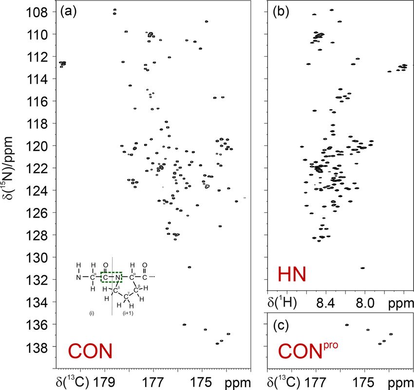
I. C. Felli et al.: IDPs by NMR: focusing on proline residues 513
Table 1. Experimental parameters used.
Dimension of acquired data Spectral width (ppm) NSa d1 (s)b
t1 t2 t3 F1 F2 F3
Experiments with α-synuclein
1 H detected 1 H−15 N HSQC 800 (15 N) 2048 (1 H) 28.1 15.0 2 1.0
13 C detected CON 512 (15 N) 1024 (13 C) 32.0 31.0 2 1.6
CONPro 128 (15 N) 1024 (13 C) 5.0 31.0 2 1.6
(H)CBCACONPro 128 (13 C) 32 (15 N) 1024 (13 C) 60.0 5.0 30.0 4 1.0
(H)CCCONPro 128 (13 C) 32 (15 N) 1024 (13 C) 70.0 5.0 30.0 4 1.0
(H)CBCANCOPro 128 (13 C) 16 (15 N) 1024 (13 C) 60.0 5.0 30.0 8 1.0
1 H and 13 C detected CON/HN 600 (15 N) 1024 (13 C) 35.0 31.0 2 1.6
(using multiple receivers) 600 (15 N) 2048 (1 H) 35.0 15.0 4
Experiments with CBP-ID4
1 H detected 1 H−15 N HSQC 800 (15 N) 2048 (1 H) 30.0 15.0 2 1.0
13 C detected CON 1024 (15 N) 1024 (13 C) 38.0 30.0 2 2.0
CONPro 170 (15 N) 1024 (13 C) 6.5 30.0 2 2.0
(H)CBCACONPro 128 (13 C) 64 (15 N) 1024 (13 C) 64.5 6.5 30.0 4 1.0
(H)CCCONPro 128 (13 C) 64 (15 N) 1024 (13 C) 75.7 6.5 30.0 4 1.0
(H)CBCANCOPro 128 (13 C) 22 (15 N) 1024 (13 C) 64.5 6.5 30.0 16 1.0
(HCA)COCONPro 96 (13 C) 64 (15 N) 1024 (13 C) 10.8 6.5 30.0 8 1.5
a Number of acquired scans. b Inter-scan delay.
shift ranges predicted for 15 N resonances of imino acids C0 i−1 − Ni coherence transfer steps to introduce the desired
such as proline are quite different from those predicted for selectivity in the proline region.
amino acids, which is expected from the different chemical As an example, the pulse sequence of the 3D
structure. The 2D CON spectra of several disordered pro- (H)CBCACONPro experiment is shown in Fig. 3. The
teins of different size and sequence complexity, reported in inclusion of the 15 N band-selective pulse in the C0 −N
Fig. 1, clearly show that proline residues are quite abundant coherence transfer step is used to generate the C0 i−1 − Ni
in IDPs/IDRs, and that 15 N resonances of proline residues antiphase coherence (2C0 y Nz ) involving the 15 N nuclear spin
indeed fall in a well-isolated spectral region. of proline residues (i); for all other amino acid types, the
Thus, 15 N resonances of proline residues in IDPs/IDRs evolution of the C0 i−1 − Ni scalar coupling (1 JC0 i−1 −Ni ) is
can be selectively irradiated, enabling us to focus on this refocused by the 180◦ band-selective pulse on the carbonyl
spectral region. This can be achieved through the use of carbon nuclei only. To achieve the desired selectivity on the
band-selective 15 N pulses, as shown for the simple case of 15 N proline resonances with respect to those of all other
the CON experiment (Murrali et al., 2018). The selective amino acids, an 8 ms RE-BURP pulse (Geen and Freeman,
CON spectrum in the proline region (CONPro ; Fig. 2) pro- 1991) was used here; this pulse may appear quite long,
vides the complementary information that is missing in 2D but it can be accommodated well in the C0 −N coherence
HN correlation experiments, even when pH and temperature transfer block that requires about 32 ms (1/21 JC0 i−1 −Ni ).
are optimized to enhance the detectability of amide protons Considering an 8–10 ppm spectral width necessary to cover
(Fig. 2). the 15 N proline region in the indirect dimension (Fig. 1),
The same strategy that exploits band-selective 15 N pulses the implementation of this 15 N band-selective pulse allows
can be used to design experimental variants of triple reso- us to reduce the spectral width by a factor of about 4 with
nance 13 C-detected experiments to focus on the 15 N proline respect to that needed to cover the whole spectral region
region and enable us to selectively detect the desired cor- in which backbone 15 N nuclear spins resonate, i.e. about
relations. When implementing this idea into these experi- 36–40 ppm. This means that the same resolution can be
ments, such as the 3D (H)CBCACON (Bermel et al., 2009), achieved in a fraction of the time since one-quarter (or less)
15 N pulses could all be substituted with band-selective ones. of the free induction decays (FIDs) should be collected,
However, instead of substituting all 15 N pulses, it is sufficient provided sensitivity is not a limiting factor. Thus, it becomes
to introduce a 180◦ band-selective 15 N pulse in one of the feasible to acquire spectra with very high resolution, ex-
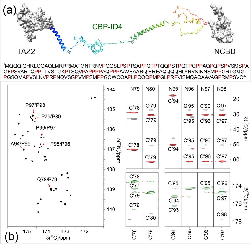
https://doi.org/10.5194/mr-2-511-2021 Magn. Reson., 2, 511–522, 2021514 I. C. Felli et al.: IDPs by NMR: focusing on proline residues Figure 1. Proline residues are abundant in IDPs/IDRs, and their 15 N resonances can be easily detected. They fall in a specific, isolated region of the 2D CON spectrum, as illustrated by the examples in the figure. From left to right: α-synuclein (140 aa, 4 % Pro; Bermel et al., 2006b), human securin (200 aa, 11 % Pro; Bermel et al., 2009), E1A (243 aa, 16 % Pro; Hošek et al., 2016), CBP-ID3 (407 aa 18 % Pro; Contreras-Martos et al., 2017), and CBP-ID4 (207 aa, 22 % Pro; Piai et al., 2016). tending the acquisition time in all the indirect dimensions to ities in all cases in which it is not possible to identify the contrast with the reduced chemical shift dispersion typical of type of amino acid preceding the proline by considering only IDPs. Non-uniform sampling strategies (Hoch et al., 2014; their 13 Cα and 13 Cβ chemical shifts. This is the case for the Kazimierczuk et al., 2010, 2011; Robson et al., 2019) can, example of Arg/Lys/Gln or Phe/Leu. of course, be implemented to reduce acquisition times; also, Similarly, the insertion of the 15 N band-selective pulse in this case, reducing the spectral complexity (the number for the proline region in the 3D (H)CBCANCO (Bermel of cross-peaks is reduced when focusing on proline 15 N et al., 2006a) enables us to detect the 13 C resonances of resonances only) is expected to contribute to a reduction the whole proline ring, providing complementary informa- in experimental times. Out of the full 3D spectrum, only tion for the sequence-specific assignment. The closed proline a small portion, the one containing the information that is ring introduces an additional heteronuclear scalar coupling completely missing in amide proton detected experiments, (1 JNi −Cδi ) that also provides the correlations with 13 Cδ and can thus be acquired with the necessary resolution to provide 13 Cγ , which is parallel to the 13 Cα and 13 Cβ chemical shifts. site-specific atomic information. Indeed, the band-selective pulses used for the 13 C aliphatic Since C0 detected experiments all exploit the C0 i−1 − Ni region also cover 13 Cγ and 13 Cδ resonances (not only 13 Cα correlation, which is an inter-residue correlation linking the and 13 Cβ ones). In addition, analysis of the observed chem- nitrogen of an amino acid (i) to the carbonyl carbon of the ical shifts for proline side chains can be correlated to the lo- previous one (i − 1) across the peptide bond (Fig. 2 inset), cal conformation, in particular to the cis/trans isomers of the focusing on proline residues can facilitate the identification peptide bond involving proline nitrogen nuclei (Schubert et of specific Xi−1 − Proi pairs through an inspection of Cα al., 2002; Shen and Bax, 2010). Finally, additional informa- and Cβ chemical shifts of the amino acid preceding the pro- tion for sequence-specific assignment can be achieved by ex- line residues. Such information can be achieved through the ploiting the same approach for the COCON experiment in its 3D (H)CBCACONPro experiment and can be very useful for 13 C start (Bermel et al., 2006b; Felli et al., 2009) as well and identifying specific pairs such as Gly/Pro, Ala/Pro, Ser/Pro, in its 1 H start variants (Mateos et al., 2020). and Thr/Pro. Acquisition of the 3D (H)CCCONPro experi- ment, in parallel to the 3D (H)CBCACONPro , provides infor- mation on aliphatic 13 C nuclear chemical shifts of the whole side chain. This contributes to narrowing down the possibil- Magn. Reson., 2, 511–522, 2021 https://doi.org/10.5194/mr-2-511-2021
I. C. Felli et al.: IDPs by NMR: focusing on proline residues                                                                            515

Figure 2. Comparison of the 2D CON (a) and 2D HN (b) spectra recorded on α-synuclein. The CONpro spectrum (c), shown below the HN
panel, clearly illustrates how this experiment provides the missing information with respect to that available in the HN-detected spectrum. In
the inset of the 2D CON spectrum, a scheme of a Glyi−1 − Proi dipeptide highlights the nuclei that give rise to the C0 i−1 − Ni correlations
detected in CON spectra (in green).

3.2   Assignment strategy                                                   Indeed, the C0 −N projections of the 3D spectra (13 C−15 N
                                                                         planes) show that the cross-peaks are well resolved in both
To illustrate the approach, experiments were acquired on                 dimensions; the selection of the 15 N proline region enables
the well-known IDP α-synuclein. Even if this protein only                us to differentiate the signals through the carbonyl car-
contains a small number of proline residues (5 out of 140),              bon chemical shifts of the preceding amino acid. There-
they are all clustered in a small portion of it (108–138) and,           fore, an inspection of the first 13 C−13 C plane of the 3D
thus, constitute about 15 % of the amino acids in this re-               (H)CBCACONPro experiment (Fig. 3a) shows the distinctive
gion. Furthermore, this terminus has a very peculiar amino               13 Cα and 13 Cβ chemical shift patterns of the residues preced-
acidic composition (36 % Asp/Glu, 9 % Tyr), and it was                   ing proline that, by comparison with the primary sequence
shown to be the part of the protein that is involved in sensing          of the protein, already suggests the identity of three residue
calcium concentration jumps associated with the transmis-                pairs to us, i.e. Ala–Pro, Asp–Pro, and Glu–Pro. These can,
sion of nerve signals (Binolfi et al., 2006; Lautenschläger et           thus, be assigned to Ala 107–Pro 108, Asp 119–Pro 120, Glu
al., 2018; Nielsen et al., 2001). Proline residues in between            138–Pro 139. Comparison with the first 13 C−13 C plane of
two negatively charged amino acids (Asp-Pro-Asp and Glu-                 the 3D (H)CCCONPro (Fig. 3b) confirms that an extra cross-
Pro-Glu) were shown to facilitate the interaction of carboxy-            peak can be detected for the Glu–Pro pair, which is as ex-
late side chains of Asp and Glu with calcium, even in a flex-            pected for amino acids that have a side chain with more than
ible and disordered state (Pontoriero et al., 2020).                     two aliphatic carbon atoms. The remaining signals derive
   Focusing on the proline 15 N region in case of α-synuclein            from the two Met–Pro pairs, in agreement with the observed
greatly simplifies the spectral complexity, enabling us to il-           chemical shifts. They can be assigned in a sequence-specific
lustrate the sequence-specific assignment of the resonances              manner by comparing these spectra with the complemen-
just by visual inspection of the first planes of the 3D spectra          tary ones based on amide proton detection. The final panel
described here (Fig. 3).

https://doi.org/10.5194/mr-2-511-2021                                                                 Magn. Reson., 2, 511–522, 2021
516 I. C. Felli et al.: IDPs by NMR: focusing on proline residues Figure 3. The implementation of the proposed strategy on α-synuclein renders NMR spectra so informative that proline resonances can be assigned just by visual inspection of the figure. To this end, the first 13 C−13 C planes of the 3D (H)CBCACONPro (a), 3D (H)CCCONPro (b), and 3D (H)CBCANCOPro (c) are shown, and the 13 C−15 N plane of the 3D (H)CBCACONPro is also shown (d). The portion of the primary sequence of α-synuclein hosting its five proline residues is also reported (below the spectra). The pulse sequence for acquiring the 3D (H)CBCACONPro experiment (e) is reported as an example of the implementation of the proposed approach (dotted box). The delays are ε = t2 (0), δ = 3.6 ms, 1 = 9 ms, 11 = 25 ms, 12 = 8 ms, 13 = 12 –14 = 5.8 ms, and 14 = 2.2 ms. The phase cycle is as follows: φ1 = x, -x; φ2 = 8(x), 8(-x); φ3 = 4(x), 4(-x); φ4 = 2(x), 2(-x); φIPAP (IP) = x; and φIPAP (AP) = -y; φrec = x, -x, -x, x, -x, x, x, -x. Quadrature detection was obtained by incrementing phase φ3 (t1 ) and φ4 (t2 ) in a States–TPPI (time-proportional phase incrementation) manner. The IPAP (in-phase/antiphase) approach was implemented for homonuclear decoupling in the direct acquisition dimension to suppress the large one bond scalar coupling constants (1 JCα−C0 ; Felli and Pierattelli, 2015); alternative approaches can be implemented that exploit band- selective homonuclear decoupling (Alik et al., 2020; Ying et al., 2014) or processing algorithms that, thus, only require the in-phase spectra (Karunanithy and Hansen, 2021; Shimba et al., 2003). shows the first 13 C−13 C plane of the 3D (H)CBCANCOPro served in Fig. 3. Chemical shifts show only minor differ- (Fig. 3c). This experiment reveals the correlations of the 15 N ences between the resonances, but these are still significant to with 13 Cα and 13 Cβ within each amino acid. In the case discriminate between the different residues, provided spectra of proline, the closed ring introduces additional scalar cou- are acquired with high resolution. plings that are responsible for two additional cross-peaks, i.e. the ones of the 15 N with 13 Cγ and 13 Cδ , as clearly ob- Magn. Reson., 2, 511–522, 2021 https://doi.org/10.5194/mr-2-511-2021
I. C. Felli et al.: IDPs by NMR: focusing on proline residues                                                                  517

3.3   A challenging case                                           very useful, as demonstrated for these two proline-rich frag-
                                                                   ments. This experiment, which includes an isotropic mixing
A compelling example of complexity is provided by the              element in the carbonyl region (MOCCA, in this case; Felli
fourth flexible linker (ID4) of CREB-binding protein (CBP),        et al., 2009; Furrer et al., 2004) enables us to detect correla-
a large transcription co-regulator (Dyson and Wright, 2016).       tions of a carbonyl with the neighbouring ones through the
CBP-ID4 connects two well-characterized globular domains           small 3 JC0 C0 scalar couplings. In case of proline residues, the
(TAZ2, 92 amino acids, and NCBD, 59 amino acids) (De               most intense cross-peak is generally observed for the preced-
Guzman et al., 2000; Kjaergaard et al., 2010) and is con-          ing amino acid (C0 i−2 ). However, additional peaks are also
stituted by 207 amino acids out of which 45 are pro-               detected with neighbouring ones and support the sequence-
line residues, including several repeated PP motifs (Piai et       specific assignment process.
al., 2016) (Fig. 4a). The 2D CONPro spectrum of CBP-ID4,              It is interesting to note that, once the sequence-specific as-
reported in Fig. 4b (left panel), shows the C0 i−1 − Ni corre-     signment becomes available, C0 i−1 − Ni correlations fall in
lations of proline residues of ID4. Interestingly, despite the     distinctive spectral regions of the 2D CONPro spectrum, as al-
small spectral region, a high number of resolved resonances        ready pointed out for selected residue pairs such as Gly–Pro,
is observed. The initial count of cross-peaks in this spectrum     Ser–Pro, Thr–Pro, and Val–Pro (Murrali et al., 2018). For ex-
reveals 42 out of the 45 expected correlations, highlighting       ample, an inspection of Fig. 1 allows us to identify Gly–Pro
the potential of this experimental strategy for the investiga-     pairs in all the CON spectra of different proteins from their
tion of IDRs/IDPs of increasing complexity. The excellent          characteristic chemical shifts (in the top-right portion of the
chemical shift dispersion of the inter-residue C0 i−1 − Ni cor-    proline region). An additional contribution towards smaller
relations is certainly one of the most important aspects for       15 N chemical shifts can also be identified in cases in which

reducing cross-peak overlap. The resolution is further en-         more than one proline follows a specific amino-acid-type,
hanced in this region by the narrow linewidths of proline 15 N     such as for Ala–Pro, Met–Pro, and Gln–Pro cross-peaks that
resonances due to the lack of the dipolar contribution of an       are shifted to lower 15 N chemical shifts when an additional
amide proton to the transverse relaxation.                         proline follows in the primary sequence. These effects likely
   These two features contribute to establishing this spectral     originate from a combination of effects deriving from the co-
region as a key one for the assignment of a large IDR. In-         valent structure (primary sequence in this case) and from lo-
deed, when passing from 2D to 3D experiments, long acqui-          cal conformations. Needless to say, the experimental investi-
sition times in the 15 N dimension are possible and enable us      gation of these aspects in more detail constitutes an important
to provide the extra contribution to the resolution enhance-       point that describes the structural and dynamic properties at
ment needed to focus on complex IDRs and to collect ad-            the atomic resolution of the proline-rich parts of highly flex-
ditional information on the proline residues and their neigh-      ible IDRs. The proposed experiments are, thus, expected to
bouring amino acids. As an example of the quality of the           become of general applicability for studies of IDPs/IDRs in
spectra, Fig. 4b reports several strips extracted from the pro-    solution.
line selective 3D experiments that were essential for the in-         The data generated on proline-rich sequences are, of
vestigation of a particularly proline-rich region of ID4, the      course, very relevant to populate databases such as the Bi-
one in between two partially populated α-helices (Piai et          ological Magnetic Resonance Data Bank (BMRB, https://
al., 2016). This is composed of 27 prolines (out of 76 amino       bmrb.io/, last access: 17 June 2021), with information on
acids) which constitute 35 % of the amino acids in this re-        proline residues in highly flexible protein regions. This, in
gion, including several proline-rich motifs (PXP, PXXP, and        turn, will generate more accurate reference data in chemi-
PP, as well as PPP and PPPP, where P stands for a pro-             cal shift databases to determine local structural propensities
line and X for any other amino acid). Figure 4b shows the          through the comparison of experimental shifts with refer-
strips, extracted from the 3D proline selective experiments        ence ones (Camilloni et al., 2012; Tamiola et al., 2010), thus
that were used to assign resonances in the two proline-rich        improving our understanding of the importance of transient
regions (78–80 and 94–98). The strips extracted from the           secondary structure elements in determining protein func-
3D (H)CBCACONPro and 3D (H)CCCONPro (Fig. 4b; right                tion. In this respect, CBP itself provides another enlighten-
panels; black and red contours respectively) are very use-         ing example with the third disordered linker of CBP (CBP-
ful for identifying the Xi−1 − Proi pairs that match, in this      ID3; residues 674-1079 of CBP), which features a high num-
case, with a Gln–Pro and an Ala–Pro pair, as well as sev-          ber of proline residues (75 out of 406 residues) represent-
eral Pro–Pro ones. The 3D (H)CBCANCOPro completes the              ing 18 % of its primary sequence. The distribution, in this
picture by providing information about 13 C resonances of          case, is along the entire sequence but less frequent toward
each proline ring (Cα , Cβ , Cγ , and Cδ ; not shown for sake of   the end, where a β strand conformation propensity is sam-
clarity in the figure). However, in regions with a high abun-      pled (Contreras-Martos et al., 2017). Also, in this case, the
dance of proline residues, additional information is needed        distribution of proline residues is important for shaping the
for their sequence-specific assignment. To this end, the 3D        conformational space accessible to the polypeptide, facilitat-
(HCA)COCONPro (Fig. 4b; right panels; green contours) is           ing the interaction with protein’s partners. Determination of

https://doi.org/10.5194/mr-2-511-2021                                                         Magn. Reson., 2, 511–522, 2021
518 I. C. Felli et al.: IDPs by NMR: focusing on proline residues Figure 4. The case of CBP-ID4 (residues 1851–2057 of human CBP). (a) One of the possible conformations of the CBP-ID4 fragment (blue to red ribbons) and the two flanking domains, TAZ2 and NCBD, clearly demonstrate that, in this region of CBP, the intrinsically disordered part is highly prevalent with respect to ordered ones. The amino acid sequence of CBP-ID4 is also reported. (b) The 2D CONPro spectrum shown on the left, which, at first sight, could seem like a 2D spectrum of a small globular protein, reports the proline fingerprint of this complex IDR. Several strips extracted from the 3D (H)CBCACONPro (grey contours), 3D (H)CCCONPro (red contours), and 3D (HCA)COCONPro (green contours) are shown to illustrate the information available for the sequence-specific resonance assignment of this proline-rich fragment of CBP-ID4. additional observables, such as the 3 JC0 C0 through the 3D investigated to access information on challenging systems (HCA)COCONPro experiment or of different ones through (such as, for example, the studies of large globular proteins modified experimental variants of these experiments, are ex- enabled by methyl transverse-relaxation-optimized spec- pected to contribute to the characterization of novel motifs in troscopy (methyl-TROSY) spectroscopy; Kay, 2011; Schütz IDRs/IDPs. and Sprangers, 2020). Interestingly, the analysis of the NMR The experimental strategy proposed here focuses on a re- spectra presented here enables one to classify the observed markably small spectral region which, however, turns out cross-peaks into residue types, also in absence of a sequence- to be one of the most interesting ones, in particular from specific assignment. This might provide interesting informa- the perspective of studying IDPs/IDRs of increasing com- tion for complex IDPs/IDRs in which one is interested in plexity, somehow reminiscent of other strategies that have the investigation of the contribution of specific residue types, been proposed in which only selected residue types are such as to monitor the occurrence of post-translational mod- Magn. Reson., 2, 511–522, 2021 https://doi.org/10.5194/mr-2-511-2021
I. C. Felli et al.: IDPs by NMR: focusing on proline residues                                                                         519

ifications or even other phenomena that are more difficult to       Supplement. The supplement related to this article is available
investigate like liquid–liquid-phase separation.                    online at: https://doi.org/10.5194/mr-2-511-2021-supplement.

                                                                    Author contributions. All authors conceived the research, de-
4   Conclusions and perspectives
                                                                    signed, carried out, and analysed the experiments, and wrote the
                                                                    paper.
Detection and assignment of proline-rich regions of highly
flexible intrinsically disordered proteins allows us to have a
glimpse of the ways in which proline residues encode spe-           Competing interests. The authors declare that they have no con-
cific properties in IDRs/IDPs by simply tuning their distribu-      flict of interest.
tion along the primary sequence. NMR spectroscopy is par-
ticularly well suited for the task, since proline residues have
attractive features from the NMR point of view, starting from       Disclaimer. Publisher’s note: Copernicus Publications remains
the peculiar chemical shifts of 15 N nuclear spins. In addition,    neutral with regard to jurisdictional claims in published maps and
the lack of the attached amide proton implies that one of the       institutional affiliations.
major contributions to relaxation of 15 N spins is absent and,
thus, proline nitrogen signals have small linewidths. These
characteristics make them a very useful starting point for se-      Special issue statement. This article is part of the special issue
quential assignment purposes and structure characterization.        “Geoffrey Bodenhausen Festschrift”. It is not associated with a con-
Furthermore, they provide a set of NMR signals with promis-         ference.
ing properties to enable high-resolution studies of increas-
ingly large IDPs/IDRs.
                                                                    Acknowledgements. The support of the CERM/CIRMMP cen-
   Several approaches, either based on HN or on Hα direct
                                                                    ter of Instruct-ERIC is gratefully acknowledged. This work has
detection, have been proposed to bypass the problem in-             been funded, in part, by the Italian Ministry of University and Re-
troduced in the sequence-specific assignment by the lack            search (FOE funding). Maria Grazia Murrali, Letizia Pontoriero,
of amide protons typical of proline residues (Hellman et            and Marco Schiavina are acknowledged for their contributions in
al., 2014; Kanelis et al., 2000; Karjalainen et al., 2020; Löhr     the early stages of the project.
et al., 2000; Mäntylahti et al., 2010; Tossavainen et al., 2020;
Wong et al., 2018). While these results can be useful for sys-
tems with moderate complexity (Hα detection) or for systems         Financial support. This research has been supported by the Fon-
that feature isolated proline residues in the primary sequence      dazione Cassa di Risparmio di Firenze (grant no. 45585).
(HN /Hα ), they are not as efficient for complex IDRs/IDPs
in which high resolution is mandatory and in which con-
secutive proline residues are often encountered. Detection of       Review statement. This paper was edited by Fabien Ferrage and
15 N-based experiments can also provide direct observation of       reviewed by two anonymous referees.
proline nuclei (Chhabra et al., 2018), but despite the excel-
lent resolution achievable with IDPs, these experiments still
suffer from sensitivity limitations.                                References
   To conclude, the experiments proposed here are crucial
to assign intrinsically disordered protein regions presenting       Ahuja, P., Cantrelle, F. X., Huvent, I., Hanoulle, X., Lopez, J., Smet,
many repeated motifs, including proline residues and poly-            C., Wieruszeski, J. M., Landrieu, I., and Lippens, G.: Proline
proline segments, to reduce spectral complexity and experi-           conformation in a functional Tau fragment, J. Mol. Biol., 428,
mental time without compromising resolution. Since NMR                79–91, https://doi.org/10.1016/j.jmb.2015.11.023, 2016.
probeheads with high 13 C sensitivity have become widely            Alik, A., Bouguechtouli, C., Julien, M., Bermel, W., Ghouil, R.,
                                                                      Zinn-Justin, S., and Theillet, F. X.: Sensitivity-Enhanced 13 C-
available, it is expected that this set of experiments will be
                                                                      NMR spectroscopy for monitoring multisite phosphorylation at
applied as an easy-to-use tool that will also complement the          physiological temperature and pH, Angew. Chem. Int. Edit., 59,
HN -based assignment.                                                 10411–10415, https://doi.org/10.1002/anie.202002288, 2020.
                                                                    Bermel, W., Bertini, I., Felli, I. C., Kümmerle, R., and Pier-
                                                                      attelli, R.: Novel 13 C direct detection experiments, includ-
Code availability. The codes of the pulse sequences used are pro-     ing extension to the third dimension, to perform the com-
vided in the Supplement.                                              plete assignment of proteins, J. Magn. Reson., 178, 56–64,
                                                                      https://doi.org/10.1016/j.jmr.2005.08.011, 2006a.
                                                                    Bermel, W., Bertini, I., Felli, I. C., Lee, Y.-M., Luchi-
Data availability. Data are available from the authors upon re-       nat, C., and Pierattelli, R.: Protonless NMR experiments
quest.                                                                for sequence-specific assignment of backbone nuclei in

https://doi.org/10.5194/mr-2-511-2021                                                             Magn. Reson., 2, 511–522, 2021
520 I. C. Felli et al.: IDPs by NMR: focusing on proline residues unfolded proteins, J. Am. Chem. Soc., 128, 3918–3919, Felli, I. C. and Pierattelli, R.: Spin-state-selective meth- https://doi.org/10.1021/ja0582206, 2006b. ods in solution- and solid-state biomolecular 13 C Bermel, W., Bertini, I., Csizmok, V., Felli, I. C., Pierat- NMR, Prog. Nucl. Magn. Res. Sp., 84–85, 1–13, telli, R., and Tompa, P.: H-start for exclusively het- https://doi.org/10.1016/j.pnmrs.2014.10.001, 2015. eronuclear NMR spectroscopy: The case of intrinsically Felli, I. C., Pierattelli, R., Glaser, S. J., and Luy, B.: Relaxation- disordered proteins, J. Magn. Reson., 198, 275–281, optimised Hartmann-Hahn transfer using a specifically Tailored https://doi.org/10.1016/j.jmr.2009.02.012, 2009. MOCCA-XY16 mixing sequence for carbonyl-carbonyl corre- Binolfi, A., Rasia, R. M., Bertoncini, C. W., Ceolin, M., Zweck- lation spectroscopy in 13 C direct detection NMR experiments, stetter, M., Griesinger, C., Jovin, T. M., and Fernández, C. O.: J. Biomol. NMR, 43, 187–196, https://doi.org/10.1007/s10858- Interaction of α-synuclein with divalent metal ions reveals key 009-9302-6, 2009. differences: A link between structure, binding specificity and Furrer, J., Kramer, F., Marino, J. P., Glaser, S. J., and fibrillation enhancement, J. Am. Chem. Soc., 128, 9893–9901, Luy, B.: Homonuclear Hartmann-Hahn transfer with re- https://doi.org/10.1021/ja0618649, 2006. duced relaxation losses by use of the MOCCA-XY16 Camilloni, C., De Simone, A., Vranken, W. F., and Vendr- multiple pulse sequence, J. Magn. Reson., 166, 39–46, uscolo, M.: Determination of secondary structure popula- https://doi.org/10.1016/j.jmr.2003.09.013, 2004. tions in disordered states of proteins using nuclear mag- Geen, H. and Freeman, R.: Band-selective radiofrequency pulses, netic resonance chemical shifts, Biochemistry, 51, 2224–2231, J. Magn. Reson., 93, 93–141, https://doi.org/10.1016/0022- https://doi.org/10.1021/bi3001825, 2012. 2364(91)90034-Q, 1991. Chaves-Arquero, B., Pantoja-Uceda, D., Roque, A., Ponte, I., Suau, Gibbs, E. B., Lu, F., Portz, B., Fisher, M. J., Medellin, B. P., P., and Jiménez, M. A.: A CON-based NMR assignment strategy Laremore, T. N., Zhang, Y. J., Gilmour, D. S., and Showal- for pro-rich intrinsically disordered proteins with low signal dis- ter, S. A.: Phosphorylation induces sequence-specific conforma- persion: the C-terminal domain of histone H1.0 as a case study, tional switches in the RNA polymerase II C-terminal domain, J. Biomol. NMR, 72, 139–148, https://doi.org/10.1007/s10858- Nat. Commun., 8, 1–11, https://doi.org/10.1038/ncomms15233, 018-0213-2, 2018. 2017. Chhabra, S., Fischer, P., Takeuchi, K., Dubey, A., Ziarek, J. J., Boes- Gil, S., Hošek, T., Solyom, Z., Kümmerle, R., Brutscher, B., zoermenyi, A., Mathieu, D., Bermel, W., Davey, N. E., Wagner, Pierattelli, R., and Felli, I. C.: NMR spectroscopic stud- G., and Arthanari, H.: 15 N detection harnesses the slow relax- ies of intrinsically disordered proteins at near-physiological ation property of nitrogen: Delivering enhanced resolution for conditions, Angew. Chem. Int. Edit., 52, 11808–11812, intrinsically disordered proteins, P. Natl. Acad. Sci. USA, 115, https://doi.org/10.1002/anie.201304272, 2013. E1710–E1719, https://doi.org/10.1073/pnas.1717560115, 2018. Haba, N. Y., Gross, R., Novacek, J., Shaked, H., Zidek, L., Contreras-Martos, S., Piai, A., Kosol, S., Varadi, M., Bekesi, A., Barda-Saad, M., and Chill, J. H.: NMR determines tran- Lebrun, P., Volkov, A. N., Gevaert, K., Pierattelli, R., Felli, I. sient structure and dynamics in the disordered C-terminal do- C., and Tompa, P.: Linking functions: An additional role for an main of WASp interacting protein, Biophys. J., 105, 481–493, intrinsically disordered linker domain in the transcriptional coac- https://doi.org/10.1016/j.bpj.2013.05.046, 2013. tivator CBP, Sci. Rep., 7, 4676, https://doi.org/10.1038/s41598- Hellman, M., Piirainen, H., Jaakola, V. P., and Permi, P.: Bridge over 017-04611-x, 2017. troubled proline: Assignment of intrinsically disordered proteins De Guzman, R. N., Liu, H. Y., Martinez-Yamout, M., Dyson, H. J., using (HCA)CON(CAN)H and (HCA)N(CA)CO(N)H experi- and Wright, P. E.: Solution structure of the TAZ2 (CH3) domain ments concomitantly with HNCO and i(HCA)CO(CA)NH, J. of the transcriptional adaptor protein CBP, J. Mol. Biol., 303, Biomol. NMR, 58, 49–60, https://doi.org/10.1007/s10858-013- 243–253, https://doi.org/10.1006/jmbi.2000.4141, 2000. 9804-0, 2014. Dunker, A. K., Oldfield, C. J., Meng, J., Romero, P., Yang, J. Y., Hoch, J. C., Maciejewski, M. W., Mobli, M., Schuyler, A. D., and Chen, J. W., Vacic, V., Obradovic, Z., and Uversky, V. N.: The un- Stern, A. S.: Nonuniform sampling and maximum entropy re- foldomics decade: An update on intrinsically disordered proteins, construction in multidimensional NMR, Acc. Chem. Res., 47, BMC Genomics, 9, S1, https://doi.org/10.1186/1471-2164-9-S2- 708–717, https://doi.org/10.1021/ar400244v, 2014. S1, 2008. Hošek, T., Calçada, E. O., Nogueira, M. O., Salvi, M., Pa- Dyson, H. J. and Wright, P. E.: Nuclear magnetic resonance meth- gani, T. D., Felli, I. C., and Pierattelli, R.: Structural and dy- ods for elucidation of structure and dynamics in disordered namic characterization of the molecular hub early region 1A states, Method. Enzymol., 339, 258–270, 2001. (E1A) from human adenovirus, Chem.-Eur. J., 22, 13010–13013, Dyson, H. J. and Wright, P. E.: Role of intrinsic protein disorder https://doi.org/10.1002/chem.201602510, 2016. in the function and interactions of the transcriptional coactiva- Hsu, S.-T. D., Bertoncini, C. W., and Dobson, C. M.: Use of pro- tors CREB-binding Protein (CBP) and p300, J. Biol. Chem., 291, tonless NMR spectroscopy to alleviate the loss of information 6714–6722, https://doi.org/10.1074/jbc.R115.692020, 2016. resulting from exchange-broadening, J. Am. Chem. Soc., 131, Emsley, L. and Bodenhausen, G.: Gaussian pulse cascades: New 7222–7223, https://doi.org/10.1021/ja902307q, 2009. analytical functions for rectangular selective inversion and in- Huang, C., Ren, G., Zhou, H., and Wang, C.: A new phase excitation in NMR, Chem. Phys. Lett., 165, 469–476, method for purification of recombinant human α-synuclein https://doi.org/10.1016/0009-2614(90)87025-M, 1990. in Escherichia coli, Protein Expres. Purif., 42, 173–177, Felli, I. C. and Pierattelli, R.: Novel methods based on 13 C detection https://doi.org/10.1016/j.pep.2005.02.014, 2005. to study intrinsically disordered proteins, J. Magn. Reson., 241, Kanelis, V., Donaldson, L., Muhandiram, D. R., Rotin, D., 115–125, https://doi.org/10.1016/j.jmr.2013.10.020, 2014. Forman-Kay, J. D., and Kay, L. E.: Sequential assignment Magn. Reson., 2, 511–522, 2021 https://doi.org/10.5194/mr-2-511-2021
I. C. Felli et al.: IDPs by NMR: focusing on proline residues                                                                             521

  of proline-rich regions in proteins: Application to modular         Lu, K. P., Finn, G., Lee, T. H., and Nicholson, L. K.: Prolyl cis-
  binding domain complexes, J. Biomol. NMR, 16, 253–259,                 trans isomerization as a molecular timer, Nat. Chem. Biol., 3,
  https://doi.org/10.1023/A:1008355012528, 2000.                         619–629, https://doi.org/10.1038/nchembio.2007.35, 2007.
Karjalainen, M., Tossavainen, H., Hellman, M., and Permi, P.: HA-     MacArthur, M. W. and Thornton, J. M.: Influence of proline
  CANCOi: a new Hα-detected experiment for backbone reso-                residues on protein conformation, J. Mol. Biol., 218, 397–412,
  nance assignment of intrinsically disordered proteins, J. Biomol.      https://doi.org/10.1016/0022-2836(91)90721-H, 1991.
  NMR, 74, 741–752, https://doi.org/10.1007/s10858-020-00347-         Mäntylahti, S., Aitio, O., Hellman, M., and Permi, P.: HA-
  5, 2020.                                                               detected experiments for the backbone assignment of intrin-
Karunanithy, G. and Hansen, D. F.: FID-Net: A versatile deep             sically disordered proteins, J. Biomol. NMR, 47, 171–181,
  neural network architecture for NMR spectral reconstruc-               https://doi.org/10.1007/s10858-010-9421-0, 2010.
  tion and virtual decoupling, J. Biomol. NMR, 75, 179–191,           Markley, J. L., Bax, A., Arata, Y., Hilbers, C. W., Kaptein,
  https://doi.org/10.1007/s10858-021-00366-w, 2021.                      R., Sykes, B. D., Wright, P. E., and Wüthrich, K.: Rec-
Kay, L. E.: Solution NMR spectroscopy of supra-molecular sys-            ommendations for the presentation of NMR structures of
  tems, why bother? A methyl-TROSY view, J. Magn. Reson., 210,           proteins and nucleic acids, J. Biomol. NMR, 12, 1–23,
  159–170, https://doi.org/10.1016/j.jmr.2011.03.008, 2011.              https://doi.org/10.1023/A:1008290618449, 1998.
Kazimierczuk, K., Stanek, J., Zawadzka-Kazimierczuk, A., and          Mateos, B., Conrad-Billroth, C., Schiavina, M., Beier, A., Kontaxis,
  Koźmiński, W.: Random sampling in multidimensional NMR               G., Konrat, R., Felli, I. C., and Pierattelli, R.: The ambivalent role
  spectroscopy, Prog. Nucl. Magn. Res. Sp., 57, 420–434,                 of proline residues in an intrinsically disordered protein: From
  https://doi.org/10.1016/j.pnmrs.2010.07.002, 2010.                     disorder promoters to compaction facilitators, J. Mol. Biol., 432,
Kazimierczuk, K., Misiak, M., Stanek, J., Zawadzka-Kazimierczuk,         3093–3111, https://doi.org/10.1016/j.jmb.2019.11.015, 2020.
  A., and Koźmiński, W.: Generalized Fourier Transform for Non-     Murrali, M. G., Piai, A., Bermel, W., Felli, I. C., and
  Uniform Sampled Data, in: Novel Sampling Approaches in                 Pierattelli, R.: Proline fingerprint in intrinsically dis-
  Higher Dimensional NMR, edited by: Billeter, M. and Orekhov,           ordered proteins, ChemBioChem, 19(15), 1625–1629,
  V., Springer, Berlin, Heidelberg, Topics in Current Chemistry,         https://doi.org/10.1002/cbic.201800172, 2018.
  316, 79–124, https://doi.org/10.1007/128_2011_186, 2011.            Nielsen, M. S., Vorum, H., Lindersson, E., and Jensen, P.
Kjaergaard, M., Teilum, K., and Poulsen, F. M.: Conformational           H.: Ca2+ Binding to α-synuclein regulates ligand bind-
  selection in the molten globule state of the nuclear coactivator       ing and oligomerization, J. Biol. Chem., 276, 22680–22684,
  binding domain of CBP, P. Natl. Acad. Sci. USA, 107, 12535–            https://doi.org/10.1074/jbc.M101181200, 2001.
  12540, https://doi.org/10.1073/pnas.1001693107, 2010.               Olsen, G. L., Szekely, O., Mateos, B., Kadeřávek, P., Ferrage,
Knoblich, K., Whittaker, S., Ludwig, C., Michiels, P., Jiang, T.,        F., Konrat, R., Pierattelli, R., Felli, I. C., Bodenhausen, G.,
  Schaffhausen, B., and Günther, U.: Backbone assignment of              Kurzbach, D., and Frydman, L.: Sensitivity-enhanced three-
  the N-terminal polyomavirus large T antigen, Biomol. NMR               dimensional and carbon-detected two-dimensional NMR of pro-
  Assign., 3, 119–123, https://doi.org/10.1007/s12104-009-9155-          teins using hyperpolarized water, J. Biomol. NMR, 74, 161–171,
  7, 2009.                                                               https://doi.org/10.1007/s10858-020-00301-5, 2020.
Kurzbach, D., Canet, E., Flamm, A. G., Jhajharia, A., Weber,          Pérez, Y., Gairí, M., Pons, M., and Bernadó, P.: Structural char-
  E. M. M., Konrat, R., and Bodenhausen, G.: Investigation               acterization of the natively unfolded N-terminal domain of
  of intrinsically disordered proteins through exchange with hy-         human c-Src kinase: insights into the role of phosphoryla-
  perpolarized water, Angew. Chem. Int. Edit., 56, 389–392,              tion of the unique domain, J. Mol. Biol., 391, 136–148,
  https://doi.org/10.1002/anie.201608903, 2017.                          https://doi.org/10.1016/j.jmb.2009.06.018, 2009.
Lautenschläger, J., Stephens, A. D., Fusco, G., Ströhl, F., Curry,    Piai, A., Calçada, E. O., Tarenzi, T., Grande, A. Del, Varadi,
  N., Zacharopoulou, M., Michel, C. H., Laine, R., Nespovitaya,          M., Tompa, P., Felli, I. C., and Pierattelli, R.: Just a flexi-
  N., Fantham, M., Pinotsi, D., Zago, W., Fraser, P., Tandon, A.,        ble linker? the structural and dynamic properties of CBP-ID4
  St George-Hyslop, P., Rees, E., Phillips, J. J., De Simone, A.,        revealed by NMR spectroscopy, Biophys. J., 110, 372–381,
  Kaminski, C. F., and Schierle, G. S. K.: C-terminal calcium bind-      https://doi.org/10.1016/j.bpj.2015.11.3516, 2016.
  ing of α-synuclein modulates synaptic vesicle interaction, Nat.     Pontoriero, L., Schiavina, M., Murrali, M. G., Pierattelli, R., and
  Commun., 9, 712 , https://doi.org/10.1038/s41467-018-03111-4,          Felli, I. C.: Monitoring the Interaction of α-synuclein with cal-
  2018.                                                                  cium ions through exclusively heteronuclear Nuclear Magnetic
Löhr, F., Pfeiffer, S., Lin, Y. J., Hartleib, J., Klimmek, O.,           Resonance experiments, Angew. Chem., 132, 18696–18704,
  and Rüterjans, H.: HNCAN pulse sequences for sequen-                   https://doi.org/10.1002/ange.202008079, 2020.
  tial backbone resonance assignment across proline residues          Robson, S., Arthanari, H., Hyberts, S. G., and Wagner, G.: Chap-
  in perdeuterated proteins, J. Biomol. NMR, 18, 337–346,                ter Ten – Nonuniform sampling for NMR spectroscopy, Methods
  https://doi.org/10.1023/A:1026737732576, 2000.                         Enzymol., 614, 263–291, 2019.
Lopez, J., Schneider, R., Cantrelle, F. X., Huvent, I., and Lip-      Schiavina, M., Murrali, M. G., Pontoriero, L., Sainati, V.,
  pens, G.: Studying intrinsically disordered proteins under true        Kümmerle, R., Bermel, W., Pierattelli, R., and Felli, I.
  in vivo conditions by combined cross-polarization and carbonyl-        C.: Taking simultaneous snapshots of intrinsically dis-
  detection NMR Spectroscopy, Angew. Chem. Int. Edit., 55,               ordered proteins in action, Biophys. J., 117, 46–55,
  7418–7422, https://doi.org/10.1002/anie.201601850, 2016.               https://doi.org/10.1016/j.bpj.2019.05.017, 2019.
                                                                      Schubert, M., Labudde, D., Oschkinat, H., and Schmieder,
                                                                         P.: A software tool for the prediction of Xaa-Pro pep-

https://doi.org/10.5194/mr-2-511-2021                                                                Magn. Reson., 2, 511–522, 2021
522 I. C. Felli et al.: IDPs by NMR: focusing on proline residues tide bond conformations in proteins based on 13 C Theillet, F., Kalmar, L., Tompa, P., Han, K., Selenko, P., Dunker, chemical shift statistics, J. Biomol. NMR, 24, 149–154, A. K., Daughdrill, G. W., and Uversky, V. N.: The alphabet https://doi.org/10.1023/A:1020997118364, 2002. of intrinsic disorder, Intrinsically Disord. Proteins, 1, e24360, Schuler, B., Lipman, E. A., Steinbach, P. J., Kumke, M., and Eaton, https://doi.org/10.4161/idp.24360, 2014. W. A.: Polyproline and the “spectroscopic ruler” revisited with Tossavainen, H., Salovaara, S., Hellman, M., Ihalin, R., and Permi, single-molecule fluorescence, P. Natl. Acad. Sci. USA, 102, P.: Dispersion from Cα or NH: 4D experiments for back- 2754–2759, https://doi.org/10.1073/pnas.0408164102, 2005. bone resonance assignment of intrinsically disordered proteins, Schütz, S. and Sprangers, R.: Methyl TROSY spectroscopy: J. Biomol. NMR, 74, 147–159, https://doi.org/10.1007/s10858- A versatile NMR approach to study challenging biolog- 020-00299-w, 2020. ical systems, Prog. Nucl. Magn. Res. Sp., 116, 56–84, Van Der Lee, R., Buljan, M., Lang, B., Weatheritt, R. J., https://doi.org/10.1016/j.pnmrs.2019.09.004, 2020. Daughdrill, G. W., Dunker, A. K., Fuxreiter, M., Gough, J., Shaka, A. J., Barker, P. B., and Freeman, R.: Computer-optimized Gsponer, J., Jones, D. T., Kim, P. M., Kriwacki, R. W., Old- decoupling scheme for wideband applications and low-level op- field, C. J., Pappu, R. V., Tompa, P., Uversky, V. N., Wright, eration, J. Magn. Reson., 64, 547–552, 1985. P. E., and Babu, M. M.: Classification of intrinsically dis- Shen, Y. and Bax, A.: Prediction of Xaa-Pro peptide bond confor- ordered regions and proteins, Chem. Rev., 114, 6589–6631, mation from sequence and chemical shifts, J. Biomol. NMR, 46, https://doi.org/10.1021/cr400525m, 2014. 199–204, https://doi.org/10.1007/s10858-009-9395-y, 2010. Williamson, M. P.: The structure and function of proline- Shimba, N., Stern, A. S., Craik, C. S., Hoch, J. C., and Dötsch, V.: rich regions in proteins, Biochem. J., 297, 249–260, Elimination of 13 Cα splitting in protein NMR spectra by decon- https://doi.org/10.1042/bj2970249, 1994. volution with maximum entropy reconstruction, J. Am. Chem. Wong, L. E., Maier, J., Wienands, J., Becker, S., and Griesinger, Soc., 125, 2382–2383, https://doi.org/10.1021/ja027973e, 2003. C.: Sensitivity-enhanced four-dimensional amide–amide corre- Szekely, O., Olsen, G. L., Felli, I. C., and Frydman, L.: lation NMR experiments for sequential assignment of proline- High-resolution 2D NMR of disordered proteins enhanced rich disordered proteins, J. Am. Chem. Soc., 140, 3518–3522, by hyperpolarized water, Anal. Chem., 90, 6169–6177, https://doi.org/10.1021/jacs.8b00215, 2018. https://doi.org/10.1021/acs.analchem.8b00585, 2018. Ying, J., Li, F., Lee, J. H., and Bax, A.: 13 Cα decoupling dur- Tamiola, K., Acar, B., and Mulder, F. A. A.: Sequence- ing direct observation of carbonyl resonances in solution NMR specific random coil chemical shifts of intrinsically dis- of isotopically enriched proteins, J. Biomol. NMR, 60, 15–21, ordered proteins, J. Am. Chem. Soc., 132, 18000–18003, https://doi.org/10.1007/s10858-014-9853-z, 2014. https://doi.org/10.1021/ja105656t, 2010. Zhou, Z., Kümmerle, R., Qiu, X., Redwine, D., Cong, R., Taha, Thakur, A., Chandra, K., Dubey, A., D’Silva, P., and Atreya, A., Baugh, D., and Winniford, B.: A new decoupling method for H. S.: Rapid characterization of hydrogen exchange accurate quantification of polyethylene copolymer composition in proteins, Angew. Chem. Int. Edit., 52, 2440–2443, and triad sequence distribution with 13 C NMR, J. Magn. Reson., https://doi.org/10.1002/anie.201206828, 2013. 187, 225–233, https://doi.org/10.1016/j.jmr.2007.05.005, 2007. Magn. Reson., 2, 511–522, 2021 https://doi.org/10.5194/mr-2-511-2021
You can also read