Structural and Nutritional Peculiarities Related to Lifespan Differences on Four Lopesia Induced Bivalve-Shaped Galls on the Single Super-Host ...
←
→
Page content transcription
If your browser does not render page correctly, please read the page content below

ORIGINAL RESEARCH
                                                                                                                                                  published: 17 May 2021
                                                                                                                                           doi: 10.3389/fpls.2021.660557

                                             Structural and Nutritional
                                             Peculiarities Related to Lifespan
                                             Differences on Four Lopesia Induced
                                             Bivalve-Shaped Galls on the Single
                                             Super-Host Mimosa gemmulata
                                             Elaine C. Costa 1 , Denis C. Oliveira 2 , Dayse K. L. Ferreira 1 and Rosy M. S. Isaias 1*
                                             1
                                                 Departamento de Botânica, Instituto de Ciências Biológicas, Universidade Federal de Minas Gerais, Belo Horizonte, Brazil,
                                             2
                                                 Instituto de Biologia, Universidade Federal de Uberlândia, Uberlândia, Brazil

                                             Super-host plants are elegant models to evaluate the peculiarities of gall structural
                                             and nutritional profiles due to the stimuli of distinct gall inducers in temporal and
                                             spatial perspectives. Galls induced by congeneric insects, Lopesia spp. (Diptera,
                                             Cecidomyiidae) on the same host plant, Mimosa gemmulata Barneby (Fabaceae)
                                             were analyzed to estimate if variations of 1 or 2 months in gall lifespans may
                          Edited by:
                                             result in differences over the accumulation of nutritional resources, and their
                Brigitte Mauch-Mani,         compartmentalization both in cell walls and protoplasm. Mimosa gemmulata hosts four
 Université de Neuchâtel, Switzerland
                                             Lopesia-induced galls: the lenticular bivalve-shaped gall (LG) with a 2-month life cycle,
                        Reviewed by:
                                             the brown lanceolate bivalve-shaped gall (BLG) and the green lanceolate bivalve-shaped
                          Valéria Maia,
  Federal University of Rio de Janeiro,      gall (GLG) with 3 month-life cycles, and the globoid bivalve-shaped gall (GG) with a
                                  Brazil     4 month-life cycle. The comparisons among the four Lopesia galls, using anatomical,
                      Man-Miao Yang,
     National Chung Hsing University,
                                             histometric, histochemical, and immunocytochemical tools, have demonstrated that the
                                Taiwan       longest lifespan of the GG related to its highest increment in structural and nutritional
                   *Correspondence:          traits compared with the LG, GLG, and BLG. The differences among the tissue
                     Rosy M. S. Isaias
                                             stratification and cell wall thickness of the galls with the 2-month and the 3-month
                     rosy@icb.ufmg.br
                                             lifespans were subtle. However, the GG had thicker cell walls and higher stratification
                    Specialty section:       of the common storage tissue, schlerenchymatic layers and typical nutritive tissue than
          This article was submitted to
                                             the other three gall morphospecies. The higher tissue thickness of the GG was followed
          Plant Pathogen Interactions,
                a section of the journal     by the formation of a bidirectional gradient of carbohydrates in the protoplasm, and the
             Frontiers in Plant Science      detection of xyloglucans in cell walls. Current data supported the presumption that the
          Received: 29 January 2021          longest the lifespan, the highest the impact over the structural and nutritional metabolism
            Accepted: 19 April 2021
            Published: 17 May 2021           of the Lopesia galls associated to M. gemmulata.
                              Citation:      Keywords: cell walls, gall anatomy, hemicelluloses, histochemistry, immunocytochemistry, Lopesia galls
                Costa EC, Oliveira DC,
  Ferreira DKL and Isaias RMS (2021)
 Structural and Nutritional Peculiarities
    Related to Lifespan Differences on
                                            INTRODUCTION
Four Lopesia Induced Bivalve-Shaped
       Galls on the Single Super-Host
                                            The gall lifespans depend on structural, cytological, and chemical traits on the host plant cells
                   Mimosa gemmulata.        stimulated by the associated galling organisms (Mani, 1964; Oliveira et al., 2010, 2016; Jorge et al.,
          Front. Plant Sci. 12:660557.      2018; Ferreira et al., 2019; Chen et al., 2020). Any changes in the galling organism behavior
       doi: 10.3389/fpls.2021.660557        may lead to the disruption of the gall life cycle (Rezende et al., 2019) and compromise gall

Frontiers in Plant Science | www.frontiersin.org                                      1                                             May 2021 | Volume 12 | Article 660557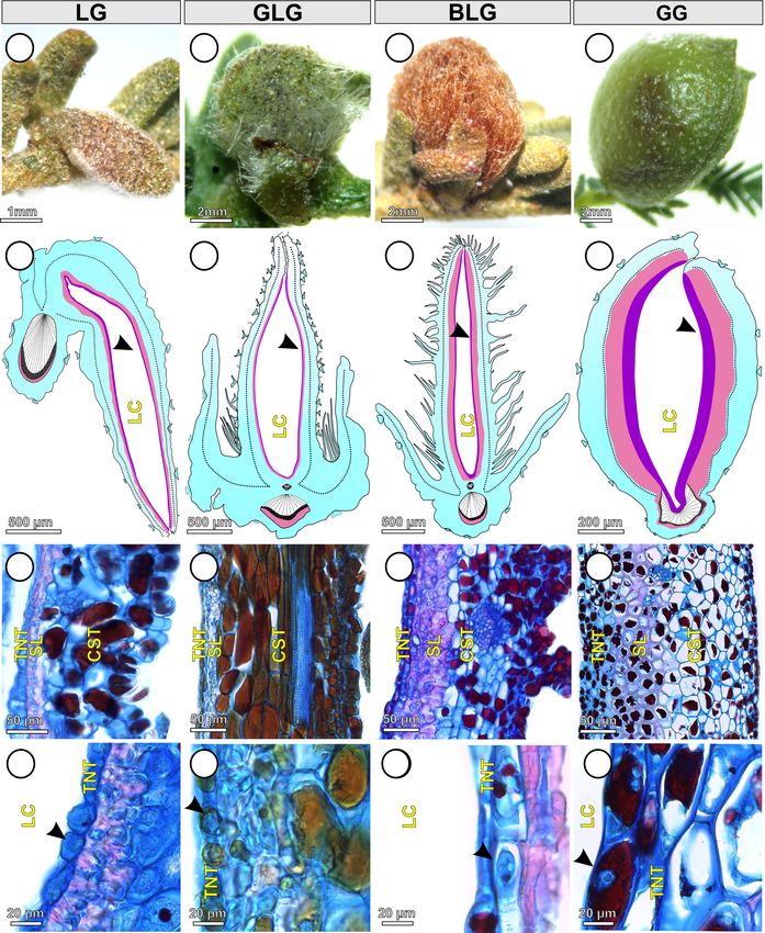
Costa et al.                                                                                                       Lopesia Galls on Mimosa gemmulata

developmental stages. In mature galls, the specialized tissues are            their compartmentalization both in cell walls and protoplasm.
established and organized in specific compartments (Bragança                  The following questions are addressed: (1) are there distinct
et al., 2017), which store energy-rich molecules that support the             peculiarities in the structural profiles among the four Lopesia
galling organism nutritional requirements (Mani, 1964; Ferreira               galls regarding tissue compartments? And (2) may the nutritional
et al., 2017). Cecidomyiidae galls, for instance, can have two types          profiles of the four Lopesia galls vary in response to the 1–2-
of storage tissues: the common storage tissue and the typical                 month-temporal distinction?
nutritive tissue, which are commonly spatially separated by
schlerenchymatic layers (Meyer and Maresquelle, 1983; Bronner,
1992; Rohfritsch, 1992). The common storage tissue is located                 MATERIALS AND METHODS
in the gall outer tissue compartment, while the typical nutritive
tissue is located in the gall inner tissue compartment, which is in           The Lifespans of Lopesia Galls on
contact with the larval chamber (Bragança et al., 2017).
                                                                              M. gemmulata
    The cells of the common storage tissue have inconspicuous
                                                                              The four Lopesia galls induced on M. gemmulata pinna-rachis
nuclei, large vacuoles, thin cytoplasm, and accumulate energetic
                                                                              are the lenticular bivalve-shaped gall (LG), the green lanceolate
metabolites related to gall growth and metabolism. The
                                                                              bivalve-shaped gall (GLG), the brown lanceolate bivalve-shaped
storage tissue also supports the nutritive tissue by cell-to-cell
                                                                              gall (BLG), and the globoid bivalve-shaped gall (GG). The four
translocation of solutes (Moura et al., 2008; Oliveira et al.,
                                                                              Lopesia spp. have multivoltine life cycles and each bivalve-shaped
2010; Ferreira and Isaias, 2014). The cells of the typical
                                                                              gall have distinct developmental times, with the maturation as the
nutritive tissue have conspicuous nuclei, fragmented vacuoles,
                                                                              longest stage of development. The LG has six life cycles a year, the
and dense protoplasm, which accumulate energetic metabolites
                                                                              BLG and GLG have four life cycles a year, and the GG has three
related to the nutrition of the gall inducer (Bronner, 1992;
                                                                              cycles a year (Costa et al., submitted). The LG has a 2-month
Oliveira et al., 2010, 2011a; Ferreira et al., 2015). The additional
                                                                              life cycle, with the maturation stage lasting ∼= 30–45 days. The
accumulation of carbohydrates in cell walls have been recently
                                                                              BLG and GLG have 3 month-life cycles, with the maturation stage
evaluated in nematode-induced galls (Ferreira et al., 2020)
                                                                              lasting ∼
                                                                                      = 45–60 days. The GG has a 4 month-life cycle, with the
and in insect-galls (Bragança et al., 2020a), with peculiarities
                                                                              maturation stage lasting ∼= 60–75 days.
regarding the type and distribution of hemicellulose epitopes.
The hemicelluloses, xyloglucans and heteromannans, integrate
the primary or secondary cell walls, may regulate cell expansion              Structural Analysis
(Cosgrove, 2016, 2018), and have also been related to the                     Samples of the non-galled pinna-rachis (control) and of the LG,
galling organism maintenance (Bragança et al., 2020a; Ferreira                GLG, BLG, and GG (mature galls with live larvae) were collected
et al., 2020). In an overall analysis, the storage and nutritive              (n = 5 for each gall system) from individuals of M. gemmulata
tissues accumulate different metabolites in Cecidomyiidae galls               (n = 5) in a Cerrado area located at Serra Geral, municipality
(Bronner, 1992; Moura et al., 2008; Oliveira et al., 2010) but can            of Caetité, state of Bahia, Brazil (14◦ 040 36.800 S, 42◦ 290 5900 W)
perform specific functions in the diverse host plant-galling insect           on March 2019. For anatomical and immunocytochemical
systems (Amorim et al., 2017; Bragança et al., 2017). Accordingly,            analyses, a set of fragments of the pinna-rachis, LG, GLG, BLG,
the longer the galling insect stay inside the gall, the higher the gall       and GG (n = 8 for each gall morphospecies) were fixed in
demand is (Carneiro et al., 2017), therefore, the long-life cycle of          2.5% glutaraldehyde and 4.5% formaldehyde in 0.1 mol.L−1
the galling insect may determine more complex gall structural                 (Karnovsky, 1965, modified to pH 7.2 phosphate buffer), for 48 h
profiles (Gonçalves et al., 2005). Such presumption may be                    at room temperature. The fixed fragments were dehydrated in an
analyzed in the nutritional perspective, and the accumulation of              ethanol series and embedded in Paraplast (Kraus and Arduin,
                                                                                                                            R

energetic resources in gall storage tissues can be higher, the longer         1997). The sections (12 µm) were obtained in a rotary microtome
the gall lifespan is, which may be elegantly evaluated in super-              (Leica BIOCUT 2035), deparaffinized in butyl acetate, and
                                                                                    R

host plants with several associated galling-insects (Formiga et al.,          hydrated in an ethanol series (Kraus and Arduin, 1997). The
2014; Amorim et al., 2017; Bragança et al., 2020a; Costa et al.,              sections (n = 5 for each category) were stained in Astra blue and
submitted).                                                                   safranin (9:1, v/v) (Bukatsch, 1972, modified to 0.5%) dehydrated
    Currently, we use anatomical, histometric, histochemical,                 in an ethanol-butyl acetate series (Kraus and Arduin, 1997), and
and immunocytochemical tools to evaluate the structural and                   mounted using colorless varnish Acrilex (Paiva et al., 2006).
                                                                                                                            R

nutritional profiles of four bivalve-shaped galls induced by                  A second set of fragments of the pinna-rachis and of the four
four undescribed new species of Lopesia (Rübsaamen, 1908)                     mature Lopesia galls (n = 17 for each gall morphospecies) was
(Diptera-Cecidomyiidae; Maia and Carvalho-Fernandes personal                  used for histochemical analysis. The histological slides were
communication) in temporal and spatial perspectives on the                    analyzed and photographed under a light microscope (Leica              R

super-host Mimosa gemmulata Barneby (Fabaceae). These galls                   DM500) with a coupled digital camera (Leica ICC50 HD).
                                                                                                                                R

have distinct lifespans and their gall-inducing Lopesia species
have multivoltine life cycles whose durations vary in 2-,                     Histometric Analysis
3-, or 4-months (Costa et al., submitted). We expect that                     The thickness of the common storage tissue, schlerenchymatic
variations of 1 or 2 months in gall lifespans may result in                   layers, and typical nutritive tissue, as well as the respective cell
differences over the accumulation of nutritional resources, and               walls, were measured in the LG, BLG, GLG, and GG (n = 5 galls,

Frontiers in Plant Science | www.frontiersin.org                          2                                       May 2021 | Volume 12 | Article 660557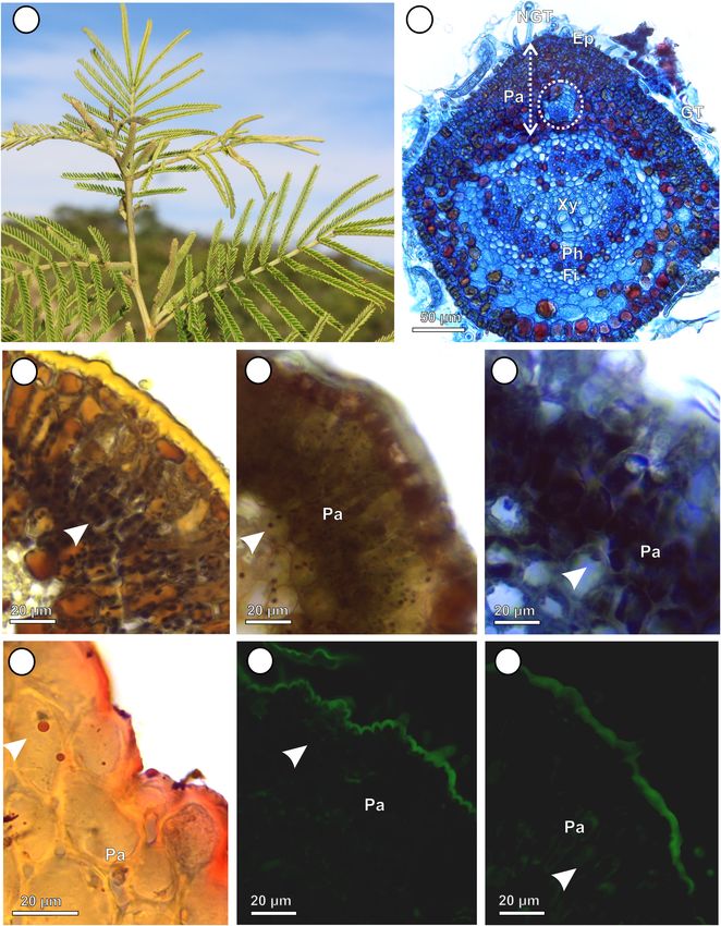
Costa et al.                                                                                                 Lopesia Galls on Mimosa gemmulata

one section per gall, 5 measurement fields per section, totalizing       RESULTS
25 measurements by tissue for each gall morphospecies). The data
were compared using one-way ANOVA followed by Tukey’s test,              Non-galled Pinna-Rachis Profile
using α = 0.05. The tests were performed with SigmaStat (Systat
                                                           R

                                                                         (Control)
Software, Inc., Chicago, Illinois) and the graphics were done with
                                                                         The pinna-rachis of M. gemmulata (Figure 1A) has uniseriated
GraphPad prism 5.0 .             R

                                                                         epidermis with glandular and non-glandular trichomes
                                                                         (Figure 1B). The adaxial cortical parenchyma is homogeneous
Histochemical Analysis                                                   with 3–4 cell layers. The vascular tissues have bicollateral
Free-handmade sections from fresh samples of the pinna-rachis,           arrangement and are surrounded by two layers of pericyclic fibers
LG, GLG, BLG, and GG (n = 7 for each gall morphospecies) and             (Figure 1B). Starch (Figure 1C), reducing sugars (Figure 1D),
Paraplast embedded sections obtained in a rotary microtome
               R

                                                                         proteins (Figure 1E), and lipidic droplets (Figure 1F) accumulate
were submitted to histochemical analyses. Starch grains were             in the protoplasm of parenchyma cells. In the pinna-rachis, the
detected with Lugol’s reagent (1% potassium iodine-iodide                epitopes of xyloglucans recognized by LM15 (24 Gy; Figure 1G)
solution) for 5 min (Johansen, 1940). Reducing sugars were               and the epitopes of heretomannans recognized by LM21 (30.4 Gy;
detected by Fehling’s reagent (Solution A: 7.9% copper sulfate,          Figure 1H) are moderately labeled in the parenchyma cell walls.
and solution B: 34.6% sodium potassium tartrate and 1%
sodium hydroxide) heated to pre-boiling temperature (Sass,
1951). Proteins were detected by 0.1% bromophenol blue in a              Profiles of Lopesia Galls
saturated solution of 10% magnesium chloride in ethanol during           Structural Profiles
15 min, washed in 0.5% acetic acid in water during 20 min,               The Lopesia galls are green (LG, GLG, and GG) or brown
and water for 3 min (Mazia et al., 1953). Lipids were detected           (BLG), isolated, pubescent (Figures 2A–D), and developed by
with a saturated solution of Sudan Red B in 70 GL ethanol                pinna-rachis cell redifferentiation and tissue reorganization. In
during 5 min (Brundett et al., 1991). Black sections were used           the four Lopesia galls, the epidermis, common storage tissue,
as controls. The sections were analyzed and photographed under           vascular tissues, and schlerenchymatic layers form the gall outer
a light microscope (Leica DM500) coupled to a digital camera
                                     R                                   compartment and the typical nutritive tissue forms the gall inner
(Leica ICC50 HD).
           R                                                             compartment (Figures 2E–H). In the LG, the common storage
                                                                         tissue has 4–5 cell layers (Figure 2I), the schlerenchyma has
Immunocytochemical Analysis                                              1–2 layers, and the typical nutritive tissue has 1–2 cell layers
The detection of hemicelluloses was performed in the sections            (Figure 2J). In the GLG, the common storage tissue has 10–
of the pinna-rachis, LG, GLG, BLG, and GG (n = 3 for each                11 cell layers (Figure 2K), the schlerenchyma has 1–2 layers,
category) obtained in a rotary microtome. The sections were              and the typical nutritive tissue has 1–2 cell layers (Figure 2L).
pre-incubated in pectate lyase at 10 µg/mL, diluted in 50 mM             In the BLG, the common storage tissue has 8–9 cell layers
N-cyclohexyl-3-aminopropane sulfonic acid (CAPS) and 2 mM                (Figure 2M), the schlerenchyma has 4–5 layers, and the typical
CaCl2 buffer, pH 10, for 2 h at room temperature (Marcus et al.,         nutritive tissue has 1–2 cell layers (Figure 2N). In the GG, the
2008). Afterward, the sections were incubated in the primary             common storage tissue has 10–11 cell layers (Figure 2O), the
monoclonal antibodies (MAbs), LM15 and LM21, diluted in                  schlerenchyma has 7–8 layers, and the typical nutritive tissue has
block solution [5% powder milk in phosphate-buffered saline-             5–6 cell layers (Figure 2P).
PBS) 0.1 mol L1 , pH 7.2 (1:5, w/v)] for the labeling of the
epitopes of xyloglucans (Marcus et al., 2008) and heteromannans          Histometric Profiles
(Marcus et al., 2010), respectively, for 90 min in the darkness.         In accordance with structural description, GG tissues are thicker
The sections were washed in PBS and incubated in the secondary           than the tissues of the other three Lopesia galls (Figure 3).
antibody anti-rat IgG linked to FITC, diluted in 5% powder               The GG common storage tissue is 177% thicker than that of
milk/PBS (1:100, w/v), for 90 min in darkness. The slides                the LG (p < 0.001), and 249% thicker than that of the BLG
were mounted in 50% glycerin, analyzed and photographed                  (p < 0.001), but there is no significant difference between the
under a fluorescence microscope (Leica DM 2500 LED), with
                                                   R                     GG and the GLG regarding the thickness of the common storage
blue excitation light (450–490 nm) and green emission light              tissue (Figure 3A). The GLG common storage tissue is 249%
(515 nm), coupled to a digital camera (Leica DFC 7000T).
                                                       R                 thicker than that of the BLG (p < 0.001). The GG schlerenchyma
The immunocytochemical images were submitted to intensity                is 2,009% thicker than that of the LG (p < 0.001), and 2,495%
measurement using ImageJ version 1.51k1 . The fluorescence               thicker than that of the GLG (p < 0.001), but there is no
intensities of the epitopes of hemicelluloses were evaluated             significant difference between the GG and the BLG regarding
by grayscale methodology (Gy = Gray value) with triplicate               the thickness of the schlerenchyma (Figure 3B). The BLG
analysis for each tissue. After the measurements, we proposed the        schlerenchyma is 348% thicker than that of the GLG (p < 0.001).
following categories: (−) negative ( = 0 Gy values); (+) weak (10–       There is no significant difference among the schlerenchyma
20 Gy values); (++) moderate (21–39.99 Gy values); and (+++)             thickness of the GL, the GLG and the BLG (Figure 3B). The
intense (≥40 Gy values).                                                 GG typical nutritive tissue is 772% thicker than that of the LG
                                                                         (p < 0.001), and 813% thicker than that of the GLG (p < 0.001),
1
    http://rsb.info.nih.gov/ij                                           but there is no significant difference between the GG and the BLG

Frontiers in Plant Science | www.frontiersin.org                     3                                     May 2021 | Volume 12 | Article 660557
Costa et al.                                                                                                                       Lopesia Galls on Mimosa gemmulata

  FIGURE 1 | Non-galled pinna-rachis of Mimosa gemmulata Barneby (Fabaceae). (A) General aspect of non-galled leaves. (B–H) Transverse sections. (B) Anatomical
  profile. (C,D) Immunocytochemical profile. (C–F) Histochemical profile. (C) Starch grains stained in black (white arrowhead). (D) Reducing sugars stained in brown
  (white arrowhead). (E) Proteins stained in blue (white arrowhead). (F) Lipidic droplets stained in red (white arrowhead). (G,H) Xyloglucans detected by LM15 in cell
  walls of parenchyma (white arrowhead). (H) Heteromannans detected by LM21 in cell walls of parenchyma (white arrowheads). Ep, Epidermis; Fi, fibers; GT,
  glandular trichomes; NGT, non-glandular trichomes; Pa, parenchyma; Ph, Phloem; Xy, Xylem.

Frontiers in Plant Science | www.frontiersin.org                                   4                                             May 2021 | Volume 12 | Article 660557

Costa et al.                                                                                                                      Lopesia Galls on Mimosa gemmulata

  FIGURE 2 | Structural profiles of the four Lopesia galls on Mimosa gemmulata Barneby (Fabaceae) pinna-rachis in transverse sections. (A–D) Macroscopic aspect
  of the galls. (A,E) Lenticular bivalve-shaped gall. (B,F) Green lanceolate bivalve-shaped gall. (C,G) Brown lanceolate bivalve-shaped gall. (D,H) Globoid
  bivalve-shaped gall. (E–H) Diagram of the Lopesia galls, evidencing common storage tissues (blue), schlerenchymatic layers (pink) in the outer tissue compartments,
  and typical nutritive tissues (purple–black arrowheads) in the inner tissue compartments. (I,J) Lenticular bivalve-shaped gall, evidencing common storage tissue,
  schlerenchymatic layer and typical nutritive cells with evident nuclei (black arrowhead). (K,L) Green lanceolate bivalve-shaped gall, evidencing common storage
  tissue, schlerenchymatic layer and typical nutritive cells with evident nuclei (black arrowhead). (M,N) Brown lanceolate bivalve-shaped gall, evidencing common
  storage tissue, schlerenchymatic layer and typical nutritive cells with evident nuclei (black arrowhead). (O,P) Globoid bivalve-shaped gall, evidencing common
  storage tissue, schlerenchymatic layer and typical nutritive cells with evident nuclei (black arrowhead). BLG, brown lanceolate bivalve-shaped gall; CST, common
  storage tissue; GG, globoid bivalve-shaped gall; GLG, green lanceolate bivalve-shaped gall; LC, larval chamber; LG, lenticular bivalve-shaped gall; SL,
  schlerenchymatic layer; TNT typical nutritive tissue.

Frontiers in Plant Science | www.frontiersin.org                                  5                                             May 2021 | Volume 12 | Article 660557

Costa et al.                                                                                                                       Lopesia Galls on Mimosa gemmulata

  FIGURE 3 | Histometry of the tissues and cell walls of the four Lopesia galls on Mimosa gemmulata Barneby (Fabaceae) pinna-rachis (A–C) Tissue thickness. (D–F)
  Cell wall thickness. BLG, brown lanceolate bivalve-shaped gall, GG, globoid bivalve-shaped gall, GLG, green lanceolate bivalve-shaped gall, LG, lenticular
  bivalve-shaped gall. n = 5 per gall systems (5 regions per section from 1 sections); p < 0.05. Same letters on bars indicate statistically equal values for the same
  variable, and different letters indicate different values.

regarding the thickness of the typical nutritive tissue (Figure 3C).                   BLG schlerenchyma are 48% thicker than the cell walls of the
There is no significant difference of the typical nutritive tissue                     GLG, and 37% thicker than the cell walls of the LG (p < 0.001).
thickness among the LG, GLG, and BLG.                                                  There is no significant statistical difference between the cell wall
   The cell walls of the schlerenchyma are thicker than the cell                       thickness of the schlerenchyma of the LG and GLG (p = 0.958).
walls of the common storage and typical nutritive tissues on                           The cell walls of the GG storage and typical nutritive tissues are
the four Lopesia galls (Figures 3D–F). The cell walls of the GG                        thicker than the cell walls of the GL and GLG (p < 0.001). There
schlerenchyma are 141% thicker than the cell walls of the LG,                          is no significant difference among in the cell wall thickness of the
160% thicker than the cell walls of the GLG, and 76% thicker                           common storage tissues (Figure 3D) and typical nutritive tissues
than the cell walls of the BLG (p < 0.001). The cell walls of the                      (Figure 3F) among the GL, GLG, and GBG.

Frontiers in Plant Science | www.frontiersin.org                                   6                                             May 2021 | Volume 12 | Article 660557
Costa et al.                                                                                                                      Lopesia Galls on Mimosa gemmulata

TABLE 1 | Histochemical profiles of the four Lopesia galls on Mimosa gemmulata (Fabaceae).

                                                           Outer compartment                                                           Inner compartment
                                 Common storage tissue                             Schlerenchymatic layer                            Typical nutritive tissue

                       LG       GLG        BLG            GG             LG       GLG        BLG             GG             LG       GLG       BLG              GG

Histochemistry
Starch                  +         +         +              +              −         −         −               +                       −          −              +
Reducing sugars         +         +         +              +              −         −         −               +              +        +          +              +
Proteins                +         +         +              +              −         −         +               +              +        +          +              +
Lipids                  +         +         +              +              +         +         −               +              +        +          +              +

BLG, brown lanceolate bivalve-shaped gall; LG, lenticular bivalve-shaped gall; GG, globoid bivalve-shaped gall; GLG, green lanceolate bivalve-shaped gall.
The symbols indicate (−) absence and (+) presence.

Histochemical Profiles                                                                 accumulation of metabolites. The Lopesia species associated
Starch, reducing sugars, proteins, and lipids are detected both in                     to the GG has the longest lifespan and distinct structural
the non-galled pinna-rachis and in the Lopesia galls (Table 1).                        and nutritional profiles compared with the other three Lopesia
Starch grains are detected only in the protoplasm of the common                        galls (Figure 6). The GG has peculiar bidirectional gradients
storage tissue of the LG (Figure 4A), GLG (Figure 4B), and                             of starch and reducing sugars, which indicate additional
BLG (Figure 4C). Starch grains (Figure 4D) are detected in the                         substrates to the synthesis of xyloglucans and heteromannans
protoplasm of the GG common storage tissue, schlerenchyma                              in cell walls. In the typical nutritive tissues, the cell wall
and typical nutritive tissue. Reducing sugars are detected in the                      xyloglucans and heteromannans together with the protoplasm
protoplasm of the common storage tissue and typical nutritive                          accumulated reducing sugars, proteins and lipids constitute
tissue of the LG (Figure 4E), GLG (Figure 4F), BLG (Figure 4G),                        the pool of energetic resources for the four galling Lopesia.
and GG (Figure 4H). There is a centripetal gradient of reducing                        Peculiarly, the GG with its 4 month-life cycle has the highest
sugars in the LG, GLG, and BLG. In the GG, the reducing sugars                         structural and nutritional investment in the storage and the
accumulate in a bidirectional gradient. Proteins are detected in                       nutritive tissues, and schlerenchymatic layers, which relates
the protoplasm of the common storage tissue, schlerenchyma and                         to the highest demand for gall development and galling
typical nutritive tissue of the LG (Figure 4I), GLG (Figure 4J),                       insect establishment.
BLG (Figure 4K), and GG (Figure 4L), forming a centripetal
gradient toward the nutritive cells. Lipidic droplets are detected
in the protoplasm of the common storage tissue and typical                             Structural Peculiarities in the Lopesia
nutritive tissue of the LG (Figure 4M), GLG (Figure 4N), BLG                           Galls
(Figure 4O), and GG (Figure 4P).                                                       Gall development and tissue compartmentalization depend on
                                                                                       the galling insect behavior and how long the galling insect
Immunocytochemical Profiles                                                            stimulus lasts (Bronner, 1992; Bragança et al., 2017; Oliveira et al.,
The epitopes of xyloglucans recognized by LM15 are moderately                          2016). The duration of the gall cycle can determine the rate of cell
labeled in the cell walls of the BLG (34 Gy) and GG (36.7 Gy)                          divisions, elongation, and tissue complexity (Carneiro et al., 2017;
common storage tissue; intensely labeled in the cell walls of the                      Costa et al., 2018), and determine the structural profile of the
LG schlerenchyma (63 Gy), and typical nutritive tissue (47.6 Gy)                       storage tissues (Rohfritsch, 1992), which are distinct among the
(Figure 5A); and moderately labeled in the cell walls of the                           Lopesia galls. The GG with the 4-month life cycle has the highest
GLG schlerenchyma (39.8 Gy), and weakly labeled in the cell                            stratification of the common storage tissue, when compared
walls of the typical nutritive tissue (17.26 Gy) (Figure 5B). The                      with the other gall morphospecies with the 2-month life cycle
xyloglucans are moderately detected in the cell walls of the                           (LG) and 3-month life cycles (GLG and BLG) due both to
BLG typical nutritive tissue (38.6 Gy; Figure 5C), and intensely                       higher hyperplasia and cell hypertrophy. In addition, the thicker
detected in the GG typical nutritive tissues (47.1 Gy; Figure 5D).                     cell walls of the GG common storage tissue indicate a distinct
The epitopes of heteromannans recognized by LM21 are weakly                            metabolic investment for the synthesis of cell wall components
labeled in the cell walls of the typical nutritive tissue of the LG                    as cellulose, hemicellulose and pectins than observed in the other
(20.6 Gy; Figure 5E), GLG (19.7 Gy; Figure 5F), BLG (12 Gy;                            three Lopesia galls. This investment in cell wall thickness is
Figure 5G), and moderately labeled in the cell walls of the GG                         important to control the turgor pressure during cell hypertrophy
typical nutritive tissue (31.6 Gy; Figure 5H and Table 2).                             (Chanliaud et al., 2002), and confers an additional support for
                                                                                       the GG. The structural and physiological traits of the common
                                                                                       storage tissue have been related to the sink and storage of
DISCUSSION                                                                             water and energetic resources (Castro et al., 2012; Oliveira et al.,
                                                                                       2017; Bragança et al., 2020b). Water and energetic molecules
The four Lopesia galls on Mimosa gemmulata have peculiarities                          accumulation increase gall succulence (tissue thickness) (Oliveira
regarding the investment in cell walls, tissue stratification, and                     et al., 2017), and can also be consequence of the longest lifespan,

Frontiers in Plant Science | www.frontiersin.org                                   7                                             May 2021 | Volume 12 | Article 660557
Costa et al. Lopesia Galls on Mimosa gemmulata FIGURE 4 | Histochemical profiles of the four Lopesia galls on Mimosa gemmulata Barneby (Fabaceae) pinna-rachis in transverse sections. (A–D) Starch grains stained in black. (E–H) Reducing sugars stained in brown. (I–L) Proteins stained in blue (M–P) Lipidic droplets stained in red. BLG, brown lanceolate bivalve-shaped gall; CST, common storage tissue; GG, globoid bivalve-shaped gall; GLG, green lanceolate bivalve-shaped gall; LG, lenticular bivalve-shaped gall; TNT, typical nutritive tissue. as evidenced in the GG on M. gemmulata with the thickest cell et al., 2019). The lignification process confers mechanical support walls as well as higher stratification of the common storage tissue. and may also protect gall tissues against the high oxidative The differentiation of the schlerenchymatic cell layers is stress originated from the galling organism respiration and cell associated with life cycle of the Lopesia galls on M. gemmulata. metabolism (Oliveira et al., 2010, 2011b, 2017; Isaias et al., 2015). Secondary walls are mainly composed of cellulose, hemicelluloses The higher thickness of the GG cell walls indicates that the (xylans and glucomannans), and lignins, whose biosynthesis is biosynthesis and deposition of the secondary wall components involved in the scavenging of free radicals (Liu et al., 2018; Zhong can be more expressive in galls with longer lifespans. The longest Frontiers in Plant Science | www.frontiersin.org 8 May 2021 | Volume 12 | Article 660557
Costa et al.                                                                                                                       Lopesia Galls on Mimosa gemmulata

  FIGURE 5 | Immunocytochemical profiles of the four Lopesia galls on Mimosa gemmulata Barneby (Fabaceae) pinna-rachis in transverse sections. (A–D)
  Xyloglucans labeled by LM15. (E–H) Heteromannans labeled by LM20. BLG, brown lanceolate bivalve-shaped gall; CST, common storage tissue; GG, globoid
  bivalve-shaped gall; GLG, green lanceolate bivalve-shaped gall; LC, larval chamber; LG, lenticular bivalve-shaped gall; TNT, typical nutritive tissue; SL,
  schlerenchymatic layer.

TABLE 2 | Average of gray value and intensity of reaction of the epitopes for hemicelluloses in the tissues of the four Lopesia galls on Mimosa gemmulata (Fabaceae).

                                                           Outer compartment                                                            Inner compartment
                                Common storage tissue                             Schlerenchymatic layer                              Typical nutritive tissue

                      LG       GLG        BLG            GG               LG        GLG        BLG            GG             LG         GLG       BLG            GG

Immunocytochemistry
Xyloglucans            0         0        34.0           36.7             63           39.8      0             0            47.6        17.2      38.6           47.1
Intensity              −         −         ++            ++              +++           ++       −              −            +++          +        ++             +++

Heteromannans          0         0        19.7             0               0            0        0             0            20.6        19.7       12            31.6
Intensity              −         −          +             −                −            −       −              −              +          +         +             ++

BLG, brown lanceolate bivalve-shaped gall; LG, lenticular bivalve-shaped gall; GG, globoid bivalve-shaped gall; GLG, green lanceolate bivalve-shaped gall.
Intensity of reaction: (−) negative (=0 Gy value); (+) weak (10–20 Gy value); (++) moderate (21–39.99 Gy value); (+++) intense (≥40 Gy value).

time the galling Lopesia associated to the GG remains inside                            cell walls and the stratification of the schlerenchymatic layers on
the gall, and the higher impact of its breathing and feeding                            the Lopesia galls associated to M. gemmulata, which support the
seem to result in the thick schlerenchymatic layers, which may                          premise that the lignification process is triggered and supported
favor the dissipation of oxidative stress and gall maintenance.                         by the galling insect activity and is more intense the longer the
Controversly, the LG and the GLG also have conspicuous                                  galling insect lifespan is.
schlerenchymatic layers, but with the thinnest cell walls amongst                           The cells of the typical nutritive tissue develop due to the
the Lopesia galls, while the BLG has an intermediate condition                          galling insect stimuli over the host plant tissues, as they are
in cell wall and schlerenchymatic layer thickness among the four                        directly impacted by the nutrition of the inducers (Bronner,
Lopesia galls on M. gemmulata. The differences in the duration of                       1992). The cell walls and stratification of the typical nutritive
the life cycles even in 1–2 months relate to the thickness of the                       tissues of the galls with 2- and the 3-month-life cycles (LG,

Frontiers in Plant Science | www.frontiersin.org                                   9                                              May 2021 | Volume 12 | Article 660557
Costa et al. Lopesia Galls on Mimosa gemmulata FIGURE 6 | Diagrams of the structural and nutritional profiles of the four Lopesia galls on Mimosa gemmulata with their respective lifespans. The color gradients in the purple, pink and blue arrows, and the symbols (– and +) indicate tissue thickness investment. The diagrams of the transverse sections illustrate the gradients of the nutritive molecules in gall tissues. BLG, brown lanceolate bivalve-shaped gall; GG, globoid bivalve-shaped gall; GLG, green lanceolate bivalve-shaped gall; LG, lenticular bivalve-shaped gall. Frontiers in Plant Science | www.frontiersin.org 10 May 2021 | Volume 12 | Article 660557
Costa et al.                                                                                                        Lopesia Galls on Mimosa gemmulata

BLG, and GLG) have similar thickness, which can indicate the                  digestion (Hatchett et al., 1990; Shukle et al., 2009; Al-Jbory et al.,
low and constant nutritional demand of the three gall-inducing                2018).
Lopesia. Differently, the primary cell walls and stratification of
the typical nutritive tissue of the GG are thicker, which seems               Nutritional Profiles in Cell Walls of
to be consequence of the greater feeding stimulus and high                    Lopesia Galls
nutritional demand of the Lopesia larvae with the longest lifespan            Xyloglucans and heteromannans are hemicelluloses involved in
(4 month-life cycle).                                                         cell expansion and rigidity (Park and Cosgrove, 2015; Cosgrove,
                                                                              2016, 2018; Voiniciuc et al., 2019), which also work out as
Nutritional Profiles in the Protoplasm of                                     reserve of carbohydrates accumulated in plant cell walls of some
                                                                              cotyledons, mainly in seeds of Fabaceae species (Buckeridge
the Lopesia Galls                                                             et al., 2000; Santos et al., 2004). Recently, the xyloglucans were
The life cycles did not alter the evaluated compounds linked to               reported as reserve carbohydrates in cell walls of nematode-
the nutritional profiles of the four Lopesia galls on M. gemmulata.           induced galls on Miconia spp. (Melastomaceae) (Ferreira et al.,
However, the histochemical gradients of carbohydrates are                     2020), and cell walls of the common storage and of the typical
bidirectional in the GG. The common storage tissues of                        nutritive tissues of Cecidomyiidae-induced galls on Inga ingoides
four Lopesia galls are starch-rich, and, unexpectedly, starch                 (Fabaceae) (Bragança et al., 2020a). Herein, the epitopes of
grains also accumulate in the GG nutritive tissue. Starch                     xyloglucans labeled by LM15 in the common storage tissue of
is an insoluble polysaccharide that must be broken down                       the BLG and the GG, and in the schlerenchymatic layers of the
in monosaccharides or disaccharides by invertases, such as                    LG and GLG can influence the dynamics of cell wall expansion
D-glucose, activity (Morris and Arthur, 1984) in gall storage                 and rigidity. Both the xyloglucans and the heteromannans are
tissues (Oliveira et al., 2010, 2011a; Bragança et al., 2017).                detected in the primary cell walls of the typical nutritive tissue
The inverse gradients of accumulation of starch and reducing                  of the four Lopesia galls on M. gemmulata. While the overall
sugars in the four Lopesia galls implies in the activity                      labeling of heteromannans in the four Lopesia galls relates to
of enzymes and may result in adequate substrate for the                       structural function, the labeling of the xyloglucans in the typical
biosynthesis of new cell wall components (Taylor, 2008; Polko                 nutritive tissues relates not only to the structural, but also to the
and Joseph, 2019), such as xyloglucans (Pauly and Keegstra,                   nutritional function.
2016) and heteromannans (Voiniciuc et al., 2019) in Lopesia                      We demonstrated that the primary walls of the GG are
galls. These hemicelluloses are synthesized by Golgi-localized                thicker, and therefore a greater investment in the synthesis
glycosyltransferases (GTs), and the resulting polysaccharides are             of xyloglucans as reserve carbohydrates occurs, which indicate
secreted to the plant cell wall via exocytosis (Driouich et al.,              the support for the galling Lopesia with the longest life cycle
2012; Voiniciuc et al., 2019). Accordingly, the bidirectional                 high nutritional demands. The xyloglucan monosaccharides
gradients of starch and reducing sugars in the common storage                 can become available for the galling Lopesia nutrition by the
and typical nutritive tissues of the GG may be associated                     activity of β-D-galactosidase and β-D-glucosidase, which may be
with additional substrates to biosynthesis of xyloglucans and                 found in salivary glands and/or midgut of Cecidomyiidae larvae
heteromannans, and the composition of the primary cell walls                  (Grover et al., 1988). Even though the heteromannans and the
(Pauly and Keegstra, 2016).                                                   xyloglucans are labeled in primary cell walls of the four Lopesia
    The proteins and lipids are energetic molecules that support              galls independently of their lifespans, the GG thickest cell walls
the four gall-inducing Lopesia nutrition and gall structural                  and typical nutritive tissue indicate a higher support for the
maintenance. The proteins can act in the scavenging of free                   4-month life cycle.
radicals and the maintenance of redox-potential homeostasis in
tissues of plants (Meyer et al., 2019; Foyer et al., 2020) and of
galls induced by different galling organisms (Schönrogge et al.,              CONCLUSION
2000; Oliveira et al., 2010; Isaias et al., 2015; Silva et al., 2019).
The protein accumulation in the nutritive cells of the Lopesia galls          The 1–2-month variation of Lopesia gall lifespans did not impact
can have antioxidant function due to highest cellular metabolism              the type of energetic molecules, but the investment in cell
in this tissue compartment. The accumulation of lipids in the                 walls and tissue stratification, especially regarding the common
storage, and/or typical nutritive tissue of Cecidomyiidae galls on            storage tissue. Moreover, cell walls have configured as sites of
different host plants are linked to the intrinsic metabolism of               additional carbohydrate accumulation not only for structural, but
the host plants (Moura et al., 2008; Oliveira et al., 2010; Ferreira          for nutritional purposes. The bivalve-shaped globoid galls with
and Isaias, 2014; Amorim et al., 2017; Nogueira et al., 2018),                the 4-month lifespan associated to M. gemmulata demanded the
as is true for M. gemmulata. The lipid droplets accumulated in                highest structural and nutritional support, as expected.
the common storage tissue are related to the maintenance of
the cellular machinery, while in the nutritive cells, they are food
resources for the Lopesia galls. In the typical nutritive tissue,             DATA AVAILABILITY STATEMENT
the lipids may become available by the activity of a lipase-
like protein expressed in the salivary glands of Cecidomyiidae                The raw data supporting the conclusions of this article will be
larvae during feeding, which can be involved in extra-oral                    made available by the authors, without undue reservation.

Frontiers in Plant Science | www.frontiersin.org                         11                                        May 2021 | Volume 12 | Article 660557
Costa et al. Lopesia Galls on Mimosa gemmulata
AUTHOR CONTRIBUTIONS (CAPES)—Finance Code 001. We thank CAPES-Brazil for
doctoral grant to EC (888877.199702/2018-00). We thank
EC, DO, and RI designed the experiments, analyzed the Conselho Nacional de Desenvolvimento Científico e Tecnológico
data, and drafted the manuscript. EC and DF performed the (CNPq) for the research scholarships to RI (304535/2019-2) and
experiments. All authors contributed to the article and approved DO (304981/2019-2).
the submitted version.
ACKNOWLEDGMENTS
FUNDING
We thank to Dra. Valéria Cid Maia and Dra. Sheila P.
This work was supported by the Coordenação de Carvalho-Fernandes for confirming the four Lopesia associated
Aperfeiçoamento de Pessoal de Nível Superior—Brazil to M. gemmulata are distinct but undescribed species.
REFERENCES leaves of Mimosa gemmulata (Leguminosae: Caesalpinioideae). Aust. J. Bot. 66,
161–172. doi: 10.1071/BT17099
Al-Jbory, Z., Anderson, K. M., Harris, M. O., Mittapalli, O., Whitworth, R. J., Driouich, A., Follet-Gueye, M. L., Bemard, S., Kousar, S., Chevalier, L., Vicré-
and Chen, M. S. (2018). Transcriptomic analyses of secreted proteins from the Gibouin, M., et al. (2012). Golgi-mediated synthesis and secretion of matrix
salivary glands of wheat midge larvae. J. Insect Sci. 18:17. doi: 10.1093/jisesa/ polysaccharides of the primary cell wall of higher plants. Front. Plant Sci. 3:79.
iey009 doi: 10.3389/fpls.2012.00079
Amorim, D. O., Ferreira, B. G., and Fleury, G. (2017). Plant potentialities determine Ferreira, B. G., Álvarez, R., Avritzer, S. C., and Isaias, R. M. S. (2017). Revisiting the
anatomical and histochemical diversity in Mikania glomerata Spreng. galls. histological patterns of storage tissues: beyond the limits of gall-inducing taxa.
Braz. J. Bot. 40, 517–527. doi: 10.1007/s40415-016-0357-9 Botany 95, 173–184. doi: 10.1139/cjb-2016-0189
Bragança, G. P., Oliveira, D. C., and Isaias, R. M. S. (2017). Compartmentalization Ferreira, B. G., Álvarez, R., Bragança, G. P., Alvarenga, D. R., Pérez-Hidalgo,
of metabolites and enzymatic mediation in nutritive cell of Cecidomyiidae galls N., and Isaias, R. M. S. (2019). Feeding and other gall facets: patterns and
on Piper arboretum Aubl. (Piperaceae). J. Plant Stud. 6, 11–22. doi: 10.5539/jps. determinants in gall structure. Bot. Rev. 85, 78–106. doi: 10.1007/s12229-019-
v6n1p11 09207w
Bragança, G. P. P., Alencar, C. F., Freitas, M. S. C., and Isaias, R. M. S. (2020a). Ferreira, B. G., Bragança, G. P., and Isaias, R. M. S. (2020). Cytological attributes
Hemicelluloses and associated compounds determine gall functional traits. of storage tissues in nematode and eriophyid galls: pectin and hemicellulose
Plant Biol. 22, 981–991. doi: 10.1111/plb.13151 functional insights. Protoplasma 257, 229–244. doi: 10.1007/s00709-019-
Bragança, G. P. P., Freitas, M. S. C., and Isaias, R. M. S. I. (2020b). The influence 01431-w
of gall position over xylem features in leaflets of Inga ingoides (Rich.) willd. Ferreira, B. G., Carneiro, R. G. S., and Isaias, R. M. S. (2015). Multivesicular bodies
(Fabaceae: Caesalpinioideae). Trees 35, 199–209. doi: 10.1007/s00468-020- differentiate exclusively in nutritive fast-dividing cells in Marcetia taxifolia galls.
02027-1 Protoplasma 252, 1275–1283.
Bronner, R. (1992). “The role of nutritive cells in the nutrition of cynipids and Ferreira, B. G., and Isaias, R. M. S. (2014). Floral-like destiny induced by a galling
cecidomyiids,” in Biology of Insect-Induced Galls, eds J. D. Shorthouse, and O. Cecidomyiidae on the axillary buds of Marcetia taxifolia (Melastomataceae).
Rohfritsch (New York, NY: Oxford University Press), 118–140. Flora 209, 391–400.
Brundett, M. C., Kendrick, B., and Peterson, C. A. (1991). Efficient lipid staining in Formiga, A. T., Silveira, F. A. O., Fernandes, G. W., and Isaias, R. M. S. (2014).
plant material with sudan red 7B or fluoral yellow 088 in polyethylene glycol– Phenotypic plasticity and similarity among gall morphotypes on a superhost,
glycerol. Biotech. Histochem. 66, 111–116. doi: 10.3109/10520299109110562 Baccharis reticularia (Asteraceae). Plant Biol. 17, 512–521.
Buckeridge, M. S., Santos, H. P., and Tiné, M. A. S. (2000). Mobilisation of storage Foyer, C. H., Baker, A., Wright, M., Sparkes, I. A., Mhamdi, A., Schippers, J. H. M.,
cell wall polysaccharides in seeds. Plant Physiol. 38, 141–156. doi: 10.1016/ et al. (2020). On the move: redox-dependent protein relocation in plants. J. Exp.
S0981-9428(00)00162-5 Bot. 71, 620–631. doi: 10.1093/jxb/erz330
Bukatsch, F. (1972). Bermerkungen zur doppelfärbung astrablau-safranin. Gonçalves, S. J. M. R., Isaias, R. M. S., Vale, F. H. A., and Fernandes, G. W. (2005).
Mikrokosmos 61:255. Sexual dimorphism of Pseudotectococcus rolliniae Hodgson & Gonçalves
Carneiro, R. G. S., Isaias, R. M. S., Moreira, A. S. F. P., and Oliveira, D. C. (2017). 2004 (Hemiptera Coccoidea Eriococcidae) influences gall morphology on
Reacquisition of new meristematic sites determines the development of a new Rollinia laurifolia Schltdl. (Annonaceae). Trop. Zool. 18, 161–169. doi: 10.1080/
organ, the Cecidomyiidae gall on Copaifera langsdorffii Desf. (Fabaceae). Front. 03946975.2005.10531219
Plant Sci. 8:1622. doi: 10.3389/fpls.2017.01622 Grover, P. B., JR., Ross, D. R., and Shukle, R. H. (1988). Identification and
Castro, A. C., Oliveira, D. C., Moreira, A. S. F. P., Lemos-Filho, J. P., and Isaias, partial characterization of digestive carbohydrases in larvae of the Hessian
R. M. S. (2012). Source-sink relationship and photosynthesis in the horn-shaped fly, Mayetiola destructor (Say) (Diptera: Cecidomyiidae). Arch. Insect Biochem.
gall and its host plant Copaifera langsdorffii Desf. (Fabaceae). S. Afr. J. Bot. 83, Physiol. 5, 59–72. doi: 10.1002/arch.940080106
121–126. doi: 10.1016/j.sajb.2012.08.007 Hatchett, J. H., Kreitner, G. L., and Elzinga, R. J. (1990). Larval mouthparts and
Chanliaud, E., Burrows, K. M., Jeronimidis, G., and Gidley, M. J. (2002). feeding mechanism of the Hessian fly (Diptera: Cecidomyiidae). Ann. Entomol.
Mechanical properties of primary plant cell wall analogues. Planta 215, 989– Soc. Am. 83, 1137–1147.
996. doi: 10.1007/s00425-002-0783-8 Isaias, R. M. S., Oliveira, D. C., Moreira, A. S. F. P., Soares, G. L. G., and Carneiro,
Chen, X., Yang, Z., Chen, H., Qian, Q., Liu, J., Wang, C., et al. (2020). A complex R. G. S. (2015). The imbalance of redox homeostasis in arthropod-induced plant
nutrient exchange between a gall-forming aphid and its plant host. Front. Plant galls: mechanisms of stress generation and dissipation. Biochim. Biophys. Acta
Sci. 11:811. doi: 10.3389/fpls.2020.00811 1850, 1509–1517. doi: 10.1016/j.bbagen.2015.03.007
Cosgrove, D. J. (2016). Plant cell wall extensibility: connecting plant cell growth Johansen, D. A. (1940). Plant Microtechnique. New York, NY: McGraw-Hill Book.
with cell wall structure, mechanics, and the action of wall modifying enzymes. Jorge, N. C., Souza-Silva, E. A., Alvarenga, D. R., Saboia, G., Soares, G. L. G.,
J. Exp. Bot. 67, 463–476. doi: 10.1093/jxb/erv511 Zini, C. A., et al. (2018). Structural and chemical profiles of Myrcia
Cosgrove, D. J. (2018). Diffuse growth of plant cell walls. Plant Physiol. 176, 16–27. splendens (Myrtaceae) leaves under the influence of the galling Nexothrips sp.
doi: 10.1104/pp.17.01541 (Thysanoptera). Front. Plant Sci. 9:1521. doi: 10.3389/fpls.2018.01521
Costa, E. C., Carneiro, G. S. C., Silva-Santos, J., and Isaias, R. M. S. (2018). Biology Karnovsky, M. J. (1965). A formaldehyde-glutaraldehyde fixative of high
and development of galls induced by Lopesia sp. (Diptera: Cecidomyiidae) on osmolarity for use in electron microscopy. J. Cell Biol. 27, 137–138.
Frontiers in Plant Science | www.frontiersin.org 12 May 2021 | Volume 12 | Article 660557Costa et al. Lopesia Galls on Mimosa gemmulata Kraus, J. E., and Arduin, M. (1997). Manual Básico de Métodos em Morfologia Park, Y. B., and Cosgrove, D. J. (2015). Xyloglucan and its interactions with Vegetal. Rio de Janeiro: Editora da Universidade Rural. other components of the growing cell wall. Plant Cell Physiol. 56, 180–194. Liu, Q., Luo, L., and Zheng, L. (2018). Lignins: biosynthesis and biological doi: 10.1093/pcp/pcu204 functions in plants. Int. J. Mol. Sci. 19:335. doi: 10.3390/ijms19020335 Pauly, M., and Keegstra, K. (2016). Biosynthesis of the plant cell wall matrix Mani, M. S. (1964). Ecology of Plant Galls. The Hague: Dr. W. Junk Publishers. polysaccharide xyloglucan. Annu. Rev. Plant Biol. 67, 235–259. doi: 10.1146/ doi: 10.1007/978-94-017-6230-4 annurev-arplant-043015-112222 Marcus, S. E., Blake, A. W., Benians, T. A. S., Lee, K. J. D., Poyser, C., Donaldson, L., Polko, J. K., and Joseph, J. K. (2019). The regulation of cellulose biosynthesis in et al. (2010). Restricted access of proteins to mannan polysaccharides in intact plants. Plant Cell 31, 282–296. doi: 10.1105/tpc.18.00760 plant cell walls. Plant J. 61, 191–203. doi: 10.1111/j.1365-313X.2010.04319.x Rezende, U. C., Custódio, J. F., Kuster, V. C., Gonçalves, L. A., and Marcus, S. E., Verhertbruggen, Y., Herve, C., Ordaz-Ortiz, J. J., Farkas, V., Oliveira, D. C. (2019). How the activity of natural enemies changes the Pedersen, H. L., et al. (2008). Pectic homogalacturonan masks abundant sets structure and metabolism of the nutritive tissue in galls? Evidence from the of xyloglucan epitopes in plant cell walls. BMC Plant Biol. 8:60. doi: 10.1186/ Palaeomystella oligophaga (Lepidoptera) –Macairea radula (Metastomataceae) 1471-2229-8-60 system. Protoplasma 256, 669–677. Mazia, D., Brewer, P. A., and Alfert, M. (1953). The cytochemistry staining and Rohfritsch, O. (1992). “Patterns in gall development,” in Biology of Insect-Induced measurement of protein with mercuric bromophenol blue. Biol. Bull. 104, Galls, eds J. D. Shorthouse, and O. Rohfritsch (New York, NY: Oxford 57–67. University Press), 60–86. Meyer, A. J., Riemer, J., and Rouhier, N. (2019). Oxidative protein folding: state-of- Rübsaamen, E. H. (1908). Beitrage zur Kenntnis aussereuropaischer Zoocecidien. the-art and current avenues of research in plants. New Phytol. 221, 1230–1246. III. Beitrag: Gallen aus Brasilien und Peru. Marcellia 7, 15–79. doi: 10.1111/nph.15436 Santos, H. P., Purgatto, E., Mercier, H., and Buckeridge, M. S. (2004). The control Meyer, J., and Maresquelle, H. J. (1983). Anatomie des Galles. Berlin: Gerbrüder of storage xyloglucan mobilization in cotyledons of Hymenaea courbaril. Plant Borntrager. Physiol. 135, 287–299. doi: 10.1104/pp.104.040220 Morris, D. A., and Arthur, E. D. (1984). Invertase and auxin-induced elongation in Sass, J. E. (1951). Botanical Microtechnique. Ames, IA: Iowa State College Press. internodal segments of Phaseolus vulgaris. Phytochemistry 23, 2163–2167. Schönrogge, K., Harper, L. J., and Lichtenstein, C. P. (2000). The protein content of Moura, M. Z. D., Soares, G. L. G., and Isaias, R. M. S. (2008). Species-specific tissues in cynipid galls (Hymenoptera: Cynipidae): similarities between cynipid changes in tissue morphogenesis induced by two arthropod leaf gallers in galls and seeds. Plant Cell Environ. 23, 215–222. doi: 10.1046/j.1365-3040.2000. Lantana camara L. (Verbenaceae). Aust. J. Bot. 56, 153–160. doi: 10.1071/ 00543.x BT07131 Shukle, R. H., Mittapalli, O., Morton, P. K., and Chen, M. S. (2009). Nogueira, R. M., Costa, E. C. C., Santos-Silva, J., and Isaias, R. M. S. (2018). Characterization and expression analysis of a gene encoding a secreted lipase- Structural and histochemical profile of Lopesia sp. Rübsaamen. 1908 pinnula like protein expressed in the salivary glands of the larval Hessian fly, Mayetiola galls on Mimosa tenuiflora (Willd.) Poir. in a Caatinga environment. Hoehnea destructor (Say). J. Insect Physiol. 55, 105–112. doi: 10.1016/j.jinsphys.2008.10. 45, 231–239. doi: 10.1590/2236-8906-80/2017 008 Oliveira, D. C., Carneiro, R. G. S., Magalhães, T. A., and Isaias, R. M. S. (2011a). Silva, A. F. D. M., Kuster, V. C., Rezende, U. C., and Oliveira, D. C. (2019). Cytological and histochemical gradients on two Copaifera langsdorffii Desf. The early developmental stages of gall-inducing insects define final gall (Fabaceae) – Cecidomyiidae gall systems. Protoplasma 248, 829–837. doi: 10. structural and histochemical profiles: the case of Bystracoccus mataybae 1007/s00709-010-0258-x galls on Matayba guianensis. Botany 97, 427–438. doi: 10.1139/cjb-2019- Oliveira, D. C., Isaias, R. M. S., Fernandes, G. W., Ferreira, B. G., Carneiro, R. G. S., 0017 and Fuzaro, L. (2016). Manipulation of host plant cells and tissues by gall- Taylor, N. G. (2008). Cellulose biosynthesis and deposition in higher plants. New inducing insects and adaptive strategies used by different feeding guilds. J. Insect Phytol. 178, 239–252. Physiol. 84, 103–113. doi: 10.1016/j.jinsphys.2015.11.012 Voiniciuc, C., Dama, M., Gawenda, N., Stritt, F., and Pauly, M. (2019). Mechanistic Oliveira, D. C., Isaias, R. M. S., Moreira, A. S. F. P., Magalhães, T. A., and Lemos- insights from plant heteromannan synthesis in yeast. Proc. Natl. Acad. Sci. Filho, J. P. (2011b). Is the oxidative stress caused by Aspidosperma spp. galls U.S.A. 116, 522–527. doi: 10.1073/pnas.1814003116 capable of altering leaf photosynthesis? Plant Sci. 180, 489–495. doi: 10.1016/j. Zhong, R., Cui, D., and Ye, Z.-H. (2019). Secondary cell wall plantsci.2010.11.005 biosynthesis. New Phytol. 221, 1703–1723. doi: 10.1111/nph. Oliveira, D. C., Magalhães, T. A., Carneiro, R. G. S., Alvim, M. N., and 15537 Isaias, R. M. S. (2010). Do Cecidomyiidae galls of Aspidosperma spruceanum (Apocynaceae) fit the pre-established cytological and histochemical patterns? Conflict of Interest: The authors declare that the research was conducted in the Protoplasma 242, 81–93. doi: 10.1007/s00709-010-0128-6 absence of any commercial or financial relationships that could be construed as a Oliveira, D. C., Moreira, A. S. F. P., Isaias, R. M. S., Martini, V., and Rezende, potential conflict of interest. U. C. (2017). Sink status and photosynthetic rate of the leaflet galls induced by Bystracoccus matayba (Eriococcidae) on Matayba guianensis (Sapindaceae). Copyright © 2021 Costa, Oliveira, Ferreira and Isaias. This is an open-access article Front. Plant Sci. 8:1249. doi: 10.3389/fpls.2017.01249 distributed under the terms of the Creative Commons Attribution License (CC BY). Paiva, J. G. A., Fank-de-Carvalho, S. M., Magalhães, M. P., and Graciano-Ribeiro, The use, distribution or reproduction in other forums is permitted, provided the D. (2006). Verniz vitral incolor 500 R : uma alternativa de meio de montagem original author(s) and the copyright owner(s) are credited and that the original economicamente viável. Acta Bot. Bras. 20, 257–264. doi: 10.1590/S0102- publication in this journal is cited, in accordance with accepted academic practice. No 33062006000200002 use, distribution or reproduction is permitted which does not comply with these terms. Frontiers in Plant Science | www.frontiersin.org 13 May 2021 | Volume 12 | Article 660557
You can also read