The critical roles of histone deacetylase 3 in the pathogenesis of solid organ injury
←
→
Page content transcription
If your browser does not render page correctly, please read the page content below
www.nature.com/cddis

REVIEW ARTICLE                        OPEN

The critical roles of histone deacetylase 3 in the pathogenesis of
solid organ injury
                                          ✉                       ✉
Li Ning1,2, Xiong Rui1,2, Wang Bo1            and Geng Qing1

© The Author(s) 2021

   Histone deacetylase 3 (HDAC3) plays a crucial role in chromatin remodeling, which, in turn, regulates gene transcription. Hence,
   HDAC3 has been implicated in various diseases, including ischemic injury, fibrosis, neurodegeneration, infections, and inflammatory
   conditions. In addition, HDAC3 plays vital roles under physiological conditions by regulating circadian rhythms, metabolism, and
   development. In this review, we summarize the current knowledge of the physiological functions of HDAC3 and its role in organ
   injury. We also discuss the therapeutic value of HDAC3 in various diseases.

   Cell Death and Disease (2021)12:734 ; https://doi.org/10.1038/s41419-021-04019-6

INTRODUCTION                                                                         (Fig. 1A) [14]. Mounting evidence suggests that HDAC3 plays a key
Solid-organ injury is among the leading causes of death globally                     role in solid organ injury [15–17]. In this review article, we provide
and significantly impacts the quality of life. Solid-organ injury can                 an overview of the current knowledge of the role of HDAC3 in the
be acute injury occurring in the perioperative period or chronic                     pathogenesis of solid organ injury, focusing on the possible
injury caused by long-term stimulation and toxic insult. Acute                       underlying molecular mechanisms.
injury includes myocardial, cerebral, renal, and hepatic ischemia-
reperfusion injury (IRI) [1, 2]. Acute organ injury is characterized by
potent proinflammatory responses involving leukocyte migration,                       HATS AND HDACS
cytokine release, microvascular thromboses, and cell death [3–5].                    Histones are critical components of nucleosomes. Post-
The proinflammatory phase of acute injury-associated systemic                         translational modifications of histones affect chromatin structure
immune responses is often followed by an immunosuppressive                           and gene expression [18]. Histones are tightly coiled by DNA to
phase [6]. During the pro-inflammatory phase, chemokines and                          form nucleosomes, contributing to the highly compact packaging
cytokines are secreted by immune cells, and damage-associated                        of the eukaryotic genome [19]. The positively charged octamers of
molecular patterns (DAMPs) activate pattern recognition receptors                    a linker histone (H1 or H5) and four highly basic histones (H3, H4,
(PRRs) [7]. Chronic injury is typically associated with metabolic                    H2A, and H2B) interact with the negatively charged DNA via
rewiring, immune imbalance, and tissue remodeling [8, 9]. Most                       electrostatic bonds. H1 and H5 are responsible for the stabilization
patients with organ injury receive temporary organ support or                        of chromosomes, promoting the formation of higher-order
replacement therapies as there are no specific treatments to                          structures. Each histone octamer is tightly coiled by 147 base
reverse or maintain individual organ functions [10]. Therefore,                      pairs of DNA [20]. These highly compact structures are dynamic
preventive and early supportive interventions are needed.                            and can be regulated by the binding of transcription factors to
   Epigenetic modifications regulate gene expression and devel-                       promoter sequences within the genome [21]. Epigenetic mod-
opmental programs in the absence of changes in the gene                              ifications can serve as markers to identify the different types of
sequence. Epigenetic modifications include histone modification,                       chromatin. For instance, heterochromatin is characterized by low
DNA methylation, chromosome remodeling, and regulation of                            acetylation levels, whereas euchromatin contains relatively high
transcription or translation by non-coding RNAs [11]. Histone                        acetylation levels. Abnormal acetylation of histones may dysre-
acetylation and deacetylation have been extensively studied in                       gulation gene expression, contributing to the occurrence of
recent years. Histones are intra-nuclear cationic proteins                           diseases [22].
expressed in eukaryotic cells. They modulate gene expression by                         Histone acetylation and deacetylation levels are tightly regu-
stabilizing chromatin structure; hence, alterations in histone                       lated by the balance between the opposing activities of HATs and
patterns have been implicated in various diseases [12]. Histone                      HDACs [23]. HATs are divided into three families: Gcn5-related
acetylation was first described by Allfrey V in 1964 [13]. By                         acetyltransferases (GNATs); p300/CREB-binding proteins (CBP); and
regulating the acetylation of the N-terminal lysine residues of                      MOZ, Ybf2/Sas3, Sas2, and Tip60 (MYST)-related HATs. HATs
histones, histone acetyltransferases (HATs) and histone deacety-                     catalyze lysine acetylation of histones by transferring the acetyl-
lases (HDACs) determine chromatin structure and gene expression                      CoA acetyl group to the ε-amino group of the internal lysine

1
 Department of Thoracic Surgery, Renmin Hospital of Wuhan University, 430060 Wuhan, China. 2These authors contributed equally: Li Ning, Xiong Rui. ✉email: rmh_wb@whu.
edu.cn; gengqingwhu@whu.edu.cn
Edited by B. Zhivotovsky

Received: 29 April 2021 Revised: 11 July 2021 Accepted: 12 July 2021

Official journal of CDDpress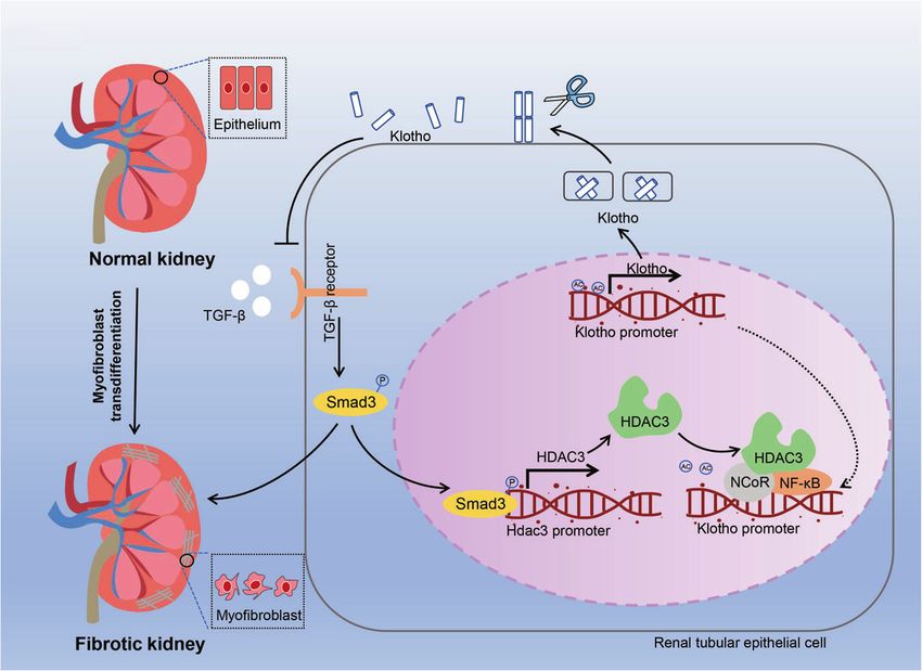
L. Ning et al.
                  2

                      Fig. 1 Schematic representation of the histone acetylation and deacetylation by HATs and HDACs. A Condensation and relaxation of
                      chromatin due to histone deacetylation and acetylation, respectively. Histone acetylation levels are determined by the interplay between HATs
1234567890();,:

                      and HDACs. Activation of HDACs leads to a net decrease of histone acetylation, chromatin condensation, and transcriptional repression.
                      Activation of HATs results in a net increase of histone acetylation, chromatin relaxation, and transcriptional activation. B The chemical formula
                      of histone acetylation and deacetylation. C Nuclear receptor co-repressor complexes containing HDAC3, GPS2, TBLX, and TBL1XR bind to
                      nuclear receptors without ligands to induce transcriptional repression via histone deacetylation. D Nuclear receptor-mediated ligand binding
                      inhibits the co-repressor complex and recruits co-activators, facilitating histone acetylation and gene transcription.

                      residue in the N-terminus. The addition of an acetyl group disrupts
                      the electrostatic interaction between the DNA and histones. By
                      neutralizing the positive lysine charge, histone acetylation alters
                      chromatin structure and gene expression (Fig. 1B) [24, 25].
                      Deacetylation of histones is usually mediated by HDACs and
                      promotes chromatin condensation and transcriptional repression
                      [23]. In mammals, HDACs are divided into four categories: class I, II,
                      III, and IV. Class I HDACs include HDAC1, HDAC2, HDAC3, and
                      HDAC8. These HDACs are predominately found in the nucleus, but
                      HDAC3 can translocate from the nucleus into the cytosol [26].
                      Class II HDACs include IIa HDACs (HDAC4, HDAC5, HDAC7, and
                      HDAC9) and IIb HDACs (HDAC6 and HDAC10). Class IIa HDACs can
                      shuttle between the nucleus and the cytosol. Class III HDACs are
                      also known as sirtuins because of their homology to the yeast
                      HDAC Sir2; this class includes SIRT1, SIRT2, SIRT3, SIRT4, SIRT5,
                      SIRT6, and SIRT7. Class IV HDACs only includes one member
                      (HDAC11), which is predominantly expressed in the nucleus
                      (Fig. 2). Class III HDACs are NAD-dependent enzymes, whereas the
                      other HDACs are zinc-dependent enzymes [27, 28].

                                                                                                 Fig. 2 The cellular localization of the four classes of histone
                      STRUCTURE AND FUNCTION OF HDAC3                                            deacetylases. Class I, IIA, VI, and part of class III HDACs are mainly
                      The unique structure of HDAC3                                              found in the nucleus. HDAC3, HDAC4, HDAC7, HDAC9, SIRT1, and
                      All class I HDAC members except HDAC8 share a similar structure,           SIRT2 shuffle between the nucleus and cytoplasm. Class IIB HDACs,
                      especially near the substrate-binding site [29]. Structural differ-        including HDAC6 and HDAC10, are localized in the cytoplasm. Class
                      ences also exist between HDAC3 and other class I HDACs. HDAC3              III HDACs, including SIRT3, SIRT4, and SIRT5, are localized in
                      possesses an aspartate residue at position 92, whereas HDAC1 and           mitochondria.
                      HDAC2 have a glutamate residue at this position at the outer rim
                      of the cavity. Furthermore, HDAC1 and HDAC2 have a tyrosine                HDAC2, enabling the development of selective inhibitors targeting
                      residue at position 199, whereas HDAC3 has phenylalanine.                  the foot pocket [32]. The biological function of HDAC3 requires
                      Particularly, position 107 of HDAC3 is a tyrosine, while in the            nuclear receptor co-repressors, including silencing mediator of
                      same position of HDAC1/2 is serine. Tyrosine 107 of HDAC3 leads            retinoic acid and thyroid hormone receptor (SMRT or NCoR2) and
                      to steric hindrance for binding to the foot pocket, precluding the         nuclear receptor co-repressor 1 (NCoR1) [33, 34]. Theoretically, the
                      binding of larger functional groups to inhibitors. Based on these          members involving 1, 2, 3, and 8 in the class I HDAC family may
                      differences, selective inhibitors against HDAC1, HDAC2, and                share similar functions and other members of the class I HDAC
                      HDAC3 can be designed [30, 31]. Hydrophobic amino acids at                 family will compensate when HDAC3 is knocked out effect.
                      positions 13 and 29 of HDAC3 also differ from those in HADC1 and           However, previous studies demonstrated that the expression of

                                                                                                                      Cell Death and Disease (2021)12:734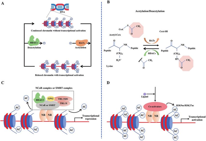
L. Ning et al.
                                                                                                                                                  3
HDAC3 is not in accordance with HDAC1, HDAC2, and HDAC8 in                acetylation in the enhancers of Ucp1 and Ppar-γ [48]. In addition,
the context of different pathological stimuli. Meanwhile, other           HDAC3 is essential for the development of vital organs. For
members also display different biological functions from HDAC3            example, mice with lung endodermal epithelium-specific Hdac3
[35–37]. Therefore, we speculate that the other members in class I        knockout displayed lethality 2 to 10 days after birth due to defects
HDAC will not compensate when HDAC3 is absent based on the                in lung sacculation and early alveologenesis [49]. Similarly, cardiac
published articles. Of course, the expression of HDAC1, 2 and             progenitor cell-specific Hdac3 knockout led to ventricular septal
8 should be also detected when HDAC3 was silenced in future               defects and underdevelopment of ventricular walls, causing
studies.                                                                  embryonic lethality [46]. Apart from lung and heart development,
                                                                          HDAC3 is also essential for the development and remodeling of
Enzymatic activity and non-enzymatic functions of HDAC3                   bones [50].
Nuclear receptors act as genetic switches regulating gene                    Moreover, HDAC3 contributes to the maintenance of intestinal
transcription by activating signal-dependent transcription factors.       homeostasis and host defense. Notably, HDAC3 loss can cause
In turn, transcription factors integrate hormonal, metabolic, and         inflammation and intestinal damage [50]. Furthermore, HDAC3-
environmental cues, recruiting various co-repressors and co-              deficiency in neural progenitor cells impaired cortical lamination
activators to specific genomic sequences [38]. HDAC3-containing            and neuronal migration, resulting in death within 16 h after birth.
nuclear receptor co-repressor complexes, including NCoR and               The critical role of HDAC3 in neuronal cell fate and function may
SMRT, bind to ligand-free nuclear receptors, which directly repress       be associated with HDAC3-mediated expression of T-box brain
gene expression. NCoR and SMRT complexes contain WD40                     protein 1 [51]. Based on these findings, we conclude that HDAC3
repeat-containing proteins, such as TBL1XR1 and TBL1X (Fig. 1C).          expression and function are essential for multiple aspects of
These complexes recruit the 19S proteasome and the ubiquityla-            mammalian physiology and homeostasis (Fig. 3). The role of
tion machinery to histones [39]. G-protein pathway suppressor 2           HDAC3 in organ injury is described below (Table 1).
(GPS2) is another core element of NCoR and SMRT complexes [40];
however, the role of GPS2 remains unclear. Noteworthily, under
certain circumstances, HDAC3 indirectly activates gene expression.        THE EMERGING ROLES OF HDAC3 IN SOLID ORGAN INJURY
By contrast, nuclear receptor-mediated ligand binding inactivates         Brain
the co-repressor complex and recruits co-activators, thereby              Ischemic stroke is a potentially deadly cardiovascular disease
facilitating gene transcription via histone acetylation [41] (Fig. 1D).   causing significant morbidity and mortality worldwide. Ischemic
                                                                          stroke is usually triggered by the thrombus in the cerebral
Enzymatic activity. The catalytic function of HDAC3 requires the          vasculature. Persistent occlusion in cerebral vasculature may
physical interaction between HDAC3 and the deacetylase-                   hinder the supply of oxygen and glucose in the local brain tissue,
activating domain (DAD) of NCoR and SMRT proteins. Crystal                eventually causing neuroinflammation, neuronal cell death, and
structure analysis of these complexes unveiled that abundant              secondary tissue injury during cerebral hypoperfusion and
protein-protein interactions between the N-terminus of HDAC3              reperfusion [52, 53]. Type-1 interferons (IFNs) are pleiotropic
and the DAD of SMRT. Inositol tetraphosphate (Ins[1,4,5,6]P4 or           cytokines regulating the expression of proinflammatory genes and
IP4) has been identified, acting as “intermolecular glue” enhancing        orchestrating innate immune responses [54]. Hence, targeting
the interaction between HDAC3 and SMRT DAD via salt bridges               IFNs or their upstream regulators may help prevent occlusion-
and hydrogen bonds [42]. Intriguingly, once HDAC3 is dissociated          induced brain injury [55, 56]. The cyclic GMP-AMP (cGAMP)
from the NCoR or SMRT complex, it becomes unstable and is                 synthase (cGAS)-stimulator of interferon genes (STING) pathway is
sequestered into a TCP1-ring complex in the cytoplasm [43]. TCP1          a key regulator of the IFN pathway and innate immunity in
promotes HDAC3 folding in the cytoplasm, facilitating the                 response to double-stranded DNA (dsDNA) [57]. Microglia are
formation of NCoR or SMRT complex containing an active HDAC3              innate immune cells residing in the central nervous system and
enzyme in an ATP-dependent manner [44].                                   serve as the principal effector cells contributing to neuroinflam-
                                                                          mation and brain injury caused by IRI. Conditional knockout of
Non-enzymatic activity. In addition to its enzymatic functions,           cGAS in microglia significantly relieved cerebral IRI. Mice with
HDAC3 also displays non-enzymatic activities. Point mutations in          microglia-specific deletion of Hdac3 displayed low expression of
Y298F, one of the active sites of HDAC3, disrupt its deacetylase          cGAS at the mRNA and protein levels, as well as decreased levels
activity. In the livers of HDAC3-deficient mice, the mutant HDAC3          of STING and IFN-γ. Mechanistically, HDAC3 deacetylates p65 at
can partially rescue hepatosteatosis and inhibit the expression of        K122 and promotes the nuclear accumulation of p65, which
lipogenic genes, suggesting that HDAC3 has non-enzymatic                  regulates the transcription of cGAS [37]. HDAC3 inhibition using
functions [44]. Moreover, global HDAC3 knockout may lead to               RGFP966 dampened the activation of melanoma 2 (AIM2)
embryonic lethality because of gastrulation defects [45]. However,        inflammasome in microglia, preventing ischemic brain injury
mutations in the DAD of SMRT and NCoR did not affect the                  [58]. Proteomic analysis revealed another mechanism of how
Mendelian ratios of offspring mice, although the mice showed              HADC3 contributes to microglial injury. Specifically, genes
little HDAC3 enzymatic activity [45, 46]. Indeed, approximately           involved in the toll-like receptor (TLR) pathway and STAT3/5
10% of enzymes with inactivating mutations in their active site are       pathway were found to be differentially expressed between
conserved in mammals, further supporting their non-catalytic              HDAC3 inhibitor-treated and untreated lipopolysaccharide (LPS)-
functions [47]. Therefore, the non-enzymatic functions of                 exposed primary microglia [59]. Diabetes mellitus significantly
HDAC3 should be taken into account in the development of                  increases the risk of cerebral vessel occlusion and is one of the
HDAC3 inhibitors.                                                         predominant risk factors for ischemic stroke [60]. HDAC3 inhibition
                                                                          mitigated cerebral IRI in diabetic mice by upregulating the brain
Physiological functions of HDAC3                                          and muscle Arnt-like 1 (Bmal1) gene [61]. However, whether the
Several genetically engineered mice with cell type-specific                ability of HDAC3 to regulate Bmal1 expression depends on histone
deletion of the Hdac3 gene have been developed recently to                deacetylation remains unclear.
investigate the role of HDAC3. Notably, HDAC3 has been shown to              Alzheimer’s disease (AD) is a leading cause of age-related
regulate metabolism by increasing fatty acid oxidation and                neuronal degeneration. AD is characterized by the deposition of
enhancing circadian histone deacetylation [47, 48]. HDAC3 also            amyloid-beta (Aβ) senile plaques in the extracellular milieu,
inhibits white adipose tissue metabolism by enhancing the futile          deposition neurofibrillary tangles encompassing acetylated and
cycle of fatty acid synthesis and oxidation and by decreasing             hyperphosphorylated tau, and synapse dysfunction [62]. Recently,

Cell Death and Disease (2021)12:734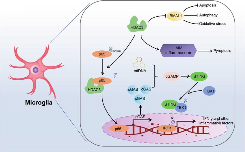
L. Ning et al.
4

    Fig. 3 The physiological functions and pathogenic effects of HDAC3. Under physiological condition, HDAC3 is mainly responsible for the
    development and homeostasis of liver, heart, brain, lung, bone, pancreas, intestine, and adipocyte. However, the abnormal expression of
    HDAC3 also contributes to organ injury including heart, brain, pancreas, kidney, lung, and liver.

    histone deacetylation at specific lysine residues has been                the infarcted myocardium to prevent further loss of cardiomyo-
    implicated in AD, and nonspecific HDAC inhibitors have been               cytes, fibrosis, and tissue damage [68]. The Hippo signaling
    used in vitro and in vivo to alleviate AD [63]. However, although        pathway regulates cellular homeostasis, immunity, heart develop-
    these nonselective HDAC inhibitors have displayed promising              ment, and regeneration. The transcription factors yes-associated
    anti-AD effects, they may cause various side effects. Interestingly,     protein (YAP) and transcriptional co-activator with PDZ-binding
    HDAC3 silencing or inhibition increased the acetylation of histones      motif (TAZ) regulate the innate and adaptive immunity in
    H3 and H4, as well as reduced the phosphorylation at Thr181,             response to Hippo pathway activation [68]. After MI, YAP/TAZ
    Ser202, and Ser396 and acetylation of tau protein. In addition,          deficiency modulates the macrophage phenotype, promoting
    HDAC3 silencing or inhibition decreased Aβ1–42 accumulation              tissue damage repair and maintaining cardiac function. Mechan-
    and β-secretase-mediate cleavage of the amyloid precursor                istically, YAP/TAZ in reparative macrophages inhibits the tran-
    protein in HEK/APPsw cells, hinting that HDAC3 may act as a              scriptional activation of Arg1 by binding to its promoter and
    critical regulator of AD-associated brain injury 36. Moreover,           interacting with the HDAC3-NCoR1 repressor complex. This
    HDAC3 inhibition inhibited the oxidation of proteins, DNA, RNA,          inhibition of Arg1 expression and the reprogramming of macro-
    and lipids in the hippocampi of mice with AD by inactivating the         phages is independent of the deacetylation activity of HDAC3 [69].
    c-Abl/MST1/YAP signaling pathway [64]. Collectively, the findings         Notably, HDAC3 knockout did not change the levels of H3K9Ac
    of these studies suggest that HDAC3 plays a crucial role in brain        and H3K27Ac in the HDAC3-binding sites in the Arg1 promoter,
    injury by regulating innate immunity, inflammation, and the               suggesting that HDAC3-mediated deacetylation is not essential for
    biological clock (Fig. 4). In addition, we found that HDAC3              the inhibition of Arg1 expression [70].
    promotes the deacetylation of both non-histones and histones in             Diabetes mellitus increases the risk of MI. Patients with diabetes
    the brain, indirectly or directly affecting the expression of target     mellitus and MI tend to have larger infarcts in the myocardium
    genes. However, considering that HDAC3 possesses both enzy-              and are at a higher risk of heart failure [71]. Circadian clock genes
    matic and non-enzymatic activities, how HDAC3 impacts the                may regulate MI and diabetes mellitus by modulating the time-of-
    target proteins has not been comprehensively investigated yet.           day dependence of cardiomyocytes [72]. HDAC3 is a vital
                                                                             regulator of the circadian rhythm and may suppress the
    Heart                                                                    expression of BMAL1 by activating the expression of the clock
    Myocardial infarction (MI) is a significant global health burden.         gene Rev-erbα [73]. In diabetic rats with or without MI, the
    Ischemic stress due to coronary artery obstruction impedes the           expression levels of HDAC3 were constant. However, ischemia-
    supply of oxygen and nutrients to the myocardium, causing tissue         reperfusion significantly increased the expression levels of HDAC3
    injury and cardiomyocyte death [65]. Cyclin-dependent kinase 2           in diabetic rats. Intriguingly, the levels of Rev-erbα and BMAL1
    (CDK2) is a serine/threonine kinase, and its dysregulated activation     displayed opposite rhythm after ischemia-reperfusion stimulation
    has been associated with MI and heart failure [66]. HDAC3 in             in diabetic rats. Subsequent studies showed that HDAC3
    cardiomyocytes can induce cardiac dysfunction and heart failure.         aggravated diabetic MI in rats by altering the oscillations of
    RGFP966 treatment in mice with MI alleviated oxidative stress and        circadian genes, thereby causing mitophagy dysfunction in
    cardiac injury. In addition, RGFP966 decreased the expression of         cardiomyocytes [15]. Interestingly, hyperglycemia significantly
    CDK2 in the myocardium by promoting miR-19a-3p expression.               increased HDAC3 protein levels. HDAC3 inhibition relieved
    However, how HDAC3 regulates the miR-19a-3p expression                   endothelial injury and dysfunction induced by type 2 diabetes
    remains unclear [67]. During MI, cardiomyocyte death activates           mellitus by blocking the interaction of Keap1 and Nrf2 in a Nox4-
    acute inflammation through the recruitment of macrophages to              dependent manner [74].

                                                                                                  Cell Death and Disease (2021)12:734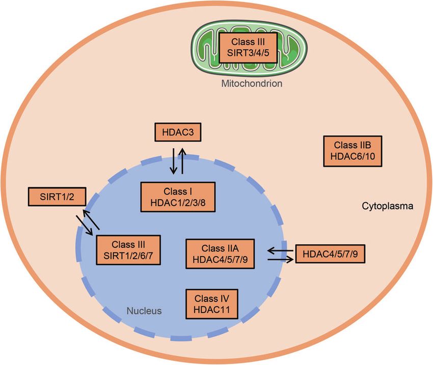
Table 1. Subcellular localization of HDAC3 and its roles in different disease models.
Models Subcellular localization Key mechanisms Activity Reference
Cerebral IRI Shuttling between the nucleus and the cytosol in Deacetylating p65 at K122 in the cytosol and interacting with Enzymatic [37]
microglia p65 in the nucleus to induce neuroinflammation by activating activity
cGAS-STING axis
Ischemic brain damage Mainly concentrating in the nucleus in microglia 24 h Deacetylating STAT1 and subsequently promoted STAT1 Enzymatic [58]
following ischemia and gradually spreading to cytoplasm phosphorylation, contributing to brain damage via the activity
72 h following ischemia regulation of AIM2
Cerebral IRI in the Not detected Promoting the cerebral infarct volume and cytotoxicity by Enzymatic [61]
diabetic state upregulating Bmal1 activity
Cell Death and Disease (2021)12:734
Ischemic stroke Distributed both in the nucleus and cytoplasm in cortical Potentiating transcriptional initiation of oxidation relative Enzymatic [133]
neurons before ischemic preconditioning; distributed genes involving Hspa1a, Bcl2l1, and Prdx2 activity
mainly in the cytoplasm after ischemic preconditioning
treatment
Chronic constriction injury Mainly concentrating in the nucleus in the hippocampus Deacetylating H3 and H4 in the hippocampus and triggering Enzymatic [35]
memory impairment activity
Alzheimer’s disease Both nucleus and the cytosol in HEK/APPsw cells. Increasing Aβ1–42 accumulation and both tau acetylation and Enzymatic [36]
phosphorylation at disease residues, thus impairing learning activity
and memory
Acute lung injury Shuttling from the cytosol to nucleus in bronchial Interacting with p65 in the cytosol and translocating to the Non-enzymatic [95]
epithelial cells nucleus, eventually triggering an inflammatory response activity
Myocardial infarction Not detected Decreasing miR-19a-3p and elevating CDK2, leading to Enzymatic [67]
myocardial infarction activity
Myocardial infarction Nucleus in macrophage Forming HDAC3-NCoR1 repressor complex and inhibiting Arg1 Non-enzymatic [69]
expression independent of its enzymatical activity activity
Diabetic myocardial IRI Nucleus in cardiomyocytes Regulating circadian gene oscillations to trigger mitophagy Non-enzymatic [15]
dysfunction and myocardial IRI activity
Diabetic endothelial Cytosol in endothelial cells Inhibiting Nrf2 signaling through the modulation of Keap1 Enzymatic [74]
L. Ning et al.
dysfunction and Nox4 activity
Renal fibrosis The nucleus of renal tubular cells Inhibiting Klotho transcription and mediating myofibroblast Enzymatic [89]
transdifferentiation by decreasing acetylations of H3K4, H3K9, activity
and H4K5 thus promotes renal fibrosis
Diabetic renal damage Not detected Epigenetically modulating miR-10a, subsequently affecting Enzymatic [92]
CREB1 and fibronectin formation activity
Diabetic hepatic damage Mainly concentrating in the cytosol in hepatocyte Decreasing Nrf2 activity by inhibiting miR-200a expression Enzymatic [107]
with a concomitant increase in Keap1 to block hepatic activity
FGF21 synthesis.
Diet-induced obesity The nucleus in intestinal epithelial cells Regulating expression of microbiota-dependent metabolic Enzymatic [108]
pathways including Chka, Mttp, Apoa1, and Pck1, thus activity
triggering diet-induced obesity
Diabetes mellitus Both nucleus and the cytosol in β-cells Affecting insulin secretion, glucose tolerance, lipotoxicity, Enzymatic [116, 117, 134]
insulin resistance, and inflammation activity
5L. Ning et al.
6
Fig. 4 The role of HDAC3 in brain injury. HDAC3 promotes neuronal cell death via multiple mechanisms. HDAC3 is upregulated in microglia
after ischemia stimulation, deacetylating p65 at K122. Deacetylated p65 translocates into the nucleus activating the transcription of cGAS.
Ischemia-induced mitochondrial DNA is recognized by cGAS, which activates the microglial cGAS-STING-IRF3 pathway and promotes
neuroinflammation. In addition, HDAC3 inhibits the activation of the AIM inflammasome and induces pyroptosis. HDAC3 also induces
apoptosis, autophagy, and oxidative stress by inhibiting the expression of BMAL1.
Heart failure is the end stage of various cardiovascular diseases pathway in this process. Specifically, TGF-β1 promotes the
and represents a critical healthcare burden. Several mechanism- recruitment of activated serine kinases, which phosphorylate
based therapies for heart failure have been developed recently Smad2 and Smad3. Phosphorylated Smad2 and Smad3 form a
[75, 76]. The reduced expression level of HDAC3 was associated complex with Smad4 and translocate into the nucleus, inducing
with better cardiac function in mice with heart failure [77]. In the expression of profibrotic genes [84, 85]. Klotho is an anti-
addition, HDAC3 has been shown to promote heart failure and fibrosis and anti-aging protein enriched in the renal tube
dietary death by exacerbating metabolic disturbances in mito- epithelium. It can be found in a soluble or membrane-bound
chondria in the cardiomyocytes of mice fed with a high-fat diet form [86]. Klotho-deficient mice spontaneously developed renal
[78]. Mechanically, HDAC3 inhibition prevented heart failure by fibrosis and displayed aging phenotypes in various organs. By
inhibiting miR-18a-targeted adrenergic-receptor β3 [79]. HDAC3 directly binding to TGF-β receptor and Wnt, Klotho inhibits the
expression and activation in cardiomyocytes were also regulated profibrotic TGF-β/Smad and Wnt/β-catenin pathways [87, 88]. In
by Ca2+/calmodulin-dependent kinase II (CaMKII). Specifically, mice with renal fibrosis induced by unilateral ureter obstruction,
CaMKII increased HDAC3 expression levels and enhanced the the expression levels of HDAC3 in the nucleus of renal tubular cells
deacetylase activity of HDAC1 and HDAC3. However, CaMKII were significantly elevated in a TGF-β/Smad signaling-dependent
hyperactivity-induced heart failure could be reversed by class I manner. The renal fibrosis-induced HDAC3 upregulation was
HDAC inhibitors [80]. Endothelial dysfunction is another critical reversed by the selective inhibition of Smad3 phosphorylation.
mechanism contributing to heart failure, diabetic cardiomyopathy, HDAC3 accumulation in the nucleus further enhanced the
and diabetic microvascular disease. In high glucose-treated transcriptional repression of Klotho. By interacting with NCoR
endothelial cells, β-hydroxybutyrate blocked the binding and and NF-κB, HDAC3 triggered myofibroblast transdifferentiation
colocalization of HDAC3 to β-catenin via increasing H3K14ac and aggravated renal fibrosis (Fig. 5) [85, 89]. The pan-HDAC
levels, thereby increasing the expression of claudin-5 and relieving inhibitor trichostatin has been reported to block TGF-β1-induced
cardiac microvascular hyperpermeability in diabetic rats [81]. epithelial-mesenchymal transition in human renal epithelial cells
However, the mechanisms underlying β-hydroxybutyrate- and IRI-induced renal fibrosis [90][25]. FK228, a class I HDACs
mediated H3K14ac regulation in endothelial cells need to be inhibitor predominately targeting HDAC1 and HDAC2, also
further investigated. These studies have provided strong evidence suppressed the activation and proliferation of renal fibroblasts
supporting that HDAC3 is activated in the myocardium during by enhancing histone H3 acetylation partially through the Smad
acute and chronic cardiac injury, aggravating the cardiac injury by pathway. Therefore, class I HDACs seem to prevent renal fibrosis
reprogramming macrophages, regulating the expression of clock by similar mechanisms.
genes, activating adrenergic receptors, and increasing microvas- Renal IRI is common during renal transplantation, cardiovascular
cular endothelial hyperpermeability. surgery, trauma, and endovascular procedures. TSA pretreatment
early after renal ischemic injury protected renal function and
Kidneys prevented renal fibrosis by upregulating miR-21. Notably, HDAC6
Renal fibrosis is a typical pathohistological characteristic of renal inhibition exhibited no significant effects on renal IRI tolerance,
aging and chronic renal injury. Renal injury can be triggered by suggesting that class II HDAC elimination is unlikely to contribute
various etiologies and is characterized by the transdifferentiation to renal IRI tolerance [90]. Diabetic nephropathy is a chronic renal
of injured renal cells to myofibroblasts [82]. In renal fibrosis, injury in patients with diabetes mellitus. Our poor understanding
transdifferentiated myofibroblasts secrete extracellular matrix of its pathogenesis makes the treatment of diabetic nephropathy
(ECM) proteins, which gradually deteriorate renal structure and challenging [91]. In mice fed a high-fat diet and treated with a low
function [83]. Although the precise mechanisms underlying renal dose of streptozotocin, HDAC3 downregulated miR-10a and
fibrosis remain primarily unclear, accumulating evidence suggests upregulated cAMP response element-binding protein 1 (CREB1),
the critical role of the transforming growth factor-β (TGF-β)/Smad thereby promoting kidney injury [92]. However, how HDAC3
Cell Death and Disease (2021)12:734L. Ning et al.
7
Fig. 5 Scheme of HDAC3-mediated renal fibrosis. Renal injury enhances the production of TGF-β, which promotes myofibroblast
differentiation and activates Hdac3 transcription and Smad signaling. Subsequently, HDAC3, together with NF-κB and NCoR, bind to and
deacetylate the Klotho promoter, downregulating Klotho and exacerbating renal fibrosis. HDAC3 inhibition preserves the expression of
Klotho, inhibiting the TGF-β receptor and alleviating renal fibrosis.
epigenetically modifies the expression of miR-10a remains can invade small airways and interfere with pulmonary gas
unknown. These data together indicate that both acute and exchange, eventually causing chronic lung injury and lung cancers
chronic kidney injury is closely associated with HDAC3 over- [99, 100]. Mechanistically, PM2.5 causes ultrastructural alterations
expression, suggesting the inhibition of HDAC3 as a promising in mitochondria and membrane lysis in alveolar epithelial cells, in
therapeutic strategy to protect renal function. addition to inducing DNA damage and the production of reactive
oxygen species (ROS) [101–103]. Furthermore, accumulating
Lungs evidence suggests that PM2.5 can also activate TLR4, TLR4/NF-
Acute lung injury is characterized by excessive damage to alveolar κB, and TGF-β/Smads, causing pulmonary inflammation and
epithelial cells and capillary endothelial cells due to infection, fibrosis [98, 104]. In this regard, HDAC3 deficiency remitted
ischemia-reperfusion, trauma, blood transfusion, and pulmonary PM2.5-induced damage and inflammation in lung epithelial cells
embolism. Lung injury eventually causes refractory hypoxemia by inhibiting TGF-β/Smad3 and NF-κB signaling [98].
and acute respiratory distress syndrome (ARDS) [93, 94]. During Although HDAC3 inhibition may prevent lung injury by
sepsis-induced acute lung injury, LPS can bind to Toll-like repressing inflammation, particular attention should be paid to
receptor-4 (TLR-4) and its co-receptor cluster of differentiation the development of HDAC3-targeting agents as the enzymatic
14 (CD14), causing neutrophil accumulation in the alveolar space activity of HDAC3 can regulate inflammation in opposing ways.
and interstitial tissue, lung parenchymal damage, increased
vascular permeability, and aggravation of pulmonary edema. Liver
Furthermore, LPS-induced TLR4 activation promoted the translo- Long-term high blood glucose or a high-fat diet can result in liver
cation of HDAC3 and NF-κB into the nucleus in alveolar epithelial damage due to increased lipid peroxidation. In turn, chronic liver
cells; nimbolide could reverse this effect [95]. In addition, HDAC3 damage can cause multiple vascular complications, including
inhibition suppressed the expression of various proinflammatory atherosclerosis [105]. Fibroblast growth factor 21 (FGF21) is a
cytokines (e.g., IL-1β, IL-6, and IL-12β) in macrophages by newly identified member of the FGF family regulating glucose and
inhibiting NF-κB, thereby preventing acute lung injury [96]. A lipid metabolism in the liver [106]. HDAC3 inhibition using
recent study showed that LPS stimulation did not change the RGFP966 mitigated diabetes-induced inflammation, aortic fibrosis,
ability of HDAC3 to bind near deacetylation-dependent genes but and pathological liver injury in mice with type 1 diabetes mellitus.
increased the binding of HDAC3 to transcription start sites in In addition, HDAC3 inhibition suppressed Keap1 translation by
deacetylation-independent genes. In bone marrow-derived upregulating miR-200a. Keap1 downregulation enhanced the
macrophages, HDAC3 promoted the expression of proinflamma- transcription of Nrf2 target genes, including heme oxygenase-1
tory cytokines by binding to ATF2-binding sites independently of (HO-1), catalase, and nicotinamide adenine dinucleotide phos-
the NCoR1/2 complex. On the contrary, ATF3-bindings sites were phate (NADPH) quinone oxidoreductase (NQO1). These antiox-
preferentially related with the deacetylase activity of HDAC3, the idants increased the level of FGF21 in the serum and alleviated
activation of which inhibited the TLR signaling pathway and oxidative stress in the liver [107]. In mice with type 2 diabetes
inflammation in macrophages [97]. Thus, during macrophage mellitus, intestinal epithelial cell-specific disruption of Hdac3
activation or acute lung injury, HDAC3 plays multifaceted roles in prevented obesity and dysregulation of glucose metabolism.
the expression of proinflammatory cytokines, which was deter- Consistently, HDAC3 inhibition prevented hepatic injury and fat
mined by the enzymatic activity of HDAC3. deposition [107]. Clinical findings also indicated that HDAC3
PM2.5 is a type of air particle with a diameter of ≤2.5 µm. expression levels in pediatric patients were positively associated
Exposure to PM2.5 poses a great threat to our health [98]. PM2.5 with bodyweight [108]. HDAC3 interacts with various nuclear
Cell Death and Disease (2021)12:734L. Ning et al.
8
proteins in hepatocytes. Particularly, HDAC3 was found to interact SELECTIVE HDAC3 INHIBITORS
with the prospero-related homeobox 1 (PROX1) in a hepatocyte Over the last 20 years, several HDAC3 inhibitors have been
nuclear factor 4α (HNF4α)-dependent manner [109]. PROX1 is a developed and tested for the treatment of various diseases. Thus
highly conserved transcription factor essential for the develop- far, the U.S. Food and Drug Administration (FDA) has approved six
ment of multiple organs in vertebrates [110]. Although Prox1 HDAC inhibitors, namely belinostat, vorinostat, romidepsin,
knockdown in adult hepatocytes did not affect the expression of chidamide, pracinostat, and panobinostat. Most of these inhibitors
HDAC3, it increased the hepatic levels of triglycerides. These data are used as anticancer agents [120]. As all class I HDACs have a
suggest that the interaction between PROX1 and HDAC3 is zinc ion in the active site, small molecule inhibitors typically
essential for maintaining hepatic lipid homeostasis and lipid possess the same zinc-binding group. In addition, most HDAC
metabolism [109]. Hence, HDAC3 acts as a double-edged sword in inhibitors possess a linker connecting the zinc-binding group to a
hepatic injury. Both overexpression and downregulation of HDAC3 capping group to mimic the lysine alkyl side chain. The most well-
trigger hepatic injury by disturbing metabolic balance. The known zinc-binding group involves o-aminoanilides and hydro-
mechanisms of how HDAC3 regulates type 1 and type 2 diabetes xamic acids. Entinostat (MS-275) and other selective class I
mellitus-induced hepatic injury are different, although HDAC3 has inhibitors are based on o-aminoanilides. The selective HDAC3
been implicated in both diseases. inhibitor RGFP966 was developed through the modification of o-
aminoanilides. PD106 and BRD3308 are also selective HDAC3
Pancreas inhibitors used for the treatment of Friedreich’s ataxia, diabetes
Type 1 diabetes mellitus is a chronic autoimmune disease mellitus, and HIV infection [121]. Considering the potential of the
caused by the immune-mediated destruction of insulin- o-aminoanilide scaffold in the development of selective HDAC3
producing β-cells in pancreatic islets, leading to insulin inhibitors, many derivatives were synthesized based on this
deficiency. By contrast, type 2 diabetes mellitus is characterized scaffold. In addition, some natural compounds can also regulate
by progressive loss of β-cell insulin secretion, which usually the expression or activity of HDAC3, exerting protective effects in
occurs in the context of insulin resistance. Loss of functional various diseases in animal and cell models. For instance, juglanin
β-cell mass, whether type 1 diabetes mellitus or type 2 diabetes prevents high-fat diet-induced renal injury by blocking the nuclear
mellitus, serves as the core mechanism in both diseases. translocation of HDAC3 and NF-κB and thereby inhibiting
Normoglycemia can be preserved as long as β-cells possess inflammation and dyslipidemia [122]. miRNAs also play critical
the ability to compensate [111, 112]. B lymphocytes and T roles in nerve injury by targeting HDAC3 [123, 124]. The most
lymphocytes participate in the destruction of β-cells and the loss common HDAC3 inhibitors and their roles in solid organ injury are
of self-tolerance in patients with type 1 diabetes mellitus [113]. summarized in Table 2. Most of the synthesized HDAC3 inhibitors
HDAC3 inhibited apoptosis in peripheral blood mononuclear lack specificity, as they partly inhibit other HDACs because of the
cells by downregulating miR-296-5p and upregulating Bcl-xl, high structural similarity of HDACs. Nearly all selective HDAC3
thereby aggravating type 1 diabetes mellitus [114]. Conversely, inhibitors are o-aminoanilide derivatives, the selectivity of which is
HDAC3 was found to be downregulated in the pancreas of mainly accessed through testing their IC50s [125]. However, this
children with type 1 diabetes mellitus. In the rat β-cell line INS-1, test method is not accurate. Alternatively, the Ki value of the o-
HDAC1 or HDAC3 knockdown decreased iNos mRNA levels in aminoanilides can be determined by testing the kon and koff values
response to cytokines. However, only HDAC1 knockdown of the inhibitors directly.
restored insulin secretion in cytokine-treated β-cells [115]. To build up a better picture of the regulatory mechanism of
HDAC3 inhibition also inhibited apoptosis, caspase 3 activations, HDAC3, the activation mechanism of HDAC3 should also be paid
and Erk1/2 phosphorylation in palmitic acid-treated NIT-1 cells, particular attention. To our knowledge, there are mainly 3 aspects
thereby restoring glucose tolerance [116]. In streptozotocin- to promote the expression or the activity of HDAC3 at present. On
induced type 1 diabetic mice, HDAC3 inhibition exerted a the one hand, HDAC3 could be transcriptionally activated by
hypoglycemic effect and improved the morphology of islets and certain transcription factors including Smad3 including Smad3
the function of β-cells [116]. through directly binding its promoter [89]. On the other hand,
MS-275, a nonspecific inhibitor of HDAC1 and HDAC3, poten- HDAC3 activity could be enhanced via deubiquitinating. For
tiated insulin secretion in the islets of rats with type 2 diabetes example, HDAC3 activity could be enhanced by ubiquitin-specific
mellitus. Bioinformatics analysis revealed that the differentially protease 38 (USP38) via deubiquitinating. Notably, USP38 knock-
expressed genes between normal islets and MS-275-treated islets down and overexpression could not change the protein level of
were enriched in calcium, cAMP, MAPK, PI3K-Akt, and HDAC3 [126]. In addition, PIWIL2 can also interact with HDAC3,
Rap1 signaling. However, the genes dysregulated by MS-275 in giving rise to the stabilization of HDAC3 from ubiquitin-mediated
rat islets were not involved in glucose oxidation [117]. Consistently, degradation via competitive relation with E3 ubiquitin ligase
SJ Lee et al. found that HDAC3 levels were elevated in palmitic Siah2. Meanwhile, PIWIL2 facilitated the interaction between CK2α
acid-treated C2C12 myotube. MS-275 pretreatment or HDAC3 and HDAC3, thus enhancing HDAC3 activity (Doi: 10.1038/s41419-
knockdown dramatically alleviated lipotoxicity and protected 018-0462-8). Last but not least, the microbiota-derived metabolite
against palmitic acid-induced insulin resistance and inflammation. [inositol-1,4,5-trisphosphate (InsP3)] could promote epithelial
In addition, MS-275 improved mitochondrial function in palmitic repair by promoting HDAC3 activity, but the detailed mechanism
acid-treated C2C12 myotubes by enhancing mitochondrial fatty remains unclear [127].
acid oxidation and upregulating mitochondrial transcription factor
A, peroxisome proliferator activator receptor γ (PPAR-γ)-co-
activator 1α (PGC1α), 3-hydroxy acyl CoA dehydrogenase, and CLINICAL VALUE OF HDAC3
enoyl-CoA hydratase [118]. HDAC3 inhibition also exerted an anti- Till now, a great many clinical studies have also established the
apoptotic effect in β-cells under glucolipotoxic conditions by clinical position of HDAC3 in solid organ injury. For instance,
alleviating endoplasmic reticulum stress [119]. Thus, HDAC3 can compared with normal-weight women, obese individuals owned
affect insulin secretion, glucose tolerance, lipotoxicity, insulin reduced levels of HDAC1, HDAC3, as well as HDAC9 in adipose
resistance, and inflammation in pancreatic cells via multiple tissues. Meanwhile, the mRNA levels and activity of these HDACs
mechanisms. Therefore, pharmacological inhibition of HDAC3 displayed an inverse correlation with inflammatory markers,
may serve as a novel therapeutic strategy to treat patients with obesity indices, and insulin levels, indicating that these HDACs
diabetes mellitus. However, more studies are required to uncover have adverse effects on the development of obesity and
the different roles of HDAC3 in type 1 and type 2 diabetes mellitus. diabetes mellitus [128]. One Chinese case-control study also
Cell Death and Disease (2021)12:734Table 2. Common HDAC3 inhibitors and their role in solid organ injury.
Inhibitor Models Key molecular signaling Major outcome Reference
RGFP966 Ischemic brain damage Acetylating and dephosphorylating STAT1, inhibiting the Protecting against inflammatory response and alleviating [58]
AIM2 inflammasome ischemic stroke
Cerebral IRI in diabetic state Inhibiting oxidative stress, apoptosis, and autophagy by Decreasing the cerebral infarct volume and inhibiting [61]
upregulating Bmal1 cytotoxicity
Chronic neuropathic injury Regulating synaptic plasticity Improving memory impairment [35]
Ischemic brain damage Inhibiting the recruitment of HDAC3 to the promoter Improving neurotoxicity and neuronal injury [133]
regions of Hspa1a, Bcl2l1, and Prdx2
Acute lung injury Repressing Hsp90-dependent RhoA activity Inhibiting endothelial barrier dysfunction and alleviating [135]
Cell Death and Disease (2021)12:734
LPS-induced lung injury
Myocardial IRI in diabetic state Regulating HDAC3/SIRT1 circuit by regulating Bmal1- Alleviating myocardial IRI [136]
mediated autophagy
Myocardial IRI in diabetic state Activating the Rev-erbα/BMAL1 circadian pathway to Alleviating myocardial IRI [15]
inhibit mitophagy
Renal fibrosis Blocking HDAC3 activity and regulating the expression Alleviating renal fibrosis and improving renal function [89]
of Klotho
MS275 Seizure-induced brain damage Downregulating p38 by decreasing histone H3 and H4 Alleviating inflammation and tissue damage [137]
methylation and increasing histone H3 and H4 acetylation
Acute lung injury Maintaining the balance between the anti-inflammatory Relieving macrophage-induced pulmonary inflammation [138]
and proinflammatory IL-10 and IL-12b
BRD3308 Type 1 diabetes Decreasing the number of apoptotic β-cells Inhibiting pancreatic islet infiltration and preventing [139]
β-cell death
MI192 Photothrombotic stroke Decreasing apoptosis and deacetylation of α-tubulin and Eliminating tissue infarct and improving motor activity [140]
upregulating GAP-43 in the cerebral cortex
Valproic acid Sepsis-induced cardiac injury Increasing histone acetylation in the PTEN promoter and Promoting cardiac autophagy and reducing mitochondrial [141]
inhibiting the AKT/mTOR pathway damage, oxidative stress, and inflammation in cardiac tissues
L. Ning et al.
Traumatic brain injury Inhibiting oxidative stress and autophagy by activating Reducing microglial activation and inflammation [142]
Nrf2/ARE signaling
Nimbolide Acute lung injury Blocking NF-κB and HDAC-3 nuclear translocation Alleviating oxidative stress, inflammation, and [95]
mediated by TNF-α pathological injury
Betaine Hypothalamic neural injury Blocking TLR4/NF-κB pathway activation and repressing Inhibiting hypothalamic astrogliosis and inflammation [143]
HDAC3 expression
Scriptaid Ischemic brain damage Increasing the acetylation of H3 and H4 Decreasing the infarct volume and neuronal degeneration, [144]
improving their neurobehavioral dysfunction
Chrysophanol Acute lung injury Promoting HMGB1/HDAC3/NF-κB/p65 complex formation Relieving lung lesions and enhancing superoxide [145]
dismutase levels
miR-193b-39 Brain injury after subarachnoid Acetylating p65 by decreasing the expression and activity Mitigating behavioral impairment, brain edema, blood-brain [146]
hemorrhage of HDAC3 barrier injury, and neurodegeneration
miR-494 Ischemic brain damage Inhibiting the expression of neuronal ataxin-3 and HDAC3 Decreasing neuronal apoptosis and infarct size [124]
and increasing acetyl-H3K9 levels
miR-19a-3p Myocardial IRI Reducing the level of CDK2 Improving cardiac function and attenuating [123]
pathological change
9L. Ning et al.
10
demonstrated that the single nucleotide polymorphism of REFERENCES
HDAC3 including rs11741808, rs2547547, and rs2547547 poly- 1. Dupont A, Rauch A, Staessens S, Moussa M, Rosa M, Corseaux D, et al. Vascular
morphism was related to type 2 diabetes mellitus [129]. And endothelial damage in the pathogenesis of organ injury in severe COVID-19.
combined HDAC3 and HDAC9 genes with diabetes mellitus Arterioscl Thrombosis Vascular Biol. 2021;41:1760–73.
worsen atherosclerosis and resulted in stroke [130, 131]. These 2. Zhou H, Ma Q, Zhu P, Ren J, Reiter R, Chen Y. Protective role of melatonin in
clinical studies suggested that the genetic change of HDAC3 is cardiac ischemia-reperfusion injury: from pathogenesis to targeted therapy. J
Pineal Res. 2018;64:e12471.
closely associated with the development of diabetes mellitus and
3. Cen M, Ouyang W, Zhang W, Yang L, Lin X, Dai M, et al. MitoQ protects against
cerebrovascular disease. However, few clinical studies have hyperpermeability of endothelium barrier in acute lung injury via a Nrf2-
reported the roles of HDAC3 in other types of solid organ injury. dependent mechanism. Redox Biol. 2021;41:101936.
Before we reach clinical attempts, we might have to obtain more 4. Peukert K, Fox M, Schulz S, Feuerborn C, Frede S, Putensen C, et al. Inhibition of
clinical data on HDAC3 to comprehensively harness the knowl- caspase-1 with tetracycline ameliorates acute lung injury. Am J Respirat Critical
edge to benefit humans. Care Med. 2021;204:53–63.
5. Tsai K, Chou W, Cheng H, Huang Y, Chang M, Chan S. Anti-IL-20 antibody
protects against ischemia/reperfusion-impaired myocardial function through
modulation of oxidative injuries, inflammation and cardiac remodeling. Anti-
FUTURE PERSPECTIVE AND CONCLUSION oxidants. 2021;10:275.
HDAC3 is a unique and important member of the HDAC family. 6. Chakraborty S, Karasu E, Huber-Lang M. Complement after trauma: suturing
The catalytic activity of HDAC3 mainly depends on the integrity of innate and adaptive immunity. Front Immunol. 2018;9:2050.
nuclear receptor co-repressor complexes. In this review, we 7. Lenz A, Franklin G, Cheadle W. Systemic inflammation after trauma. Injury.
summarized the enzymatic and non-enzymatic functions of 2007;38:1336–45.
HDCA3. The non-enzymatic functions of HDAC3 should be taken 8. Ritchie R, Abel E. Basic mechanisms of diabetic heart disease. Circulation Res.
into consideration when developing new HDAC3-targeting 2020;126:1501–25.
9. Alaeddine L, Harb F, Hamza M, Dia B, Mogharbil N, Azar N, et al. Pharmacological
strategies. We also briefly discussed the role of HDAC3 in the
regulation of cytochrome P450 metabolites of arachidonic acid attenuates
integration of various signals from the environment to regulate cardiac injury in diabetic rats: the role of cytochromes P450 metabolites in
cellular fate, development, metabolism, and energy homeostasis. diabetic cardiomyopathy. Transl Res. 2021;S1931-5244:00073–6
We also described the role of HDAC3 in the injury of solid organs, 10. Zhao H, Jaffer T, Eguchi S, Wang Z, Linkermann A, Ma D. Role of necroptosis in
including the brain, heart, kidneys, liver, lungs, and pancreas. the pathogenesis of solid organ injury. Cell Death Dis. 2015;6:e1975.
Although HDAC3 is beneficial under physiological conditions, 11. Sodum N, Kumar G, Bojja S, Kumar N, Rao C. Epigenetics in NAFLD/NASH: targets
pathological HDAC3 upregulation is detrimental in the pathogen- and therapy. Pharmacol Res. 2021;167:105484
esis of solid organ injury. 12. Ghoneim M, Fuchs H, Musselman C. Histone tail conformations: a fuzzy affair
No human disease-causing mutations have been identified in with DNA. Trends Biochem Sci. 2021;46:564–78
13. Allfrey V, Faulkner R, Mirsky AS. Acetylation and methylation of histones and
the HDAC3 gene thus far, possible because germline mutations in
their possible role in the regulation of rna synthesis. Proc Natl Acad Sci USA.
HDAC3 may be may embryonically lethal in humans. However, 1964;51:786–94.
numerous single-nucleotide polymorphisms (SNPs) in HDAC3 14. Harrison I, Dexter D. Epigenetic targeting of histone deacetylase: therapeutic
genes have been reported [130, 132]. We speculate that some potential in Parkinson’s disease? Pharmacol. Therapeut. 2013;140:34–52.
SNPs may lie within enhancers and that these SNPs may hinder 15. Qiu Z, Ming H, Lei S, Zhou B, Zhao B, Yu Y, et al. Roles of HDAC3-orchestrated
the recruitment of HDAC3 because of impaired binding of nuclear circadian clock gene oscillations in diabetic rats following myocardial ischaemia/
receptors or transcription factors, dysregulating HDAC3 expres- reperfusion injury. Cell Death Dis. 2021;12:43.
sion. Another glaring question is how to overcome the high 16. Ghiboub M, Zhao J, Li Yim A, Schilderink R, Verseijden C, van Hamersveld P, et al.
structural similarity of zinc-dependent HDAC isoenzymes to HDAC3 mediates the inflammatory response and LPS tolerance in human
monocytes and macrophages. Front Immunol. 2020;11:550769.
develop specific HDAC3 inhibitors rather than pan-HDAC inhibi-
17. Lin W, Zhang Q, Liu L, Yin S, Liu Z, Cao W. Klotho restoration via acetylation of
tors. The biological functions of HDAC3 should also be further peroxisome proliferation-activated receptor γ reduces the progression of
explored in future studies. chronic kidney disease. Kidney Int. 2017;92:669–79.
18. Xu H, Wu M, Ma X, Huang W, Xu Y. Function and mechanism of novel histone
posttranslational modifications in health and disease. BioMed Res Int.
FACTS 2021;2021:6635225.
19. Campos E, Reinberg D. Histones: annotating chromatin. Annu Rev Genet.
Epigenetic modifications could regulate gene activity, as well as
2009;43:559–99.
the development of an organism without changing gene 20. Stevens K, Swadling J, Hocher A, Bang C, Gribaldo S, Schmitz R, et al. Histone
sequence. variants in archaea and the evolution of combinatorial chromatin complexity.
The reversible acetylation on the N-terminal lysine residues of Proc Natl Acad Sci USA. 2020;117:33384–95.
histone proteins by histone HATs and histone deacetylases 21. Tolsma T, Hansen J. Post-translational modifications and chromatin dynamics.
HDACs synergistically determine chromatin structure and gene Essays Biochem. 2019;63:89–96.
expression. 22. Neganova M, Klochkov S, Aleksandrova Y, Aliev G. Histone modifications in
HDAC3 is closely correlated with acute/chronic injury in the epigenetic regulation of cancer: Perspectives and achieved progress. Semin
brain, heart, kidney, liver, lung, and pancreas. Cancer Biol. 2020;S1044-579X:30176-0.
23. Zhou Y, Peng J, Jiang S. Role of histone acetyltransferases and histone deace-
tylases in adipocyte differentiation and adipogenesis. Eur J Cell Biol.
2014;93:170–7.
OPEN QUESTIONS 24. Kumar V, Thakur J, Prasad M. Histone acetylation dynamics regulating plant
What is the physiological function of HDAC3 in development and development and stress responses. Cell Mol Life Sci. 2021;78:4467–86.
homeostasis? 25. Gomathi K, Akshaya N, Srinaath N, Rohini M, Selvamurugan N. Histone acetyl
What are the precise molecular mechanisms of HDAC3 in solid transferases and their epigenetic impact on bone remodeling. Int J Biol Mac-
organ injury? romolecules. 2021;170:326–35.
26. Zhang K, Liu Z, Yao Y, Qiu Y, Li F, Chen D, et al. Structure-based design of a
What are the current situations and challenges in developing
selective class I histone deacetylase (HDAC) near-Infrared (NIR) probe for epi-
novel drugs targeting HDAC3 for solid organ injury? genetic regulation detection in triple-negative breast cancer (TNBC). J Med
Chem. 2021.64:4020–33.
27. Lee J, Bollschweiler D, Schäfer T, Huber R. Structural basis for the regulation of
DATA AVAILABILITY nucleosome recognition and HDAC activity by histone deacetylase assemblies.
No applicable resources were generated or analyzed during this study. Sci Adv. 2021;7:eabd4413.
Cell Death and Disease (2021)12:734L. Ning et al.
11
28. Chen X, He Y, Fu W, Sahebkar A, Tan Y, Xu S, et al. Histone Deacetylases (HDACs) 55. MZ A, CED A, CHYW B, KMB A, GA A, JG C, et al. Type-I interferon signalling
and atherosclerosis: a mechanistic and pharmacological review. Front Cell Dev through IFNAR1 plays a deleterious role in the outcome after stroke. Neu-
Biol. 2020;8:581015. rochemistry Int. 2017;108:472–80.
29. Maolanon A, Madsen A, Olsen C. Innovative strategies for selective inhibition of 56. Li L, Qin J, Guo S, Zhang P, Gong J, Zhang X, et al. Attenuation of cerebral
histone deacetylases. Cell Chem Biol. 2016;23:759–68. ischemic injury in interferon regulatory factor 3-deficient rat. J Neurochem.
30. Methot J, Chakravarty P, Chenard M, Close J, Cruz J, Dahlberg W, et al. 2016;136:871–83.
Exploration of the internal cavity of histone deacetylase (HDAC) with selective 57. Bai J, Liu F. The cGAS-cGAMP-STING pathway: a molecular link between
HDAC1/HDAC2 inhibitors (SHI-1:2). Bioorg Med Chem Lett. 2008;18:973–8. immunity and metabolism. Diabetes. 2019;68:1099–108.
31. Wagner FF, Weïwer M, Steinbacher S, Schomburg A, Reinemer P, Gale JP, et al. 58. Zhang M, Zhao Q, Xia M, Chen J, Chen Y, Cao X, et al. The HDAC3 inhibitor
Kinetic and structural insights into the binding of histone deacetylase 1 and 2 RGFP966 ameliorated ischemic brain damage by downregulating the AIM2
(HDAC1, 2) inhibitors. Bioorganic Med Chem. 2016;24:4008–15. inflammasome. FASEB J. 2020;34:648–62.
32. Bressi JC, Jennings AJ, Skene R, Wu Y, Melkus R, Jong RD, et al. Exploration of the 59. Xia M, Zhao Q, Zhang H, Chen Y, Yuan Z, Xu Y, et al. Proteomic analysis of
HDAC2 foot pocket: synthesis and SAR of substituted N-(2-aminophenyl)ben- HDAC3 selective inhibitor in the regulation of inflammatory response of primary
zamides. Bioorg Medicinal Chem Lett. 2010;20:3142–5. microglia. Neural Plasticity. 2017;2017:6237351.
33. Li J, Wang J, Wang J, Nawaz Z, Wong J. Both corepressor proteins SMRT and N- 60. Shou J, Zhou L, Zhu S, Zhang X. Diabetes is an independent risk factor for
CoR exist in large protein complexes containing HDAC3 [In Process Citation]. stroke recurrence in stroke patients: a meta-analysis. J Stroke Cerebrovasc Dis.
EMBO J. 2000;19:4342–50. 2015;24:1961–8.
34. Li J. Both corepressor proteins SMRT and N‐CoR exist in large protein complexes 61. Zhao B, Yuan Q, Hou J, Xia Z, Zhan L, Li M, et al. Inhibition of HDAC3 ameliorates
containing HDAC3. EMBO J. 2014;19:4342–50. cerebral ischemia reperfusion injury in diabetic mice in vivo and in vitro. J
35. Zhang G, Zhou Z, Guo J, Gu H, Su M, Yu B, et al. Histone deacetylase 3 in Diabetes Res. 2019;2019:8520856.
hippocampus contributes to memory impairment after chronic constriction 62. Selkoe D, Hardy J. The amyloid hypothesis of Alzheimer’s disease at 25 years.
injury of sciatic nerve in mice. Pain. 2021;162:382–95. EMBO Mol Med. 2016;8:595–608.
36. Janczura K, Volmar C, Sartor G, Rao S, Ricciardi N, Lambert G, et al. Inhibition of 63. Kilgore M, Miller CA, Fass DM, Hennig KM, Haggarty SJ, Sweatt JD, et al. Inhibitors
HDAC3 reverses Alzheimer’s disease-related pathologies in vitro and in the of class 1 histone deacetylases reverse contextual memory deficits in a mouse
3xTg-AD mouse model. Proc Natl Acad Sci USA. 2018;115:E11148–E11157. model of Alzheimer’s disease. Neuropsychopharmacology. 2010;35:870–80.
37. Liao Y, Cheng J, Kong X, Li S, Li X, Zhang M, et al. HDAC3 inhibition ameliorates 64. Yu L, Liu Y, Jin Y, Cao X, Chen J, Jin J, et al. Lentivirus-mediated HDAC3 inhibition
ischemia/reperfusion-induced brain injury by regulating the microglial cGAS- attenuates oxidative stress in APPswe/PS1dE9 Mice. J Alzheimer’s Dis.
STING pathway. Theranostics. 2020;10:9644–62. 2018;61:1411–24.
38. Everett LJ, Lazar MA. Cell-specific integration of nuclear receptor function at the 65. Borrelli M, Turnquist H, Little S. Biologics and their delivery systems: trends in
genome. Wiley Interdiscip Rev Syst Biol Med. 2013;5:615–29. myocardial infarction. Adv Drug Delivery Rev. 2021;173:181–215
39. Yoon H, Chan D, Huang Z, Li J, Fondell J, Qin J, et al. Purification and functional 66. Liem D, Zhao P, Angelis E, Chan S, Zhang J, Wang G, et al. Cyclin-dependent
characterization of the human N-CoR complex: the roles of HDAC3, TBL1 and kinase 2 signaling regulates myocardial ischemia/reperfusion injury. J Mol Cell
TBLR1. EMBO J. 2003;22:1336–46. Cardiol. 2008;45:610–6.
40. Zhang J, Kalkum M, Chait B, Roeder R. The N-CoR-HDAC3 nuclear receptor 67. Song K, Li L, Quan Q, Wei Y, Hu S. Inhibited histone deacetylase 3 ameliorates
corepressor complex inhibits the JNK pathway through the integral subunit myocardial ischemia-reperfusion injury in a rat model by elevating
GPS2. Mol Cell. 2002;9:611–23. microRNA-19a-3p and reducing cyclin-dependent kinase 2. IUBMB Life.
41. Emmett M, Lazar M. Integrative regulation of physiology by histone deacetylase 2020;72:2696–709.
3. Nat Rev Mol Cell Biol. 2019;20:102–15. 68. Hulsmans M, Sager HB, Roh JD, Valero-Muñoz M, Nahrendorf M. Cardiac mac-
42. Watson P, Fairall L, Santos G, Schwabe J. Structure of HDAC3 bound to co- rophages promote diastolic dysfunction. J Exp Med. 2018;215:jem.20171274.
repressor and inositol tetraphosphate. Nature. 2012;481:335–40. 69. Mia M, Cibi D, Abdul Ghani S, Song W, Tee N, Ghosh S, et al. YAP/TAZ deficiency
43. Guenther M, Yu J, Kao G, Yen T, Lazar M. Assembly of the SMRT-histone dea- reprograms macrophage phenotype and improves infarct healing and cardiac
cetylase 3 repression complex requires the TCP-1 ring complex. Genes Dev. function after myocardial infarction. PLoS Biol. 2020;18:e3000941.
2002;16:3130–5. 70. Mullican S, Gaddis C, Alenghat T, Nair M, Giacomin P, Everett L, et al. Histone
44. Sun Z, Feng D, Fang B, Mullican S, You S, Lim H, et al. Deacetylase-independent deacetylase 3 is an epigenomic brake in macrophage alternative activation.
function of HDAC3 in transcription and metabolism requires nuclear receptor Genes Dev. 2011;25:2480–8.
corepressor. Mol Cell. 2013;52:769–82. 71. Yahagi K, Kolodgie FD, Lutter C, Mori H, Virmani R. Pathology of human coronary
45. Montgomery RL, Potthoff MJ, Haberland M, Qi X, Olson EN. Maintenance of and carotid artery atherosclerosis and vascular calcification in diabetes mellitus.
cardiac energy by histone deacetylase 3 metabolism in mice. J Clin Investig. Arterioscler Thromb Vasc Biol. 2017;37:191–204.
2008;118:3588–97. 72. Hsieh P, Zhang L, Jain M. Coordination of cardiac rhythmic output and circadian
46. You SH, Lim HW, Sun Z, Broache M, Won KJ, Lazar MA. Nuclear receptor co- metabolic regulation in the heart. Cell Mol Life Sci. 2018;75:403–16.
repressors are required for the histone-deacetylase activity of HDAC3 in vivo. 73. Ikeda R, Tsuchiya Y, Koike N, Umemura Y, Inokawa H, Ono R, et al. REV-ERBα and
Nat Struct Mol Biol. 2013;20:182–87. REV-ERBβ function as key factors regulating mammalian circadian output. Sci
47. Adrain C, Freeman M. New lives for old: evolution of pseudoenzyme function Rep. 2019;9:10171.
illustrated by iRhoms. Nat Rev Mol Cell Biol. 2012;13:489–98. 74. Huang S, Chen G, Sun J, Chen Y, Wang N, Dong Y, et al. Histone deacetylase 3
48. Ferrari A, Longo R, Fiorino E, Silva R, Mitro N, Cermenati G, et al. HDAC3 is a inhibition alleviates type 2 diabetes mellitus-induced endothelial dysfunction
molecular brake of the metabolic switch supporting white adipose tissue via Nrf2. Cell Commun Signal. 2021;19:35.
browning. Nat Commun. 2017;8:93. 75. Zhou H, Ren J, Toan S, Mui D. Role of mitochondrial quality surveillance in
49. Wang Y, Frank D, Morley M, Zhou S, Wang X, Lu M, et al. HDAC3-dependent myocardial infarction: from bench to bedside. Ageing Res Rev. 2021;66:101250.
epigenetic pathway controls lung alveolar epithelial cell remodeling and 76. Pellicori P, Platz E, Dauw J, Ter Maaten J, Martens P, Pivetta E, et al. Ultrasound
spreading via miR-17-92 and TGF-β signaling regulation. Dev Cell. imaging of congestion in heart failure: examinations beyond the heart. Eur J
2016;36:303–15. Heart Failure. 2020;23:703–12
50. Carpio L, Bradley E, McGee-Lawrence M, Weivoda M, Poston D, Dudakovic A, 77. Sharifi-Sanjani M, Shoushtari A, Quiroz M, Baust J, Sestito S, Mosher M, et al.
et al. Histone deacetylase 3 supports endochondral bone formation by con- Cardiac CD47 drives left ventricular heart failure through Ca2+-CaMKII-regu-
trolling cytokine signaling and matrix remodeling. Sci Signal. 2016;9:ra79. lated induction of HDAC3. J Am Heart Assoc. 2014;3:e000670.
51. Norwood J, Franklin J, Sharma D, D’Mello S. Histone deacetylase 3 is necessary 78. Sun Z, Singh N, Mullican S, Everett L, Li L, Yuan L, et al. Diet-induced lethality
for proper brain development. J Biol Chem. 2014;289:34569–82. due to deletion of the Hdac3 gene in heart and skeletal muscle. J Biol Chem.
52. Duan J, Gao S, Tu S, Lenahan C, Shao A, Sheng J. Pathophysiology and ther- 2011;286:33301–9.
apeutic Potential of NADPH oxidases in ischemic stroke-induced oxidative 79. Na J, Jin H, Wang X, Huang K, Sun S, Li Q, et al. The crosstalk of HDAC3, microRNA-
stress. Oxid Med Cell Longev. 2021;2021:6631805. 18a and ADRB3 in the progression of heart failure. Cell Biosci. 2021;11:31.
53. Taskiran-Sag A, Yemisci M, Gursoy-Ozdemir Y, Erdener S, Karatas H, Yuce D, et al. 80. Zhang M, Yang X, Zimmerman R, Wang Q, Ross M, Granger J, et al. CaMKII
Improving microcirculatory reperfusion reduces parenchymal oxygen radical exacerbates heart failure progression by activating class I HDACs. J Mol Cell
formation and provides neuroprotection. Stroke. 2018;49:1267–75. Cardiol. 2020;149:73–81.
54. Minter M, Zhang M, Ates R, Taylor J, Crack P. Type-1 interferons contribute to 81. Li B, Yu Y, Liu K, Zhang Y, Qi J. β-Hydroxybutyrate inhibits histone deacetylase 3
oxygen glucose deprivation induced neuro-inflammation in BE(2)M17 human to promote claudin-5 generation and attenuate cardiac microvascular hyper-
neuroblastoma cells. J. Neuroinflammation. 2014;11:43. permeability in diabetes. Diabetologia. 2021;64:1–14.
Cell Death and Disease (2021)12:734You can also read