THE INNATE IMMUNE SYSTEM IN VENOUS THROMBOSIS - ZACHARY ZIMMERMAN MD PHD 7/13/2012
←
→
Page content transcription
If your browser does not render page correctly, please read the page content below
The Innate Immune System
  in Venous Thrombosis

 ‘Monocytes, neutrophils, and platelets cooperate to initiate and
propagate venous thrombosis in mice in vivo.Von Bruhl ML, et al. J
             Exp Med 2012 Apr 9;209(4):819-35

                Zachary Zimmerman           MD PhD

                            7/13/2012
Venous Thrombosis: A Serious and
               Common Problem

Venous thrombosis is a major cause of morbidity and
mortality.

        VTE Incidence ~300,000-2,000,000/year in the
        US.

Though common, relatively little is understood about
the cellular and molecular events which initiate the
acute phase of venous clot formation.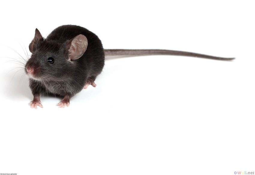
Risk Factors for Venous Thrombosis

Virchow’s Triad (1856)

  • Endothelial Injury

  • Venous Stasis

  • Hypercoagulable state

    Pregnancy, Malignancy, Surgery, Trauma, Age,
    Drugs, Obesity, Smoking, Hereditary
    Thrombophilias, Smoking, Inflammation.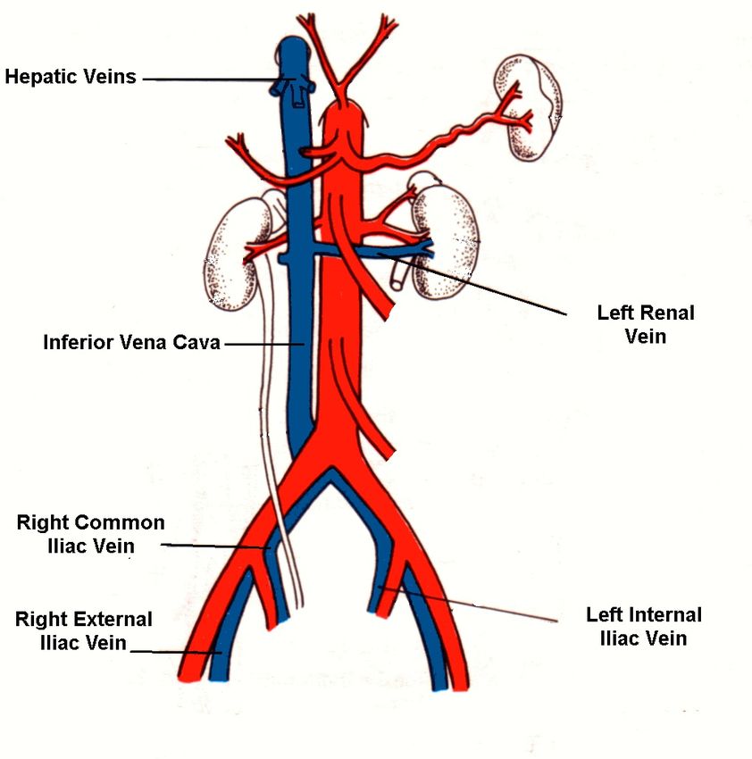
Inflammation and VTE:
       Increased Risk of VTE in autoimmune disease.

VTE Is a well-described complication of Crohn’s Disease and
Ulcerative Colitis

       Recent study of 13,756 patients with IBD, risk of VTE was
       increased compared to controls (HR 3.4, CI 2.7-4.3). The risk
       was amplified at time of disease flare (HR 8.4, CI 5.5-12.8).
       Lancet. 2010;375(9715):657.

Many studies have demonstrated a higher incidence of VTE in
patients with other systemic autoimmune diseases.


Patients with connective tissue diseases had an increased
incidence of VTE (IRR 2.3%) within 1 year post diagnosis compared
to controls. Risks were highest with SLE and JRA. J Thromb Haemost. 2012
May;10(5):815-21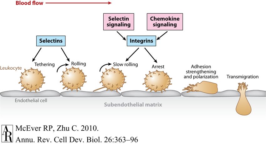
Inflammation and VTE:
               Risk of VTE and Acute Infection


Smeeth et al examined cases of first DVT (n= 7278) or first PE (n=3755)
for association with URI or UTI in patients from the UK Health Improvement
Network. Risk of VTE was increased proximal to both infections (IR= 2.1%
and 1.9% for UTI and URI respectively). Lancet. 2006 Apr 1;367(9516):1075-9

     Data from the Danish National Registry of Patients supported an
increased risk of VTE in patients recently diagnosed with an infection in both
        834 Tichelaar et al. Inflammatory diseases, infections and venous thrombosis
community of hospital settings. Respiratory tract, urinary tract, skin, intra-
abdominal and bacteremia were associated with at least two fold increased
VTE risk. J InternTable 2: Summary
                   Med. 2012          of risks of venous thrombosis.
                             Jun;271(6):608-18.

                             Discussion
                                                   Relative risk   Absolute risk 1
                                                                                       There is now convincin


              Inflammatory bowel disease          1.5 – 8.4     2.6 – 9.0           clinical epidemiologica
                 ANCA-associated vasculitis          n/a           43 – 70             thrombosis are related.


     Human immunodeficiency virus        1.2 – 1.4     1.3 – 5.8           when time between the
                 Pneumonia                           1.9 – 2.7     n/a                 when the inflammatory
                                                                                       cently or, more specific


     Urinary tract infections            1.8 – 2.1     n/a
                                                                                       disease was active (flare
                 Infections NOS                      1.7 – 2.5     n/a
                                                                                       of the range of the relati


              1 Cases per 1,000 person-years
                                                                                       which convincing evide
                                              Thromb Haemost. 2012 May 2;107(5):827-37.

bosis was found in this
Inflammation and VTE:
Biomarkers of Inflammation
•
JOURNAL OF VASCULAR SURGERY
702 Roumen-Klappe et al April 2002
VTEs are often
associated with A
elevated levels acute IL-6
phase reactants such
as C reactive
protein.
• Other inflammatory
biomarkers have B C
been associated with IL-8
CRP
VTE including IL-6,
IL-8, MCP-1, and
TNF- .
J Vasc Surg. 2002 Apr;35(4):701-6.
Fig 1. Plasma levels in control subjects and patients with deep vein thrombosis (DVT) of (A) interleukin-6 (IL-6), (B) interleukin-Inflammation, Leukocyte Adhesion, and Thrombosis Early endothelial inflammation is associated with the release of Weibel-Palade bodies which contain vWF and P-selectin. P-Selectin is required for venous clot formation in numerous animal models. P-selctin mediates leukocyte rolling through interaction with P- selectin glycoprotein ligand 1
Clinical, epidemiologic, pathologic, and experimental
evidence suggests a role for inflammation and innate
immune cells early in venous thrombosis.

Endothelial injury is rarely demonstrable in VTE (except
post-surgical).


What role does inflammation and the innate immune
system play in the initiation and propagation of venous
thrombosis in the absence of endothelial injury?
Inflammation

              ?
Stasis

            Hypoxia

    Venous Thrombosis
Published March 26, 2012
Article
Monocytes, neutrophils, and platelets
cooperate to initiate and propagate venous
thrombosis in mice in vivo
Marie-Luise von Brühl,1,4 Konstantin Stark,1,4 Alexander Steinhart,1,4
Sue Chandraratne,1,4 Ildiko Konrad,1,4 Michael Lorenz,1,4
Alexander Khandoga,1,4 Anca Tirniceriu,1,4 Raffaele Coletti,1,4
Maria Köllnberger,1,4 Robert A. Byrne,1,4 Iina Laitinen,2 Axel Walch,5
Alexander Brill,6 Susanne Pfeiler,7 Davit Manukyan,7 Siegmund Braun,1
Philipp Lange,8 Julia Riegger,1,4 Jerry Ware,9 Annekathrin Eckart,1,4
Selgai Haidari,1,4 Martina Rudelius,3 Christian Schulz,1,4,10
Katrin Echtler,1,4 Volker Brinkmann,11 Markus Schwaiger,2
Klaus T. Preissner,12 Denisa D. Wagner,6 Nigel Mackman,13
Downloaded from jem.rupre
Bernd Engelmann,7 and Steffen Massberg1,4
1Deutsches Herzzentrum and I. Medizinische Klinik, 2Nuklearmedizinische Klinik und Poliklinik, Klinikum rechts der Isar, and 3Institut für
Allgemeine Pathologie und Pathologische Anatomie, Technische Universität München (TUM), 80333 Munich, Germany
4Munich Heart Alliance, Munich, Germany
5Helmholtz Zentrum München, Deutsches Forschungszentrum für Umwelt und Gesundheit, Institut für Pathologie,
85764 Neuherberg, Germany
6Immune Disease Institute, Program in Cellular and Molecular Medicine, Children’s Hospital, Boston,
and Department of Pediatrics, Harvard Medical School, Boston, MA 02115
7Institut für Klinische Chemie and 8Medizinische Klinik I, Ludwig-Maximilians-Universität, 81377 Munich, Germany
9Study Objectives
1)
Develop a clinically relevant mouse model to
establish venous thrombosis resulting from ‘low flow’
state in the absence of endothelial damage.
II) Use model to investigate role of innate immune
response and inflammation early in thrombus
formation.Study Model Mice undergo ‘atraumatic’ laparotomy with placement of a permanent narrowing ligature on the IVC below left renal vein. Post-procedure, blood flow velocity is reduced ~80%. Genetic mouse models were used in conjunction with intravital 2 photon microscopy to interrogate cellular and molecular events in early clot formation.
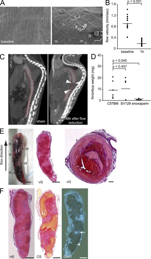
Fig 1: Model induces DVTs similar to those from
human patients

                                                  Figure 1. A
                                                  model of DV
                                                  images of the
                                                  (baseline) and
                                                  illustrate the
                                                  without endo
                                                  a representat
                                                  ment of blood
                                                  ligation (n = 1
                                                  ments; lines s
                                                  (C) Evaluation
                                                  tomography (
                                                  IVC (also see
                                                  sagittal proje
                                                  Right: animal
                                                  The dotted lin
                                                  cavity. Arrow
                                                  agent, indicat
                                                  animal with s
                                                  contrast fillin
                                                  of n = 3 expe
                                                  harvested IVC
                                                  C57BL/6 (n =
                                                  enoxaparin tr
                                                  vidual experim
                                                  group. (E) Ma
e v g o C t ( a ( e 1 n o a d s s c i ( i fl p t e c t
fl
Fig 2: Leukocytes are recruited early to ph
th
endothelial surface
Published March 26, 2012
20
n
an
C
Figure 2. Leukocytes are recruited during the early c
phase of venous thrombosis to the intact endothelial sur- m
face. (A) Leukocyte accumulation in DVT induced by 48 h of cy
flow restriction. vG (top left) and immunohistochemical stain- th
ings (middle and right, top and bottom) for Ly6G+ MPO+ neu- c
trophils and F4/80+ monocytes. Nuclei are counterstained with ac
DAPI. Bars, 50 µm. The bottom left shows the quantification of
D
neutrophils and monocytes. Results are mean ± SEM (n = 3).
(B) Scanning electron microscopic images taken directly after
partial IVC ligation showing the intact endothelium. After 6 h, L
a carpet composed of cell aggregates and fibrin can be visual- o
ized on the endothelial surface. Bars, 5 µm. (C) TEM images of S
venous vessels showing the anticoagulant endothelial cell n
lining (pseudocolored in yellow). Bar, 5 µm. Detail is shown in in
the right image. (D) Histological analyses of the IVC 6 h after u
flow reduction examining leukocyte recruitment in the early
tr
phase of venous thrombus formation. Histological sections in
three different stainings (HE, vG, and MSB). Bars: (top row) C
20 µm; (bottom row) 10 µm. Data are representative of th
P
Downloaded from jem.rupress.o
n = 3 experiments per group.
h
b
and Video 4). We then examined heterozygous ce
CX3CR1-eGFP mice to specifically define mono- m
cyte recruitment. This revealed that CX3CR1+ re
monocytes constitute Y15% of the recruited leuko- re
cytes (Fig. the
To dissect 3, D and F; subsets
leukocyte and Video 5). Together,
recruited in response to adopts a pr
these findings
depressed indicate
blood flow, we that
nextneutrophils
induced flowandrestriction
mono- in endothelial
cytes are the
LysM-eGFP predominant
reporter mice. Weleukocyte subsets
observed that >80%thatof all We then fo
actively
cells accumulate
recruited during theatfirst
the6 vascular surface
h were eGFP during
hi (Fig. 3 D). In P-selectin–
DVT development.What are the kinetics of leukocyte
recruitment by cell subset?
IVC Ligature
Imaging by
2 photon microscopy
Acradine orange (Leukocytes)
LysM-eGFP (Neutophils)
CX3CR1-eGFP (Monocytes)Fig 3: Neutrophils and Monocytes are the main
Published March 26, 2012
leukocyte subsets which accumulate early after DVT
Published March 26, 2012
induction.
Figure 3. Neutroph
are the
Figure 3. main leukocy
Neutrophils
arelating
the main during the ini
leukocyte
(A) Neutrophils
lating during the (green initiat
(A) vessel wall (red)
Neutrophils of the
(green) cr
induction
vessel wall (red) visualized
of the IVb
copy. Tracks
induction of individ
visualized by tw
shown
copy. Tracks in of
white (also se
individual
Shown
shown is a representa
in white (also see V
ments.
Shown is a(B) Time course
representative
endothelial
ments. (B) Timeinteractio
course of
flow restriction
endothelial interactionas eva w
flowmicroscopy
restrictioninasWT anim
evalua
orange (WT
microscopy in WT+ acr oran
animal
were(WT
orange taken before
+ acr orange)liga
wereweretakenused to evaluate
before ligation
wereCXused
3CR1-eGFP mice neu
to evaluate we
CX3ate monocytes.
CR1-eGFP mice Bars,
were 1
aterepresentative
monocytes. Bars, of n100 =
(C) Dynamics
representative ofofn= the5 ex
re
(C) subsets
Dynamics of of the recru
innate imm
subsets of innate
initiation immune
determined i
initiation determined
copy. Rolling and in vi
firm
copy. Rolling
kocytes areand firmasadn
given
kocytes are given
millimeter. as num
Results are
millimeter.
SEM (n =Results are sh
5 per group)microscopy in WTbefo
were taken an
orangewere(WTused
+ acr
toora
ev
wereCX taken before lig
3CR1-eGFP m
Fig 3: Neutrophils and Monocytes are the main wereateused
CX3CR1-eGFP
to evaluat
monocytes.
mice w
representative o
leukocyte subsets which accumulate early after DVT ate monocytes.
(C) Dynamics
representative
Bars
n=
of
subsets ofofinna
induction.
(C) Dynamics
initiationof the
determ
subsets of innate im
copy. Rolling an
initiation determined
kocytes are give
copy. Rolling and firm
millimeter. Resu
kocytes are given as
SEM (n = 5 per
millimeter. Results a
frequency of ne
SEM (n = 5 per grou
6 h of flow rest
frequency of neutrop
vital video micr
6 h of flow restrictio
SEM (n = 5 per
vital video microscop
tative images ta
SEM (n = 5 per grou
tativecence
imagesmicrosco
taken
cencerecruitment
microscopy6shh
IVC. Neutrophils
recruitment 6 h afte
mice. The numb
IVC. Neutrophils were
mice.wasThe assessed
number of u
In both strains
was assessed using C
(irrespective
In both strains of of
mi
by counterstain
(irrespective of their
Acridine orange
by counterstaining w
Bars,orange
Acridine 100 µm.(pse(E
Bars,LysM-eGFP
100 µm. (E)andFAC
IVC ligation
LysM-eGFP and CX3usi
Ly6G antibody.
IVC ligation using a n
Ly6Gmicroscopy
antibody. (F)ofInL
eGFP with
microscopy PE-la
of LysM-
leukocyte recruitment or thrombus formation (Fig. 4 F). Hence, only minimal (Published March 26, 2012
Fig 4: Leukocyte Adhesion Depends on Endothelial
Published March 26, 2012
P-Selectin
Figure 4.
Figure 4.
leukocyte
leukocyte
of traffickin
of flow
to trafficki
res
to flow
after DVTresi
aftershown
are DVT i
are RT-PCR
(B) shown
(B) RT-PCo
baseline
baseline
(n = 5 perog
(n = 5 per gd
standard
standard
immunohi
immunoh
endotheliu
endotheliu
ing P-selec
ing P-selec
surface. Nu
surface.
Bars, N
50 µm
Bars, 50ofµm
images
images
and SELPof
and SELP
Leukocytes
Leukocytes
orange and
orange an
microscop
microscop
arrows ind
arrows
Bars, indµ
100
Bars,
of firm100
leuµ
of firm leu
restriction.
number
restrictionpe
(n = 5) and
number peendothelium 48 h after DVT ind
Bars, 50 µm. (D, Left
ing P-selectin and vWF on the
images of adherent
surface. Nuclei are counterstai
µm. SELP
Bars, 50 and
/ mice 6
Fig 4: Leukocyte Adhesion Depends on Endothelial
images of
(D, Left) Represen
Leukocytes were sta
adherent leukocytes
and SELPorange and6 hvisualize
P-Selectin
/ mice after ind
microscopy
Leukocytes were stained (arrowhe
with A
orange and visualized
arrows indicateby intrav
sing
microscopy
Bars,(arrowhead
100 µm. (D, indicat
Rig
arrows indicate
of firm single adheren
leukocyte ad
Bars, 100 µm. (D, Right) Quant
restriction. Firm cell
of firm leukocyte adhesion, 6 h
number per square m
restriction. Firm cell adhesion i
number (n per=square SELP
5) andmillimeters /
as mean
(n = 5) and SELP/ ± (n
SEM.
= 7).(E,
Da
as meanimages
± SEM. of (E, the
Left)excise
Repre
images ofthrombus
the excised after
IVC48 h
inclu
thrombus mice.
afterBars,
48 h 1inmm.
C57BL/ (E
mice. Bars, 1 mm. (E,(n
in C57BL/6 Right)
= 8)Thr an
in C57BL/6
48 (n
h =after
8) andDVTSELP
/
induc
48 h after DVT induction.
individual experimen Dots
individual experiments; lines sh
of each group. (F) Hi
of each group. (F) Histological
harvested IVC throm
harvested IVC thrombi 48 h aft
tion
tion given as given
thrombus as thromb
load in
limeters limeters (n platelets.
(n = 5). plt, = 5). plt,D
as meanas mean ± SEM.
± SEM.
formation was significant
formation was si
compared with WT
compared anim
with W
ing theingmajor
the role
majorof mro
cell–derived TF for DVT d
cell–derived TF fo
(Fig. 5 D). The procoagu
(Fig.cells
of myeloid 5 D). Thefro
isolated
of mice
TFflox/flox myeloid cells is
is significanFig 5: Is Blood Cell TF Involved in DVT Formation?
Low-hTF-No mouse TF (Fig 5: Is Blood Cell TF Involved in DVT Formation?
Low-hTF
Bone Marrow
HCV
Chimera expressing
little TF on blood cellsAre Neutrophil Extracellular Traps
(NETs)involved in DVT induction?
Fuchs et al Neutrophil Extracellular Trap Impact on Deep Vein Thrombosis 3
Neutrophil NET formation is a process by which Neutrophils
actively extrude DNA associated complexes which include granular
proteins and anti-bacterial molecules.
NETs serve a putative innate anti-microbial immune function
NET formation has also been implicated in pathologic conditions
such as autoimmune disease, TRALI, and VTE.
Fuchs et al Neutrophil Extracell
Arterioscler
Figure 2. Neutrophil extracellular trap (NET) formation and function. A, Electron micrograph Thromb
of NETs with trapped SalmonellaVasc Biol. 2012
typhimurium; May 31Fig 6. NETs in DVT induction.
Published March 26, 2012
Figure 6. NET
(A) Leukocyte ac
flow restriction i
Figure 6. NETs treated with DVT
propagate con
(A) Leukocyte accumulation
Ly6G mAb to in vivo
dep
flow restriction in the IVC of LysM
aggregated
treated with control antibodyneutor th
ent cells.
Ly6G mAb to deplete Bars, 10
neutrophils.
aggregated neutrophils; arrows: si
48 h after DVT in
ent cells. Bars, 100 µm. (B) Thromb
Ly6G–treated
48 h after DVT induction WT
in isotype
Ly6G–treated WT mice (n =individ
represent 6 per g
represent individual experiments;
the mean of each thegroup.
mean(C)ofRepre
each
image of nof=intra
image of n = 3 experiments 3 ex
copy 3 or 48 h after flow reduction
copy 3 or 48 h a
Sytox Green+ NETs in the IVC. Bars
(D) VisualizationSytox Green
in vivoNE
+
of NETs by
microscopy. Ly6G-positive neutrop
(D) Visualization
FITC anti-Ly6G antibody) attached
microscopy.
sel wall (blue) release Ly6G
Sytox orange
FITC inside
(red) NET structures anti-Ly6G
the IVC a
flow reduction (also see Video 7). S
sel wall (blue) re
orange–positive nuclei correspond
(red)have
neutrophils, which NETnotstructu
(yet) e
their DNA to the extracellular
flow reduction spac
(a
head: extracellular DNA; arrow: ne
orange–positive
Bar, 50 µm. (E) Immunohistochem
ization of NETs neutrophils,
by staining forwhic
DNA
MPO, NE, and histones (H2A-H2B-
their DNA to the
the IVC of WT mice 48 h after indu
DVT. Hoechst+ DNAhead: extracellul
originating from
Bar, 50
neutrophils (arrows) µm.be(E)
could Im
dete
Arrows, nuclei; arrowheads, NET fi
Bars, 10 µm. (F)ization
Numberof of NETs
NETs in b
MPO, NE,inand
nic mice were quantified throm histheir DNA to the
head: extracellu
Bar, 50 µm. (E) I
Fig 6. NETs in DVT induction.
ization of NETs
MPO, NE, and h
the IVC of WT m
DVT. Hoechst+ DN
neutrophils (arr
Arrows, nuclei; a
Bars, 10 µm. (F)
nic mice were q
treatment with
antibody (n = 3
mean ± SEM. (G
thrombi stained
treatment. Arrow
Shown is a repr
ments. (H) After
bus weight (left
after 48 h of flo
individual exper
of each group. Q
shown (right) as
tained in WT inj
(n = 14) or DNa
of NETs at 48 h
animals (n = 4 p
revealing a typical NET morphology (Fig. 6 C and Video 6). whether NETs con- mean ± SEM.
Intravital 2-PIVM revealed that extracellular DNA originates centrate prothrom-
from Ly6G+ neutrophils (Fig. 6 D and Video 7). Extracel- botic factors on their
lular DNA was located in close proximity to neutrophils and surfaces. In fact, the
stained positive for the neutrophil granule proteins MPO, NETs were decorated with TF and
neutrophil elastase (NE), and the histone proteins H2A-H2B erase, an enzyme implicated in theFig 7. Platelets and Leukocytes Interact to Support
ed March 26, 2012
DVT Formation
AFig 7. Platelets and Leukocytes Interact to Support
DVT Formation
7. Platelet recruitment supports DVT formation in vivo. (A) Immunohistological cross sections of the IVC 48 h after DVT induction display p
+ch 26, 2012
Fig 7. Platelets Induce NET FormationSince Factor XII can be activated by negatively charged surfaces, do NETs activate factor XII in DVT formation?
Fig 7. NETs serve as Factor XII scaffold and
                              facilitate Factor XII activation.

                                                                                                                                                                                                                                                                  Downloaded from jem.rupress.org on June 15, 2012
                                                                                                                                                                                                               Downloaded from jem.rupress.org on June 15, 2012
                                                                                                                                                            Downloaded from jem.rupress.org on June 15, 2012
Figure
 Figure8.8. Platelets
             Platelets
         Figure
                       induce
                8. Platelets
                                NET
                                NET formation,
                        induceinduce formation, which
                                                which
                                     NET formation,
                                                        triggers
                                                       triggers
                                                    which
                                                                  FXIIa-dependent
                                                                FXIIa-dependent
                                                          triggers
                                                                                     thrombus
                                                                   FXIIa-dependent thrombus
                                                                                               propagation.
                                                                                             propagation.
                                                                                   thrombus propagation. (A)(A)
                                                                                                                (A) Freshly
                                                                                                                 Freshly
                                                                                                             Freshly
                                                                                                                             isolated
                                                                                                                         isolated
                                                                                                                     isolated human
                                                                                                                                      human
                                                                                                                                   human      neutrophils
                                                                                                                                          neutrophils
                                                                                                                                     neutrophils were were
                                                                                                                                                          were
Summary/ Interpretation Study provides novel insight into how restricted venous flow can initiate an inflammatory response during DVT induction. à Flow restriction upregulates several chemokines in vessel wall. Leukocytes are recruited early, predominately neutrophils. à Innate immune cells play an active role in thrombus formation both by the delivery of TF and NETs. Platelets enhance this process (in the absence of endothelial disruption). à Data suggests NET formation forms a prothrombotic scaffold for activation of factor XII.
Inflammation

              ?
Stasis

            Hypoxia

    Venous Thrombosis
Proposed Model of Innate Immune Cell
2
Involvement in Venous Thrombosis
Arterioscler Thromb Vasc Biol August 2012
2 Arterioscler Thromb Vasc Biol August 2012
2 Arterioscler Thromb Vasc Biol August 2012
A.
B.
C.
Figure 1.Figure 1. Neutrophil
Figure
Neutrophil extracellular
1. extracellular
Neutrophil traps traps
extracellular(NETs) (NETs)
traps thein the
in(NETs) time time
in theline lineline
time of deep
of deep
Arterioscler
of deep vein
vein thrombosis
vein thrombosis
Thromb
thrombosis (DVT):
(DVT):
(DVT): model.A,
a model.
a amodel.
Vasc Biol. A,DVT
A, DVTisis
2012
DVT initiated
is initiated
May
by
initiated local
byby local
31
hypoxia
hypoxia
local hypoxia
and activation
and activation of endothelial cellscells
of endothelial (ECs) as aasresult
(ECs) a resultof flow restriction/disturbances.
of flow restriction/disturbances. Activated
Activated endothelium
endotheliumreleases
releasesultralarge
ultralargevonvonWille-
Wille-
and activation
brand of endothelial
factor
brand (ULVWF)
factor
cells
and and
(ULVWF)
(ECs)
P-selectin as from
P-selectin
a result
from
of flow restriction/disturbances.
Weibel-Palade
Weibel-Palade bodies
bodies (WPB) which
(WPB) which
Activated
mediate
mediateplateletendothelium
and
platelet neutrophil
and
releases
neutrophiladhesion.
ultralarge
adhesion.Activated
vonplatelets
Wille-
Activatedplatelets
brand factor (ULVWF)
recruit tissue
recruit and
factor
tissue P-selectin
(TF)–containing
factor frommicroparticles
(TF)–containing Weibel-Palade
microparticles bodies
thatthat
enhance (WPB)
enhance thrombinwhich
thrombin mediateinplatelet
generation
generation thethe
in and thrombus.
growing
growing neutrophil
thrombus.B,adhesion.
ActivatedActivated
B,Activated platelets
plateletsandplatelets
and
recruit tissue factor (TF)–containing
endothelium
endothelium or other
or other stimulus microparticles
stimulus
induceinduce
NETNET that enhance
formation
formation in adherent thrombin
in adherent generation
neutrophils.
neutrophils. NETs
NETs inprovide
the growing
provide anan thrombus.
additional
additional B,
scaffold
scaffold Activated
for
for plateletand
platelet platelets
and and
redblood
red blood
endothelium or other
cell
cell (RBC) (RBC) stimulus
adhesion,
adhesion, induce
promote
promote NET
fibrin formation
fibrin formation,
formation, andinandadherent
exacerbate
exacerbate neutrophils.
platelet
platelet andandNETs provide
endothelial
endothelial an additional
activation.
activation. C,C, scaffold
Plasmin,
Plasmin, for platelet
a adisintegrin
disintegrin and red blood
andmetallopro-
and metallopro-
cell (RBC) adhesion,
teinase
teinase apromote
with with fibrin formation,
a thrombospondin
thrombospondin typetype 1 and
1 motif,motif,exacerbate
member
member 13 13 platelet
(ADAMTS13),
(ADAMTS13), and endothelial
andand activation.
deoxyribonuclease
deoxyribonuclease C, Plasmin,
(DNase)
(DNase) mediate
mediate athrombolysis
disintegrin
thrombolysisand by metallopro-
bydegrading
degrading
teinase with afibrin,
fibrin, ULVWF, ULVWF,and and
thrombospondin DNA, DNA,
type respectively. Monocytes/macrophages
1 motif,Monocytes/macrophages
respectively. member 13 (ADAMTS13), (MØ)(MØ)
and releaseanan additional
deoxyribonuclease
release additional source ofofDNase
(DNase)
source DNase
mediateandgenerate
and generateplasmin
thrombolysisplasmin and
byand
degrading
fibrin, ULVWF, promote
promote and restoration
restoration of blood
of blood flow.
DNA, respectively. flow.
Monocytes/macrophages (MØ) release an additional source of DNase and generate plasmin andQuestions for Further Investigation Neutrophils appear to be important for early venous thrombosis formation. What is the contribution of immune cell subsets to thrombolysis? Are the mechanisms described in this study clinically relevant to human patients with VTE? Do these mechanisms apply to different clinical scenarios such as clots that form in neutropenic patients? Are any of the mechanisms of clot initiation/ propagation described in the study hyperactive in patients with unidentified thrombophilias?
Questions for Further Investigation

Do these results suggest novel ways to intervene in
VTE?

  • DNAse?

  • Neutrophil Elastase (NE) and peptidylarginine
    deiminase 4 (PAD4)?

  • RAF-MEK-ERK pathway? (GW5074, U0126)
Questions?
Additional References:

Lancet. 2010;375(9715):657.

J Thromb Haemost. 2012 May;10(5):815-21

Lancet. 2006 Apr 1;367(9516):1075-9

J Intern Med. 2012 Jun;271(6):608-18.

Thromb Haemost. 2012 May 2;107(5):827-37.

J Vasc Surg. 2002 Apr;35(4):701-6.

J Cell Biol. 2010;191:677–691

J Cell Biol. 2009;184:205–213.

Nat Chem Biol 2011 Feb;7(2):75-7.

J Clin Invest. 2012 Jul 2;122(7):2661-71.

Blood. 2012 Jun 28;119(26):6335-43.
You can also read