A brainstem circuit for nausea suppression - Cell Press
←
→
Page content transcription
If your browser does not render page correctly, please read the page content below
Report

A brainstem circuit for nausea suppression
Graphical abstract                                                 Authors
                                                                   Chuchu Zhang, Lindsay K. Vincelette,
                                                                   Frank Reimann, Stephen D. Liberles

                                                                   Correspondence
                                                                   stephen_liberles@hms.harvard.edu

                                                                   In brief
                                                                   Nausea is an unpleasant sensation of
                                                                   visceral malaise that remains poorly
                                                                   understood at a molecular and cellular
                                                                   level. Zhang et al. uncover brainstem
                                                                   inhibitory neurons that suppress nausea-
                                                                   related behaviors through selective
                                                                   blockade of nausea-promoting, GDF15
                                                                   responsive-excitatory neurons. Nausea-
                                                                   suppressing neurons can be
                                                                   pharmacologically engaged by the gut
                                                                   hormone glucose insulinotropic peptide
                                                                   (GIP).

Highlights
d   A transcriptome-defined area postrema inhibitory neuron
    type expresses GIPR

d   GIPR neurons project locally and selectively inhibit nausea-
    related GFRAL neurons

d   GIP activates inhibitory neurons, blocks GFRAL neurons, and
    suppresses nausea

d   Ablating brainstem GIPR neurons removes anti-nausea
    effects of GIP

           Zhang et al., 2022, Cell Reports 39, 110953
           June 14, 2022 ª 2022 The Author(s).
           https://doi.org/10.1016/j.celrep.2022.110953                                          ll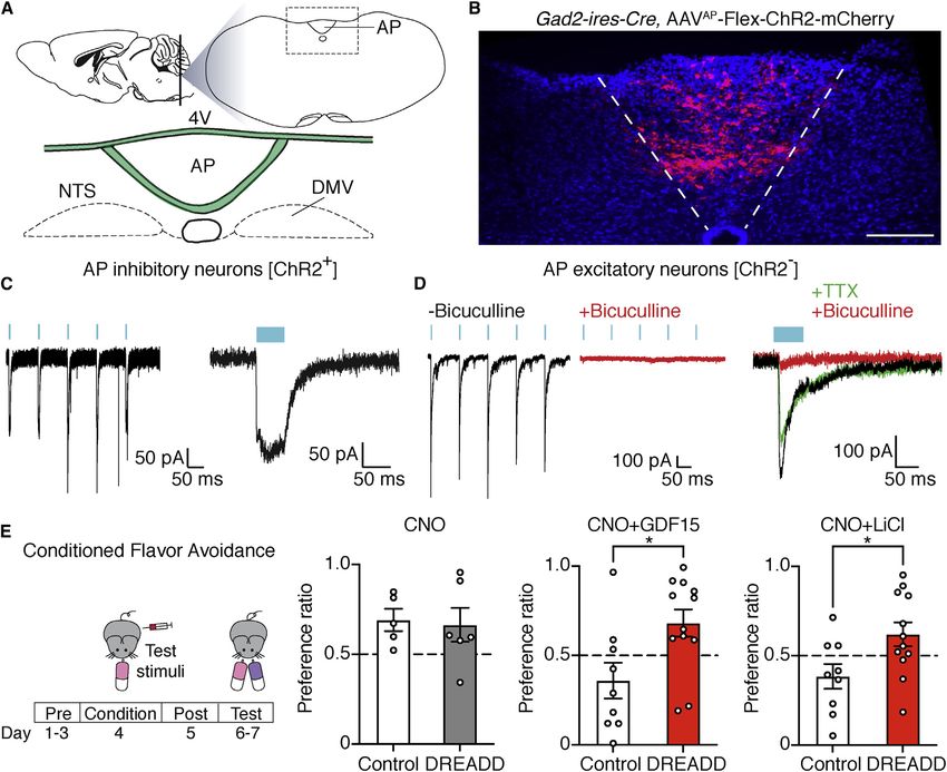
ll
                                                                                                                        OPEN ACCESS

Report
A brainstem circuit for nausea suppression
Chuchu Zhang,1 Lindsay K. Vincelette,1 Frank Reimann,2 and Stephen D. Liberles1,3,*
1Howard   Hughes Medical Institute, Department of Cell Biology, Harvard Medical School, Boston, MA 02115, USA
2Wellcome    Trust - MRC Institute of Metabolic Science, Metabolic Research Laboratories, Addenbrooke’s Hospital, Hills Road, Cambridge
CB2 0QQ, UK
3Lead contact

*Correspondence: stephen_liberles@hms.harvard.edu
 https://doi.org/10.1016/j.celrep.2022.110953

SUMMARY

Nausea is a discomforting sensation of gut malaise that remains a major clinical challenge. Several visceral
poisons induce nausea through the area postrema, a sensory circumventricular organ that detects blood-
borne factors. Here, we use genetic approaches based on an area postrema cell atlas to reveal inhibitory
neurons that counteract nausea-associated poison responses. The gut hormone glucose insulinotropic
peptide (GIP) activates area postrema inhibitory neurons that project locally and elicit inhibitory currents
in nausea-promoting excitatory neurons through g-aminobutyric acid (GABA) receptors. Moreover, GIP
blocks behavioral responses to poisons in wild-type mice, with protection eliminated by targeted area
postrema neuron ablation. These findings provide insights into the basic organization of nausea-associ-
ated brainstem circuits and reveal that area postrema inhibitory neurons are an effective pharmacological
target for nausea intervention.

INTRODUCTION                                                           and inhibitory pathways that lower their activity may reduce
                                                                       symptoms of gut malaise.
Nausea is one of the most encountered symptoms in healthcare,             The functions of area postrema inhibitory neurons are unclear.
with current anti-nausea medications displaying variable clinical      All three area postrema inhibitory neuron types (and none of the
success (Hesketh, 2008). New strategies for nausea intervention        excitatory neurons) are marked in Gad2-ires-Cre mice, and ge-
are needed and may be enabled by a mechanistic understanding           netic tracing previously revealed their projections to be local
of how the sensation of nausea arises. Classical studies involving     and largely confined to the area postrema, with minor projections
brain lesion and stimulation revealed a tiny brainstem structure       observed in the adjacent nucleus of the solitary tract (NTS) and
termed the area postrema that mediates nausea responses to             not in other brain regions that receive excitatory area postrema
several visceral threats (Borison, 1989). The area postrema is a       inputs (Zhang et al., 2021). Based on this observation, we hy-
sensory circumventricular organ with a privileged anatomical           pothesized that at least some inhibitory neuron types may sup-
location containing a reduced blood-brain barrier, which allows        press the activity and function of area postrema excitatory neu-
resident neurons to sample hormones and other chemicals in             rons, including those involved in nausea.
the circulatory system (Borison, 1989).
   Single-cell cDNA sequencing approaches recently provided a          RESULTS
cell atlas of the area postrema, revealing four excitatory and three
inhibitory neuron types (Zhang et al., 2021). One excitatory           Area postrema inhibitory neurons suppress local
neuron type expresses multiple receptors for nausea-inducing           excitatory neurons and poison responses
stimuli, including the growth/differentiation factor 15 (GDF15) re-    We used channelrhodopsin (ChR2)-assisted circuit mapping
ceptor (GFRAL), the glucagon-like peptide 1 (GLP1) receptor            (CRACM) to investigate the connectivity patterns of area postrema
(GLP1R), and the calcium-sensing receptor (CaSR). GFRAL,               inhibitory neurons. Gad2-ires-Cre, Rosa26-lsl-L10GFP mice were
GLP1R, and CaSR agonists cause nausea and/or vomiting in               injected in the area postrema with an AAV containing a Cre-depen-
large animals and in mice, which are incapable of vomiting, evoke      dent ChR2-mCherry allele (AAV-Flex-ChR2-mCherry); post hoc
a characteristic behavioral response termed conditioned flavor         histological analysis confirmed that viral infection was largely
avoidance in which paired administration of a poison and a novel       restricted to the area postrema (Figures 1A and 1B). Whole-cell re-
flavor causes future avoidance of that flavor (Andrews, 1992; Pa-      cordings in area postrema tissue slices revealed robust light-gated
tel et al., 2019; Zhang et al., 2021). Chemogenetic activation of      currents in mCherry-positive inhibitory neurons (Figure 1C). Post-
area postrema GFRAL neurons promotes flavor avoidance, while           synaptic responses were then measured in area postrema excit-
their ablation eliminates avoidance imposed by several visceral        atory neurons, which were identified by neuronal morphology
poisons (Sabatini et al., 2021; Zhang et al., 2021). Thus, area        and a lack of GFP fluorescence in Gad2-ires-Cre, Rosa26-lsl-
postrema GFRAL neurons are a key node in nausea circuits,              L10GFP mice. Optogenetic activation of area postrema inhibitory

                                                                         Cell Reports 39, 110953, June 14, 2022 ª 2022 The Author(s). 1
                                    This is an open access article under the CC BY license (http://creativecommons.org/licenses/by/4.0/).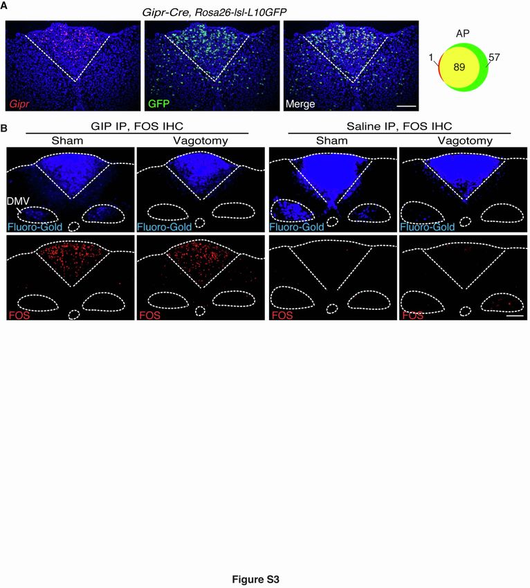
ll
      OPEN ACCESS                                                                                                                                        Report

Figure 1. Area postrema inhibitory neurons suppress local excitatory neurons and nausea-associated behavior
(A) Cartoon indicating area postrema (AP) location in the brainstem. 4V, fourth ventricle; DMV, dorsal motor nucleus of the vagus; NTS, nucleus of the solitary
tract.
(B) Native mCherry fluorescence in coronal area postrema cryosections of Gad2-ires-Cre mice with area-postrema-localized injection of AAV-Flex-ChR2-
mCherry. Scale bar: 100 mm.
(C) Whole-cell, voltage-clamp recordings of responses to light (blue bar) in mCherry-expressing area postrema neurons in brainstem slices from Gad2-ires-Cre;
AAV-Flex-ChR2-mCherry mice. Responses were observed in 5/5 mCherry-positive neurons from 4 mice.
(D) Light-induced postsynaptic currents were measured in GFP-negative area postrema neurons from Gad2-ires-Cre, Rosa26-lsl-L10GFP mice previously in-
jected in the area postrema with AAV-Flex-ChR2-mCherry. Whole-cell, voltage-clamp recordings of photostimulation-induced IPSCs were made at -60 mV using
a high-chloride intracellular solution to reveal a chloride conductance. Some recordings were made in the presence of bicuculline (10 mm, red) or tetrodotoxin
(TTX; 100 nM, green). Responses observed in 16/18 neurons, 5 mice.
(E) Gad2-ires-Cre mice were injected in the area postrema with (DREADD) or without (control) AAV-Flex-GsDREADD-mCherry. CNO was administered on the
flavor-conditioning day prior to LiCl (right), GDF15 (middle), or saline injection (left), and the effect on subsequent flavor avoidance was measured. n (left to right) =
5, 6, 9, 12, 9, and 12; mean ± SEM; circles: individual mice; Mann-Whitney test, *p < 0.05.

neurons produced large outward chloride currents (using high-                          consistent with a direct monosynaptic connection from inhibitory
chloride intracellular solution) in area postrema excitatory neurons                   neurons. Post-synaptic responses were also observed with
(resting membrane potential: -49.3 ± 1.1 mV; Figure S1A) that were                     reduced frequency in 27% (6/22) of area postrema inhibitory neu-
abolished by application of the GABAA receptor antagonist bicu-                        rons (mCherry-negative, GFP-positive neurons) and 38% (8/21) of
culline (Figure 1D). Light-evoked inhibitory post-synaptic currents                    excitatory neurons in adjacent NTS regions (Figures S1C and S1D).
(IPSCs) in most excitatory neurons (89%, 16/18) displayed onset                        Together, these findings indicate that most area postrema excit-
kinetics and insensitivity to tetrodotoxin (Figures 1D and S1B),                       atory neurons receive local inhibitory input, with area postrema

2 Cell Reports 39, 110953, June 14, 2022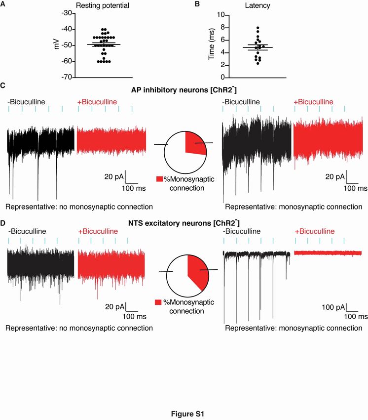
ll
Report                                                                                                                                  OPEN ACCESS

A

B                                                    C                                                              D

E

Figure 2. GIP activates area postrema inhibitory neurons with local projections
(A) Expression of Gipr was compared with Gad2 (top) and Glp1r (bottom). Uniform manifold approximation and projection (UMAP) plots (left) are derived from
published single-cell transcriptome data (Zhang et al., 2021). Two-color expression analysis (middle) was performed in coronal area postrema cryosections of
Gad2-ires-Cre; Rosa26-lsl-L10GFP (top) and Glp1r-ires-Cre; Rosa26-lsl-L10GFP (bottom), and involved fluorescent RNA in situ hybridization for Gipr (red) and
native GFP fluorescence (green). Scale bar: 100 mm. Co-labeled (yellow) or individually labeled (red, green) area postrema neurons were counted (right) from
images in middle.
(B) cAMP transients evoked by GIP (100 nM) and forskolin (25 mM) were measured in tdTomato-positive (GIPR+, red) and tdTomato-negative (GIPR-, black) area
postrema neurons cultured from Gipr-Cre; Rosa26-lsl-tdTomato mice.

                                                                                                                         (legend continued on next page)

                                                                                                           Cell Reports 39, 110953, June 14, 2022 3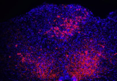
ll
      OPEN ACCESS                                                                                                                              Report

inhibitory neurons also forming functional connections with some                 GIP inhibits GFRAL neurons through a monosynaptic
NTS excitatory neurons and other area postrema inhibitory                        connection
neurons.                                                                         We focused on one subpopulation of area postrema inhibitory
   We activated area postrema inhibitory neurons using chemo-                    neurons that express the receptor (GIPR) for glucose insulino-
genetic approaches and observed the consequences on mouse                        tropic peptide (GIP). GIP is a gut-derived hormone and incretin
behavior. We injected Gad2-ires-Cre mice in the area postrema                    released upon nutrient intake that rapidly promotes insulin
with or without adeno-associated viruses (AAVs) containing Cre-                  release (Baggio and Drucker, 2007). Small molecules that acti-
dependent genes encoding designer Gas-coupled receptors                          vate the receptor for another incretin, GLP1, are clinical main-
(AAV-Flex-GsDREADD-mCherry) activated by the synthetic                           stays for diabetes treatment but induce nausea as an adverse
agonist clozapine-N-oxide (CNO) (Roth, 2016); Gas-coupled re-                    side effect through area postrema excitatory neurons (Drucker
ceptors were used based on commonly expressed area post-                         and Nauck, 2006; Zhang et al., 2021). Recent studies involving
rema receptors (see below). Chemogenetic activation of area                      paired administration of both incretins—GIP and GLP1—
postrema inhibitory neurons was validated by measuring CNO-                      observed that GIP suppressed some adverse behavioral re-
induced Fos expression in mCherry-labeled neurons (Fig-                          sponses to GLP1 (Borner et al., 2021); furthermore, GIPR ago-
ure S2A). Proper AAV targeting of the area postrema was                          nists reduced morphine- and cancer-drug-induced vomiting in
confirmed in every animal post hoc by blinded histological anal-                 ferrets (Asami et al., 2018). The neuronal basis for GIP responses
ysis of mCherry expression; we sometimes observed targeting of                   remains unclear, with several gut-brain communication routes
a few nearby NTS neurons (Figure S2B), and animals lacking                       possible.
labeled area postrema neurons were excluded from analysis                           We previously revealed by single-cell cDNA sequencing that
(see STAR Methods for more information).                                         Gipr is expressed in a subset of area postrema inhibitory neurons
   We used an established behavioral paradigm to measure                         (neuron cluster 6; Figure 2A) (Zhang et al., 2021). Since we
conditioned flavor avoidance (Patel et al., 2019; Zhang et al.,                  observed here that area postrema inhibitory neurons suppress
2021). Briefly, on a conditioning day, water-restricted mice                     poison responses, we hypothesized that cluster 6 neurons may
were given access to a novel flavored saccharin solution (either                 directly mediate or contribute to the anti-nausea effects of GIP
cherry or grape flavored) and then immediately injected with sa-                 and that targeting these neurons could represent a general strat-
line (control), poisons, and/or CNO. Subsequently, on a testing                  egy for nausea intervention. Moreover, GIPR expression pro-
day, behavioral preference for cherry- or grape-flavored solution                vides a selective molecular handle for both genetic and pharma-
was measured using a two-choice assay and expressed as a                         cological control of cluster 6 neurons. We obtained Gipr-Cre
preference index where the time drinking the conditioned flavor                  mice (Adriaenssens et al., 2019) and validated efficient targeting
was divided by total time drinking. In the absence of malaise in-                of cluster 6 area postrema neurons by two-color expression
duction, mice displayed a modest preference for the experi-                      analysis involving dual visualization of a Cre-driven fluorescent
enced flavor, with similar results observed for cherry and grape                 reporter and Gipr mRNA by in situ hybridization (Figure S3A);
(Zhang et al., 2021). In contrast, robust behavioral avoidance                   as is common with Cre tools, we noted some reporter-positive,
of the experienced flavor was evoked by various poisons (Zhang                   Gipr-negative cells, more so in the NTS than the area postrema,
et al., 2021), as well as the GFRAL agonist GDF15, as reported                   which may have resulted from Gipr expression that is transient or
previously (Patel et al., 2019). CNO-induced activation of area                  too low for detection by RNA in situ hybridization.
postrema inhibitory neurons did not evoke flavor aversion or                        We asked whether GIP evoked responses in area postrema
attraction in this paradigm in the absence of poison induction                   cluster 6 neurons marked in Gipr-Cre, R26-lsl-tdTomato mice;
(Figure 1E). Strikingly, however, when chemogenetic activation                   as a note, we used [D-Ala2]-GIP as a stable GIP analog in all ex-
of area postrema inhibitory neurons was paired with GDF15 or                     periments. GIPR is a Gas-coupled receptor, and we observed
lithium chloride injection, imposed aversion was lost (Figure 1E).               GIP-evoked cAMP transients in dissociated GIPR neurons
CNO silenced GDF15 and lithium chloride responses in mice                        (71.4%, 15/21 cells) using the genetically encoded fluorescent
with DREADD expression in inhibitory neurons but had no effect                   cAMP sensor cADDis (Figure 2B) (Tewson et al., 2016).
in control mice lacking DREADD expression, either in the pres-                   Increasing cAMP levels depolarizes some, but not all, neurons,
ence or absence of poisons. Together with anatomical and con-                    so we asked whether GIP also evokes electrical responses in
nectivity-mapping studies, these findings indicate that at least                 cluster 6 neurons. Whole-cell, patch-clamp analysis in area post-
some area postrema inhibitory neurons project to and inhibit                     rema slices revealed robust GIP-evoked depolarization and ac-
the activity and function of nausea-promoting excitatory                         tion potentials in tdTomato-positive neurons (9/17 cells) from
neurons.                                                                         Gipr-Cre, R26-lsl-tdTomato mice (Figures 2C and 2D). For

(C) Whole-cell, current-clamp responses of tdTomato-positive (GIPR+, red) and tdTomato-negative (GIPR-, black) area postrema neurons in brainstem slices
from Gipr-Cre; Rosa26-lsl-tdTomato mice to GIP (100 nM) with ionotropic glutamate receptor (iGluR) antagonists CNQX (10 mM) and D-AP5 (50 mM) or GABAA
receptor antagonist bicuculline (bic, 10 mM).
(D) GIP-evoked changes in membrane potential in tdTomato-positive (left, GIPR+, red) or tdTomato-negative (right, GIPR-, black) neurons from recordings in (C).
n= 17 (left) and 74 (right); circles: individual data points.
(E) AAV-Flex-GFP and AAV-Flex-Syn-mCherry were injected into the area postrema of Gipr-Cre (top) and Glp1r-ires-Cre (bottom) mice. GFP-positive neurons
and mCherry-positive synaptic terminals were visualized by immunohistochemistry. A GFP fluorescence intensity was used, which enables visualization of
neuronal cell bodies but not axons. VLM, ventrolateral medulla; PBN, parabrachial nucleus; scp, superior cerebellar peduncle. Scale bar: 100 mm.

4 Cell Reports 39, 110953, June 14, 2022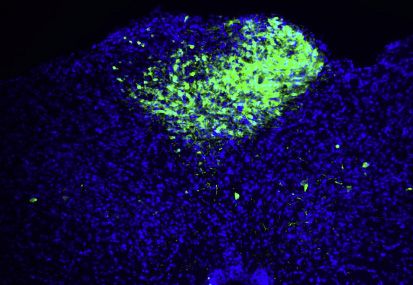
ll
Report                                                                                                                   OPEN ACCESS

comparison, GIP rarely depolarized (2/74 cells) or evoked cAMP         circuits may provide an effective way to attenuate poison re-
transients (3/29 cells) in other area postrema neurons lacking         sponses. Pharmacological access to cluster 6 neurons is facili-
tdTomato expression; instead, hyperpolarization of some tdTo-          tated as the area postrema contains a reduced blood-brain bar-
mato-negative neurons was observed (23%, 17/74 neurons).               rier and local neurons can directly detect circulating peptides like
Intraperitoneal (i.p.) injection of GIP also induced Fos expression    GIP. We tested whether GIP could suppress nausea-related be-
in area postrema neurons, with effects persisting after vagot-         haviors and whether GIP-evoked behavioral responses required
omy, suggesting that vagal inputs were not required for area           area postrema inhibitory circuits. Using the conditioned-flavor-
postrema responses to GIP (Figure S3B). Taken together, GIP            avoidance paradigm described above, wild-type mice were
evokes characteristic and direct responses in GIPR-expressing          administered either GIP or saline on the conditioning day and
area postrema inhibitory neurons that include increased cAMP           20 min later were exposed to GDF15 or LiCl. GDF15 and LiCl
levels, depolarization, and cell firing.                               evoked robust flavor-avoidance responses in saline-injected
   We used genetic approaches in Gipr-Cre mice to map the anat-        control mice, but responses were abolished in mice receiving a
omy of area postrema cluster 6 neurons and, for comparison, used       prophylactic GIP injection (Figure 4A).
Glp1r-ires-Cre mice to map area postrema excitatory neuron clus-          We asked whether area postrema cluster 6 neurons were
ters 2–4 (Figure 2E) (Williams et al., 2016; Zhang et al., 2021). We   required for the suppression of nausea-related behavior by
injected AAV-Flex-GFP and AAV-Flex-Synaptophysin-mCherry               GIP. We used a genetic approach involving diphtheria toxin
into the area postrema to label neuronal cell bodies and synaptic      (DT) to ablate Cre-expressing GIPR neurons in the area post-
terminals, respectively. As we observed previously, area postrema      rema. Mouse cells are DT resistant but can be made susceptible
excitatory neurons project to multiple brain regions including the     by Cre-guided expression of the DT receptor (DTR) (Buch et al.,
NTS, parabrachial nucleus (PBN), and autonomic motor nuclei            2005). DT was injected into the area postrema of Gipr-Cre;
(Zhang et al., 2021). In contrast, we observed that area postrema      Rosa26-lsl-DTR mice (Gipr-ABLATEAP mice) or of Cre-negative
GIPR neurons, like inhibitory neurons in bulk, displayed dense ar-     Rosa26-lsl-DTR mice (non-ABLATE mice) as a control. Gipr-
borizations within the area postrema and minor projections to          ABLATEAP mice displayed near-complete loss of cluster 6 neu-
proximal NTS, but GIPR neuron projections were not observed            rons, but not other area postrema neuron types, and some
in the PBN or autonomic motor nuclei. These findings raised the        loss of GIPR neurons in the NTS but not loss of GIPR neurons
possibility that GIPR neurons form inhibitory contacts with some       in other brain regions such as the hypothalamus (Figures 4B
or all area postrema excitatory neurons.                               and 4C). Behavioral responses of Gipr-ABLATEAP mice and
   We asked whether area postrema cluster 6 neurons evoked             non-ABLATE mice were examined using the conditioned-fla-
inhibitory postsynaptic currents in particular classes of area         vor-avoidance paradigm. We observed that GIP protected
postrema excitatory neurons. Various excitatory neuron types           against GDF15-conditioned flavor avoidance in non-ABLATE
were marked in Calcr-ires-Cre; Rosa26-lsl-tdTomato (cluster            mice but no longer suppressed GDF15 responses in Gipr-ABLA-
1), Slc6a2-p2a-Cre; Rosa26-lsl-tdTomato (cluster 2), Agtr1a-           TEAP mice (Figure 4D). Thus, GIP suppresses nausea-associated
t2a-Cre; Rosa26-lsl-tdTomato (cluster 3), Gfral-p2a-Cre;               behavior through area postrema inhibitory neurons.
Rosa26-lsl-tdTomato (cluster 4), and Glp1r-ires-Cre; Rosa26-
lsl-tdTomato (clusters 2–4) mice. We obtained area postrema            DISCUSSION
tissue slices from mice of each genotype and examined GIP-
evoked responses by whole-cell, patch-clamp electrophysi-              Nausea evoked by visceral poisons can be counterproductive,
ology in the presence of ionotropic glutamate receptor antago-         causing patients to forego life-saving medications for cancer,
nists (D-AP5, CNQX) to block excitatory transmission (Figure 3).       diabetes, and other diseases. Studies here establish area post-
GIP induced acute hyperpolarization (-6.4 ± 0.9 mV) of GFRAL-          rema inhibitory neurons (cluster 6, GIPR neurons) as a target
expressing area postrema neurons (8/13 cells) but failed to hy-        for suppressing behavioral responses to at least some nausea-
perpolarize most other excitatory neurons (SLC6A2: 2/15 cells;         inducing toxins (Figure 4E). GIP is a gut hormone and incretin
AGTR1A: 1/12 cells, CALCR: 1/14 cells). GIP hyperpolarized             that activates GIPR neurons, and GIPR neurons in turn suppress
20% (4/20) of GLP1R neurons, consistent with GLP1R expres-             the activity of nearby GFRAL-expressing and nausea-promoting
sion in multiple neuron types with differing GIP sensitivity. The      excitatory neurons. In addition to GIPR, cluster 6 neurons ex-
GABAA receptor antagonist bicuculline did not impact GIP-              press other cell surface receptors (Zhang et al., 2021), including
evoked depolarization of GIPR neurons but did eliminate GIP-           mu-opioid receptor and neuropeptide Y receptor 2; it is possible
evoked hyperpolarization of GFRAL neurons. Taken together,             that activating these receptors stimulates or inhibits GIPR
these findings indicate that GIP directly stimulates area post-        neurons, with other stimulating agonists potentially providing
rema cluster 6 neurons, which in turn form local and selective         additional avenues for pharmacological modulation of nausea-
GABAergic inhibitory connections with area postrema GFRAL              related behaviors with variable associated side effects. Further-
neurons (cluster 4).                                                   more, the strategy of targeting inhibitory circuits will likely be
                                                                       applicable to other area postrema neurons, including SLC6A2
GIP suppresses nausea behaviors through area                           neurons, which also condition flavor avoidance (Zhang et al.,
postrema inhibitory circuits                                           2021) and receive distinct local inhibitory input from GAD2-pos-
GFRAL neurons mediate nausea-associated responses to                   itive, GIPR-negative neurons. Other inhibitory neurons (cluster 5)
several visceral threats (Zhang et al., 2021), so we reasoned          express the ghrelin receptor (Zhang et al., 2021), raising the pos-
that inhibiting GFRAL neurons through local area postrema              sibility that several gut hormones act in concert to toggle the

                                                                                               Cell Reports 39, 110953, June 14, 2022 5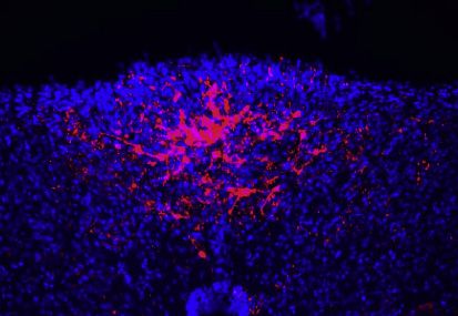
ll
      OPEN ACCESS                                                                                                                             Report

A

B

C

D

Figure 3. GIPR neurons selectively inhibit area postrema GFRAL neurons
(A) UMAP plots derived from published single-cell transcriptome data (Zhang et al., 2021) indicate expression of marker genes in different area postrema
excitatory neurons.
(B) Whole-cell, current-clamp responses to GIP (100 nM) of tdTomato-positive area postrema neurons in brainstem slices from either (1) Calcr-ires-Cre; Rosa26-
lsl-tdTomato, (2) Slc6a2-p2a-Cre; Rosa26-lsl-tdTomato, (3) Agtr1a-t2a-Cre; Rosa26-lsl-tdTomato, (4) Gfral-p2a-Cre; Rosa26-lsl-tdTomato, or (5) Glp1r-ires-Cre;
Rosa26-lsl-tdTomato mice. Recordings were done with either iGluR antagonists (red) CNQX (10 mM) and D-AP5 (50 mM) or GABAA receptor antagonist bic (10 mM,
blue).
(C) Maximal GIP-evoked membrane potential change with (blue) or without (red) bic from individual neurons (circles) from (B).
(D) Frequency of neurons showing GIP-induced hyperpolarization from area postrema and NTS in mice from (B).

balance of area postrema inhibition and excitation. GIP is                      consummatory decisions based on need, reward value, and
released following nutrient-rich meals (Baggio and Drucker,                     toxin risk. Together, experiments here reveal that GIP sup-
2007), and it may be beneficial under certain physiological con-                presses poison responses through a dedicated neuronal
ditions for animals to consume calorie-rich foods containing                    pathway, key insights into area postrema circuit organization,
small amounts of harmful chemicals. Area postrema circuits                      and a potential strategy for nausea intervention that involves tar-
that integrate reward and punishment signals could guide future                 geting of brainstem inhibitory neurons.

6 Cell Reports 39, 110953, June 14, 2022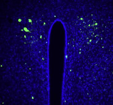
ll
Report                                                                                                                                     OPEN ACCESS

        A                                                                         B

        C

        D                                                                E

Figure 4. GIP suppresses nausea-associated behavior through area postrema inhibitory circuits
(A) Flavor avoidance conditioned by GDF15 (left) or LiCl (right) in wild-type mice was measured with prophylactic injection of GIP (374 mg/mL, blue) or saline
(white). n = 7, 8 (left) and 7, 7 (right); mean ± SEM; circles: individual data points; Mann-Whitney test, **p < 0.01.
(B and C) The area postrema of Gipr-Cre; Rosa26-lsl-DTR mice was injected with DT (+DT) or saline (-DT). Immunohistochemistry for DTR (B) and quantification of
DTR-expressing cells (C) in coronal brain cryosections containing area postrema, paraventricular hypothalamus (PVH), and arcuate nucleus (ARC). Scale bar:
100 mm; n = 4–46 sections from 4 mice (-DT) or 12 mice (+DT); mean ± SEM; circles: individual data points; ****p < 0.0001.
(D) GDF15-induced flavor avoidance was measured with prophylactic GIP injection (374 mg/mL) in non-ABLATE and Gipr-ABLATEAP mice. n = 16, 12; mean ±
SEM; circles: individual data points; *p < 0.05, Mann-Whitney test.
(E) Cartoon depicting the mechanism of GIP action involving inhibitory GIPR neurons and excitatory GFRAL neurons in the area postrema.

Limitations of the study                                                         arrive at the brainstem to stimulate area-postrema-localized
GIP directly stimulates cluster 6 neurons in slice preparations                  GIPR. Area postrema GIPR neurons are required for GIP re-
(Figure 2B), and peripheral GIP injection induces area postrema                  sponses reported here (Figure 4D), yet a less parsimonious
Fos expression (Figure S3B), but it has not been demonstrated                    model is that GIP acts through an upstream neuronal route to
that a sufficient concentration of meal-induced GIP will naturally               engage area postrema GIPR neurons indirectly. GIP does not

                                                                                                             Cell Reports 39, 110953, June 14, 2022 7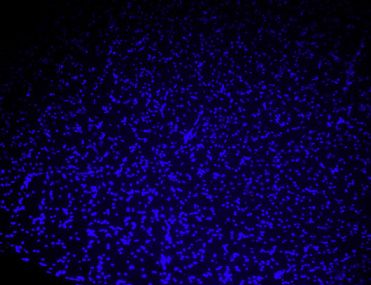
ll
        OPEN ACCESS                                                                                                                                    Report

activate GIPR-negative area postrema neurons (Figures 2D and                         Andrews, P.L. (1992). Physiology of nausea and vomiting. Br. J. Anaesth. 69,
3), and while the area postrema receives predominant input from                      2S–19S. https://doi.org/10.1093/bja/69.supplement_1.2s.
the vagus nerve, area postrema GIP responses persist in vago-                        Asami, T., Nishizawa, N., Niida, A., Kanematsu, Y., Adachi, M., Takekawa, S.,
tomized mice (Figure S3B). Additional studies are needed to un-                      and Morimoto, T. (2018). Patent Application WO2018181864A1 (Takeda Phar-
                                                                                     maceutical Company Limited).
derstand when GIPR neurons are naturally engaged and whether
they are activated by meal consumption, vagal inputs, top-down                       Baggio, L.L., and Drucker, D.J. (2007). Biology of incretins: GLP-1 and GIP.
                                                                                     Gastroenterology 132, 2131–2157. https://doi.org/10.1053/j.gastro.2007.03.
inputs, and/or other stimuli.
                                                                                     054.
                                                                                     Borison, H.L. (1989). Area postrema: chemoreceptor circumventricular organ
STAR+METHODS
                                                                                     of the medulla oblongata. Prog. Neurobiol. 32, 351–390. https://doi.org/10.
                                                                                     1016/0301-0082(89)90028-2.
Detailed methods are provided in the online version of this paper
                                                                                     Borner, T., Geisler, C.E., Fortin, S.M., Cosgrove, R., Alsina-Fernandez, J.,
and include the following:                                                           Dogra, M., Doebley, S., Sanchez-Navarro, M.J., Leon, R.M., Gaisinsky, J.,
                                                                                     et al. (2021). GIP receptor agonism attenuates GLP-1 receptor agonist-
    d   KEY RESOURCES TABLE                                                          induced nausea and emesis in preclinical models. Diabetes 70, 2545–2553.
    d   RESOURCE AVAILABILITY                                                        https://doi.org/10.2337/db21-0459.
        B Lead contact                                                               Buch, T., Heppner, F.L., Tertilt, C., Heinen, T.J.A.J., Kremer, M., Wunderlich,
        B Materials availability                                                     F.T., Jung, S., and Waisman, A. (2005). A Cre-inducible diphtheria toxin recep-
        B Data and code availability                                                 tor mediates cell lineage ablation after toxin administration. Nat. Methods 2,
    d   EXPERIMENTAL MODEL AND SUBJECT DETAILS                                       419–426. https://doi.org/10.1038/nmeth762.
    d   METHOD DETAILS                                                               Chang, R.B., Strochlic, D.E., Williams, E.K., Umans, B.D., and Liberles, S.D.
        B Electrophysiology and circuit mapping                                      (2015). Vagal sensory neuron subtypes that differentially control breathing.
        B cAMP imaging of area postrema neurons                                      Cell 161, 622–633. https://doi.org/10.1016/j.cell.2015.03.022.
        B AAV/DT injections                                                          Choi, H.M.T., Schwarzkopf, M., Fornace, M.E., Acharya, A., Artavanis, G.,
        B Analysis of RNA, protein, and reporter expression                          Stegmaier, J., Cunha, A., and Pierce, N.A. (2018). Third-generation. Develop-
                                                                                     ment 145, dev.165753. https://doi.org/10.1242/dev.165753.
        B Behavioral assays
    d   QUANTIFICATION AND STATISTICAL ANALYSIS                                      Choi, H.M.T., Schwarzkopf, M., and Pierce, N.A. (2020). Multiplexed quantita-
                                                                                     tive in situ hybridization with subcellular or single-molecule resolution within
                                                                                     whole-mount vertebrate embryos: qHCR and dHCR imaging (v3.0). Methods
SUPPLEMENTAL INFORMATION
                                                                                     Mol. Biol. 2148, 159–178. https://doi.org/10.1007/978-1-0716-0623-0_10.

Supplemental information can be found online at https://doi.org/10.1016/j.           Drucker, D.J., and Nauck, M.A. (2006). The incretin system: glucagon-like
celrep.2022.110953.                                                                  peptide-1 receptor agonists and dipeptidyl peptidase-4 inhibitors in type 2 dia-
                                                                                     betes. Lancet 368, 1696–1705. https://doi.org/10.1016/s0140-6736(06)
ACKNOWLEDGMENTS                                                                      69705-5.
                                                                                     Hesketh, P.J. (2008). Chemotherapy-induced nausea and vomiting. N. Engl. J.
We thank Brad Lowell, David Ginty, Chen Ran, and Zhikai Liu for manuscript           Med. 358, 2482–2494. https://doi.org/10.1056/nejmra0706547.
comments. The work was funded by NIH grants to S.D.L. (RO1 NS122767,                 Oh, S.W., Harris, J.A., Ng, L., Winslow, B., Cain, N., Mihalas, S., Wang, Q., Lau,
DP1 AT009497, and R01 DK103703). Gipr-Cre mice were generated with fund-             C., Kuan, L., Henry, A.M., et al. (2014). A mesoscale connectome of the mouse
ing from MRC-UK (MRC_MC_UU_12012/3) and the Wellcome Trust (106263/                  brain. Nature 508, 207–214. https://doi.org/10.1038/nature13186.
Z/14/Z). C.Z. is a fellow of the Damon Runyon Cancer Research Foundation.
                                                                                     Pan, W., Adams, J.M., Allison, M.B., Patterson, C., Flak, J.N., Jones, J., Stroh-
S.D.L. is an investigator of the Howard Hughes Medical Institute.
                                                                                     behn, G., Trevaskis, J., Rhodes, C.J., Olson, D.P., and Myers, M.G., Jr. (2018).
                                                                                     Essential role for hypothalamic calcitonin ReceptorExpressing neurons in the
AUTHOR CONTRIBUTIONS
                                                                                     control of food intake by leptin. Endocrinology 159, 1860–1872. https://doi.
                                                                                     org/10.1210/en.2017-03259.
C.Z. and S.D.L. designed the study, analyzed data, and wrote the manuscript;
F.R. generated Gipr-Cre mice; C.Z. performed all experiments, except some            Patel, S., Alvarez-Guaita, A., Melvin, A., Rimmington, D., Dattilo, A., Miedzy-
immunohistochemistry performed by L.K.V.                                             brodzka, E.L., Cimino, I., Maurin, A.C., Roberts, G.P., Meek, C.L., et al.
                                                                                     (2019). GDF15 provides an endocrine signal of nutritional stress in mice and
DECLARATION OF INTERESTS                                                             humans. Cell Metab. 29, 707–718.e8. https://doi.org/10.1016/j.cmet.2018.
                                                                                     12.016.
S.D.L. is a consultant for Kallyope, Inc.                                            Roth, B.L. (2016). DREADDs for neuroscientists. Neuron 89, 683–694. https://
                                                                                     doi.org/10.1016/j.neuron.2016.01.040.
Received: January 12, 2022                                                           Sabatini, P.V., Frikke-Schmidt, H., Arthurs, J., Gordian, D., Patel, A., Rupp,
Revised: April 8, 2022                                                               A.C., Adams, J.M., Wang, J., Beck Jorgensen, S., Olson, D.P., et al. (2021).
Accepted: May 23, 2022                                                               GFRAL-expressing neurons suppress food intake via aversive pathways.
Published: June 14, 2022                                                             Proc. Natl. Acad. Sci. U. S. A. 118, e2021357118. https://doi.org/10.1073/
                                                                                     pnas.2021357118.
REFERENCES
                                                                                     Schindelin, J., Arganda-Carreras, I., Frise, E., Kaynig, V., Longair, M., Pietzsch,
Adriaenssens, A.E., Biggs, E.K., Darwish, T., Tadross, J., Sukthankar, T., Gir-      T., Preibisch, S., Rueden, C., Saalfeld, S., Schmid, B., et al. (2012). Fiji: an
ish, M., Polex-Wolf, J., Lam, B.Y., Zvetkova, I., Pan, W., et al. (2019). Glucose-   open-source platform for biological-image analysis. Nat. Methods 9,
dependent insulinotropic polypeptide receptor-expressing cells in the hypo-          676–682. https://doi.org/10.1038/nmeth.2019.
thalamus regulate food intake. Cell Metab. 30, 987–996.e6. https://doi.org/          Slotnick, B. (2009). A simple 2-transistor touch or lick detector circuit. J. Exp.
10.1016/j.cmet.2019.07.013.                                                          Anal. Behav. 91, 253–255. https://doi.org/10.1901/jeab.2009.91-253.

8 Cell Reports 39, 110953, June 14, 2022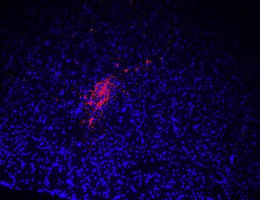
ll
Report                                                                                                                                  OPEN ACCESS

Tewson, P.H., Martinka, S., Shaner, N.C., Hughes, T.E., and Quinn, A.M.        the digestive system. Cell 166, 209–221. https://doi.org/10.1016/j.cell.2016.
(2016). New DAG and cAMP sensors optimized for live-cell assays in auto-       05.011.
mated laboratories. J. Biomol. Screen 21, 298–305. https://doi.org/10.1177/
1087057115618608.                                                              Zhang, C., Kaye, J.A., Cai, Z., Wang, Y., Prescott, S.L., and Liberles, S.D.
Williams, E.K., Chang, R.B., Strochlic, D.E., Umans, B.D., Lowell, B.B., and   (2021). Area postrema cell types that mediate nausea-associated behaviors.
Liberles, S.D. (2016). Sensory neurons that detect stretch and nutrients in    Neuron 109, 461–472.e5. https://doi.org/10.1016/j.neuron.2020.11.010.

                                                                                                           Cell Reports 39, 110953, June 14, 2022 9
ll
OPEN ACCESS Report
STAR+METHODS
KEY RESOURCES TABLE
REAGENT or RESOURCE SOURCE IDENTIFIER
Antibodies
Rabbit-anti-RFP Rockland Cat# 600-401-379; RRID:AB_2209751
Goat-anti-HB-EGF (anti-DTR) R&D Systems Cat# AF-259-NA, RRID:AB_354429
Rabbit-anti-Fos Synaptic Systems Cat# 226 003, RRID:AB_2231974
Chick-anti-GFP Aves Labs Cat# GFP-1020, RRID:AB_10000240
Bacterial and virus strains
AAV9-pCAG-Flex-EGFP Oh et al., 2014 Addgene, Cat #51502-AAV9
AAV9-Ef1a-DIO-hChR2(H134R)- Gift from Karl Deisseroth Addgene, Cat #20297-AAV9
mCherry
pAAV-hSyn-DIO-rM3D (Gas)- Gift from Bryan Roth Addgene, Cat #50458
mCherry
AAV8.2-hEF1a-DIO-synaptophysin- MGH Viral Core Cat #AAV-RN1
mCherry
Green Up cADDis cAMP sensor Montana Molecular Cat #U0200G
Chemicals, peptides, and recombinant proteins
[D-Ala2]-GIP Tocris Cat #6699
Clozapine N-oxide dihydrochloride Tocris Cat #6329
Bicuculline methiodide Tocris Cat #2503
Cyanquixaline Tocris Cat #1045
Forskolin Tocris Cat #1099
Diphtheria Toxin from Corynebacterium Sigma-Aldrich Cat #D0564
diphtheriae
D-AP5 Tocris Cat #0106
GDF15 R&D systems Cat #957-GD-025/CF
Tetrodotoxin Tocris Cat #1078
Gipr B3 probe Molecular Instruments Lot #PRL060
FluoroGold Santa Cruz Biotechnology Cat #sc-358883
Critical commercial assays
Papain dissociation system Worthington Biochemical Cat # LK003150
Deposited data
Raw images This study Mendeley data https://doi.org/10.17632/
h3fztb3ynh.1
Experimental models: Organisms/strains
Glp1r-ires-Cre Chang et al., 2015; Williams Deposited in Jackson laboratory
et al.,2016 (Cat#029282)
Calcr-ires-Cre Pan et al., 2018 N/A
Gipr-Cre Adriaenssens et al., 2019 N/A
Wild-type C57BL/6J Jackson Laboratory Cat #000664
Rosa26-LSL-L10GFP Jackson Laboratory Cat #024750
Agtr1a-t2a-Cre Jackson Laboratory Cat #031487
Rosa26-LSL-tdTomato Jackson Laboratory Cat #007908
Gad2-ires-Cre Jackson Laboratory Cat #010802
Rosa26-LSL-DTR Jackson Laboratory Cat #007900
Slc6a2-p2a-Cre Zhang et al., 2021 N/A
Gfral-p2a-Cre Zhang et al., 2021 N/A
(Continued on next page)
e1 Cell Reports 39, 110953, June 14, 2022ll
Report OPEN ACCESS
Continued
REAGENT or RESOURCE SOURCE IDENTIFIER
Software and algorithms
MATLAB R2019a MathWorks RRID: SCR_001622 https://www.mathworks.com/
products/matlab.html
PRISM 9 GraphPad RRID: SCR_002798 https://www.graphpad.com/
scientific-software/prism/
FIJI Schindelin et al., 2012 RRID:SCR_002285 https://fiji.sc
pClamp 10.2 Axon Instruments RRID: SCR_011323 https://www.moleculardevices.
com/products/software/pclamp.html
BioRender BioRender.com RRID: SCR_018361 https://biorender.com
RESOURCE AVAILABILITY
Lead contact
Further information and requests for resources and reagents should be directed and will be fulfilled by the Lead Contact, Stephen
Liberles (Stephen_liberles@hms.harvard.edu).
Materials availability
All mouse lines are available as previously described (Zhang et al., 2021), except Gipr-Cre which is available from Frank Reimann
upon reasonable request.
Data and code availability
d All primary images and data have been deposited at Mendeley Data (https://doi.org/10.17632/h3fztb3ynh.1).
d This paper does not report original code.
d Any additional information related to data reported is available from the lead contact upon request.
EXPERIMENTAL MODEL AND SUBJECT DETAILS
All animal husbandry and procedures were performed in compliance with institutional animal care and use committee guidelines. All
animal husbandry and procedures followed the ethical guidelines outlined in the NIH Guide for the Care and Use of Laboratory An-
imals (https://grants.nih.gov/grants/olaw/guide-for-the-care-and-use-of-laboratory-animals.pdf), and all protocols were approved
by the institutional animal care and use committee (IACUC) at Harvard Medical School. Slc6a2-p2a-Cre (Zhang et al., 2021),
Gfral-p2a-Cre (Zhang et al., 2021), Glp1r-ires-Cre (Chang et al., 2015; Williams et al., 2016), Calcr-ires-Cre (Pan et al., 2018), and
Gipr-Cre (Adriaenssens et al., 2019) mice were described before; wild-type C57BL/6J (000664), Rosa26-lsl-L10GFP (024750),
Rosa26-lsl-tdTomato (007908), Gad2-ires-Cre (010802), Agtr1a-t2a-Cre (031487), and Rosa26-lsl-DTR (007900) mice were pur-
chased (Jackson Laboratory). Both male and female mice between 8-24 weeks old were used for all other studies, and no differences
based on sex were observed.
METHOD DETAILS
Electrophysiology and circuit mapping
Brain tissue was dissected from mice (6-10 weeks of age) following anesthesia (isoflurane) and decapitation, and brain slices (200 mm)
were cut with a Leica VT1000S vibratome in slicing solution (4 C) [slicing solution: (in mM) 110 choline chloride, 2.5 KCl, 25 NaHCO3,
5 sodium ascorbate, 1.25 NaH2PO4, 7 MgCl2, 25 D-glucose, 0.5 CaCl2, 2 Ethyl pyruvate, continually bubbled with carbogen gas (95%
O2/5%CO2)]. Then, slices were maintained and recorded at room temperature in oxygenated artificial cerebral spinal fluid (ACSF)
solution [ACSF solution: (in mM) 126 NaCl, 3 KCl, 20 NaHCO3, 1.25 NaH2PO4, 2 MgCl2, 10 D-glucose, 2 CaCl2, continually bubbled
with carbogen gas]. Inhibitory synaptic currents were recorded using a cesium methanesulfonate-based intracellular solution with
high chloride, which contained (in mM): 120 CsCl, 15 CeMeSO3, 8 NaCl, 0.5 EGTA, 10 HEPES, pH 7.3, 290 mOsm. Photostimula-
tion-evoked IPSCs were recorded in the whole-cell, voltage-clamp mode, with membrane potential clamped at -60 mV. To photo-
stimulate channelrhodopsin-positive cells, an LED light source (473 nm; CoolLED, Andover, UK) was used and controlled by the
pClamp 10.2 software (Axon Instruments), with a photostimulation involving five 50 ms blue light laser pulses administered 1 s apart.
In some experiments, 10 mM bicuculline methiodide (Tocris 2503) and 100 nM tetrodotoxin (Tocris 1078) were applied during record-
ings. GIP (100 nM, [D-Ala2]-GIP, Tocris 6699) responses were measured by whole-cell recordings in current-clamp mode and zero
holding current. Intracellular solution contained (in mM) 130 K-Gluconate, 15 KCl, 4 NaCl, 0.5 CaCl2, 10 HEPES, 1 EGTA, pH 7.2,
Cell Reports 39, 110953, June 14, 2022 e2ll
OPEN ACCESS Report
290 mOsm. As indicated, some experiments involved administration of cyanquixaline (CNQX, 10 mM, Tocris 1045), D-AP5 (50 mM,
Tocris 0106), and bicuculline methiodide (10 mM). Recordings were obtained using borosilicate glass microelectrodes (2-5 MU)
and Molecular Device 700B amplifier with data filtered at 1 kHz.
cAMP imaging of area postrema neurons
Neuronal responses were measured in acutely dissociated area postrema neurons. Brains were acutely harvested from Gipr-Cre,
Rosa26-lsl-tdTomato mice. Coronal brain slices (200 mm, Leica VT1000S vibratome) containing the area postrema were obtained
in slicing solution (4 C): (in mM) 110 choline chloride, 2.5 KCl, 25 NaHCO3, 5 sodium ascorbate, 1.25 NaH2PO4, 7 MgCl2, 25
D-glucose, 0.5 CaCl2, 2 Ethyl pyruvate, continually bubbled with carbogen gas (95%O2/5%CO2)]. The area postrema was visualized
by microscopy and harvested based on anatomical landmarks. Harvested tissues were digested with a papain dissociation system
(Worthington Biochemical, LK003150, 60 min, 37 C), washed [Earle’s Balanced Salt Solution containing ovomucoid protease inhib-
itor (1 mg/mL), BSA (1 mg/mL), and DNase (100 units/mL)], and gently triturated with three pipettes of decreasing diameters. Isolated
single cells were centrifuged (100g, 5 min, 4 C) and resuspended in culture medium [Neurobasal medium with 2.5% fetal bovine
serum, N-2 (Thermo Fisher 17502048), B-27 (Thermo Fisher 17504044), Glutamax (Thermo Fisher 35050061), and Antibiotic-Antimy-
cotic (Thermo Fisher 15240062)]. Cells were plated on laminin-coated coverslips (neuVitro, GG-12-Laminin), and transfected (over-
night, 37 C) with a viral vector encoding Green Up cADDis cAMP sensor (Montana Molecular U0200G). Real-time cAMP transients
were imaged using a Leica SP5 II confocal microscope with cells under continuous gravity-based perfusion (0.6 mL/min flow rate,
1 mL chamber reservoir) of Ringer’s solution (in mM 140 NaCl, 5 KCl, 2 MgCl2, 2 CaCl2, 10 HEPES, 10 glucose, pH 7.4) alone or con-
taining 100 nM [D-Ala2]-GIP or 25 mM forskolin (Tocris 1099). Responses were processed with Matlab (MathWorks), and for display,
smoothened with the smoothdata function, as a moving average with an automated window length. The baseline activity for each
neuron was defined as the average green fluorescence intensity over a five-minute period preceding stimulus delivery, and cells
were excluded if they failed to display responses to positive controls.
AAV/DT injections
Mice were anaesthetized (avertin) and placed on a stereotaxic frame (David Kopf Instruments) with heads facing down 45 . The
fourth ventricle and area postrema were surgically exposed after removal of the meninges, and a Nanoject III Injector (Drummond)
was positioned directly into the area postrema for injections of virus (titer R 1013 vg/mL, 35 nL, 2 nL/second) or DT (Sigma D0564,
5 mg/mL in saline, 35 nL, 2 nL/second). Animals recovered for 3-4 weeks for behavioral analysis. Injected viruses included AAV-Flex-
ChR2-mCherry (Addgene, AAV9-EF1A-DIO-hChR2(H134R)-mCherry, prep #20297-AAV9), AAV-Flex-Gas-DREADD (Addgene,
AAV-SYN-DIO-rM3D (Gas)-mCherry, plasmid 50458, custom virus (Zhang et al., 2021), serotype 9), AAV-Flex-GFP (Addgene,
AAV9-pCAG-Flex-EGFP (Oh et al., 2014), prep #51502-AAV9), AAV-Flex-synaptophysin-mCherry (MGH GDT core, AAV-RN1:
AAV8.2-EF1a-DIO-synaptophysion-mCherry). After behavioral measurements, tissue was collected for analysis of 1) ablation effi-
ciency by DTR immunohistochemistry or 2) viral reporter expression. Animals with incomplete DT-induced ablation or that lacked
AAV-mediated reporter expression in the area postrema were determined by an investigator blind to behavioral results, and were
excluded from subsequent analysis; the numbers of excluded animals were (left to right) 0, 3, 0, 3, 0, 9 in Figure 1E and 0, 17 in
Figure 4D.
Analysis of RNA, protein, and reporter expression
Hybridization chain reaction (HCR) RNA in situ hybridization was performed on cryosections of unfixed brain (25 mm) following the
HCR-FISH 3.0 protocol (Choi et al., 2018, 2020). Gipr probe, probe hybridization buffer, probe wash buffer, amplification buffer,
and fluorescent HCR hairpins were purchased from Molecular Instruments (Los Angeles, USA). Gipr probe was designed to be asso-
ciated with the B3 initiator sequence and detected by hairpins labeled with Alexa Fluor 647 (Lot# PRL060). Fluorescent images were
analyzed with a Leica SP5 II confocal microscope.
Immunohistochemistry and reporter fluorescence analysis was performed on cryosections (40 mm) of tissues fixed by intracardial
perfusion with cold fixative (4% paraformaldehyde, PBS) and cryopreserved (30% sucrose, PBS, 4 C, two days). Slide-mounted sec-
tions were blocked (1 hr, RT, blocking solution: PBS, 5% donkey serum, 0.1% Triton X-100), incubated with primary antibody (block-
ing solution, 4 C, overnight), washed (300:1, PBST, 3 3 5 min, RT), incubated with fluorophore-conjugated secondary antibodies
(PBST, 5% donkey serum, 1-2 hours, RT). Antibody solutions were Rabbit-anti-RFP (for mCherry, Rockland AB_2209751,
1:1000), Chick-anti-GFP (for GFP, Aves Labs GFP-1020, 1:1000), Goat-anti-HB-EGF (for DTR, R&D Systems AF-259-NA, 1:500).
Fluorescent images were analyzed with a Leica SP5 II confocal microscope and processed with FIJI (Schindelin et al., 2012).
Behavioral assays
Conditioned flavor avoidance assays involved a seven-day protocol with daily 30-minute introductions to a test arena containing two
water bottles soon after dark onset. Mice had no access to water in the home arena but given ad libitum water access in the test
arena. In the first three days (habituation days), both water bottles contained unflavored water. On day four (conditioning day),
both water bottles were filled with either grape-flavored or cherry-flavored water (grape or cherry Kool-Aid) sweetened with 0.2%
saccharin, each on half of the trials. Immediately after test arena occupancy on the conditioning day, mice were injected with saline
(10 mL/g, IP) alone or containing either CNO (1 mg/kg, Tocris 6329), GDF15 (20 mg/kg, R&D systems 957-GD-025/CF), [D-Ala2]-GIP
e3 Cell Reports 39, 110953, June 14, 2022ll
Report OPEN ACCESS
(374 mg/kg), or LiCl (168 mg/kg). On day five (recovery day), both water bottles contained unflavored water. On day six (testing day 1),
one water bottle contained cherry-flavored water, while the other contained grape-flavored water on a randomized arena side, and
consumption from each water bottle scored using an automated lickometer based on a published design (Slotnick, 2009). On day
seven (testing day 2), the positions of the two bottles containing either grape- or cherry-flavored water were switched. Preference
index was calculated as time drinking the conditioned flavor water divided by total time drinking and was based on the first test
day results.
Fos studies involved IP injection of CNO (1 mg/kg, Tocris 6329) or [D-Ala2]-GIP (374 mg/kg) 2.5 hours before tissue harvest. For
Figure S3B, bilateral vagotomy was performed to remove the right cervical trunk and left subdiaphragmatic vagal trunk; seven
days later, Fluoro-Gold (Santa Cruz sc-358883, 1% in saline, 5 mL/g) was injected IP and after another three days, GIP-induced
Fos was measured.
QUANTIFICATION AND STATISTICAL ANALYSIS
All data points are derived from different mice except for some data points related to expression quantification (Figures 4C and S2B)
which include multiple sections per mouse, and recordings (Figures 2D, 3C and S1A) which include multiple cells per mouse. Sample
sizes from left to right: Figure 1C (5 cells from 5 mice), Figure 1D (18 cells from 5 mice), Figure 1E (5, 6, 9, 12, 9, 12), Figure 2D (17, 74),
Figure 3C (14, 7, 15, 15, 12, 10, 13, 11, 20, 15), Figure 4A (7, 8, 7, 7), Figure 4C (6, 46, 7, 46, 4, 11, 4, 9), Figure 4D (16, 12), Figure S1A
(32), Figure S1B (16), Figure S1C (22 cells from 5 mice), Figure S1D (21 cells from 5 mice), Figure 2D (15, 15, 15, 15). All graphs report
mean ± SEM as indicated in figure legends.
In neuronal imaging experiments, cells were counted as responsive if stimulus-evoked DF/F exceeded three standard deviations
above the baseline mean. Statistical significance was measured on Prism 9 software (Graphpad) using a Mann-Whitney test
(Figures 1E, 4A and 4D) and a Mann-Whitney test with Bonferroni correction (Figure 4C).
Cell Reports 39, 110953, June 14, 2022 e4Cell Reports, Volume 39 Supplemental information A brainstem circuit for nausea suppression Chuchu Zhang, Lindsay K. Vincelette, Frank Reimann, and Stephen D. Liberles
Supplementary Figure 1

Figure S1. Connectivity patterns of area postrema inhibitory neurons, related to Figure 1.
(A) Resting membrane potential of area postrema excitatory neurons recorded using whole-cell, patch-clamp with a
potassium gluconate-based intracellular solution, n=32, mean ± sem, circles: individual data points.
(B) Latency of postsynaptic currents in area postrema excitatory neurons after onset of light stimulation to activate
area postrema inhibitory neurons, from data in Figure 1D, n=16, mean ± sem, circles: individual data points.
(C) Light-induced postsynaptic currents were measured in GFP-positive mCherry-negative area postrema neurons
from Gad2-ires-Cre, Rosa26-lsl-L10GFP mice previously injected in the area postrema with AAV-Flex-ChR2-
mCherry. Whole-cell, voltage-clamp recordings of photostimulation-induced IPSCs were made at -60 mV using a
high chloride intracellular solution to reveal a chloride conductance. Recordings were made with (red) or without
(black) bicuculline (10 µM). Representative traces indicating the presence (right) or absence (left) of monosynaptic
connections and quantification (middle, monosynaptic connections observed in 6/22 neurons from 5 mice).
(D) Light-induced postsynaptic currents were measured in GFP-negative NTS neurons from Gad2-ires-Cre,
Rosa26-lsl-L10GFP mice previously injected in the area postrema with AAV-Flex-ChR2-mCherry. Recordings
were made with (red) or without (black) bicuculline (10 µM). Whole-cell, voltage-clamp recordings of

                                                                                                                    1
photostimulation-induced IPSCs were made at -60 mV using a high chloride intracellular solution to reveal a
chloride conductance. Representative traces indicating the presence (right) or absence (left) of monosynaptic
connections and quantification (middle, monosynaptic connections observed in 8/21 neurons from 5 mice).
2Supplementary Figure
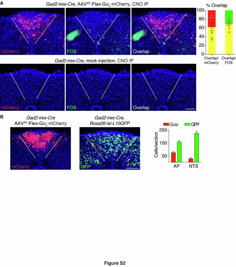
Figure S2. Validation of chemogenetic approaches to activate area postrema inhibitory neurons, related to
Figure 1.
(A) Gad2-ires-Cre mice were injected in the area postrema with (top) or without (bottom) AAV-Flex-GsDREADD-
mCherry. CNO was injected IP, and two-color expression analysis was subsequently performed in fixed coronal area
postrema cryosections for native mCherry fluorescence (red) and cFos immunofluorescence (green), scale bar: 100
µm. Fos responses were observed in mCherry-labeled area postrema neurons of mice injected with AAV-Flex-
GsDREADD-mCherry (quantification on the right), but were not observed in control mice lacking AAV-Flex-
GsDREADD-mCherry.
(B) Gad2-ires-Cre mice were either injected in the area postrema with AAV-Flex-GsDREADD-mCherry (left) or
crossed to mice with a Rosa26-lsl-L10GFP allele (middle), scale bar: 100 µm. Quantification (right) of area
postrema (AP) or NTS cells displaying native reporter fluorescence per coronal brainstem cryosection (40 µm) from
Gad2-ires-Cre mice injected in the AP with AAV-Flex-GsDREADD-mCherry mice (red) or Gad2-ires-Cre;
Rosa26-lsl-L10GFP mice (green), n=15 sections from 10 (Gas) or 7 (GFP) mice.
3Supplementary Figure 3
Figure S3. Validation of Gipr-Cre mice, related to Figure 2.
(A) Two-color expression analysis in coronal area postrema cryosections of Gipr-Cre, Rosa26-lsl-L10GFP mice,
with RNA in situ hybridization to detect Gipr transcripts (red) and native GFP fluorescence (green), scale bar: 100
µm. The numbers of co-labeled (yellow) or individually labeled (red, green) cells were counted (right).
(B) Mice underwent either sham surgery or bilateral vagotomy (unilateral cervical vagotomy and unilateral
subdiaphragmatic vagotomy), and were injected (IP) with Fluoro-Gold to label uncut neurons in the dorsal motor
nucleus of the vagus (DMV). Mice were later injected (IP) with either GIP or saline, and fixed coronal brainstem
sections were harvested for Fos immunochemistry and visualization of native Fluoro-Gold fluorescence, scale bar:
100 µm.
4You can also read