MOTS-c is an exercise-induced mitochondrial-encoded regulator of age-dependent physical decline and muscle homeostasis - The USC ...
←
→
Page content transcription
If your browser does not render page correctly, please read the page content below
ARTICLE
                  https://doi.org/10.1038/s41467-020-20790-0                OPEN

                  MOTS-c is an exercise-induced mitochondrial-
                  encoded regulator of age-dependent physical
                  decline and muscle homeostasis
                  Joseph C. Reynolds 1, Rochelle W. Lai1, Jonathan S. T. Woodhead2,3, James H. Joly4, Cameron J. Mitchell                                           5,6,

                  David Cameron-Smith5, Ryan Lu1, Pinchas Cohen 1,7, Nicholas A. Graham 4,7, Bérénice A. Benayoun                                                  1,7,8,

                  Troy L. Merry5,6 & Changhan Lee 1,7,9 ✉
1234567890():,;

                  Healthy aging can be promoted by enhanced metabolic fitness and physical capacity.
                  Mitochondria are chief metabolic organelles with strong implications in aging that also
                  coordinate broad physiological functions, in part, using peptides that are encoded within their
                  independent genome. However, mitochondrial-encoded factors that actively regulate aging
                  are unknown. Here, we report that mitochondrial-encoded MOTS-c can significantly enhance
                  physical performance in young (2 mo.), middle-age (12 mo.), and old (22 mo.) mice. MOTS-c
                  can regulate (i) nuclear genes, including those related to metabolism and proteostasis, (ii)
                  skeletal muscle metabolism, and (iii) myoblast adaptation to metabolic stress. We provide
                  evidence that late-life (23.5 mo.) initiated intermittent MOTS-c treatment (3x/week) can
                  increase physical capacity and healthspan in mice. In humans, exercise induces endogenous
                  MOTS-c expression in skeletal muscle and in circulation. Our data indicate that aging is
                  regulated by genes encoded in both of our co-evolved mitochondrial and nuclear genomes.

                  1 Leonard Davis School of Gerontology, University of Southern California, Los Angeles, CA 90089, USA. 2 Discipline of Nutrition, Faculty of Medical and

                  Health Sciences, The University of Auckland, Auckland, New Zealand. 3 Maurice Wilkins Centre for Molecular Biodiscovery, The University of Auckland,
                  Auckland, New Zealand. 4 USC Mork Family Department of Chemical Engineering and Materials Science, Los Angeles, CA 90089, USA. 5 Liggins Institute,
                  The University of Auckland, Auckland, New Zealand. 6 School of Kinesiology, University of British Colombia, Vancouver, BC, Canada V6T 1Z3. 7 USC Norris
                  Comprehensive Cancer Center, Los Angeles, CA 90089, USA. 8 USC Stem Cell Initiative, Los Angeles, CA 90089, USA. 9 Biomedical Science, Graduate
                  School, Ajou University, Suwon 16499, Korea. ✉email: changhan.lee@usc.edu

                  NATURE COMMUNICATIONS | (2021)12:470 | https://doi.org/10.1038/s41467-020-20790-0 | www.nature.com/naturecommunications                              1
ARTICLE                                                                NATURE COMMUNICATIONS | https://doi.org/10.1038/s41467-020-20790-0

T
        he progressive loss of metabolic homeostasis is a hallmark            absolute quantification values. ELISA revealed that relative levels
        of aging, which impedes parenchymal function and ulti-                of circulating endogenous MOTS-c also significantly increased
        mately diminishes physical capacity1,2. In fact, aging is a           during (1.6-fold) and after (1.5-fold) exercise, which then
leading risk factor for a myriad of non-communicable chronic                  returned to baseline after 4 h of resting (Fig. 1d and Supple-
diseases3–9. Organismal fitness requires continuous adaptive cel-              mentary Fig. 1b). These findings suggest that exercise induces the
lular stress responses to the ever-shifting internal and external             expression of mitochondrial-encoded regulatory peptides in
environment. Mitochondria not only produce the bulk of cellular               humans.
energy, but also coordinate adaptive cellular homeostasis by
dynamically communicating to the nucleus10 and other sub-
                                                                              MOTS-c treatment improves physical performance in young
cellular compartments11. Mitochondrial communication is
                                                                              mice. We next probed if MOTS-c functions as an exercise-
mediated by multiple nuclear-encoded proteins, transient mole-
                                                                              induced mitochondrial signal that improves physical capacity by
cules, and mitochondrial metabolites12.
                                                                              treating young mice (CD-1; outbred) daily with MOTS-c [5 mg/
   Mitochondria possess a distinct circular genome that has been
                                                                              kg/day; intraperitoneal injections (IP)] for 2 weeks; we previously
traditionally known to host only 13 protein-coding genes. How-
                                                                              reported that 7 days of MOTS-c treatment improved skeletal
ever, short open reading frames (sORFs) encoded in the mito-
                                                                              muscle insulin sensitivity in young and older C57BL/6J mice15.
chondrial genome have been recently identified. Such sORFs
                                                                              The rotarod performance test, whereby mice are placed on a
produce bioactive peptides, collectively referred to as
                                                                              rotating rod, revealed that daily MOTS-c significantly improved
mitochondrial-derived peptides (MDPs), with broad physiologi-
                                                                              physical capacity and/or motor coordination (Supplementary
cal functions13,14. MOTS-c (mitochondrial ORF of the 12S rRNA
                                                                              Fig. 2a), but not grip strength (Supplementary Fig. 2b) in young
type-c) is an MDP that promotes metabolic homeostasis, in part,
                                                                              mice. Because the rotarod test can also be affected by cognitive
via AMPK15,16 and by directly regulating adaptive nuclear gene
                                                                              capacity, we assessed learning and memory using the Barnes
expression following nuclear translocation17,18. MOTS-c expres-
                                                                              maze and found no improvement (Supplementary Fig. 2c, d).
sion is age-dependent and detected in multiple tissues, including
                                                                                 A treadmill running test confirmed that MOTS-c treatment can
skeletal muscle, and in circulation15,16,19, thus it has been dubbed
                                                                              enhance physical performance. Because MOTS-c is a regulator of
a “mitochondrial hormone”16 or “mitokine”20,21. In fact, we
                                                                              metabolic homeostasis that prevented high-fat diet (HFD)-
previously reported that systemic MOTS-c treatment reversed
                                                                              induced obesity and insulin resistance15, we tested if MOTS-c
diet-induced obesity and diet- and age-dependent insulin resis-
                                                                              also improved running performance under metabolic (dietary)
tance in mice15. We tested if MOTS-c functions as a
                                                                              stress. We fed young mice (CD-1) a HFD (60% calories from fat)
mitochondrial-encoded regulator of physical capacity and
                                                                              and treated them with 2 doses of MOTS-c (5 and 15 mg/kg/day;
performance2,22,23 in young (2 mo.), middle-aged (12 mo.), and
                                                                              IP; Supplementary Fig. 3a). Mice on the higher dose of MOTS-c
old (22 mo.) mice.
   Here, we show that exercise induces mtDNA-encoded MOTS-c                   showed significantly superior running capacity (Fig. 2a–c) and
                                                                              power output (joules; Supplementary Fig. 4a) following 10 days of
expression in humans. MOTS-c treatment significantly (i)
improves physical performance in young, middle-age, and old                   treatment, but not following 7 days of treatment (Supplementary
                                                                              Fig. 5a). Notably, 7 days of MOTS-c treatment significantly
mice, (ii) regulates skeletal muscle metabolism and gene expres-
sion, and (iii) enhances adaptation to metabolic stress in C2C12              improved skeletal muscle insulin sensitivity in young and older
                                                                              C57BL/6J mice also fed a high-fat diet15. We progressively
cells in an HSF1-dependent manner.
                                                                              increased the treadmill speed to test both endurance and speed.
                                                                              The final stage, which required mice to sprint (23 m/min), was
Results                                                                       reached by 100% of mice on the higher dose of MOTS-c, but only
Endogenous MOTS-c levels increase upon exercise in humans.                    16.6% in the lower dose and control (vehicle) groups (Fig. 2d).
To determine if endogenous MOTS-c responds to physical                        Body composition analysis using a time-domain NMR analyzer
exertion, and thus may be involved in driving adaptation to                   revealed that both doses of MOTS-c significantly retarded fat gain
enhance physical capacity, we collected skeletal muscle and                   and that the high dose significantly increased lean mass in young
plasma from sedentary healthy young male volunteers (24.5 ± 3.7               mice (CD-1; Supplementary Fig. 6a–c), in accord with prior
years old and BMI 24.1 ± 2.1) that exercised on a stationary                  reports15. There was no correlation between body weight and
bicycle (Fig. 1a). Samples were collected before, during (plasma              running time (Supplementary Fig. 7a). Taken together, perfor-
only), and after exercise and following a 4-h rest. Western blot-             mance improvements in treadmill running and rotarod test,
ting for endogenous MOTS-c in skeletal muscle revealed that                   paired with lack of changes in the grip strength and Barnes Maze,
relative levels (i.e. individual changes based on pre-exercise                indicate an enhancement in physical capacity.
values) significantly increased after exercise (11.9-fold) and                    In young CD-1 mice, we simultaneously initiated MOTS-c
remained elevated after a 4-h rest, albeit exhibiting a trend to              treatment and a HFD (Supplementary Fig. 3a). To test if MOTS-c
return to baseline (Fig. 1b, c); see Supplementary Fig. 1a for                can improve physical performance in mice that have been on a

Fig. 1 MOTS-c responds to and regulates exercise in young subjects. a Schedule of exercise on a stationary bicycle and blood and skeletal muscle
collection in young male subjects (n = 10). b, c Representative western blot of MOTS-c from skeletal muscle and quantification (n = 10; P = 0.0098). d
Quantification of serum MOTS-c levels by ELISA (n = 10; P = 0.0011; P = 0.0021, respectively). Data expressed as mean ± SEM. Wilcoxon matched-pairs
two-sided signed rank test was used for (c, d).

2                NATURE COMMUNICATIONS | (2021)12:470 | https://doi.org/10.1038/s41467-020-20790-0 | www.nature.com/naturecommunications
NATURE COMMUNICATIONS | https://doi.org/10.1038/s41467-020-20790-0 ARTICLE Fig. 2 MOTS-c treatment increases physical capacity in young mice regardless of diet. a–c Treadmill performance of 12-week-old male CD-1 (outbred) mice fed a normal diet (n = 5, Control and 5 mg/kg MOTS-c; n = 6, 15 mg/kg MOTS-c); a running curves (P = 0.0329), b total time on treadmill (P = 0.0069, 0 vs. 15; P = 0.0081, 5 vs. 15), c total distance ran (P = 0.0418, 0 vs. 15; P = 0.0081, 5 vs.15), and d percent capable of reaching the highest speed (sprint). e–h Treadmill performance of 12-week-old male C57BL/6J (inbred) mice fed a HFD (n = 8); e running curves (P = 0.0003), f total time on treadmill (P = 0.0237, normal diet control vs. normal diet + MOTS-c; P = 0.0084, HFD control vs. HFD + MOTS-c) g total distance ran (P = 0.0326, normal diet control vs. normal diet + MOTS-c; P = 0.0087, HFD Control vs. HFD + MOTS-c) h percent capable of reaching the highest speed (final stage). i PCA and MSEA on metabolomic data from skeletal muscle and liver of C57BL/6J mice that were fed a HFD, treated with MOTS-c, and exercised. Data expressed as mean ± SEM. Log-rank (Mantel-Cox) test was used for a, e. Otherwise, all statistics were performed using the two-sided Student’s t test. *P < 0.05, **P < 0.01, ***P < 0.001. HFD, we fed young C57BL/6J mice a HFD, or a normal diet, for MOTS-c treatment enhances physical capacity in old mice. 2 weeks before initiating daily MOTS-c injections (15 mg/kg/day) Aging is accompanied by a progressive decline in mitochondrial for 2 weeks prior to a treadmill running test (Supplementary function1,24 and loss of metabolic homeostasis, in which MOTS-c Fig. 3b). MOTS-c treatment significantly enhanced running may play a role2,10. Aging is associated with reduced MOTS-c performance and power output (joules; Supplementary Fig. 4b) levels in certain tissues, including the skeletal muscle, and in on the treadmill regardless of the diet (Fig. 2e–h and circulation15,19. We previously showed that an acute one-week Supplementary Fig. 5b). MOTS-c treatment enabled 25% of the MOTS-c treatment reversed age-dependent insulin resistance in young C57BL/6J mice to enter the final running stage (highest mouse skeletal muscle15. Thus, we investigated if promoting speed) on a normal diet, but none on a HFD (Fig. 2h). Consistent metabolic homeostasis by MOTS-c treatment could reverse age- with our prior study15, MOTS-c treatment curbed HFD-induced dependent decline in physical capacity. Middle-aged (12 mo.) and weight gain in C57BL/6J mice (Supplementary Fig. 6d), which old (22 mo.) C57BL/6N mice were treated daily with MOTS-c was largely driven by reduced fat accumulation (Supplementary (15 mg/kg/day; IP) for 2 weeks, then subjected to a treadmill Fig. 6e), but not loss of lean mass (Supplementary Fig. 6f), as running test (Fig. 3a). Both middle-aged and old mice ran sig- determined by an NMR-based body composition analysis. nificantly longer with increased power output (joules) following However, there was no correlation between body weight and MOTS-c treatment (Fig. 3b and Supplementary Fig. 4c). Old mice running time (Supplementary Fig. 7b). Also, consistent with our ran longer (2-fold; Fig. 3c) and farther (2.16-fold; Fig. 3d) when prior study15, there were no differences in food intake between treated with MOTS-c. Further, MOTS-c enabled 17% of the old control diet or HFD groups (Supplementary Fig. 8). Further, mice to enter the final running stage (highest speed), whereas targeted metabolomics revealed that MOTS-c treatment signifi- none in the untreated group were successful (Fig. 3e). Notably, cantly regulated (i) glycolysis/PPP (pentose phosphate pathway) MOTS-c treatment enabled old mice to outperform untreated and (ii) amino acid metabolism (Fig. 2i; Supplementary Figs. 3b middle-aged mice, suggesting a more pervasive physical repro- and 9a; and Supplementary Data 1 and 2) in skeletal muscle, but gramming rather than just rejuvenation. Respiratory exchange not in liver, consistent with our previous study15. Together, these ratio (RER), measured using a metabolic cage, indicates fuel data indicate that MOTS-c treatment can improve overall preference (1.0: carbohydrates, 0.7: fat). “Metabolic flexibility”, physical performance, in part, by targeting skeletal muscle which refers to the overall adaptive capacity to a shift in metabolic metabolism in young mice. supply-demand equilibrium (e.g. exercise), declines with age25,26. NATURE COMMUNICATIONS | (2021)12:470 | https://doi.org/10.1038/s41467-020-20790-0 | www.nature.com/naturecommunications 3
ARTICLE                                                                 NATURE COMMUNICATIONS | https://doi.org/10.1038/s41467-020-20790-0

Fig. 3 Acute MOTS-c treatment enhances physical capacity in old mice. a Schedule of MOTS-c treatment and assays in middle-aged and old C57BL/
6N mice (n = 10, MA groups; n = 19, old control; n = 18, old MOTS-c), including b treadmill running curves (P = 0.000000046), c total time on
treadmill (P = 0.0458, MA control vs. old control; P = 0.0497, MA control vs. MA MOTS-c; P = 0.000002, old control vs. old MOTS-c), d total
distance ran on treadmill (n = 19, old control; n = 18, Old MOTS-c; P = 0.0464, MA control vs. old control; P = 0.0462, MA control vs. MA MOTS-c;
P = 0.000002, old control vs. old MOTS-c), and e percent capable of reaching the highest speed on a treadmill (final stage). f Respiratory exchange
ratio (RER) following 2 weeks of daily MOTS-c injection (n = 4; P = 0.0055). g, h Skeletal muscle from treadmill-exercised old mice (22.5 months)
treated daily with MOTS-c (15 mg/kg/day) for 2 weeks (n = 10) were subject to g metabolomics and analyzed using PCA and MSEA and h GSEA
analysis of muscle RNA-seq analysis. Balloon plots of select enriched terms using Gene Ontology Biological Process (GO_BP) database at false
discovery rate (FDR) < 15%. NES Normalized Enrichment Score. Full GSEA results are available in Supplementary Data 5. Data expressed as mean ±
SEM. Log-rank (Mantel–Cox) test was used for b and two-way ANOVA (repeated measures) was used for f. GSEA statistics from R package
‘clusterProfiler’ were used for h. Otherwise, all statistics were performed using the two-sided Student’s t test. *P < 0.05, **P < 0.01, ***P < 0.001. MA
middle-age.

4                 NATURE COMMUNICATIONS | (2021)12:470 | https://doi.org/10.1038/s41467-020-20790-0 | www.nature.com/naturecommunications
NATURE COMMUNICATIONS | https://doi.org/10.1038/s41467-020-20790-0                                                                ARTICLE

Indeed, we observed a shift in RER between daytime and night-
time, which trended more towards statistical significance for
middle-aged mice (P = 0.076) compared to aged mice (P = 0.39;
Fig. 3f and Supplementary Fig. 10), indicating reduced metabolic
flexibility with age. RER during daytime, but not nighttime, was
significantly different between middle-aged vs. old mice, whereby
old mice preferred to continue to utilize carbohydrates more than
their middle-aged counterparts (Fig. 3f and Supplementary
Fig. 10). This phenomena was fully reversed by MOTS-c treat-
ment and the RER of old mice exhibited a similar circadian
pattern to that of middle-aged mice (Fig. 3f and Supplementary
Fig. 10). Such circadian-dependent effects of MOTS-c RER may
be affected by food intake, as mice feed mostly during the night
(Supplementary Fig. 11). There was no correlation between body
weight and running time upon MOTS-c treatment (Supplemen-
tary Fig. 7c). Metabolomic analysis on skeletal muscle collected
immediately post-exercise (a 30-min run at a fixed moderate
speed) in MOTS-c-treated (2 weeks) mice revealed that MOTS-c
significantly regulated glycolysis and amino acid metabolism
(Fig. 3g, Supplementary Fig. 9b, and Supplementary Data 3 and
4); the skeletal muscles of non-exercised mice did not show sig-
nificant alterations in response to MOTS-c (Supplementary
Fig. 12), suggesting that MOTS-c induces an adaptive metabolic
response to exercise. To begin to understand the molecular
mechanisms underlying the effects of MOTS-c, we performed
RNA-seq analysis on the same skeletal muscles used for meta-
bolomics. Although individual-to-individual variability was high,
Gene Set Enrichment Analysis (GSEA) using the KEGG pathway
database revealed that MOTS-c regulated processes related to (i)
metabolism, including those known to be regulated by MOTS-c
(e.g. AMPK signaling, glycolysis, and central carbon
metabolism15,17, and (ii) longevity (FDR < 15%; select pathways
in Fig. 3h; full DESeq2 analysis in Supplementary Data 5 and full
GSEA analysis in Supplementary Data 6). Gene Ontology Bio-
logical Process (GO_BP) analysis revealed a broader range of
processes, including metabolism (lipid, carbohydrate, amino acid,
and nucleotides), oxidative stress response, immune response,
and nuclear transport (FDR < 15%; select pathways in Supple-
mentary Fig. 13; full analysis in Supplementary Data 6), again,
consistent with our previous studies15,17. The rotarod perfor-
mance test reinforced that MOTS-c treatment improved physical
capacity in old mice (Supplementary Fig. 14a), while learning and
memory was not affected as determined using the Y-maze test
(Supplementary Fig. 14b), consistent with our observations in
young mice (Supplementary Fig. 2). Together, these data suggest
that MOTS-c treatment can significantly improve physical
capacity in old mice, in part, by regulating skeletal function and
improving “metabolic flexibility”.

Late-life MOTS-c treatment improves mouse healthspan. Anti-
aging interventions that are applied later in life would be more
translationally feasible compared to life-long treatments27–29.                   Independent lines of research have shown that MOTS-c is a
Building on the treadmill running tests, we tested if a late-life              mitochondrial-encoded metabolic regulator at the cellular and
initiated (~24 mo.) intermittent (LLII) MOTS-c treatment (3x/                  organismal level15,17,19,31–35. We posited that LLII MOTS-c
week; 15 mg/kg/day) would improve healthy lifespan (Fig. 3a). To               treatment would cause metabolic reprogramming in old mice.
assess healthspan, towards the end-of-life (>30 mo.), we per-                  Consistent with our previous report15, non-fasting blood glucose
formed a battery of physical tests to further probe the effect of              was better maintained in LLII MOTS-c-treated old mice (30 mo.;
MOTS-c on reversing age-dependent physical decline (Fig. 3a).                  Fig. 4d). Over course of their life, LLII MOTS-c-treated mice
LLII MOTS-c improved (i) grip strength (Fig. 4a), (ii) gait,                   showed comparable body weight to their untreated counterparts
assessed by stride length (Fig. 4b), and (iii) physical performance,           (Fig. 4e, f). However, total food intake was significantly reduced
assessed by a 60-second walking test (running was not possible at              (Fig. 4g, h and Supplementary Fig. 11), whereas total activity was
this age; Fig. 4c). In humans, reduced stride length and walking               significantly higher (Supplementary Fig. 15). Yet, the activity
capacity are strongly linked to mortality and morbidity30. Toge-               changes at 30 months of age was marginal and may not account
ther, these data indicate that LLII MOTS-c treatment improves                  for considerable increase in energy expenditure that could explain
physical capacity in old mice.                                                 the effect of MOTS-c on body weight at different age groups

NATURE COMMUNICATIONS | (2021)12:470 | https://doi.org/10.1038/s41467-020-20790-0 | www.nature.com/naturecommunications                        5
ARTICLE NATURE COMMUNICATIONS | https://doi.org/10.1038/s41467-020-20790-0
Fig. 4 MOTS-c regulates aging metabolism and healthspan. Life-long without glucose (0 g/L). As expected, most control cells died
measurements on male C57BL6/N mice treated intermittently (3x/week) without glucose even with lipid supplementation, whereas
with MOTS-c (15 mg/kg/day) starting at middle and old age (13.5 and 23.5 MOTS-c treatment provided significant protection (~2-fold;
mo.) as described in Fig. 2a. a grip strength test (n = 11; run in triplicate; Fig. 5c). Real-time metabolic flux analysis revealed that MOTS-c
P = 0.000078), b gait analysis (stride length; n = 5; run in triplicate; P = treatment significantly increased lipid utilization capacity through
0.0038), c 60-s walking test (n = 11, control; n = 12, MOTS-c; P = 0.0428), increasing OCR, but not ECAR, in the presence of palmitate in
and d blood glucose levels (n = 11; P = 0.0397). e, f Body weight e as a C2C12 cells (Fig. 5d and Supplementary Fig. 17).
function of time and f the total sum (∑; n = 19, old control; n = 18, old We previously reported that endogenous MOTS-c translocates
MOTS-c; P = 0.0013, MA control vs. MA MOTS-c; P = 0.001, old control to the nucleus to directly regulate adaptive nuclear gene
vs. old MOTS-c); g, h Food intake g as a function of time and h the total expression in response to cellular stress17. Using fluorescently
sum (∑; n = 19, old control; n = 18, old MOTS-c; P = 0.0276, MA control labeled MOTS-c peptide (MOTS-c-FITC), we confirmed that
vs. MA MOTS-c; P = 0.00005, old control vs. old MOTS-c); i, j Percent fat exogenously treated MOTS-c also dynamically translocated to the
mass i as a function of time and j the total sum (∑; n = 19, old control; n = nucleus in a time-dependent manner (Fig. 5e)17, indicating a
18, old MOTS-c; P < 1E-50); k, l Percent lean mass k as a function of time direct nuclear role. We performed RNA-seq on C2C12 cells
and l the total sum (∑; P = 0.026, MA control vs. MA MOTS-c; P = treated with MOTS-c or vehicle control (10 µM) under GR/SD for
0.0003, old control vs. old MOTS-c). m Lifespan curve; P = 0.05 until 48 h and found (i) separation of groups in PC1 (first principle
31.8 months of age. Overall curve trended towards increased median and component) using a principal component analysis (PCA; Fig. 5f)
maximum lifespan (P = 0.23). Data expressed as mean ± SEM. Log-rank and (ii) 69 genes that were differentially regulated at FDR < 5%
(Mantel–Cox) test was used for m. Otherwise, all statistics were performed (Fig. 5g). Further, using the STRING (Search Tool for the
using the two-sided Student’s t test. *P < 0.05, **P < 0.01, ***P < 0.001. MA Retrieval of Interacting Genes/Proteins) database to assess
middle-age. putative changes in protein-protein interaction networks based
on our RNA-seq results, we found that a cluster related to heat-
shock responses, including Hsp40 (DNAJ) and Hsp70s (HSPA),
(Supplementary Fig. 15). A likely possibility that could increase were prominently regulated by MOTS-c in C2C12 cells under
energy expenditure is, consistent with previous reports from our GR/SD; we also identified previously reported MOTS-c targets,
lab and others15,36, increased heat production. Body composition including Atf3, Jun, Fosl1, and Mafg17 (Fig. 5h and Supplemen-
analysis using a time-domain NMR analyzer revealed significant tary Data 5). Indeed, select GO_BP analysis revealed that protein
reduction of fat mass (Fig. 4i, j) and a modest reduction in age- homeostasis (i.e. proteostasis) processes were significantly
dependent loss of lean mass (Fig. 4k, l). The RER, measured using targeted with protein folding being the most significantly affected
metabolic cages, at 30 mo. revealed increased fat utilization, pathway in metabolically stressed myoblasts (Supplementary
consistent with that obtained at ~23.5 mo. (Fig. 3f), but with a Fig. 18). Next, to identify common pathways in in vitro and
circadian shift compared to the same mice measured 6 months in vivo models, we overlaid RNA-seq data from MOTS-c-treated
before (Supplementary Fig. 16); this is also consistent with (i) old mouse skeletal muscle and (ii) metabolically stressed (GR/
reduced total fat mass (Fig. 4i, j and Supplementary Fig. 6b, e) SD) C2C12. Consistently, select GO_BP analysis revealed several
and increased lipid utilization15,35. Ultimately, LLII MOTS-c commonly targeted pathways by MOTS-c in both exercised
treatment showed a trend towards increased median (6.4%) and skeletal muscle and metabolically stressed myoblasts that were
maximum (7.0%) lifespan and reduced hazard ratio (0.654); P = related to proteostasis, including protein regulation and protein
0.05 until 31.8 months (Fig. 4m). Larger cohorts will be needed to metabolism (select terms in Fig. 5i; full results in Supplementary
confirm the broader significance of MOTS-c treatment on overall Data 5 and 8); notably, commonly targeted pathways also
longevity. These data suggest that LLII MOTS-c treatment provided a specific cellular context that reflects metabolic stress,
improves overall physical capacity in old mice and may compress including oxidative stress, mitochondria, cellular metabolism. To
morbidity and increase healthspan. identify putative transcription factors involved in differential gene
regulation upon MOTS-c treatment, we performed a statistical
enrichment analysis inferred from the ChEA knowledge base43
MOTS-c promotes metabolic adaptations in vitro. Skeletal (FDR < 5%). Consistent with the STRING and GSEA (GO_BP)
muscle must adapt to various exercise-induced challenges37, analyses, we identified heat shock factor 1 (HSF1) as a putative
including nutrient (e.g. metabolic supply-demand imbalance)38, transcription factor that was significantly enriched in both mouse
oxidative39,40, and heat stress37,41, which share mitochondria as a skeletal muscle and myoblasts and could potentially regulate gene
common denominator. Because MOTS-c enhanced cellular expression upon MOTS-c treatment (Fig. 5j). HSF1 is a master
resistance against metabolic/oxidative stress17, we tested if nutrient sensor and regulator of stress adaptation and proteos-
MOTS-c treatment improved skeletal muscle adaptation to tasis, in part, by inducing the expression of a myriad of heat shock
metabolic stress using C2C12 mouse myoblast cells. Using crystal proteins, including Hsp40 (DNAJ) and Hsp70s (HSPA), con-
violet staining to determine cellular viability, we found that sistent with the effects of MOTS-c (Fig. 5h)44,45. Exercise induces
MOTS-c (10 µM) treatment significantly protected C2C12 cells HSF1 activation in skeletal muscle46 and cardiac muscle47 in
(~2-fold) from 48 h of metabolic stress [glucose restriction (GR; rodents. Indeed, siRNA-mediated HSF1 knockdown reversed the
0.5 g/L) and serum deprivation (SD; 1% FBS)] (Fig. 5a). Next, we protective effects of MOTS-c against GR/SD in myoblasts (Fig. 5k,
tested the replicative capacity of C2C12 cells following prolonged l). Together, these data suggest that MOTS-c improves metabolic
metabolic stress as a functional marker of protection. C2C12 cells homeostasis/flexibility and protein homeostasis in skeletal muscle
were metabolically stressed (GR/SD) for one week with daily under exercise-induced stress conditions.
MOTS-c (10 µM) treatment, then replenished with complete
medium for 2 days and stained with crystal violet. MOTS-c-
treated C2C12 cells showed significantly enhanced proliferative Discussion
capacity within 2 days (~6-fold; Fig. 5b). Because MOTS-c pro- We previously reported that MOTS-c prevented diet-induced
motes fat utilization, which may underly its effect on “metabolic obesity and insulin resistance and reversed age-dependent skeletal
flexibility” (Figs. 3f and 4i, j and Supplementary Fig. 6)15,35,42, we muscle insulin resistance in C57BL/6J mice, indicating a role in
tested if MOTS-c-treated C2C12 cells could survive on lipids metabolic conditions such as diabetes15,48,49. MOTS-c functions
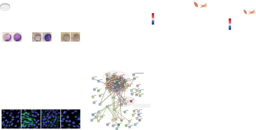
6 NATURE COMMUNICATIONS | (2021)12:470 | https://doi.org/10.1038/s41467-020-20790-0 | www.nature.com/naturecommunicationsNATURE COMMUNICATIONS | https://doi.org/10.1038/s41467-020-20790-0 ARTICLE Fig. 5 MOTS-c regulates myoblast gene expression and enhances adaptation to metabolic stress. a–c Survival of MOTS-c-treated (10 µM; equal-volume vehicle control) C2C12 myoblasts assessed by crystal violet staining following a 48 h of glucose restriction (GR; 0.5 g/L) and serum deprivation (SD; 1% FBS) with MOTS-c treated only once initially (n = 12, duplicates; P = 2.57E-25), b 7 days of GR/SD with daily MOTS-c treatment, followed by a 2-day recovery in full media with MOTS-c (n = 10, duplicates; P = 8.95E-22), and c 48 h of complete GR (0 g/L) with chemically defined lipid supplementation and daily MOTS-c treatment (n = 6, duplicates; P = 1.17E-09). d Real-time oxygen consumption rate (OCR) in response to fatty acid (palmitate-BSA) in C2C12 myoblasts treated with MOTS-c (10 µM) for 48 h (n = 11, BSA baseline; n = 12, palmitate addition; P = 6.27E-07). e Time-dependent subcellular localization pattern of exogenously treated MOTS-c-FITC (10 µM) in C2C12 myoblasts. Scale bar: 10 µm. Images represent two biological replicates and three technical replicates. f–i RNA-seq was performed on C2C12 myoblasts following 48 h of GR/SD with/without a unique initial MOTS-c (10 µM) treatment (n = 6). f Principle Component Analysis (PCA) and g heatmap of significantly differentially regulated genes by MOTS-c at false discovery rate (FDR) < 5% by DESeq2 analysis. h Protein–protein interaction network analysis based on genes that were significantly differentially regulated by MOTS-c (FDR < 5%) using the STRING (Search Tool for the Retrieval of Interacting Genes/Proteins) database version 11.079. i Balloon plots of common biological processes derived from RNA-seq data between MOTS-c-treated (i) skeletal muscle from old mice (see Fig. 3) and (ii) C2C12 myoblasts, based on gene set enrichment analysis (GSEA) using gene ontology biological process (GO_BP). j Balloon plots of common transcription factors derived from RNA-seq data between MOTS-c-treated (i) skeletal muscle from old mice (see Fig. 3) and (ii) C2C12 myoblasts, based on ChEA database. k, l Relative cell number at k 48 vs 72 h of GR/SD assessed by crystal violet staining (n = 6; P = 0.00027, siCtrl + MOTS-c vs. siHsf1 + MOTS-c; P = 0.00017, siCtrl vs. siCtrl + MOTS- c.) and l 72 h of GR/SD assessed by flow cytometry (n = 3, siCtrl; n = 6, siHsf1; P = 0.0053). Data expressed as mean ± SEM. Two-sided Student’s t test. *P < 0.05, **P < 0.01, ***P < 0.001. have been shown to be dependent on the master nutrient sensor (Fig. 5e)17, and appears to be highly regulated in a context- AMPK15,17,34,36,50–54, including its nuclear translocation in dependent manner (unpublished observation). Notably, anti- response to metabolic imbalance55. Notably, MOTS-c treatment microbial peptides (AMPs), which are small amphiphilic and in mice activated AMPK in skeletal muscle and increased the cationic peptides (as small as 15–20 amino acids) like MOTS-c17, expression of its downstream glucose transporter (i.e GLUT4)15, provide an excellent example of bioactive peptides that can enter suggesting improved muscle metabolism and physical capacity56. cells without being degraded and retainbiological activity60; the Notably, MOTS-c treatment improved running capacity inde- cellular entry of peptides is still largely unclear and known to adopt pendent of body weight, based on total work (joules) output and a wide range of mechanisms in a concentration-dependent fashion, lack of correlation between body weight and running time including energy-dependent transport via endocytosis61,62. (Supplementary Figs. 4 and 7). This indicates that MOTS-c Mitochondria are strongly implicated in aging at multiple treatment enhanced physical performance by improving whole levels1,2,24,63–65 and MOTS-c may contribute to longevity by body energy metabolism, in part, by promoting adaptive promoting cellular homeostasis2,10,57–59. Here, we present evi- responses to exercise-related stress conditions (e.g. metabolic dence that the mitochondrial genome encodes for instructions to imbalance and heat shock) in skeletal muscle. Further, the maintain physical capacity (i.e. performance and metabolism) treatment duration of MOTS-c was important to enhance per- during aging and thereby increase healthspan. MOTS-c treatment formance (Fig. 2a–h and Supplementary Fig. 5), suggesting the initiated in late-life, proximal to the age at which the lifespan need to reach a certain physiological state with an improved curve rapidly descends for C57BL/6N mice, significantly delayed metabolic profile. the onset of age-related physical disabilities, suggesting “com- MOTS-c acts as an intracellular and endocrine factor, thus has pression of morbidity” in later life66. Interestingly, an excep- been dubbed a mitochondrial-encoded hormone or a mitochondrial tionally long-lived Japanese population harbors a mitochondrial cytokine (mitokine)2,10,16,49,57–59. Understanding the detailed DNA (mtDNA) SNP (m.1382A>C) that yields a functional var- molecular details of MOTS-c function as an endocrine factor is an iant of MOTS-c67,68. ongoing investigation at multiple ends, including its cellular uptake. Our study shows that exogenously treated MOTS-c enters the The cellular entry of MOTS-c is likely a crucial step in exerting its nucleus and regulates nuclear gene expression, including those effects and can occur rapidly, within 30 minutes of treatment involved in heat shock response and metabolism. Thus, age- NATURE COMMUNICATIONS | (2021)12:470 | https://doi.org/10.1038/s41467-020-20790-0 | www.nature.com/naturecommunications 7
ARTICLE                                                                            NATURE COMMUNICATIONS | https://doi.org/10.1038/s41467-020-20790-0

related gene networks are comprised of integrated factors enco-                          Barnes maze. In all, 12-week-old male CD-1 mice were tested twice daily for 7 days.
ded by both genomes, which entails a bi-genomic basis for the                            Mice were placed in a start chamber in the middle of the maze and allowed to
                                                                                         habituate (30 s), then the mouse was released to explore the maze and find the
evolution of aging. Although the detailed molecular mechanism                            escape box (EB). Latency (time to enter the EB) and number of errors (nose pokes
(s) underlying the functions of MOTS-c is an active field of                              and head deflections over false holes) were recorded. A maximum of 2 min was
research we provide a proof-of-principlestudy that realizes the                          allowed for each trial72.
mitochondrial genome as a source for instructions that can reg-
ulate physical capacity and healthy aging.                                               In vivo metabolism assessment
                                                                                         Metabolic cages. Metabolic activity in mice was measured using the PhenoMaster
                                                                                         system (TSE-Systems) equipped to detect indirect calorimetry, measure food and
Methods                                                                                  water intake, and monitor activity. Prior to metabolic analysis, mice were housed
Mouse care. All animal work was approved by and performed in accordance with
                                                                                         3–4 per cage in a facility with a 12:12 h light–dark cycle (light period 0600-1800) at
the University of Southern California (USC) Institutional Animal Care and Use
                                                                                         24 °C. Food and water were available ad libitum. For metabolic assessment, nice
Committee. Mice were housed 3–4 per cage in a facility with a 12:12 h light–dark
                                                                                         were moved into individual PhenoMaster cages in an isolated room under the same
cycle (light period 0600-1800) at 24 °C. Food and water were available ad libitum.
                                                                                         environmental conditions. Mice were automatedly monitored for 36 h to record
MOTS-c (New England Peptide, USA) was administered daily at 5 or 15 mg/kg via
                                                                                         physiological parameters. To measure O2 intake and CO2 production, gas sensors
intraperitoneal injections. In all, 12-week-old male CD-1 (outbred) mice (Charles
                                                                                         were calibrated prior to the study using primary gas standards of known con-
River, USA), 12-week old male C57BL/6J mice (Jackson Laboratory) and 8- and 18-
                                                                                         centrations of O2, CO2, and N2. Room air was passed through the animal chambers
month-old male C57BL/6N mice (National Institute on Aging; NIA) were
                                                                                         at a rate of 0.5 L/min. Exhaust air from individual cages were sampled at 30-min
obtained. All mice were fed either a HFD (60% calories from fat) or matching
                                                                                         intervals for 3 min. Sample air was passed through sensors to determine oxygen
control diet (Research Diets, USA, #D12492 and D12450J, respectively). NIA mice
                                                                                         consumption (VO2) and carbon dioxide production (VCO2). The respiratory
were sufficiently acclimated for 4 months in our vivarium until they were con-
                                                                                         exchange ratio (RER) was calculated as the ratio of carbon dioxide produced to
sidered middle-aged (12 mo.) and old (22 mo.) at the start of MOTS-c injections.
                                                                                         oxygen consumption. The PhenoMaster system allows for activity monitoring
Body weight and food consumption were recorded daily, while body composition
                                                                                         using a triple beam IR technology system. Breaking the IR beams through move-
was analyzed twice weekly using an LF90II time-domain NMR minispec (Bruker,
                                                                                         ment was considered a “count”. The three-beam system allows XYZ monitoring
USA). After eight weeks of injections (23.5 months of age), mice were transitioned
                                                                                         that considers both ambulatory activity around the cage as well as rearing activity.
to receive MOTS-c injections three times weekly. No live mouse was censored.
                                                                                         All data are expressed as the mean of three 24-hour acquisition cycles.

Physical tests in mice                                                                   Blood glucose. Blood was collected via a single tail-nick and immediately analyzed
Running test. Prior to running training/testing, mice were acclimated to the sta-        using a glucometer (Freestyle, Abbott). Blood collection was performed by trained
tionary treadmill apparatus (TSE-Systems, USA) for 10 min on two consecutive             professionals and in accordance with the University of Southern California Insti-
days (Days 1 and 2). Both the high intensity test and training protocols were            tutional Animal Care and Use Committee.
adapted from published protocols69,70. Running training was given twice on non-
consecutive days and consisted of a fixed speed run of 10 m/min for 20 min (Days 4
and 6) on a level treadmill. The treadmill test on Day 10 consisted of three stages.     Western blots. Protein samples were lysed in 1% Triton X-100 (Thermo Fisher
Stage one was a five-minute run at 13 m/min. For the next 5 min, the speed was            Scientific, USA, #21568-2500) with 1 mM EDTA (Promega Life Sciences, USA,
increased by 1 m/min. The mice run at a fixed speed of 18 m/min for the next              #V4231) and 100 mM Tris-HCl pH 7.5 (Quality Biological, USA, #351-006-101)
30 min. Finally, after 40 min of total run time, the running speed is increased to       and protease inhibitors (Roche, Germany, #118636170001) and sonicated using a
23 m/min until exhaustion is reached. All training and testing were done on a level      Sonic Dismembrator (Fisher Scientific, USA). Samples were heated at 95 °C for 5
treadmill. Mice resting on the platform were gently prodded to encourage re-             min. Samples were ran on 4–20% gradient tris-glycine gels (TGX; Bio-Rad, USA,
engagement. Any mouse that resisted prodding and remained on the platform for            #456-1104) and transferred onto 0.2 µM PVDF membranes (Bio-Rad #162-0184)
30 s was considered to be exhausted, and time was recorded.                              using a Transblot Turbo semi-dry transfer system (Bio-Rad) at 9 volts for 15 min.
                                                                                         Membranes were blocked for 1 h using 5% BSA (Akron Biotech, USA, #AK8905-
Walking test. When the mice reached 30 months of age, they were no longer                0100) in tris-buffered saline containing 0.05% Tween-20 (Bio-Rad #161-0781)
capable of performing the same treadmill routine. We developed a measure of              and incubated in primary antibodies against MOTS-c (1:250, rabbit polyclonal;
mobility in the aged mice consisting of a 60-s walking test. The treadmill was set at    YenZym, USA) and GAPDH (1:1,000, cat# 5174; Cell Signaling, USA) overnight at
13 m/min for 60 s. We recorded whether the mouse was able to walk, or not, on the        4 °C. Secondary HRP-conjugated antibodies (#7074; Cell Signaling, USA) were
treadmill for 60 s, with gentle prodding as needed. Mice remaining on the sta-           then added (1:30,000) for one hour at room temperature. Chemiluminescence was
tionary platform, refusing to engage in the treadmill walking, for more than 5 s         detected and imaged using Clarity western ECL substrate (Bio-Rad #1705060) and
were considered to have failed the test.                                                 Chemidoc XRS system (Bio-Rad). Western blots were quantified using ImageJ
                                                                                         version 1.52k.
Rotarod. The Rotarod test was performed by placing the mice on the apparatus
(TSE-Systems), all facing the opposite direction of rotation. The initial speed of       Cell studies
rotation was 24 rpm and accelerated at 1 rpm every 10 s. Time to fall was recorded       Cell culture. C2C12 cells were cultured in DMEM with 4.5 g/L glucose (Corning,
for each mouse, and three trials per mouse was run. Mice received no
NATURE COMMUNICATIONS | https://doi.org/10.1038/s41467-020-20790-0 ARTICLE
protein concentration using a BCA protein assay kit (Thermo Fisher Scientific detected and quantified as area under the curve based on retention time and
#23227). accurate mass (≤5 ppm) using the TraceFinder 3.3 (Thermo Scientific) software.
Confocal microscopy. Confocal images were obtained using a Zeiss Confocal Laser
Scanning Microscope 700 (Zeiss, Germany). C2C12 myoblasts were cultured on RNA-seq
glass coverslips (Chemglass, USA, #CLS-1760-015). Cells were treated with FITC- RNA purification from tissue and cells. Total RNA extraction from skelatal muscle
MOTS-c (New England Peptide) for either 0 h (immediate), 30 min, 4 h, or 24 h. tissue or C2C12 mouse myoblasts was done using TRI Reagent (Millipore-Sigma
All cells were treated with Hoeschst (Biotium, USA, #40045) for 15 min and then #T9424). Muscle tissue samples were flash-frozen in liquid nitrogen until further
washed three times with PBS. Cells were fixed in 10% formalin (Millipore-Sigma processing. Tissues were resuspended in 600 μL of TRI Reagent, then homogenized
#EM-R04586-82) and washed an additional three times in PBS. Coverslips were on Lysing Matrix D 2 mL tubes (MP Biomedicals) on a BeadBug homogenizer
affixed to glass slides (VWR, USA, #48300-025) using ProLong Gold antifade (Benchmark Scientific). For both skeletal muscle and C2C12 cells, total RNA was
reagent (Life Technologies Corporation, USA, #P36934). purified using the Direct-zol RNA MiniPrep (Zymo Research #R2052).
RNA-seq library preparation. Total RNA was subjected to rRNA depletion using
Human studies the NEBNext rRNA Depletion Kit (New England Biolabs), according to the
Study outline. Participants gave written consent before the commencement of the manufacturer’s protocol. Strand specific RNA-seq libraries were then constructed
study, which was approved by the Northern Health and Disability Ethics Com- using the SMARTer Stranded RNA-Seq Kit (Clontech # 634839), according to the
mittee, and carried out in accordance with guidelines for human research (New manufacturer’s protocol. Based on rRNA-depleted input amount, 13–15 cycles of
Zealand; 16/STH/116/AM01). In total, 10 sedentary ( 20. All reads were also trimmed by 6 bp from their 5′ end to avoid
output set to increase by 1 W every 4 s (15 W/min) continuously until the parti- poor qualities or biases. cDNA sequences of protein coding and lincRNA genes
cipant was unable to maintain cycling workload (cycling cadenceARTICLE NATURE COMMUNICATIONS | https://doi.org/10.1038/s41467-020-20790-0
The RNA-seq analytical code will be made available on the Benayoun lab github plasma samples for doping control purposes. Rapid Commun. Mass Spectrom.
(https://github.com/BenayounLaboratory/MOTSc_Exercise). 33, 371–380 (2019).
24. Jang, J. Y., Blum, A., Liu, J. & Finkel, T. The role of mitochondria in aging. J.
Reporting summary. Further information on research design is available in the Nature Clin. Investig. 128, 3662–3670 (2018).
Research Reporting Summary linked to this article. 25. Goodpaster, B. H. & Sparks, L. M. Metabolic flexibility in health and disease.
Cell Metab. 25, 1027–1036 (2017).
26. Smith, R. L., Soeters, M. R., Wüst, R. C. I. & Houtkooper, R. H. Metabolic
Data availability flexibility as an adaptation to energy resources and requirements in health and
All data are available in the main manuscript and supplementary material and all disease. Endocr. Rev. 39, 489–517 (2018).
relevant data is available from the authors. RNA-seq data have been uploaded to the 27. Harrison, D. E. et al. Rapamycin fed late in life extends lifespan in genetically
NCBI SRA database accession: PRJNA556045 https://doi.org/10.5281/zenodo.4267090. A heterogeneous mice. Nature 460, 392–395 (2009).
Source Data file is available for this article. Source data are provided with this paper. 28. Mao, K. et al. Late-life targeting of the IGF-1 receptor improves healthspan
and lifespan in female mice. Nat. Commun. https://doi.org/10.1038/s41467-
018-04805-5 (2018).
Code availability: 29. Rae, M. J. et al. The demographic and biomedical case for late-life
The RNA-seq analytical code will be made available on the Benayoun lab github (https://
interventions in aging. Sci. Transl. Med. 2, 40cm21–40cm21 (2010).
github.com/BenayounLaboratory/MOTSc_Exercise). Source data are provided with
30. Neufer, P. D. et al. Understanding the cellular and molecular mechanisms of
this paper.
physical activity-induced health benefits. Cell Metab. 22, 4–11 (2015).
31. Cataldo, L. R., Fernandez-Verdejo, R., Santos, J. L. & Galgani, J. E. Plasma
Received: 24 December 2019; Accepted: 18 December 2020; MOTS-c levels are associated with insulin sensitivity in lean but not in obese
individuals. J. Investig. Med. 66, 1019–1022 (2018).
32. Du, C. et al. Circulating MOTS-c levels are decreased in obese male children
and adolescents and associated with insulin resistance. Pediatr. Diabetes
https://doi.org/10.1111/pedi.12685 (2018).
33. Li, Q. et al. Earlier changes in mice after D-galactose treatment were improved
References by mitochondria derived small peptide MOTS-c. Biochem. Biophys. Res.
1. Lopez-Otin, C., Blasco, M. A., Partridge, L., Serrano, M. & Kroemer, G. The Commun. https://doi.org/10.1016/j.bbrc.2019.03.194 (2019).
hallmarks of aging. Cell 153, 1194–1217 (2013). 34. Lu, H. et al. MOTS-c peptide regulates adipose homeostasis to prevent
2. Lopez-Otin, C., Galluzzi, L., Freije, J. M., Madeo, F. & Kroemer, G. Metabolic ovariectomy-induced metabolic dysfunction. J. Mol. Med. https://doi.org/
control of longevity. Cell 166, 802–821 (2016). 10.1007/s00109-018-01738-w (2019).
3. Kaeberlein, M., Rabinovitch, P. S. & Martin, G. M. Healthy aging: the ultimate 35. Ramanjaneya, M. et al. Lipids and insulin regulate mitochondrial‐derived
preventative medicine. Science 350, 1191–1193 (2015). peptide (MOTS‐c) in PCOS and healthy subjects. Clin. Endocrinol. https://doi.
4. Campisi, J. et al. From discoveries in ageing research to therapeutics for org/10.1111/cen.14007 (2019).
healthy ageing. Nature 571, 183–192 (2019). 36. Lu et al. Mitochondrial-derived peptide MOTS-c increases adipose thermogenic
5. Kennedy, B. K. et al. Geroscience: linking aging to chronic disease. Cell 159, activation to promote cold adaptation. Int. J. Mol. Sci. 20, 2456 (2019).
709–713 (2014). 37. Hawley, J. A., Hargreaves, M., Joyner, M. J. & Zierath, J. R. Integrative biology
6. De Magalhães, J. P., Stevens, M. & Thornton, D. The business of anti-aging of exercise. Cell 159, 738–749 (2014).
science. Trends Biotechnol. 35, 1062–1073 (2017). 38. Egan, B. & Juleen Exercise metabolism and the molecular regulation of skeletal
7. Dzau, V. J., Inouye, S. K., Rowe, J. W., Finkelman, E. & Yamada, T. Enabling muscle adaptation. Cell Metab. 17, 162–184 (2013).
healthful aging for all — the national academy of medicine grand challenge in 39. Packer, L., Cadenas, E. & Davies, K. J. A. Free radicals and exercise: an
healthy longevity. New Engl. J. Med. https://doi.org/10.1056/nejmp1912298 introduction. Free Radic. Biol. Med. 44, 123–125 (2008).
(2019). 40. Merry, T. L. & Ristow, M. Do antioxidant supplements interfere with skeletal
8. Barzilai, N., Cuervo, A. M. & Austad, S. Aging as a biological target for muscle adaptation to exercise training? J. Physiol. https://doi.org/10.1113/
prevention and therapy. JAMA 320, 1321 (2018). jp270654 (2015).
9. Olshansky, S. J. From lifespan to healthspan. JAMA 320, 1323 (2018). 41. Chrétien, D. et al. Mitochondria are physiologically maintained at close to 50 °
10. Quirós, P. M., Mottis, A. & Auwerx, J. Mitonuclear communication in C. PLoS Biol. 16, e2003992 (2018).
homeostasis and stress. Nat. Rev. Mol. Cell Biol. 17, 213–226 (2016). 42. Kim, S. J. et al. The mitochondrial‐derived peptide MOTS‐c is a regulator of
11. Gottschling, D. E. & Nyström, T. The upsides and downsides of organelle plasma metabolites and enhances insulin sensitivity. Physiol. Rep. https://doi.
interconnectivity. Cell 169, 24–34 (2017). org/10.14814/phy2.14171 (2019).
12. Matilainen, O., Quirós, P. M. & Auwerx, J. Mitochondria and epigenetics – 43. Kuleshov, M. V. et al. Enrichr: a comprehensive gene set enrichment analysis
crosstalk in homeostasis and stress. Trends Cell Biol. 27, 453–463 (2017). web server 2016 update. Nucleic Acids Res. 44, W90–W97 (2016).
13. Kim, S.-J., Xiao, J., Wan, J., Cohen, P. & Yen, K. Mitochondrially derived 44. Li, J., Labbadia, J. & Morimoto, R. I. Rethinking HSF1 in stress, development,
peptides as novel regulators of metabolism. J. Physiol. 595, 6613–6621 (2017). and organismal health. Trends Cell Biol. 27, 895–905 (2017).
14. Lee, C., Yen, K. & Cohen, P. Humanin: a harbinger of mitochondrial-derived 45. Anckar, J. & Sistonen, L. Regulation of HSF1 function in the heat stress
peptides? Trends Endocrinol. Metab. https://doi.org/10.1016/j.tem.2013.01.005 response: implications in aging and disease. Annu. Rev. Biochem. 80,
(2013). 1089–1115 (2011).
15. Lee, C. et al. The mitochondrial-derived peptide MOTS-c promotes metabolic 46. Vasilaki, A., McArdle, F., Iwanejko, L. M. & McArdle, A. Adaptive responses
homeostasis and reduces obesity and insulin resistance. Cell Metab. 21, of mouse skeletal muscle to contractile activity: the effect of age. Mech. Ageing
443–454 (2015). Dev. 127, 830–839 (2006).
16. Zarse, K. & Ristow, M. A mitochondrially encoded hormone ameliorates 47. Sakamoto, M. et al. Upregulation of heat shock transcription factor 1
obesity and insulin resistance. Cell Metab. 21, 355–356 (2015). plays a critical role in adaptive cardiac hypertrophy. Circ. Res. 99, 1411–1418
17. Kim, K. H., Son, J. M., Benayoun, B. A. & Lee, C. The mitochondrial-encoded (2006).
peptide mots-c translocates to the nucleus to regulate nuclear gene expression 48. Hesselink, M. K. C., Schrauwen-Hinderling, V. & Schrauwen, P. Skeletal
in response to metabolic stress. Cell Metab. 28, 516–524 e517 (2018). muscle mitochondria as a target to prevent or treat type 2 diabetes mellitus.
18. Mangalhara, K. C. & Shadel, G. S. A mitochondrial-derived peptide exercises Nat. Rev. Endocrinol. 12, 633–645 (2016).
the nuclear option. Cell Metab. 28, 330–331 (2018). 49. Priest, C. & Tontonoz, P. Inter-organ cross-talk in metabolic syndrome. Nat.
19. Ramanjaneya, M. et al. Mitochondrial-derived peptides are down regulated in Metab. 1, 1177–1188 (2019).
diabetes subjects. Front. Endocrinol. https://doi.org/10.3389/fendo.2019.00331 50. Ming, W. et al. Mitochondria related peptide MOTS-c suppresses
(2019). ovariectomy-induced bone loss via AMPK activation. Biochem. Biophys. Res.
20. Yong, C. Q. Y. & Tang, B. L. A Mitochondrial encoded messenger at the Commun. 476, 412–419 (2016).
nucleus. Cells https://doi.org/10.3390/cells7080105 (2018). 51. Yin, X. et al. The intraperitoneal administration of MOTS-c produces
21. Alis, R., Lucia, A., Blesa, J. R. & Sanchis-Gomar, F. The role of mitochondrial antinociceptive and anti-inflammatory effects through the activation of AMPK
derived peptides (MDPs) in metabolism. J. Cell. Physiol. 230, 2903–2904 pathway in the mouse formalin test. Eur. J. Pharmacol. 870, 172909 (2020).
(2015). 52. Xinqiang, Y., Quan, C., Yuanyuan, J. & Hanmei, X. Protective effect of MOTS-
22. Li, S. & Laher, I. Exercise pills: at the starting line. Trends Pharmacol. Sci. 36, c on acute lung injury induced by lipopolysaccharide in mice. Int.
906–917 (2015). Immunopharmacol. 80, 106174 (2020).
23. Knoop, A., Thomas, A. & Thevis, M. Development of a mass spectrometry 53. Wei, M. et al. Mitochondrial-derived peptide MOTS-c attenuates vascular
based detection method for the mitochondrion-derived peptide MOTS-c in calcification and secondary myocardial remodeling via adenosine
10 NATURE COMMUNICATIONS | (2021)12:470 | https://doi.org/10.1038/s41467-020-20790-0 | www.nature.com/naturecommunicationsNATURE COMMUNICATIONS | https://doi.org/10.1038/s41467-020-20790-0 ARTICLE
monophosphate-activated protein kinase signaling pathway. Cardiorenal. 80. Yu, G., Wang, L.-G., Han, Y. & He, Q.-Y. clusterProfiler: an R Package for
Med. 10, 42–50 (2020). comparing biological themes among gene clusters. OMICS 16, 284–287
54. Yan, Z. et al. MOTS-c inhibits Osteolysis in the Mouse Calvaria by affecting (2012).
osteocyte-osteoclast crosstalk and inhibiting inflammation. Pharmacol. Res. 81. Benayoun, B. A. et al. Remodeling of epigenome and transcriptome landscapes
147, 104381 (2019). with aging in mice reveals widespread induction of inflammatory responses.
55. Kim, K. H., Son, J. M., Benayoun, B. A. & Lee, C. The mitochondrial-encoded Genome Res. https://doi.org/10.1101/gr.240093.118 (2019).
peptide MOTS-c translocates to the nucleus to regulate nuclear gene 82. Subramanian, A. et al. Gene set enrichment analysis: A knowledge-based
expression in response to metabolic stress. Cell Metab. https://doi.org/10.1016/ approach for interpreting genome-wide expression profiles. Proc. Natl Acad.
j.cmet.2018.06.008 (2018). Sci. USA 102, 15545–15550 (2005).
56. Li, S. & Laher, I. Exercise pills: at the starting line. Trends Pharmacol. Sci. 36,
906–917 (2015).
57. Galluzzi, L., Yamazaki, T. & Kroemer, G. Linking cellular stress responses to Acknowledgements
systemic homeostasis. Nat. Rev. Mol. Cell Biol. 19, 731–745 (2018). We thank the USC Leonard Davis School of Gerontology Mouse Phenotyping core and
58. Mottis, A., Herzig, S. & Auwerx, J. Mitocellular communication: Shaping Seahorse Bioanalyzer core and the USC Genomics core for experimental assistance.
health and disease. Science 366, 827–832 (2019). Funding was provided by the American Federation for Aging Research (AFAR), the
59. Tan, J. X. & Finkel, T. Mitochondria as intracellular signaling platforms in National Institute on Aging (T32 AG052374), and the USC Manning Endowed Fel-
health and disease. J. Cell Biol. https://doi.org/10.1083/jcb.202002179 (2020). lowship to J.C.R., a Mork Graduate Fellowship from the USC Viterbi School of Engi-
60. Mookherjee, N., Anderson, M. A., Haagsman, H. P. & Davidson, D. J. neering to J.H.J., the USC Viterbi School of Engineering to N.A.G., the NIA
Antimicrobial host defence peptides: functions and clinical potential. Nat. Rev. (P01AG034906) to P.C., the NIA (R00AG049934), an innovator grant from the Rose
Drug Discov. https://doi.org/10.1038/s41573-019-0058-8 (2020). Hills foundation, a seed grant the NAVIGAGE foundation, and the Hanson-Thorell
61. Kauffman, W. B., Guha, S. & Wimley, W. C. Synthetic molecular evolution of Family to B.A.B., Rutherford Discovery Fellowship and a Marsden Fund Fast Start Grant
hybrid cell penetrating peptides. Nat. Commun. https://doi.org/10.1038/ to T.L.M., and the NIA (R01AG052258), Ellison Medical Foundation (EMF), AFAR, and
s41467-018-04874-6 (2018). the Hanson-Thorell Family to C.L.
62. Ruseska, I. & Zimmer, A. Internalization mechanisms of cell-penetrating
peptides. Beilstein J. Nanotechnol. 11, 101–123 (2020). Author contributions
63. Wang, Y. & Hekimi, S. Mitochondrial dysfunction and longevity in animals: J.C.R., T.L.M., B.A.B., P.C., N.A.G., D.C-S., and C.L. conceived the experiments. J.R.,
untangling the knot. Science 350, 1204–1207 (2015). J.S.T.W., B.A.B., R.L., J.H.J., C.J.M., and D.C-S. performed experiments. J.R., T.L.M.,
64. Kauppila, T. E. S., Kauppila, J. H. K. & Larsson, N.-G. Mammalian J.S.T.W., B.A.B., R.L., P.C., N.A.G., J.H.J., and C.L. analyzed the data. J.R. and C.L. wrote
mitochondria and aging: an update. Cell Metab. 25, 57–71 (2017). the manuscript. All authors approved the manuscript.
65. Son, J. M. & Lee, C. Mitochondria: multifaceted regulators of aging. BMB Rep.
52, 13–23 (2019).
66. Crimmins, E. M. Lifespan and healthspan: past, present, and promise. Competing interests
Gerontologist 55, 901–911 (2015). P.C. and C.L. are consultants and shareholders of CohBar, Inc. All other authors declare
67. Zempo, H. et al. Relation between type 2 diabetes and m. 1382 A> C no competing interests.
polymorphism which occurs amino acid replacement (K14Q) of
mitochondria-derived MOTS-c. FASEB J. 30, 956.951–956.951 (2016).
68. Fuku, N. et al. The mitochondrial-derived peptide MOTS-c: a player in
Additional information
Supplementary information is available for this paper at https://doi.org/10.1038/s41467-
exceptional longevity? Aging Cell 14, 921–923 (2015).
020-20790-0.
69. Das, A. et al. Impairment of an endothelial NAD(+)-H2S signaling network is
a reversible cause of vascular aging. Cell 173, 74–89 e20 (2018).
Correspondence and requests for materials should be addressed to C.L.
70. Reynolds, J. C. & Lee, C. Mouse fitness as determined through treadmill
running and walking. Methods Mol. Biol. 2144, 57–65 (2020).
Peer review information Nature Communications thanks Julian Griffin and the other,
71. Carter, R. J., Morton, J. & Dunnett, S. B. Motor coordination and balance in
anonymous, reviewer(s) for their contribution to the peer review of this work.
rodents. Curr. Protoc. Neurosci. https://doi.org/10.1002/0471142301.
ns0812s15 (2001).
Reprints and permission information is available at http://www.nature.com/reprints
72. Brandhorst, S. et al. A periodic diet that mimics fasting promotes multi-system
regeneration, enhanced cognitive performance, and healthspan. Cell Metab.
Publisher’s note Springer Nature remains neutral with regard to jurisdictional claims in
22, 86–99 (2015).
published maps and institutional affiliations.
73. Hedges, C. P. et al. Peripheral blood mononuclear cells do not reflect skeletal
muscle mitochondrial function or adaptation to high-intensity interval
training in healthy young men. J. Appl Physiol. 126, 454–461 (2019).
74. Bergstrom, J. Percutaneous needle biopsy of skeletal muscle in physiological Open Access This article is licensed under a Creative Commons
and clinical research. Scand. J. Clin. Lab Invest. 35, 609–616 (1975). Attribution 4.0 International License, which permits use, sharing,
75. Bray, N. L., Pimentel, H., Melsted, P. & Pachter, L. Near-optimal probabilistic adaptation, distribution and reproduction in any medium or format, as long as you give
RNA-seq quantification. Nat. Biotechnol. 34, 525–527 (2016). appropriate credit to the original author(s) and the source, provide a link to the Creative
76. Leek, J. T. & Storey, J. D. Capturing heterogeneity in gene expression studies Commons license, and indicate if changes were made. The images or other third party
by surrogate variable analysis. PLoS Genet. 3, e161 (2007). material in this article are included in the article’s Creative Commons license, unless
77. Leek, J. T. et al. sva: Surrogate Variable Analysis. R package version 3.32.1. indicated otherwise in a credit line to the material. If material is not included in the
(2019). article’s Creative Commons license and your intended use is not permitted by statutory
78. Love, M. I., Huber, W. & Anders, S. Moderated estimation of fold change and regulation or exceeds the permitted use, you will need to obtain permission directly from
dispersion for RNA-seq data with DESeq2. Genome Biol. https://doi.org/ the copyright holder. To view a copy of this license, visit http://creativecommons.org/
10.1186/s13059-014-0550-8 (2014). licenses/by/4.0/.
79. Szklarczyk, D. et al. STRING v11: protein–protein association networks with
increased coverage, supporting functional discovery in genome-wide
experimental datasets. Nucleic Acids Res. 47, D607–D613 (2019). © The Author(s) 2021
NATURE COMMUNICATIONS | (2021)12:470 | https://doi.org/10.1038/s41467-020-20790-0 | www.nature.com/naturecommunications 11You can also read