ADVANCEMENTS IN DETECTION OF SARS-COV-2 INFECTION FOR CONFRONTING COVID-19 PANDEMICS - NATURE
←
→
Page content transcription
If your browser does not render page correctly, please read the page content below
www.nature.com/labinvest

REVIEW ARTICLE
Advancements in detection of SARS-CoV-2 infection for
confronting COVID-19 pandemics
                                                 ✉                     1,3,4 ✉
Yuan Zhou1, Li Zhang1, You-Hua Xie1,2                and Jian Wu

© The Author(s), under exclusive licence to United States and Canadian Academy of Pathology 2021

   As one of the major approaches in combating the COVID-19 pandemics, the availability of specific and reliable assays for the SARS-
   CoV-2 viral genome and its proteins is essential to identify the infection in suspected populations, make diagnoses in symptomatic
   or asymptomatic individuals, and determine clearance of the virus after the infection. For these purposes, use of the quantitative
   reverse transcriptase polymerase chain reaction (qRT-PCR) for detection of the viral nucleic acid remains the most valuable in terms
   of its specificity, fast turn-around, high-throughput capacity, and reliability. It is critical to update the sequences of primers and
   probes to ensure the detection of newly emerged variants. Various assays for increased levels of IgG or IgM antibodies are available
   for detecting ongoing or past infection, vaccination responses, and persistence and for identifying high titers of neutralizing
   antibodies in recovered individuals. Viral genome sequencing is increasingly used for tracing infectious sources, monitoring
   mutations, and subtype classification and is less valuable in diagnosis because of its capacity and high cost. Nanopore target
   sequencing with portable options is available for a quick process for sequencing data. Emerging CRISPR-Cas-based assays, such as
   SHERLOCK and AIOD-CRISPR, for viral genome detection may offer options for prompt and point-of-care detection. Moreover,
   aptamer-based probes may be multifaceted for developing portable and high-throughput assays with fluorescent or
   chemiluminescent probes for viral proteins. In conclusion, assays are available for viral genome and protein detection, and the
   selection of specific assays depends on the purposes of prevention, diagnosis and pandemic control, or monitoring of vaccination
   efficacy.

   Laboratory Investigation (2022) 102:4–13; https://doi.org/10.1038/s41374-021-00663-w

INTRODUCTION                                                                           known, currently available tests fall into two categories: (1) nucleic
According to the World Health Organization (WHO), cases of                             acid-based tests and (2) serology-based tests for detection of viral
pneumonia of unknown etiology were reported during late 2019                           antigens or host antibodies. Nucleic acid tests directly probe for
and early 2020 in several regions. This pneumonia was later                            viral RNA in throat or nasal swabs collected from individuals,
named coronavirus infectious disease (COVID-19)1, and its                              whereas serological tests detect antibodies present in serum or
pathogen was identified as severe acute respiratory syndrome                            viral antigen in tissues, secretions, or eliminations from individuals
coronavirus 2 (SARS-CoV-2)2. Among the foremost priorities to                          with ongoing or past infections3.
facilitate public interventions is reliable laboratory testing. A valid                   The delineation of the molecular characteristics of the virus
test is the most effective approach to identify cases in a mass                        helps to develop reliable assays for the detection of viral genomic
population, including asymptomatic infections, to trace transmis-                      RNA and proteins. As illustrated in Fig. 1, SARS-CoV-2 is classified
sion routes and carriers, to evaluate the efficacy of therapeutic                       as a new β-coronavirus and possesses a genome composed of
approaches, and to determine the eradication of the infection.                         positive single-stranded RNA of approximately 30,000 bp nucleo-
Therefore, as one of the critical tools in tracing, isolating, and                     tides. SARS-CoV-2 encodes four structural proteins and sixteen
treating COVID-19 pandemics, it is a priority for each country to                      nonstructural proteins (NSPs). Structural proteins, including the
invest in cutting-edge technologies and to provide financial                            nucleocapsid (N), envelope glycoprotein spike (S), envelope (E),
support for the development and validation of reliable tests for                       and transmembrane (M), constitute the envelope and the capsid4.
COVID-19. To date, all available tests generally satisfy the demands                   The nonstructural proteins encoded by ORF1ab, such as RNA-
of mass screening, individual diagnosis or mutation identification,                     dependent RNA polymerase (RdRp) and helicase (Hel), are mainly
although the capacity varies between countries, regions or races                       required for viral replication5. Most molecular diagnoses of COVID-
largely because of differences in economic status and healthcare                       19 worldwide involve quantitative reverse-transcription polymer-
systems. Since the pathogen for COVID-19 is known and the viral                        ase chain reaction (RT-PCR) assays, and several conserved regions
genome, transmission routes and host receptor for viral entry are                      in the SARS-CoV-2 genome have been chosen as reliable targets

1
 Department of Microbiology & Parasitology, MOE/NHC/CAMS Key Laboratory of Medical Molecular Virology, School of Basic Medical Sciences, Fudan University Shanghai
Medical College, Shanghai, China. 2Shanghai Institutes of Infectious Disease and Biosecurity, Fudan University Shanghai Medical College, Shanghai, China. 3Department of
Gastroenterology & Hepatology, Zhongshan Hospital of Fudan University, Shanghai, China. 4Shanghai Institute of Liver Diseases, Fudan University Shanghai Medical College,
Shanghai, China. ✉email: yhxie@fudan.edu.cn; jian.wu@fudan.edu.cn

Received: 7 May 2021 Revised: 5 August 2021 Accepted: 6 August 2021
Published online: 8 September 2021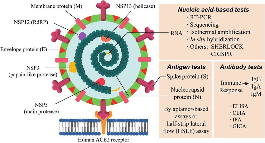
Y. Zhou et al.
                                                                                                                                                                    5

                  Fig. 1 Illustration of the SARS-CoV-2 viral genome, proteins and corresponding assays. The genome of SARS-CoV-2 is a positive single-
                  stranded RNA with more than 30,000 bp nucleotides. The capsid outside the genome is formed by the nucleocapsid protein (N) and is further
                  wrapped by an envelope composed of three structural proteins: membrane protein (M), spike protein (S), and envelope protein (E). The entry
                  of coronavirus into host cells is mediated by the S protein, which is a homotrimer protruding from the viral envelope that recognizes the
                  receptor angiotensin-converting enzyme 2 (ACE2) via the S1 receptor-binding domain (RBD) and uses the S2 domain for fusion with the host
                  cell membrane to enter host cells. In addition to these four structural proteins, SARS-CoV-2 contains sixteen nonstructural proteins (NSPs).
                  Four NSPs responsible for viral replication or transcription are shown in this illustration. NSP3 separates the translated protein. NSP5 is
                  responsible for cleaving the viral polyprotein into functional units during replication. NSP12 contains the RNA-dependent RNA polymerase
1234567890();,:

                  (RdRp). NSP13 participates in viral replication or transcription via the zinc-binding domain. ACE2 angiotensin-converting enzyme 2, E
                  envelope protein, M transmembrane protein, N nucleocapsid protein, NSP nonstructural protein, ORF open reading frame, RdRp RNA-
                  dependent RNA polymerase, S spike protein.

                  for primer design in various PCR assays. In clinical practice, at least    complementary for prevention, patient care and follow-up, as well
                  two targets are recommended to avoid potential genetic mutation            as have use in basic and translational research for combating this
                  of SARS-CoV-2 or cross-reaction with other coronaviruses, accord-          global threat (Table 1). The present review aims to provide an
                  ing to the WHO6. Three conserved regions (the E, N, and ORF1ab             overview regarding the major advantages, disadvantages, and
                  genes) are usually selected as the standard targets for the design         particular applications of currently available assays for detection,
                  of primers and probes (Fig. 2). Moreover, sequencing of the viral          prevention, mass screening, and follow-up in combating this
                  genome helps to identify new variants of coronavirus that occur            worldwide health crisis.
                  over time. Compared to traditional sequencing methods that are
                  usually very costly, newly emerging portable or quantitative
                  sequencing methods, such as nanopore target sequencing (NTS),              NUCLEIC ACID TESTING FOR COVID-19
                  may offer accurate high-throughput diagnosis during pandemics.             Quantitative reverse-transcription polymerase chain reaction
                     For serological assays, the N and S proteins are the most               As an RNA virus, the large genome needs to be reverse-
                  important targets for immunologic detection among the four                 transcribed to cDNA for PCR amplification. Hence, quantitative
                  structural proteins7. For direct detection of viral products, the N        reverse-transcription polymerase chain reaction (qRT-PCR) has
                  protein, which functions as a structural component of the helical          been deemed to be the “gold standard” for COVID-19 diagnosis,
                  nucleocapsid and plays a vital role in viral replication, is often         because it has been shown to be very sensitive for accurately
                  detected in COVID-19 patients8,9. The S protein, which is encoded          detecting the viral genome, able to detect a single copy of the
                  by the S gene, consists of two subunits, the S1 domain for receptor        viral RNA10. Three highly conserved regions have been found in
                  binding and the S2 domain for fusion, and is critical for receptor         the SARS-CoV-2 viral genome, including the RdRp, E and N gene11.
                  recognition, interaction and internalization; therefore, this protein      The assays are designed as a two-target system in which one
                  is a particular focus for studies assessing viral mutation and             primer universally detects numerous coronaviruses, including
                  spread4.                                                                   SARS-CoV-2, and a second primer set exclusively detects SARS-
                     For most individuals during the first few days of infection, viral       CoV-2. For a routine workflow, it is recommended that the E gene
                  titers are high, and a single nasopharyngeal swab may harbor up            be used as the first-line screening target, followed by confirmatory
                  to 1 million SARS-CoV-2 viral particles. However, patient IgG and          testing of the RdRp gene11. A number of RT-PCR primer and probe
                  IgM antibody production typically occurs 5–10 days after the               sets11–14 approved for SARS-CoV-2 detection by the Center of
                  onset of initial symptoms9. Therefore, nucleic acid tests offer the        Disease Control (CDC) in different countries are listed in Table 2.
                  earliest and most sensitive detection for the presence of SARS-               The general workflow of RT-PCR tests includes three main steps:
                  CoV-2 infection. For research purposes, viral proteins in infected         sample collection and transport, lysis, and RNA purification and
                  tissues or cells are solid evidence of viral replication, in addition to   amplification. These standard RT-PCR tests take approximately 3 h
                  in situ hybridization for the detection of the viral genome in             to complete3. Efforts have been made to eliminate an RNA
                  particular cell types. The titer changes in specific antibodies             purification step, which may dramatically reduce the overall
                  against viral proteins allow for monitoring the patient response to        workflow duration in several commercial kits15 (Table 3). Notably,
                  the infection, and the persistence or fluctuation in antibody levels        the high-throughput TaqPath COVID-19 Combo Kit from Thermo
                  over time postinfection. As a valid assay, immunologic detection           Fisher Scientific detects two copies of the viral genome in one µl
                  of positivity and titer changes in specific antibodies in a selected        of sample, and the detection sensitivity appears to be higher than
                  population help to determine the mass infection rate, vaccine              that of the other kits listed in the table.
                  response, and general immunity against the virus or its variants.             A study including 1014 patients found that the average interval
                  Therefore, all these assays have particular usages and may be              between the initial positive and negative RT-PCR results was 6.9 ±

                  Laboratory Investigation (2022) 102:4 – 13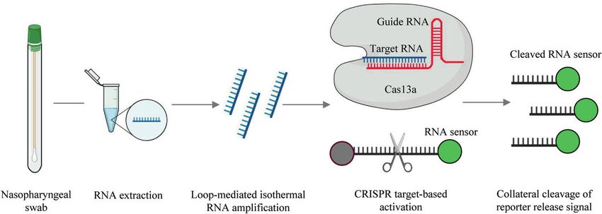
Y. Zhou et al.
6
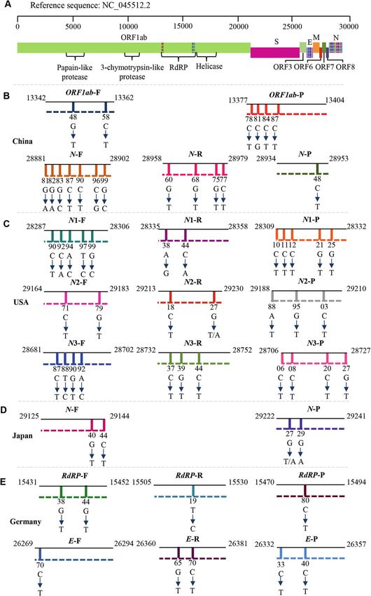
Fig. 2 Viral open reading frames (ORFs) and mutations in primer or probe regions. A Four open reading frames (ORFs) in the viral genome
are indicated for encoding viral structural or nonstructural proteins. B–E Mutations have been found in primer and probe regions in various
countries and regions. As indicated, most mutations occur in the N protein region, and the efficiency of primers and probes against the N
protein coding region will be affected by these mutations. Specific point mutations are indicated in primer or probe regions used in various
countries, and these mutations may hamper the detection efficiency of RT-PCR kits and cause false-negative results. Therefore, for the
detection of newly occurring mutated variants of the SARS-CoV-2 virus, updating specific primers and probes is essential for the reliability of
the kits. E envelope protein, F forward primer, N nucleocapsid protein; ORF open reading frame, P probe, R reverse primer, RdRp RNA-
dependent RNA polymerase.
Laboratory Investigation (2022) 102:4 – 13Y. Zhou et al.
7
Table 1. Common features of various testing methods for SARS-COV-2 infection.
Testing type Time duration (Short or longa) Suitable population (Large or small) Accuracy (High or low)
Nucleic acid testing
Quantitative RT-PCR Long Large High
Sequencing for diagnosis Long Small High
Isothermal amplification Short Large High
In situ hybridization Long Small High
Protein testing
Antibody testing Short Large High
Antigen testing Short Large Low
a
Short and long are defined by the duration (usually approximately 3 h) for the RT-PCR assay for nucleotide detection. If the test duration is longer than 3 h, it
is defined as “long”; otherwise, it is defined as “short”.
2.3 days16. In addition to upper respiratory tract nasopharyngeal log10 RNA copies/test) and nasal swabs (2 log10 RNA copies/test)21.
swabs, which are mostly used in nucleic acid detection, an In respiratory samples, viral load increases with disease severity,
observation study detected samples from the digestive tract, and the viral loads of severe patients are at a peak level (6 log10
including fecal and anal swabs, and found that the clearance time RNA copies/mL) in the third to fourth week after the onset of
of SARS-CoV-2 in the gastrointestinal tract was longer than that in symptoms, while mild patients reach a peak (5 log10 RNA copies/
the respiratory tract17, which may last up to 33 days or even mL) within one week and slowly decrease22, which suggested that
longer after a negative respiratory PCR report18. Although anal high viral load might be a risk factor for severe manifestation and
swabs have not been the official standard for diagnosis, they are a predictor of worse clinical outcome, such as death23. On the
complementary to the current detection methods to monitor other hand, the detection of viral load is a critical parameter in
clearance of the virus postinfection. Therefore, it is recommended evaluating the efficacy of newly developed medications. On
that inactivation of possible virus-infected human elimination February 9, 2021, the U.S. Food and Drug Administration issued an
samples in toilets is needed for patient management. It is crucial emergency use authorization (EUA) for two monoclonal antibodies
to choose appropriate sample types and collection routes for (bamlanivimab and etesevimab), which are two anti-spike
monitoring the clearance of SARS-CoV-2 virus. It is equally neutralizing monoclonal antibodies derived separately from two
important to determine how long infected individuals should be patients that recovered from COVID-19 in North America24 and
isolated depending on serum or fecal sample negativity. China25, to be administered together for the treatment of mild to
It is worth mentioning that to meet the needs for completion of moderate COVID-19 in adults and pediatric patients26. In clinical
nucleic acid tests in a large population, the China CDC adopted a trials, the primary outcome for characterizing the efficacy of these
pooling method in which samples from at most five different two neutralizing antibodies was the reduction in SARS-CoV-2 viral
individuals were mixed in one test tube for RT-PCR assays. Once load down to baseline, as measured by quantitative RT-PCR27.
an abnormality is detected in the results of the pooling samples,
all individual samples added to the mixed sample are tested Detection of mutated variants with standard RT-PCR kits
separately, which significantly enhances efficiency, as demon- The viral genome mutates constantly as it replicates. New variants
strated by the testing of over 10 million people that was with genetic mutations may lead to new waves of SARS-CoV-2
completed within only 19 days in a city in China19. It has been pandemic episodes. Since most of the PCR primers have been
recommended that ten samples might be combined together and designed based on the early isolated virions5, particularly the
subjected to nucleic acid extraction and RT-PCR analysis, and the reference genome (SARS-CoV-2, NC_045512.2)28, even a single
numbers of tests required are estimated based on the incidence of mutation in the middle of a primer sequence might contribute to
COVID-19 in their respective countries/regions. Compared to $58 the lower amplification efficiency of qRT-PCR tests and result in
million for the routine screening of 1 million people, the false negative results in detection29. Studies have analyzed
recommended large-scale population screening method sequencing samples submitted to GenBank and GISAID and
decreases this cost to $9.1 million20. This cost-effective and found that mutations in Germany and China have mainly occurred
time-saving approach has been widely adopted in mass screening in the ORF1ab region30,31.
tests in large populations during recent local community-spread Interestingly, another study based on 31,421 SARS-CoV-2
episodes in several cities in China. genome samples found that most of the mutations were within
False negativity in RT-PCR tests often occurs, which was the targets of the various N gene primers and probes32, and might
observed particularly during the early period of the pandemics, affect the efficiency of PCR amplification that is designed to probe
most likely due to improper sample collection, nonstandardized the N gene in RT-PCR assays. Cases have been reported that
RNA extraction, and an assessment time that was too early for detection might be interfered with due to mutations in the N
detection of positivity after contact with virus. To avoid false gene33,34. Mutations have been found in all targets of the COVID-
negative results, repeated sample collections are recommended. 19 diagnostic primers recommended by the US CDC, whereas the
For successful control of small-scale community spread, up to targets of N gene primers and probes used in Japan, Thailand, and
three nucleic acid tests within 2 weeks have identified potentially China have shown multiple mutations in different clusters, which
contagious “asymptomatic carriers”, who are to be quarantined to suggested that the N gene might not be a stable target for RT-PCR
completely end occasional episodes of local spread in a short kits and that these N gene-based kits should be updated
period. periodically for emerging alpha, beta, gamma, delta variants34
In addition to qualitative detection, viral load can also be (Fig. 2).
calculated by plotting CT values onto the standard curve provided
by the commercial RT-PCR kits. Differences exist in the viral load of Sequencing for diagnosis
different sample types, as the average viral load in sputum (4 log10 Compared to RT-PCR, viral genome sequencing has the dis-
RNA copies/test) is usually higher than that in throat swabs (3 advantages of a higher cost, larger amount of data analysis, and
Laboratory Investigation (2022) 102:4 – 13Y. Zhou et al.
8
Table 2. Sequences of RT-PCR primers and probes approved by CDCs in different countries.
Country Target gene Primer and probe Sequence (5′-3′) Position (Reference sequence: NC_045512.2)
China12 ORF1ab ORF1ab-F ccctgtgggttttacacttaa 13,342–13,362
ORF1ab-R acgattgtgcatcagctga 13,442–13,460
ORF1ab-P FAM-ccgtctgcggtatgtggaaaggttatgg-BHQ1 13,377–13,404
N N-F ggggaacttctcctgctagaat 28,881–28,902
N-R cagacattttgctctcaagctg 28,958–28,979
N-P FAM-ttgctgctgcttgacagatt-TAMRA 28,934–28,953
USA13 N N1-F gaccccaaaatcagcgaaat 28,287–28,306
N1-R tctggttactgccagttgaatctg 28,335–28,358
N1-P FAM-accccgcattacgtttggtggacc-BHQ1 28,309–28,332
N N2-F ttacaaacattggccgcaaa 29,164–29,183
N2-R gcgcgacattccgaagaa 29,213–29,230
N2-P FAM-acaattttgcccccagcgcttcag-BHQ1 29,188–29,210
N N3-F gggagccttgaatacaccaaaa 28,681–28,702
N3-R tgtagcacgattgcagcattg 28,732–28,752
N3-P FAM-aycacattggcacccgcaatcctg-BHQ1 28,706–28,727
Japan14 N N-F aaattttggggaccaggaac 29,125–29,144
N-R tggcagctgtgtaggtcaac 29,280–29,299
N-P FAM-atgtcgcgcattggcatgga-BHQ 29,222–29,241
Germany11 RdRP RdRp-F gtgaratggtcatgtgtggcgg 15,431–15,452
RdRP-R caratgttaaasacactattagcata 15,505–15,530
RdRp-P FAM-caggtggaacctcatcaggagatgc-BBQ 15,470–15,494
E E-F acaggtacgttaatagttaatagcgt 26,269–26,294
E-R atattgcagcagtacgcacaca 26,360–26,381
E-P FAM-acactagccatccttactgcgcttcg-BBQ 26,332–26,357
E envelope protein, F forward primer, N nucleocapsid protein, ORF open reading frame, P probe, R reverse primer, RdRp RNA-dependent RNA polymerase.
lower clinical efficiency, which is unsuitable for rapid detection in The combination of genomic and epidemiological analysis
mass populations. However, the first genome sequence of SARS- accelerates the detection of potential transmission events, and
CoV-2 was precisely achieved using metagenomic RNA sequen- helps to take timely measures to control and prevent widespread
cing technology28. According to the report by the WHO and China, of the virus. In addition, a possible pathogenic mechanism might
104 SARS-CoV-2 strains have been isolated and sequenced using be revealed when NTS is employed to analyze deletions and
Illumina and Nanopore technologies from the end of December other mutations in the SARS-CoV-2 genome in infected indivi-
2019 to mid-February 202035. The genome and proteome duals. Patients infected with virus with deletions mainly in ORF3a
compositions of SARS-CoV-2 have been determined, and over and ORF7a of the SARS-CoV-2 genome were observed to be
1000 similar sequences have been made available in the GISAID associated with interferon antagonism41. Moreover, a novel
and GenBank databases14. The advantage of sequencing-based molecular diagnostic tool based on Sanger sequencing technol-
detection is that viral mutations can be tracked by collecting ogy was able to detect SARS-CoV-2 RNA from viral particles
information on new strains. Sequencing of the viral genome helps suspended in transmission medium (directly added to the PCR
to identify and classify new strains of coronavirus over time35. As master mix), suggesting that RNA extraction may be skipped
the virus replicates and spreads, random mutations in the genome completely without reducing performance at a testing speed of
accumulate at a rate of approximately two per month, based on more than 1,000,000 tests per day42. With this capacity, one may
the data of closely tracking the viral evolution36. New mutant imagine that natural mutations in mass populations can be
viruses have been reported, including alpha (B.1.1.7), beta mapped at overall genome levels or specific sites during a
(B.1.351), gamma (P.1) and delta (B.1.617.2), which may pose the particular time period or within a geographic area, allowing the
risk of a much quicker spread of the virus37,38. sources and origins of the variants to be traced when analytic
Due to increasing demand, high-throughput methods or capability is in place.
portable rapid sequencing technology have been developed as
diagnostic tools for COVID-19. Nanopore target sequencing (NTS) Isothermal amplification
is fast, highly portable, and sensitive, making it attractive for RT-PCR is performed in a thermal cycle device, which is under
clinical testing. An NTS method sequencing 11 viral regions enable precise temperature control and needs a power supply. In
the detection of as few as ten viral copies/mL in 1 h of contrast, isothermal amplification technology is carried out at a
sequencing39. Compared to traditional sequencing methods, constant temperature using a specific enzyme for rapid nucleic
which are usually very costly, these newly emerging portable or acid amplification. The reaction takes place generally at 60–65 °C
quantitative methods may provide accurate high-throughput and is completed within 1 h43, conferring an analytic sensitivity
diagnosis during pandemics. A prospective genomic surveillance similar to PCR without special laboratory equipment such as a
study in the UK used NTS, enabling sample-to-report in less than thermal cycler44. The isothermal technique utilizes a recombinant
24 h, to establish real-time genomic surveillance of SARS-CoV-240. polymerase and helicase-dependent or loop-mediated isothermal
Laboratory Investigation (2022) 102:4 – 13Y. Zhou et al.
9
Table 3. Features of commercial kits for COVID-19 nucleic acid detection.
Kit name Manufacturer Target genes LOD Accuracy (%) TAT (min)
(copies/μL)
TaqPath COVID-19 high throughput Thermo Fisher S&N 2 100.0 Can run up to 8000 reactions
combo15 Scientific per day
Real-time fluorescent RT-PCR kit for BGI Genomics ORF1ab 150 100.0 180
2019- nCoV15
TRUPCR SARS-CoV-2 RT-qPCR15 Black Biotech E, N & RdRp 10 100.0 Unavailable
Allplex 2019-nCoV assay15 Seegene E, N & RdRp 4167 100.0 110
FOSUN COVID-19 RT-PCR15 Fosun E, N & ORF1ab 300 96.15 120
LabGun real-time PCR15 Lab Genomics E & RdRp 20 94.3 Unavailable
E envelope protein, LOD limit of detection, M transmembrane protein, N nucleocapsid protein, ORF open reading frame, RdRp RNA-dependent RNA
polymerase, S spike protein, TAT turn-around time.
Table 4. SARS-CoV-2 RT-LAMP tests in different laboratories.
Authors Target genes LOD (copy/reaction) Sensitivity and specificity TAT (min)
Lu et al.46 N, S and RdRp 118.6 94 and 90% 20
Huang et al.47 N, S and RdRp 20 100 and 100% 30
Yan et al.48 ORF1a/b 20 100 and 100% 60
Baek et al.49 N 100 100 and 98.70% 30
Lu et al.50 RdRp 30 100%/– 40
Jiang et al.51 ORF1b and N 500 copies/mL 91.4 and 99.5% 30
Kitawaya et al.52 N and S 10 copies/μL 100 and 97.6% 35
LOD limit of detection, N nucleocapsid protein, ORF open reading frame, RdRp RNA-dependent RNA polymerase, S spike protein, TAT turn-around time.
amplification (LAMP) and allows fast, sensitive, portable, and 19 in a maximal capacity of 100 samples per hour57. On the basis
point-of-care applications. of the two-step process, a streamlined assay that combines the
Reverse transcription LAMP (RT-LAMP) has been validated for simplified extraction of viral RNA with isothermal amplification
the detection of several RNA viruses, including influenza, Zika, and CRISPR-mediated detection, designated as SHERLOCK Testing
Ebola, and MERS. The positive reaction of LAMP is quantified in One Pot (STOP), has been developed54. A common reaction
visually by turbidity, colorimetric and fluorometric parameters, buffer that accommodates both steps has been developed, and
because the insoluble byproduct magnesium pyrophosphate the test is performed at a single temperature in less than an hour
formed during the LAMP reaction is visible to the naked eye45. with a low risk of cross-contamination.
RT-LAMP tests for SARS-CoV-2 have already been developed and Other CRISPR-based detection assays are also currently in use.
clinically validated worldwide with the shortest reaction time of One method that utilizes a custom CRISPR-Cas12a/gRNA complex,
20 min and the lowest detection limit of 20 copies of the virion and a fluorescent probe to detect target amplicons produced by
(Table 4)46–52. The advantage of RT-LAMP is point-of-care testing standard RT-PCR allows sensitive and robust detection of SARS-
without the requirement for specific equipment, which is CoV-2-positive samples, with a sample-to-answer time within 50
particularly valuable in a remote region or a large population. min and a limit of detection (LOD) of two copies per reaction58.
For the demands of a high-throughput capacity, automation for The AIOD-CRISPR system uses a pair of Cas12a-crRNA complexes,
sample handling, assay performance and result reports are the which may bind to corresponding sites close to the primer
keys for wide acceptance in pandemic epicenters. recognition sites in the target sequence, and is visually detected
by fluorescence or color change59. A plasmid containing a cDNA
Other nucleic acid-based assays representing 384 nucleotides of the N gene has already been used
SHERLOCK (specific high sensitivity enzymatic reporter unlocking) as the target to develop an all-in-one dual CRISPR-Cas12a (AIOD-
is a system using Cas13a ribonuclease for RNA detection53. The CRISPR) assay, which was shown to detect 1.3 copies of the SARS-
SHERLOCK procedure consists of two main steps: isothermal CoV-2 N gene plasmid in a visual and real-time mode within 40
target nucleic acid amplification and CRISPR-Cas13 nucleic acid min60. Another assay, known as DNA endonuclease-targeted
detection. Cas13 is an RNA-guided RNase that produces multiple CRISPR trans-reporter (DETECTR), has been designed to simulta-
cleavage sites in single-stranded areas of an RNA target with a neously reverse transcribe and isothermally amplify the RNA
specific base preference and has been shown to be useful for extracted from nasopharyngeal swabs, followed by Cas12-
nucleic acid detection in CRISPR-based systems54. CRISPR-Cas13 mediated viral detection61. The DETECTR assay provides a visual
enzymes are programmed by CRISPR RNA (crRNA) and exhibit and rapid (Y. Zhou et al.
10
Fig. 3 A schematic illustration of the SHERLOCK detection assay. Using nasopharyngeal swabs as an example, conventional RNA extraction
is used as the input and is followed by reverse transcription and loop-mediated isothermal amplification. The CRISPR-Cas13-RNA complex is
activated by binding to a complementary RNA target, while CRISPR-Cas13 exhibits nonspecific endonuclease activity, which activates and
cleaves fluorescent RNA sensors. The fluorescent RNA sensor is quenched when it is intact, whereas it emits fluorescent signals when it is
cleaved by the activated CRISPR-Cas13 complex.
they have several limitations for clinical validation. Since no
commercial kit is available, the existing CRISPR-based assays rely
Table 5. Comparison of CRISPR-based and RT-PCR assays.
on the preparation and testing of reaction components, such as
bacterial transformation and large-scale protein expression. These Parameters or CRISPR- RT-PCR assays
empirical adjustments of parameters require expertize in protein aspects based assays
purification and RNA biology. Compared to RT-PCR, the multistep Target gene E and N ORF1ab, N, E, and
nucleic acid amplification process may affect precise target RdRP
quantification in CRISPR-based assays. Raw nucleic acid extrac- TAT 30–40 min 4h
tions may be contaminated with nucleases, which may degrade
Assay results Qualitative Quantitative
viral samples and generate false-negative signals; at the same
time, they may degrade the sensing molecules and lead to false Specific instrument No Yes
positive signals. When designing crRNA, overlapping the recom- required
binase and the polymerase coding regions should be avoided so FDA or EUA approval Not yet Yes
as not to detect off-target products. Because of the preamplifica- E envelope protein, N nucleocapsid protein, EUA European emergency use
tion reaction, the risk of surface contamination might increase in authorization, FDA US Food and Drug Administration, ORF open reading
the multistep assays. Thus, for applications that do not demand frame, RdRp RNA-dependent RNA polymerase, TAT turn-around time.
speed but do demand quantitative detection, a clinically validated
detection assay, such as RT-PCR, might be a better choice. A
comparison of the CRISPR-based assays with the qRT-PCR assay is particularly to the occurrence of false negative results when using
listed in Table 5. primers and probes for the N gene target, given that previous
studies have confirmed that mutations in the N gene might lead
to interference in the test. Viral genome sequencing takes a longer
In situ hybridization for viral RNA detection in tissues or cells time at a higher expense than qRT-PCR assays and makes it
To determine which tissue or cell types are susceptible to SARS- unsuitable for diagnosis and patient screening. Instead, the
CoV-2 infection, the presence of the ACE2 receptor is the key for availability of whole viral sequence information from different
selective viral entry. ACE2 is widely present in the upper regions allows for tracing the source and mutations of SARS-CoV-2
respiratory tract and lungs; thus, the lungs are typically highly in a specific region or population. The main challenge for using
infected and are the main site for viral replication. To determine sequencing as a tool for combating the pandemic is how to
whether other tissues, such as the heart, liver, and kidneys, are reduce the detection duration and cost. RT-LAMP usually allows
also targets of infection, in addition to detection of the ACE2 detection within 1 h under isothermal conditions without the
receptor and viral proteins in these tissues by immunohistochem- need for special equipment. Moreover, the results are visible to
ical staining, detection of viral genomic RNA by in situ hybridiza- the naked eye, which is suitable for viral detection in remote areas
tion is a classic and reliable approach necessary to pinpoint viral or other places lacking thermal cyclers. In addition, new molecular
replication in the cell types of the infected tissue. For example, detection methods, such as CRISPR-based assays, focus on how to
viral RNA positivity was observed in the placenta in a SARS-CoV-2- further reduce time and cost while maintaining high sensitivity
positive pregnant woman, and maternal-fetal transmission was and specificity. For research purposes, traditional in situ hybridiza-
confirmed after infection63. Thus, in situ hybridization as a tion validates the presence of the viral genome in particular tissue
molecular approach may be used as a research tool in verifying or cell types and provides direct evidence of viral replication in
viral replication in particular cell types or tissues, and yields tissue or cell types. The selection of particular methods or modes
additional histological evidence of viral infection after antibody of detection depends largely on the population size and the
detection of viral protein by immunohistochemical staining. More purpose, such as for diagnosis, tracing, or laboratory research.
solid and direct evidence of infection would be electronic
microscopy observation of viral particles in the tissue or cells64–66.
In summary, among all the nucleic acid detection methods of PROTEIN-BASED COVID-19 DIAGNOSIS
SARS-CoV-2, qRT-PCR is more commonly used due to its higher Antibody testing
sensitivity and higher specificity, which enables rapid screening of As an increasing number of people worldwide have insisted on
a large number of specimens within a short time. As summarized maintaining social distance and staying at home, the focus of
in Tables 2–4, several commercial RT-PCR kits have been epidemic prevention and control has now shifted to extensive
developed and widely used. Special attention should be paid serological antibody testing of the population to monitor
Laboratory Investigation (2022) 102:4 – 13Y. Zhou et al.
11
Table 6. Features of different antibody tests.
Kit name Manufacturer Test type Antibody tested Sensitivity and specificity
Platelia SARS-CoV-2 total antibody assay84 Bio-Rad ELISA Total antibody 100.0 and 99.6%
COVID-19 antibody test84 Mount Sinai ELISA IgG 92.0 and 100.0%
LIAISON® SARS-CoV-2 S1/S2 IgG84 DiaSorin CLIA IgG 97.56 and 99.3%
Ortho’s VITROS® COVID-19 antigen test84 Ortho-Clinical Diagnostic CLIA IgG 75 and 100%
Eugene® SARS-CoV2 IgG/IgM rapid test85 Shanghai Eugene Biotech GICA Combo IgM/IgG 96.4 and 98.7%
Standard™ Q COVID-19 IgM/IgG duo test85 SD Biosensor GICA Combo IgM/IgG 100% and IgM 91.7%, IgG 79.2%
COVID-19 IgG/IgM rapid test85 Healgen Scientific GICA Combo IgM/IgG IgM 87.9%, IgG 97.2% and 100%
SARS-CoV-2 antibody test85 Biologix Corporation GICA Total antibody 86.43% and 99.57%
CLIA chemiluminescent immunoassay, ELISA enzyme-linked immunosorbent assay, GICA colloidal gold immunochromatographic assay, IFA immunofluorescent
assay, IgG immunoglobulin G, IgM immunoglobulin M.
population infection status, vaccine efficacy, immunity persis- the target; however, it may have better sensitivity and more
tence, and high-titer neutralizing antibody screening and collec- options for the development of assays for different purposes.
tion. These tests, such as enzyme-linked immunosorbent assay Through a SELEX selection strategy76, a specific ssDNA aptamer
(ELISA), chemiluminescent immunoassay (CLIA), immunofluores- that binds to the N protein has been suggested to be a sensitive
cent assay (IFA) and colloidal gold immune chromatographic assay and alternative probe for the detection of SARS-CoV-277. More-
(GICA), are based on targeting S protein and N protein antigens for over, another study reported that four DNA aptamers with an
the rapid detection of SARS-CoV-2 through IgM and/or IgG affinity below 5 nM were identified to bind to the N protein in a
antibodies in serum or body fluid samples (Table 6). One study sandwich-type interaction with an LOD of 1 ng/mL78. Compared to
investigated the presence of SARS-CoV-2-specific antibodies in using antibodies alone in ELISA with LODs ranging from 50 to 100
959 blood samples collected from a prospective pulmonary cancer ng/mL79,80, the LOD of aptamer-based technologies was much
screening among asymptomatic individuals in Italy between lower than that of common immunoassays in a short turn-around
September 2019 and March 2020, several months before the first time (TAT) with high repeatability and reusability76,81. Therefore,
patient was reported67. Testing indicates that SARS-CoV-2 infec- aptamer-based antigen detection may be superior to antibody-
tion was present in approximately 11.6% of the local population based assays in terms of detection sensitivity, versatility in
before COVID-19 was initially reported. biosensor conjugation for chemiluminescent or fluorescent
The usefulness of antibody tests is for population exposure detection, and much lower variation in aptamer production76,81.
studies to investigate the exposure rate before and after a major Finally, it is worth mentioning that the sensitivity of rapid
epidemic episode in a region, and to determine whether antigen detection is 103 times lower than that of virus culture and
neutralizing antibodies are developing in individuals, who have 105 times lower than that of RT-PCR82. Previous studies reported
been exposed to the virus and the duration and titer changes in that the sensitivity of the rapid antigen test is approximately only
neutralizing antibodies over time. It is critical to follow-up on 30% of nucleotide acid testing83, suggesting that antigen testing
neutralizing antibody development after vaccination since various is not a rapid method but may be used as a confirmation or
types of vaccines against SARS-CoV-2 infection are available for research assay for specific patient samples.
general populations.
Antigen testing PERSPECTIVES AND CONCLUSION
In SARS-CoV, N protein and S protein are the main immunogens, The global COVID-19 pandemic is one of the most devastating
and antibodies against these two proteins may last for 30 weeks in infectious diseases in history in terms of infection numbers and
the serum of SARS patients68. A novel antigen-based rapid test for mortality in humans, and heavily hit areas are still in combating
diagnosis showed high sensitivity and specificity mainly in the first the overwhelming hospitalization rate and fatality, although
week among symptomatic patients and samples with high viral vaccination is ongoing for high-risk populations. The emergence
load69, while a rapid method based on a fluorescent immune and epidemic of new variants in more than 20 countries have led
chromatographic assay detecting N protein demonstrated high to a high surge and more rapid transmission in affected regions.
sensitivity only in an early phase of infection70. Mass spectrometry Mutated variants may bring about new challenges in false
analysis reported the presence of N protein in gargle solution negativity in currently available diagnostic nucleic acid detection
samples of COVID-19 patients71. A fluorescent immune chromato- and in the efficacy of currently available mRNA-based, recombi-
graphic assay detected N protein in urine samples in 73.6% of nant, or inactivated vaccines. Recently, re-emerging community-
diagnosed COVID-19 patients70. Due to its late appearance, S acquired transmission in China due to the foreign travel of people
protein is more suitable for detection during the recovery or the import of goods has led to the implementation of large-
period72, and an ultrasensitive antigen test for S protein is scale population screening and tracing, which has resulted in the
conveniently performed with a microplate reader73. timely control of community-acquired and small-scale epidemics.
In addition to these common methods, the SARS-CoV-2 Therefore, identifying infectious sources, such as asymptomatic
coronavirus nucleocapsid antigen-detecting half-strip lateral flow individuals, infected individuals or contaminated goods, has
(HSLF) assay has been developed, which displays better clinical become an effective measure in containing community spread.
sensitivity than traditional serology assays, as the LOD for the As discussed, among the nucleic acid-based methods, quanti-
commercially available Genscript N protein is 3.03 ng/mL74. A tative RT-PCR is the most specific and the fastest method for
novel nanozyme-based chemiluminescent paper assay is feasible screening and diagnosis in a large population, and the sequencing
using the camera of a standard smartphone, with a LOD for of the viral genome is the most reliable method in tracing
recombinant spike antigen of SARS-CoV-2 of 0.1 ng/mL75. infectious sources, monitoring mutations, and determining
For antigen detection, a specific nucleotide aptamer against the genome types with limited capacity for selected individuals. Viral
N protein has the same specificity as an antibody for recognizing load determination by quantitative RT-PCR is valid for monitoring
Laboratory Investigation (2022) 102:4 – 13Y. Zhou et al.
12
disease progression, therapeutic efficacy, and prognosis. Emerging 11. Corman, V. M. et al. Detection of 2019 novel coronavirus (2019-nCoV) by real-time
new technologies, such as RT-LAMP and CRISPR-based assays, RT-PCR. Euro Surveill. 25, 2000045 (2020).
offer fast point-of-care testing for heavily infected or remote areas. 12. China CDC. The detection primer and probe sequences of SARS-CoV-2. http://
For research purposes, in situ RNA hybridization for direct ivdc.chinacdc.cn/kyjz/202001/t20200121_211337.html (2020).
13. US CDC. Information about emergency use authorization for 2019 novel cor-
detection of the viral RNA genome yields convincing evidence
onavirus (2019-nCoV) real-time RT-PCR diagnostic panel. https://www.cdc.gov/
of infection in specific tissue or cell types. Using specific csels/dls/locs/2020/information_about_emergency_use_authorization_for_
antibodies to detect viral proteins, such as the N or S proteins, 2019_novel_coronavirus_real_time_rt-pcr_diagnostic_panel.html (2020).
in tissue by immunohistochemical assays allows the quick 14. Udugama, B. et al. Diagnosing COVID-19: the disease and tools for detection. ACS
observation of virus-infected tissue distribution in particular Nano 14, 3822–3835 (2020).
organs, tissues or cell types. For viral protein detection, the half- 15. Garg, A. et al. Evaluation of seven commercial RT-PCR kits for COVID-19 testing in
strip lateral flow (HSLF) assay is a state-of-the-art advancement in pooled clinical specimens. J. Med. Virol. 93, 2281–2286 (2020).
providing convenient point-of-care detection in remote regions or 16. Ai, T. et al. Correlation of chest CT and RT-PCR testing for coronavirus dis-
self-service at home. Moreover, aptamer-based assays may have ease 2019 (COVID-19) in China: a report of 1014 cases. Radiology 296, E32–E40
(2020).
the same level of specificity as antibody-based assays, but possess
17. Wu, J. et al. Detection and analysis of nucleic acid in various biological samples of
a lower LOD with more options for high-throughput capacity. For COVID-19 patients. Travel Med. Infect. Dis. 37, 101673 (2020).
antibody testing, although its sensitivity is lower than that of 18. Cheung, K. S. et al. Gastrointestinal manifestations of SARS-CoV-2 infection and
quantitative RT-PCR, various methods, such as ELISA and virus load in fecal samples from a Hong Kong cohort: systematic review and
fluorescent or luminescent assays, have been developed to detect meta-analysis. Gastroenterology 159, 81–95 (2020).
IgG or IgM postinfection. These assays have wide applications in 19. The State Council Information Office of the People’s Republic of China. The 104th
tracing infected individuals in a large population and monitoring press conference on the prevention and control of COVID-19 in Hubei province.
specific antibody development and persistence. Moreover, they http://www.scio.gov.cn/xwfbh/gssxwfbh/xwfbh/hubei/document/1681627/
are useful for selecting individuals with high titers of neutralizing 1681627.htm (2020).
20. Sahajpal, N. S. et al. Proposal of RT-PCR-based mass population screening for
antibodies for therapy or prevention as a passive immunity
severe acute respiratory syndrome Coronavirus 2 (Coronavirus disease 2019). J.
approach. It is known that antibody assays are a primary method Mol. Diagn. 22, 1294–1299 (2020).
to determine immunity development and effectiveness, as well as 21. Yu, F. et al. Quantitative detection and viral load analysis of SARS-CoV-2 in
persistence after vaccination. Newly emerging assays, such as RT- infected patients. Clin. Infect. Dis. 71, 793–798 (2020).
LAMP, SHERLOCK, AIOD-CRISPR, and DETECTR, are required to 22. Zheng, S. et al. Viral load dynamics and disease severity in patients infected with
combat pandemics and evolve for specific applications. To select SARS-CoV-2 in Zhejiang province, China, January–March 2020: retrospective
the right assay for a particular usage, understanding its principles, cohort study. BMJ 369, m1443 (2020).
the advantages and disadvantages is essential to fulfill this task. 23. Chu, C. M. et al. Initial viral load and the outcomes of SARS. CMAJ 171, 1349–1352
With the availability of multiple options for the detection of viral (2004).
24. FDA. Coronavirus (COVID-19) Update: FDA authorizes monoclonal antibodies for
nucleic acid and protein or host antibody production, specific and
treatment of COVID-19, https://www.fda.gov/news-events/press-announcements/
effective assays aid in identifying infectious sources, assigning coronavirus-covid-19-update-fda-authorizes-monoclonal-antibodies-treatment-
isolation or diagnosis, and meeting research demands in covid-19-0 (2021).
combating this global pandemics, which is expected to be under 25. Jones, B. E. et al. LY-CoV555, a rapidly isolated potent neutralizing antibody,
control due to widespread vaccination in general populations in provides protection in a non-human primate model of SARS-CoV-2 infection.
the coming months or years. bioRxiv, Preprint posted online 9 October 2020, https://doi.org/10.1101/
2020.09.30.318972 (2020).
26. Shi, R. et al. A human neutralizing antibody targets the receptor-binding site of
DATA AVAILABILITY SARS-CoV-2. Nature 584, 120–124 (2020).
No original data are presented in this review article. Summaries of published data are 27. Gottlieb, R. L. et al. Effect of bamlanivimab as monotherapy or in combination
presented in Tables 1–6. with etesevimab on viral load in patients with mild to moderate COVID-19: a
randomized clinical trial. JAMA 325, 632–644 (2021).
28. Wu, F. et al. A new coronavirus associated with human respiratory disease in
China. Nature 579, 265–269 (2020).
REFERENCES
29. Bru, D., Martin-Laurent, F. & Philippot, L. Quantification of the detrimental effect
1. Zhou, P. et al. A pneumonia outbreak associated with a new coronavirus of of a single primer-template mismatch by real-time PCR using the 16S rRNA gene
probable bat origin. Nature 579, 270–273 (2020). as an example. Appl. Environ. Microbiol. 74, 1660–1663 (2008).
2. Coronaviridae Study Group of the International Committee on Taxonomy of 500 30. Farkas, C., Fuentes-Villalobos, F., Garrido, J. L., Haigh, J. & Barría, M. I. Insights on
Viruses. The species Severe acute respiratory syndrome-related coronavirus: classi- early mutational events in SARS-CoV-2 virus reveal founder effects across geo-
fying 2019-nCoV and naming it SARS-CoV-2. Nat. Microbiol. 5, 536–544 (2020). graphical regions. PeerJ 8, e9255 (2020).
3. Esbin, M. N. et al. Overcoming the bottleneck to widespread testing: a rapid 31. Koyama, T., Platt, D. & Parida, L. Variant analysis of SARS-CoV-2 genomes. Bull.
review of nucleic acid testing approaches for COVID-19 detection. RNA 26, World Health Organ. 98, 495–504 (2020).
771–783 (2020). 32. Wang, R., Hozumi, Y., Yin, C. & Wei, G. W. Mutations on COVID-19 diagnostic
4. Cui, J., Li, F. & Shi, Z. L. Origin and evolution of pathogenic coronaviruses. Nat. Rev. targets. Genomics 112, 5204–5213 (2020).
Microbiol. 17, 181–192 (2019). 33. Ziegler, K. et al. SARS-CoV-2 samples may escape detection because of a single
5. Chan, J. F. et al. Improved molecular diagnosis of COVID-19 by the novel, highly point mutation in the N gene. Euro Surveill. 25, 2001650 (2020).
sensitive and specific COVID-19-RdRp/Hel real-time reverse transcription-PCR 34. Kaden, R. Early phylogenetic diversification of SARS-CoV-2: determination of
assay validated in vitro and with clinical specimens. J. Clin. Microbiol. 58, variants and the effect on epidemiology, immunology, and diagnostics. J. Clin.
e00310–e00320 (2020). Med. 9, 1615 (2020).
6. Holshue, M. L. et al. First case of 2019 novel coronavirus in the United States. N. 35. Lu, R. et al. Genomic characterisation and epidemiology of 2019 novel cor-
Engl. J. Med. 382, 929–936 (2020). onavirus: implications for virus origins and receptor binding. Lancet 395, 565–574
7. Tang, Y. W., Schmitz, J. E., Persing, D. H. & Stratton, C. W. Laboratory diagnosis of (2020).
COVID-19: current issues and challenges. J. Clin. Microbiol. 58, e00512–e00520 36. Hadfield, J. et al. Nextstrain: real-time tracking of pathogen evolution. Bioinfor-
(2020). matics 34, 4121–4123 (2018).
8. Chan-Yeung, M. & Xu, R. H. SARS: epidemiology. Respirology 8, S9–14 (2003). 37. Tang, J. W., Tambyah, P. A. & Hui, D. S. Emergence of a new SARS-CoV-2 variant in
9. Liu, Y., Eggo, R. M. & Kucharski, A. J. Secondary attack rate and superspreading the UK. J. Infect. 82, e27–e28 (2020).
events for SARS-CoV-2. Lancet 395, e47 (2020). 38. Makoni, M. South Africa responds to new SARS-CoV-2 variant. Lancet 397, 267
10. Wang, J. et al. Novel one-step single-tube nested quantitative real-time PCR assay (2021).
for highly sensitive detection of SARS-CoV-2. Anal. Chem. 92, 9399–9404 (2020).
Laboratory Investigation (2022) 102:4 – 13Y. Zhou et al.
13
39. Wang, M. et al. Nanopore target sequencing for accurate and comprehensive 68. Qiu, M. et al. Antibody responses to individual proteins of SARS coronavirus and
detection of SARS-CoV-2 and other respiratory viruses. Small 16, e2002169 their neutralization activities. Microbes Infect. 7, 882–889 (2005).
(2020). 69. Porte, L. et al. Evaluation of a novel antigen-based rapid detection test for the
40. Meredith, L. W. et al. Rapid implementation of SARS-CoV-2 sequencing to diagnosis of SARS-CoV-2 in respiratory samples. Int. J. Infect. Dis. 99, 328–333
investigate cases of health-care associated COVID-19: a prospective genomic (2020).
surveillance study. Lancet Infect. Dis. 20, 1263–1272 (2020). 70. Diao, B. et al. Accuracy of a nucleocapsid protein antigen rapid test in the
41. Moore, S. C. et al. Amplicon-based detection and sequencing of SARS-CoV-2 in diagnosis of SARS-CoV-2 infection. Clin. Microbiol. Infect. 27, 289.e1–289.e4 (2020).
nasopharyngeal swabs from patients with COVID-19 and identification of dele- 71. Ihling, C. et al. Mass spectrometric identification of SARS-CoV-2 proteins from
tions in the viral genome that encode proteins involved in interferon antagon- gargle solution samples of COVID-19 patients. J. Proteome Res. 19, 4389–4392
ism. Viruses 12, 1164 (2020). (2020).
42. Chandler-Brown, D., Bueno, A. M., Atay, O. & Tsao, D. S. A highly scalable and 72. Tan, Y. J. et al. Profiles of antibody responses against severe acute respiratory
rapidly deployable RNA extraction-free COVID-19 assay by quantitative Sanger syndrome coronavirus recombinant proteins and their potential use as diagnostic
sequencing. bioRxiv https://doi.org/10.1101/2020.04.07.029199 (2020). markers. Clin. Diagn. Lab. Immunol. 11, 362–371 (2004).
43. Notomi, T. et al. Loop-mediated isothermal amplification of DNA. Nucleic Acids 73. Kyosei, Y. et al. Proposal of de novo antigen test for COVID-19: ultrasensitive
Res. 28, E63 (2000). detection of Spike proteins of SARS-CoV-2. Diagnostics 10, 594 (2020).
44. Craw, P. & Balachandran, W. Isothermal nucleic acid amplification technologies 74. Grant, B. D. et al. SARS-CoV-2 coronavirus nucleocapsid antigen-detecting half-
for point-of-care diagnostics: a critical review. Lab Chip 12, 2469–86 (2012). strip lateral flow assay toward the development of point of care tests using
45. Mori, Y. & Notomi, T. Loop-mediated isothermal amplification (LAMP): a rapid, commercially available reagents. Anal. Chem. 92, 11305–11309 (2020).
accurate, and cost-effective diagnostic method for infectious diseases. J. Infect. 75. Liu, D. et al. Nanozyme chemiluminescence paper test for rapid and sensitive
Chemother. 15, 62–69 (2009). detection of SARS-CoV-2 antigen. Biosens. Bioelectron. 173, 112817 (2020).
46. Lu, R. et al. A novel reverse transcription loop-mediated isothermal amplification 76. Li, H. Y. et al. Advances in detection of infectious agents by aptamer-based
method for rapid detection of SARS-CoV-2. Int. J. Mol. Sci. 21, 2826 (2020). technologies. Emerg. Microbes Infect. 9, 1671–1681 (2020).
47. Huang, W. E. et al. RT-LAMP for rapid diagnosis of coronavirus SARS-CoV-2. 77. Cho, S. J., Woo, H. M., Kim, K. S., Oh, J. W. & Jeong, Y. J. Novel system for detecting
Microb. Biotechnol. 13, 950–961 (2020). SARS coronavirus nucleocapsid protein using an ssDNA aptamer. J. Biosci. Bioeng.
48. Yan, C. et al. Rapid and visual detection of 2019 novel coronavirus (SARS-CoV-2) 112, 535–540 (2011).
by a reverse transcription loop-mediated isothermal amplification assay. Clin. 78. Zhang, L. et al. Discovery of sandwich type COVID-19 nucleocapsid protein DNA
Microbiol. Infect. 26, 773–779 (2020). aptamers. Chem. Commun. 56, 10235–10238 (2020).
49. Baek, Y. H. et al. Development of a reverse transcription-loop-mediated iso- 79. Mak, G. C. K. et al. Evaluation of rapid antigen detection kit from the WHO
thermal amplification as a rapid early-detection method for novel SARS-CoV-2. emergency use list for detecting SARS-CoV-2. J. Clin. Virol. 134, 104712 (2021).
Emerg. Microbes Infect. 9, 998–1007 (2020). 80. Mak, G. C. et al. Analytical sensitivity and clinical sensitivity of the three rapid
50. Lu, R. et al. Development of a novel reverse transcription loop-mediated iso- antigen detection kits for detection of SARS-CoV-2 virus. J. Clin. Virol. 133, 104684
thermal amplification method for rapid detection of SARS-CoV-2. Virol. Sin. 35, (2020).
344–347 (2020). 81. Li, H. Y. et al. Novel aptasensor-based assay of sonic hedgehog ligand for
51. Jiang, M. et al. Development and validation of a rapid, single-step reverse tran- detection of portal vein invasion of hepatocellular carcinoma. Biosens. Bioelectron.
scriptase loop-mediated isothermal amplification (RT-LAMP) system potentially to 174, 112738 (2021).
be used for reliable and high-throughput screening of COVID-19. Front. Cell Infect. 82. Mak, G. C. et al. Evaluation of rapid antigen test for detection of SARS-CoV-2 virus.
Microbiol. 10, 331 (2020). J. Clin. Virol. 129, 104500 (2020).
52. Kitagawa, Y. et al. Evaluation of rapid diagnosis of novel coronavirus disease 83. Scohy, A. et al. Low performance of rapid antigen detection test as frontline
(COVID-19) using loop-mediated isothermal amplification. J. Clin. Virol. 129, testing for COVID-19 diagnosis. J. Clin. Virol. 129, 104455 (2020).
104446 (2020). 84. Lisboa, B. M. et al. Diagnostic accuracy of serological tests for covid-19: systematic
53. Kellner, M. J., Koob, J. G., Gootenberg, J. S., Abudayyeh, O. O. & Zhang, F. review and meta-analysis. BMJ 370, m2516 (2020).
SHERLOCK: nucleic acid detection with CRISPR nucleases. Nat. Protoc. 14, 85. Zainol, R. Z., Othman, S. N., Abdul, S. M. N., Ali, U. K. & Wong, K. K. Diagnostic
2986–3012 (2019). performance of COVID-19 serology assays. Malays. J. Pathol. 42, 13–21 (2020).
54. Chen, J. A.-O. et al. CRISPR-Cas12a target binding unleashes indiscriminate single-
stranded DNase activity. Science 360, 436–439 (2018).
55. O’Connell, M. R. Molecular mechanisms of RNA targeting by Cas13-containing ACKNOWLEDGEMENTS
type VI CRISPR-Cas systems. J. Mol. Biol. 431, 66–87 (2019). This work is supported by the National Natural Science Foundation of China (NSFC: #
56. Gootenberg, J. S. et al. Nucleic acid detection with CRISPR-Cas13a/C2c2. Science 81871997 and # 82170624 to J.W.).
356, 438–442 (2017).
57. Joung, J. et al. Point-of-care testing for COVID-19 using SHERLOCK diagnostics.
medRxiv https://doi.org/10.1101/2020.05.04.20091231 (2020).
58. Huang, Z. et al. Ultra-sensitive and high-throughput CRISPR-p owered COVID-19 AUTHOR CONTRIBUTIONS
diagnosis. Biosens. Bioelectron. 164, 112316 (2020). Y.Z.: manuscript preparation; L.Z.: participation in the literature analysis and
59. Jeon, Y. et al. Direct observation of DNA target searching and cleavage by manuscript preparation; Y.X. and J.W.: overall supervision, concept development,
CRISPR-Cas12a. Nat. Commun. 9, 2777 (2018). and manuscript finalization.
60. Ding, X., Yin, K., Li, Z. & Liu, C. All-in-one dual CRISPR-Cas12a (AIOD-CRISPR) assay:
a case for rapid, ultrasensitive and visual detection of novel coronavirus SARS-
CoV-2 and HIV virus. bioRxiv https://doi.org/10.1101/2020.03.19.998724 (2020). COMPETING INTERESTS
61. Broughton, J. A.-O. et al. CRISPR-Cas12-based detection of SARS-CoV-2. Nat. The authors declare no competing interests.
Biotechnol. 38, 870–874 (2020).
62. Myhrvold, C. A.-O. X. et al. Field-deployable viral diagnostics using CRISPR-Cas13.
ETHICAL APPROVAL
Science 360, 444–448 (2018).
No human subjects or experimental animals were involved in this manuscript.
63. Facchetti, F. et al. SARS-CoV2 vertical transmission with adverse effects on the
newborn revealed through integrated immunohistochemical, electron micro-
scopy and molecular analyses of Placenta. EBioMedicine 59, 102951 (2020).
64. Nicholls, J. M. et al. Lung pathology of fatal severe acute respiratory syndrome.
ADDITIONAL INFORMATION
Lancet 361, 1773–1778 (2003). Correspondence and requests for materials should be addressed to You-Hua Xie or
65. Bradley, B. T. et al. Histopathology and ultrastructural findings of fatal COVID-19 Jian Wu.
infections in Washington State: a case series. Lancet 396, 320–332 (2020).
66. Varga, Z. et al. Endothelial cell infection and endotheliitis in COVID-19. Lancet Reprints and permission information is available at http://www.nature.com/
395, 1417–1418 (2020). reprints
67. Apolone, G. et al. Unexpected detection of SARS-CoV-2 antibodies in the pre-
pandemic period in Italy. Tumori https://doi.org/10.1177/0300891620974755 Publisher’s note Springer Nature remains neutral with regard to jurisdictional claims
(2020). in published maps and institutional affiliations.
Laboratory Investigation (2022) 102:4 – 13You can also read