An ECM-Mimetic Hydrogel to Promote the Therapeutic Efficacy of Osteoblast-Derived Extracellular Vesicles for Bone Regeneration
←
→
Page content transcription
If your browser does not render page correctly, please read the page content below
ORIGINAL RESEARCH
                                                                                                                                                published: 30 March 2022
                                                                                                                                          doi: 10.3389/fbioe.2022.829969

                                           An ECM-Mimetic Hydrogel to Promote
                                           the Therapeutic Efficacy of
                                           Osteoblast-Derived Extracellular
                                           Vesicles for Bone Regeneration
                                           Kenny Man 1, Mathieu Y. Brunet 1, Angelica S. Federici 2,3,4, David A. Hoey 2,3,4 and
                                           Sophie C. Cox 1*
                                           1
                                            School of Chemical Engineering, University of Birmingham, Birmingham, United Kingdom, 2Trinity Centre for Biomedical
                                           Engineering, Trinity Biomedical Sciences Institute, Trinity College Dublin, Dublin, Ireland, 3Dept. of Mechanical, Manufacturing,
                                           and Biomedical Engineering, School of Engineering, Trinity College Dublin, Dublin, Ireland, 4Advanced Materials and
                                           Bioengineering Research Centre, Trinity College Dublin and RCSI, Dublin, Ireland

                                           The use of extracellular vesicles (EVs) is emerging as a promising acellular approach for
                                           bone regeneration, overcoming translational hurdles associated with cell-based therapies.
                                           Despite their potential, EVs short half-life following systemic administration hinders their
                           Edited by:      therapeutic efficacy. EVs have been reported to bind to extracellular matrix (ECM) proteins
                      Nuno M. Neves,       and play an essential role in matrix mineralisation. Chitosan and collagen type I are
         University of Minho, Portugal
                                           naturally-derived pro-osteogenic biomaterials, which have been demonstrated to control
                        Reviewed by:
                             Dake Hao,
                                           EV release kinetics. Therefore, this study aimed to develop an injectable ECM-mimetic
        University of California, Davis,   hydrogel capable of controlling the release of osteoblast-derived EVs to promote bone
                         United States     repair. Pure chitosan hydrogels significantly enhanced compressive modulus (2.48-fold)
                      Lisa Jane White,
            University of Nottingham,      and osteogenic differentiation (3.07-fold), whilst reducing gelation times (2.09-fold) and
                      United Kingdom       proliferation (2.7-fold) compared to pure collagen gels (p ≤ 0.001). EV release was strongly
                   *Correspondence:        associated with collagen concentration (R2 > 0.94), where a significantly increased EV
                        Sophie C. Cox
                  s.c.cox@bham.ac.uk
                                           release profile was observed from chitosan containing gels using the CD63 ELISA (p ≤
                                           0.001). Hydrogel-released EVs enhanced human bone marrow stromal cells (hBMSCs)
                    Specialty section:     proliferation (1.12-fold), migration (2.55-fold), and mineralisation (3.25-fold) compared to
         This article was submitted to
                                           untreated cells (p ≤ 0.001). Importantly, EV-functionalised chitosan-collagen composites
 Tissue Engineering and Regenerative
                              Medicine,    significantly promoted hBMSCs extracellular matrix mineralisation when compared to the
                a section of the journal   EV-free gels in a dose-dependent manner (p ≤ 0.001). Taken together, these findings
       Frontiers in Bioengineering and
                         Biotechnology
                                           demonstrate the development of a pro-osteogenic thermosensitive chitosan-collagen
       Received: 06 December 2021
                                           hydrogel capable of enhancing the therapeutic efficacy of osteoblast-derived EVs as a
          Accepted: 11 March 2022          novel acellular tool for bone augmentation strategy.
         Published: 30 March 2022
                                           Keywords: extracellular vesicle, bone, controlled release, hydrogel, tissue engineering, drug delivery
                              Citation:
      Man K, Brunet MY, Federici AS,
Hoey DA and Cox SC (2022) An ECM-
    Mimetic Hydrogel to Promote the
                                           1 INTRODUCTION
   Therapeutic Efficacy of Osteoblast-
      Derived Extracellular Vesicles for
                                           There is a critical calling for therapeutic strategies to repair bone damage caused by traumatic injury,
                  Bone Regeneration.       tumour resection, or other age-associated diseases such as osteoporosis (Baroli, 2009; Dimitriou
Front. Bioeng. Biotechnol. 10:829969.      et al., 2011). Approximately 10 million people suffer from musculoskeletal disorders in the
     doi: 10.3389/fbioe.2022.829969        United Kingdom, costing the National Health Service £5 billion annually (Chance-Larsen et al.,

Frontiers in Bioengineering and Biotechnology | www.frontiersin.org                 1                                            March 2022 | Volume 10 | Article 829969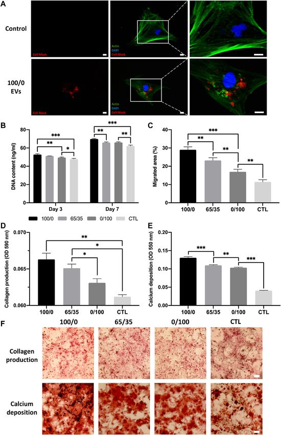
Man et al.                                                                                ECM-Mimetic Hydrogel Promotes Vesicle-Induced Mineralisation

 GRAPHICAL ABSTRACT |

2019). Alarmingly, this is expected to escalate further in the future       transplantation of MSC-based therapies, however are associated
as a result of the growing ageing population and demand for                 with several complications including their uncontrolled
continued quality of life, exacerbating the medical, and socio-             differentiation, immunological rejection, their inherent
economic burden worldwide. Traditional treatments such as                   heterogeneity, functional tissue engraftment, and neoplasm
autogenous or allogenous bone grafts have been seen as the                  formation (Amariglio et al., 2009; Herberts et al., 2011).
gold standard for many decades, however, their use is                       Moreover, the translation of cell-based therapies to the clinical
associated with limitations such as donor site morbidity,                   setting is hindered by significant hurdles associated with intensive
limited availability and other complications (Betz, 2002;                   cost, government regulations, and ethical issues (Heathman et al.,
Roberts and Rosenbaum, 2012). Bone graft substitutes                        2015). Consequently, there is great precedence to develop new
combined with hyper-concentrated osteoinductive growth                      treatments that retain the therapeutic effects of cell-based
factors have been utilised clinically with positive results                 therapies.
(Roberts and Rosenbaum, 2012). For example, bone                                In recent years, growing evidence has shown that bioactive
morphogenic protein 2 (BMP2) loaded collagen scaffolds                      factors secreted from cells play a critical role in the activation of
(INFUSE®) has been widely used by orthopaedic surgeons to                   stem/progenitor cell-mediated tissue repair (Kim et al., 2010; Li
promote fracture repair (Gresham et al., 2021). However, these              et al., 2016). Extracellular vesicles (EVs) are considered one of the
supraphysiological doses of BMP2 can result in severe                       most important secretory products of cells, involved in numerous
complications including heterotopic ossification, hematoma,                  trophic, and immunomodulatory processes. EVs are cell-secreted
inflammation, and myelopathy that may require further                        lipid nanoparticles that contain a diverse biological cargo
surgical interventions (Epstein, 2013; Hustedt and Blizzard,                including nucleic acids, proteins, and bioactive molecules
2014; James et al., 2016). Hence, there is a tremendous need                (Raposo and Stoorvogel, 2013; Börger et al., 2017; Man et al.,
for new approaches to rapidly regenerate bone, overcoming the               2020). These nano-sized vesicles are thought to be heavily
limitations of current clinical strategies (Giannoudis et al., 2013).       involved in intercellular communications, regulating tissue
   Tissue engineering approaches for bone regeneration have                 homeostasis and development (Yoon et al., 2014). The
been subject to extensive research in recent decades, with                  beneficial effects once attributed to cells are now thought to be
methods combining the use of cells with osteoinductive                      partially due to the bioactive factors delivered by EVs (Gnecchi
biomaterials as a promising strategy to promote osteogenesis.               et al., 2005; Xin et al., 2014). Extensive research has investigated
Cell-based therapies commonly utilise mesenchymal stromal cells             the use of EV-based therapeutics due to several advantageous
(MSCs) due to their ease of procurement from numerous tissues               properties when compared to cell-based treatments. For example,
and their multipotency (Marolt Presen et al., 2019; Man et al.,             when compared to cells, EVs nanoscale size promotes
2021a). The use of these treatments are attractive as they attempt          administration, decreases vascular occlusion, and macrophage
to mimic the body’s endogenous repair mechanisms, with some                 phagocytosis (van den Boorn et al., 2011; EL Andaloussi et al.,
promising results observed (Tatara and Mikos, 2016). The direct             2013). Additionally, EVs possess high physiochemical stability

Frontiers in Bioengineering and Biotechnology | www.frontiersin.org     2                                      March 2022 | Volume 10 | Article 829969
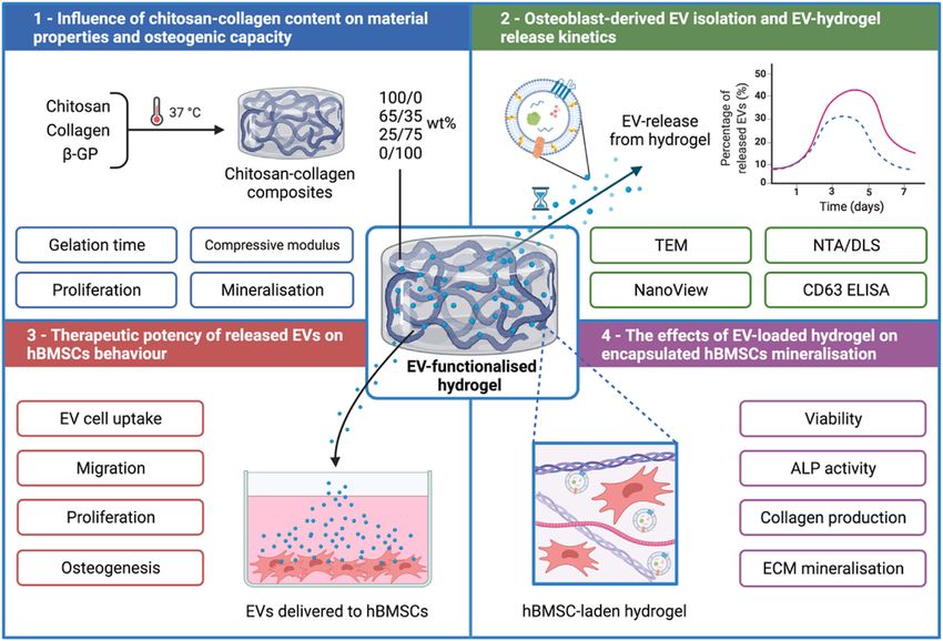
Man et al.                                                                                                  ECM-Mimetic Hydrogel Promotes Vesicle-Induced Mineralisation

 FIGURE 1 | Experimental outline detailing the development of a chitosan-collagen composite hydrogel for promoting EV therapeutic efficacy for site specific bone
 regeneration. 1) The influence of chitosan-collagen hydrogel content on material properties and osteogenic differentiation. 2) EV isolation, characterisation, and hydrogel-
 EV release kinetics. 3) The biological efficacy of hydrogel-released EVs on hBMSCs behaviour. 4) The influence of EV-functionalised hydrogel on encapsulated hBMSCs
 mineralisation. Created with BioRender.com.

and innate biocompatibility when compared to similarly sized                              involved in bone regeneration (Grosso et al., 2017; Marrella
delivery vehicles such as artificial nanoparticles (Clayton et al.,                        et al., 2018). Previously, we reported that EVs derived from
2003; Man et al., 2020). These naturally-derived nanoparticles are                        mineralising osteoblast were enriched with pro-mineralising
thought to be fundamentally involved in bone development, as                              proteins such as calcium channelling Annexins proteins, and
matrix-bound EVs are critical for endochondral ossification                                several pro-osteogenic microRNA species (Davies et al., 2017;
(Ferreira and Porter, 2018; Ansari et al., 2021). Moreover, EVs                           Man et al., 2021b). Hence, there have been intensive
derived from bone cells have been found to play a role in                                 investigations into the role EVs may play as novel acellular
mediating intercellular communication, regulating bone                                    tools that support natural bone regeneration, overcoming the
homeostasis (Gao et al., 2018; Tao and Guo, 2019).                                        tremendous regulatory hurdles associated with the translation of
Specifically, it has been shown that osteoblast-EVs contain                                cell-based therapies (Sensebé et al., 2013; Heathman et al., 2015).
RANKL, can stimulate RANKL-RANK signalling to promote                                         Several studies have demonstrated the osteoinductive capacity
osteoclast formation (Deng et al., 2015). In addition, RANK-                              of EVs derived from stem/progenitor cells as an acellular
containing osteoclast-EVs may obstruct RANKL-containing                                   approach to bone tissue engineering (BTE) (Davies et al.,
osteoblast-EVs function, thus inhibiting osteoclast formation                             2017; Yan et al., 2020; Man et al., 2021b; Man et al., 2021c).
(Huynh et al., 2016).Thus, harnessing these nanoparticles for                             Although the potential of these nanoparticles have been shown, as
regenerative medicine is an attractive acellular, but biological                          with the administration of any bioactive molecules, controlling
approach to recapitulate endogenous bone repair. Moreover, due                            the half-life of EVs at the defect site is essential to therapeutic
to the limitations of current single growth factor treatments (e.g.,                      efficacy (Man et al., 2020). Previous studies have reported the
BMP2) (Hustedt and Blizzard, 2014), the diverse biological cargo                          rapid clearance of systemically administrated EVs, which
delivered by EVs presents a multitargeted strategy to stimulate                           accumulate in the liver and lungs and are sequestered by
tissue repair, an approach which is needed given the known                                circulating macrophages in the reticuloendothelial systems and
synergistic role of angiogenesis, osteogenesis, innervation                               cleared from the body (Imai et al., 2015). Moreover, the direct

Frontiers in Bioengineering and Biotechnology | www.frontiersin.org                   3                                             March 2022 | Volume 10 | Article 829969
Man et al.                                                                                ECM-Mimetic Hydrogel Promotes Vesicle-Induced Mineralisation

administration of EVs into the site of injury has only transient             found in the bone matrix and has been widely utilised as a
effects as they are rapidly cleared from the defect site, often              biomaterial since it supports cell adhesion, proliferation, and
requiring subsequent injections to be clinically effective (Zhang            differentiation (Yang et al., 2004; Glowacki and Mizuno, 2008).
et al., 2016). As such, there are growing investigations exploring           Moreover, the integrin-mediated adhesion to collagen has been
the delivery of EVs within biomaterials to promote their                     shown to enhance stem cell osteogenic differentiation (Salasznyk
bioavailability in vivo, ultimately improving their therapeutic              et al., 2004; Kundu and Putnam, 2006). This interaction has also
efficacy (Brennan et al., 2020). The use of injectable polymeric              been used to immobilise EVs within collagen hydrogels for
biomaterials presents an attractive platform to tailor the release of        different clinical applications (Buzás et al., 2018; Ramírez
these naturally-derived nanoparticles for different clinical                 et al., 2020). Additionally, it has been reported that the
requirements, in addition to allowing for minimally invasive                 calcium binding Annexin proteins found on the surface of
delivery (Kearney and Mooney, 2013). For example, stem cell-                 mineralising vesicles are essential in mediating proteoliposome
derived EVs substantially improved cardiac function in mice                  attachment to collagen fibrils (Kirsch et al., 2000; Davies et al.,
following myocardial infarction when delivered in a gelatin                  2017). Hence, an ECM-mimetic composite hydrogel consisting of
methacryloyl (GelMA) hydrogel when compared to saline                        chitosan and type I collagen could control EV release kinetics and
solution (Tang et al., 2021). Although several reports have                  provide an osteoinductive delivery system to promote the
demonstrated the sustained release kinetics of EVs from                      therapeutic efficacy for EVs for bone repair.
biomaterial systems (Mol et al., 2019; Nikravesh et al., 2019),                  Therefore, in this present study, we developed an injectable
there have been limited studies investigating the delivery of these          thermo-responsive chitosan-collagen composite hydrogel with
nanoparticles within pro-osteoinductive materials to facilitate              sustained EV release kinetics to promote bone formation.
EV-induced bone formation. Hence, there is a current unmet                   Initially, the influence of augmenting chitosan-collagen content
clinical need to locally deliver these naturally-derived                     on the hydrogel’s gelation time, mechanical strength and
nanoparticles within biomaterials systems that control their                 osteoinduction was investigated (Figure 1). EVs were isolated
release kinetics in vivo, in addition to promoting EV-induced                from mineralising osteoblasts and their release kinetics from
bone formation.                                                              these composite gels were analysed via a CD63 ELISA. The
    Numerous natural and synthetic polymeric materials have                  biological efficacy of hydrogel-released EVs on human bone
been shown to exhibit pro-osteoinductive properties (Bai et al.,             marrow stromal cells (hBMSCs) proliferation and osteogenic
2018; Zhang et al., 2020). It has been reported that secreted EVs            differentiation was investigated. Finally, the effects of EV-
are sequestered by extracellular matrix (ECM) components                     functionalised     hydrogels     on    encapsulated      hBMSCs
(Huang et al., 2016; Man et al., 2020), therefore, the delivery              mineralisation was assessed.
of these nanoparticles within an ECM-mimetic biomaterial could
provide a template for EV-induced mineralisation. Chitosan, a
natural polysaccharide derived from deacetylated chitin, a                   2 MATERIALS AND METHODS
structural component of crustacean exoskeletons (Saravanan
et al., 2019), has been used for several applications due to its             2.1 Preparation of Chitosan-Collagen
biocompatibility, antibacterial activity and low immunogenicity              Hydrogels
(Chung et al., 2012; Ahmadi et al., 2015). The polysaccharide unit           Rat tail collagen type I (Corning, United Kingdom) was diluted in
of chitosan structurally resembles glycosaminoglycans (GAGs), a              0.02 M acetic acid to obtain a 4 mg/ml concentration. Medium
major component of the bone matrix (Khor and Lim, 2003). The                 molecular weight chitosan (Sigma-Aldrich, United Kingdom)
cationic nature of chitosan is attributed to its biological properties       was dissolved in 0.1 M acetic acid to obtain a 2 wt% chitosan
such as antimicrobial activity, hemostasis and osteoconduction               stock solution. These two solutions were mixed at various
(Peschel et al., 2012). Additionally, the existence of hydroxyl and          chitosan/collagen ratios of 100/0, 65/35, 25/75, or 0/100 wt%.
amino groups has been extensively exploited for the delivery of              Pre-cooled 58 wt% β-GP (Sigma-Aldrich, United Kingdom)
various growth factors or drugs (Zhang et al., 2010; Saravanan               solution was added to the above chitosan/collagen solutions to
et al., 2019). Several studies have reported the enhanced                    obtain a final β-GP concentration of 8%. All procedures were
therapeutic administration of EVs within thermosensitive                     conducted on ice to maintain a liquid state before initiating
chitosan hydrogels for numerous applications (Li et al., 2018;               gelation at 37°C. Gelation was determined by the mobility of
Shen et al., 2020; Zhao et al., 2021), harnessing the electrostatic          chitosan-collagen solutions after inverting a test tube and the
interactions between cationic chitosan and the anionic EVs. To               transition from clear to opaque transparency. The pH of the
induced thermo-gelation within chitosan, the addition of β-                  hydrogels was recorded before and after gelation.
glycerophosphate (β-GP) has commonly be employed to
facilitate its sol-gel transition at physiological temperatures              2.2 Mechanical Testing
(Kong et al., 2018; Rahmanian-Devin et al., 2021). Moreover,                 The Young’s modulus of the composite gels was assessed via
it has been previously reported that EVs require a phosphate-rich            cyclic testing using the Instron 5542 mechanical tester (Instron,
environment to stimulate hBMSCs mineralisation (Davies et al.,               United States). Cylindrical hydrogels (Ø8 mm × 2 mm) were
2017). In BTE, chitosan has been combined with other materials               prepared as previously described and incubated in Phosphate
to increase its osteoconductivity and mechanical strength (Di                buffered saline (PBS, Lonza, United Kingdom) for 4 h prior to
Martino et al., 2005). Type I collagen is the fundamental protein            testing. Compression testing was performed at a rate of 1 mm/

Frontiers in Bioengineering and Biotechnology | www.frontiersin.org      4                                     March 2022 | Volume 10 | Article 829969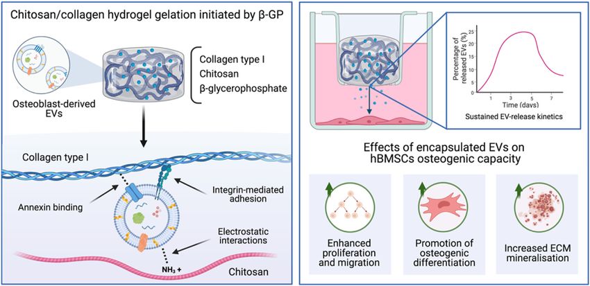
Man et al.                                                                               ECM-Mimetic Hydrogel Promotes Vesicle-Induced Mineralisation

min to a maximum strain of 60% of the original height by                   EVs. The supernatant was removed, and the pellet was washed in
performing 8 cycles of loading/unloading. The load (N) and                 sterile PBS and centrifuged at 120,000 g for 70 min and the
compressive strain (mm) was assessed using the Bluehill 3                  resultant pellet was re-suspended in 500 μL PBS. All
software. The Young’s modulus was calculated from the slope                ultracentrifugation steps were performed utilising the Sorvall
of the linear region of the stress (kPa)/strain (mm/mm) curves             WX Ultra Series Ultracentrifuge (Thermo Scientific,
from the 8th cycle. Samples were tested in triplicate for each             United Kingdom) and a Fiberlite, F50L-8×39 fixed angle rotor
condition.                                                                 (Piramoon Technologies Inc., United States).

2.3 Cell Culture and Reagents                                              2.5.2 Particle Size and Concentration Analysis
MC3T3 pre-osteoblasts were acquired from American Type                     Nanoparticle tracking analysis was performed on isolated EVs to
Culture Collection (ATCC, United Kingdom) and hBMSCs                       determine particle concentration using a ZetaView® instrument
were purchased from Lonza (Lonza, United Kingdom). Cells                   (Particle Metrix, Germany). EV samples were diluted at 1:100 in
were cultured in basal media consisting of minimal essential               PBS and injected into the ZetaView®, where 4 × 40 s videos were
medium (α-MEM; Sigma-Aldrich, United Kingdom)                              obtained of particles in motion. Particle size and concentration
supplemented with 10% foetal bovine serum (FBS), 1%                        were determined with the ZetaView® software. Dynamic Light
penicillin/streptomycin (Sigma-Aldrich, United Kingdom), and               Scattering (DLS) (Zetasizer Nano ZS, Malvern Instruments,
L-glutamine (Sigma-Aldrich, United Kingdom). hBMSCs were                   United Kingdom) was used to analyse the size distribution of
used at passage 4. Osteogenic medium was comprised of basal                EVs before and after incorporation within the different hydrogels.
media supplemented with 50 μg/ml L-ascorbic acid (Sigma-
Aldrich, United Kingdom) and 10 mM β-GP (Sigma-Aldrich,                    2.5.3 Transmission Electron Microscopy
United Kingdom). The medium used for EV isolation and the                  The morphology of isolated EVs was conducted via a JEOL
culture of EV-functionalised hydrogels was depleted of FBS-                JEM1400 transmission electron microscope (TEM) coupled
derived EVs via ultracentrifugation at 120,000 g for 16 h prior            with an AMT XR80 digital acquisition system. Samples were
to use.                                                                    physiosorbed to 200 mesh carbon-coated copper formvar grids
                                                                           (Agar Scientific, United Kingdom) and negatively stained with
                                                                           1% uranyl acetate.
2.4 The Biological Efficacy of
Chitosan-Collagen Composite Hydrogel                                       2.5.4 EV Marker Analysis
The influence of hydrogel composition on proliferation was                  The presence of tetraspanin markers on the surface of EVs was
assessed via quantification of DNA content. Briefly, MC3T3s
were mixed at low density (5 × 10⁵ cell/ml) in the hydrogel prior
                                                                           assessed using the ExoView        ™ Tetraspanin Kit (NanoView
                                                                           Biosciences, United States) according to the manufacturers’
to gelation. Following sol-gel transition, cell-laden hydrogels were       protocol and as previously described (Gupta et al., 2020; Luo
cultured in basal medium for 2 weeks with media changes every              et al., 2021). Briefly, 35 µL of EV suspension (1:1,000 dilution in
3 days. The cellular morphology was assessed at day 3 by
incubated cell-laden hydrogels with Calcein-AM (1 μg/ml in
                                                                           Incubation Solution) was incubated on the ExoView chip for
                                                                           16 h. The chip was washed with Incubation Solution for 3 min,
                                                                                                                                        ™
PBS, Sigma-Aldrich, United Kingdom) and Propidium iodide                   three times using Wash Solution and then with deionized water.
(1 μg/ml in PBS, Sigma-Aldrich, United Kingdom) in the dark for            The chip was then dried and analysed using the ExoView R100
30 min. Samples were observed under an EVOS fluorescent                     (NanoView Biosciences, United States) with the nScan software
inverted microscope (Thermo Scientific, United Kingdom).                    (version 2.8.10). Using single particle interferometric reflectance
The DNA content was assessed using PicoGreen (Life                         imaging sensing (SP-IRIS), CD9 and/or CD81 tetraspanin-
Technologies, United Kingdom) according to the                             positive nanoparticles were detected and counted spot by spot
manufacturer’s protocol.                                                   as they were immuno-captured on the chip. Rat IgG spots were
   To evaluate the hydrogel effect on osteoinduction, MC3T3s               used as an isotype control. The data were analysed using the
were mixed at high density (1 × 10⁶ cell/ml) in the hydrogel prior         NanoViewer software (version 2.8.10) with sizing thresholds set
to gelation. Following sol-gel transition, hydrogels were                  to 50–200 nm diameter.
incubated in basal medium for 24 h. The media was replaced
with osteogenic medium and gels were cultured for 2 weeks, with            2.6 EV Release Kinetics From Hydrogels
media changes occurring every 3 days.                                      The in vitro release kinetics of EVs within the chitosan-collagen
                                                                           hydrogels was assessed as previously reported (Nikravesh et al.,
2.5 EV Isolation and Characterization                                      2019). Briefly, EVs were introduced into the different hydrogel
2.5.1 EV Isolation                                                         formulations at the concentration of 100 µg/ml of EV protein
EVs were isolated from MC3T3s as previously reported (Man                  prior to gelation. EV-functionalised gels were then incubated in
et al., 2021b). Briefly, osteoblasts were cultured at scale in T175         sterile PBS at 37°C. At day 1, 3, 5, and 7, the receiving medium was
flasks in osteogenic medium for 14 days. The conditioned media              collected, and replaced by an equal volume of fresh PBS. Previous
was collected every two days. EVs were isolated from the                   studies have shown that EVs derived from mineralising
conditioned media by differential centrifugation: 2000 g for               osteoblasts were CD63 positive and as such we selected this
20 min, 10,000 g for 30 min, and 120,000 g for 70 min to pellet            marker for the release study (Man et al., 2021b; Man et al.,

Frontiers in Bioengineering and Biotechnology | www.frontiersin.org    5                                      March 2022 | Volume 10 | Article 829969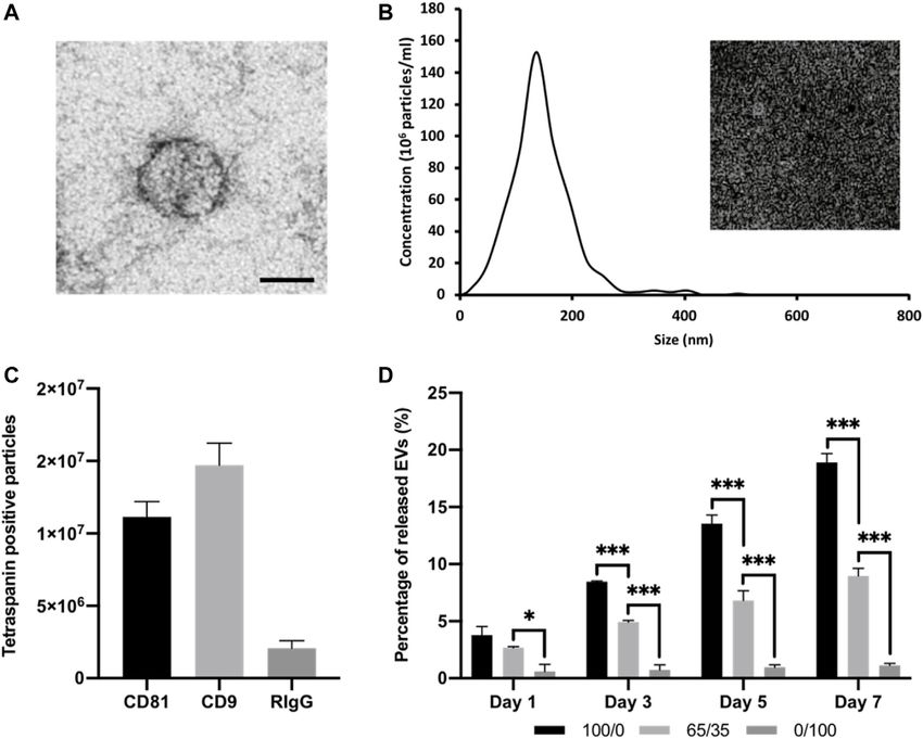
Man et al.                                                                                            ECM-Mimetic Hydrogel Promotes Vesicle-Induced Mineralisation

 FIGURE 2 | Physiochemical properties of chitosan-collagen hydrogels. (A) pH of composite hydrogels before and after β-GP addition. (B) Gelation time of
 hydrogels at 37°C. (C) Compressive modulus. (D) Macroscopic image of hydrogels after gelation. Data are expressed as mean ± SD (n = 3). *p ≤ 0.05, **p ≤ 0.01, and
 ***p ≤ 0.001.

2022). The EV concentration in the collected medium was                               with EV-free hydrogels were used as the control. After 24 h, cells
evaluated using the CD63 ExoELISA-ULTRA complete kit                                  were fixed with 10% (v/v) neutral buffered formalin (NBF,
(System Biosciences, United States) following the                                     Cellpath, United Kingdom), stained with Alexa Fluor 488
manufacturer’s protocol. Percentage of EVs released was                               phalloidin,      1:20    (Cell      Signalling      Technology,
calculated from the initial quantity of EVs added prior to gelation.                  United Kingdom), and mounted with Prolong
                                                                                      Antifade Mountant with DAPI (Thermo Scientific,
                                                                                                                                                  Gold     ™
                                                                                      United Kingdom) to label the actin cytoskeleton and nuclei,
2.7 The Impact of Hydrogel-Released EVs                                               respectively. Samples were imaged with an EVOS fluorescent
on hBMSCs Proliferation, Migration, and                                               inverted      microscope    (M5000,      Thermo        Scientific,
Mineralisation                                                                        United Kingdom).
2.7.1 EV Cell Uptake
EVs were labelled using Cell Mask                 ™
                                            Deep Red Plasma
Membrane Stain, 1:1,000 in PBS (Thermo Scientific,
                                                                                      2.7.2 Proliferation
                                                                                      hBMSCs were plated at low density (1 × 10⁴ cells/cm2) in basal
United Kingdom), and incubated for 10 min. Labelled EVs                               medium within a 48 well plate. After 24 h, media was replaced
were washed twice with PBS via ultracentrifugation at                                 with fresh basal medium and transwell inserts (0.4 µm pore size,
120,000 g for 70 min, then incorporated within the 100/0                              Greiner Bio-One, United Kingdom), containing EV-
hydrogel before gelation. hBMSCs were seeded at 3 × 10³                               functionalised hydrogels, were placed into each well. Media
cells/cm2 in a 48 well plate for 24 h, then media was replaced                        was replaced every 3 days. DNA content was assessed using
with fresh basal medium and transwell inserts (0.4 µm pore size,                      the PicoGreen (Life Technologies, United Kingdom) according
Greiner Bio-One, United Kingdom) containing Cell Mask-                                to the manufacturer’s protocol. Cells cultured without EV-
labelled EVs encapsulated within the hydrogel. Cells cultured                         functionalised hydrogels were used at the control. An EV only

Frontiers in Bioengineering and Biotechnology | www.frontiersin.org               6                                          March 2022 | Volume 10 | Article 829969
Man et al. ECM-Mimetic Hydrogel Promotes Vesicle-Induced Mineralisation
FIGURE 3 | The effects of chitosan-collagen composite hydrogel on proliferation and osteogenic differentiation. (A) Schematic representation of functional
assessments. The influence of different hydrogel formulations on (B) proliferation, (C) ALP activity and (D,E) calcium deposition. Black staining indicates mineral nodules.
Scale bar = 200 µm. Data are expressed as mean ± SD (n = 3). *p ≤ 0.05, **p ≤ 0.01, and ***p ≤ 0.001.
group was not included, as we focused on assessing the biological was applied with a 200 µL pipette tip and the width was measured
potency of EVs release from these hydrogel systems on recipient as the baseline. Cells were incubated with transwell inserts
hBMSCs behaviour. (0.4 µm pore size, Greiner Bio-One, United Kingdom)
containing EV-functionalised hydrogels for 3 days. Cells
2.7.3 Migration cultured without hydrogels were used as the control. The area
The migration area was calculated by performing the wound of wound closure from day 0 was assessed using fluorescent
healing assay. Briefly, cells at a density of 30 × 103 cells/cm2 in a 48 microscopy. Briefly, cells were labelled with Calcein-AM (1 μg/ml
well plate were plated and allowed to adhere for 24 h. A scratch in PBS, Sigma-Aldrich, United Kingdom) in the dark for 30 min.
Frontiers in Bioengineering and Biotechnology | www.frontiersin.org 7 March 2022 | Volume 10 | Article 829969Man et al. ECM-Mimetic Hydrogel Promotes Vesicle-Induced Mineralisation
FIGURE 4 | Characterisation of isolated osteoblast-derived EVs and hydrogel-EV release kinetics. (A) TEM image of EVs. Scale bar = 50 nm. (B) Size distribution of
isolated EV by NTA. Insert shows snapshot of particles during analysis. (C) Detection of tetraspanin-positive nanoparticles (CD81 and/or CD9) via interferometry after
immuno-capture onto ExoView™ chip. (D) Quantification of EVs released from chitosan-collagen hydrogels assessed via CD63 positive ELISA. Data are expressed as
mean ± SD (n = 3). *p ≤ 0.05, **p ≤ 0.01, and ***p ≤ 0.001.
Samples were observed under an EVOS fluorescent inverted hydrogels were incubated with Calcein-AM (1 μg/ml in PBS,
microscope (Thermo Scientific, United Kingdom). Sigma-Aldrich, United Kingdom), and Propidium iodide (1 μg/
ml in PBS, Sigma-Aldrich, United Kingdom) in the dark for
2.7.4 hBMSCs Osteoinduction 30 min. Samples were observed under an EVOS fluorescent
hBMSCs were seeded at high density (3 × 10³ cells/cm2) in basal inverted microscope (Thermo Scientific, United Kingdom).
medium within a 48 well plate. After 24 h, media was replaced The proliferation of cell-laden EV-hydrogels was assessed via
with osteogenic medium and transwell inserts (0.4 µm pore size, quantifying DNA content following culture in basal medium for
Greiner Bio-One, United Kingdom) containing EV- 7 days.
functionalised hydrogels were placed in each well. Cells The capacity of EV-functionalised hydrogels to stimulate
cultured without hydrogels were used as the control. Media encapsulated hBMSCs (1 × 10⁶ cell/ml) osteogenic
was replaced every 3 days. differentiation and mineralisation was evaluated after culture
in osteogenic medium for 3 weeks. Osteogenic differentiation
was assessed by quantifying alkaline phosphatase activity,
2.8 EV-Functionalised Hydrogels on collagen production and mineral deposition, detailed below.
hBMSCs Proliferation and Mineralisation Cell-free hydrogels of each composition were cultured in the
The viability and proliferation of hBMSCs (5 × 10⁵ cell/ml) within same conditions as described above and used as an acellular
the EV-functionalised 65/35 composite hydrogels was assessed control for the following analysis.
via live/dead staining. Briefly, hBMSCs (5 × 10⁵ cell/ml) were
mixed with the EV-functionalised hydrogel (0, 50, or 100 µg/ml of 2.9 Alkaline Phosphatase Activity
EV protein) prior to gelation. Following sol-gel transition, ALP activity was determined using the 4-nitrophenyl
hydrogels were culture in basal medium. At day 7, cell-laden colourimetric phosphate liquid assay (pNPP, Sigma-Aldrich,
Frontiers in Bioengineering and Biotechnology | www.frontiersin.org 8 March 2022 | Volume 10 | Article 829969Man et al. ECM-Mimetic Hydrogel Promotes Vesicle-Induced Mineralisation
United Kingdom) as previously reported (Man et al., 2021a). when compared to chitosan-containing gels (16.9 ± 0.25 min
Briefly, cell lysate was isolated from cell-laden hydrogels by five (100/0), 12.65 ± 0.66 (65/35), 9.93 ± 0.26 (25/75), and 8.13±
freeze/thaw cycles between −80 and 37°C, with homogenisation 0.41 min (0/100) (p ≤ 0.01) (Figure 2B). The compressive
via passing through syringe/needles and sonication in between. modulus of chitosan-collagen hydrogels is shown in
10 μL of cell lysate was added to 90 μL of pNPP and incubated for Figure 2C. There was a chitosan-dependent increase in
60 min at 37°C. The absorbance at 405 nm was read on a SPARK hydrogel stiffness, with compressive moduli of 7.43 ± 0.25
spectrophotometer (TECAN, CH). ALP activity was normalised (100/0), 4.97 ± 0.49 (65/35), 3.71 ± 0.46 (25/75), and 2.99 ±
with DNA content. 0.28 kPa (0/100) (p ≤ 0.05–0.001). Macroscopic images of
chitosan-collagen hydrogels following sol-gel transition are
2.10 Collagen Production shown in Figure 2D, where the gels have transitioned from a
Extracellular matrix collagen deposition was evaluated with clear to opaque transparency and displayed no mobility during
picrosirius red staining. Briefly, samples were washed twice in inversion testing.
PBS and fixed in 10% NBF for 30 min, prior to staining with
Picro-Sirius Red Solution (ScyTek Laboratories, Inc.,
United States) for 1 h. The unbound dye was removed by
3.2 The Effects of Chitosan-Collagen
washing in 0.5 M acetic acid followed by distilled water wash Hydrogel Composition on Osteogenic
and left to air dry prior to imaging using light microscopy (EVOS Differentiation.
XL Core, Invitrogen, United Kingdom). To quantify collagen The biological effects of altering chitosan-collagen content within
staining, 0.5 M sodium hydroxide was used to elute the bound dye the composite hydrogel on cell behaviour was evaluated
and absorbance were read at 590 nm using the SPARK (Figure 3A). The morphology of encapsulated cells within the
spectrophotometer (TECAN, CH). different hydrogel compositions were initially assessed. At day 3
in culture, cells in the 0/100 gels exhibited a rounded morphology,
2.11 Mineral Deposition while the encapsulated cells in the collagen-containing gels
To investigate mineralisation, calcium deposition was assessed displayed a spindle-shaped morphology (Supplementary
via alizarin red staining. Cells were washed twice in PBS and fixed Figure S1). Moreover, these images indicated cell proliferation
in 10% NBF for 30 min. Following fixation, samples were washed was increased in the collagen-containing gels, which was then
in distilled water and then incubated with alizarin red solution evaluated through DNA quantification. Hydrogels containing
(Sigma-Aldrich, United Kingdom) for 10 min. The unbound dye increased collagen proportions exhibited enhanced DNA
was removed by washing in distilled water. Staining was content, where the 0/100 gels displayed a 1.70-fold (p ≤ 0.01),
visualised using light microscopy (EVOS XL Core, Invitrogen, and 2.71-fold (p ≤ 0.001) significant increase compared to the 65/
United Kingdom). For quantification, samples were eluted with 35 and 100/0 groups respectively after 2 weeks of culture
10% cetylpyridinium chloride (Sigma-Aldrich, United Kingdom) (Figure 3B). The osteogenic capacity of chitosan-collagen
for 1 h and then absorbance was read at 550 nm using the SPARK composites was initially assessed by quantifying ALP activity.
spectrophotometer (TECAN, CH). Cells cultured within hydrogels containing more chitosan
displayed enhanced ALP activity, with the 100/0 group
2.12 Statistical Analysis exhibiting a 1.87 (p ≤ 0.001) and 6.61-fold (p ≤ 0.001)
For all data presented, experiments were performed in triplicate. significant increase compared to the 65/35 and 0/100 gels
All statistical analysis was undertaken using ANOVA multiple respectively (Figure 3C). A similar profile on calcium
comparisons test with Tukey modification using IBM SPSS deposition was observed, with the cells cultured within the
software (IBM Analytics, version 21). p values equal to or 100/0 gels exhibiting a 1.32 (p ≤ 0.001) and 3.07-fold (p ≤
lower than 0.05 was considered as significant. *p ≤ 0.05, **p ≤ 0.001) enhancement in calcium content when compared to the
0.01, ***p ≤ 0.001. All experiments were repeated independently 65/35 and 0/100 groups (Figures 3D, E). An increased density of
at least three times. mineral-like nodules (black staining) were observed in the gels
containing increased chitosan.
3 RESULTS
3.3 Characterisation of Isolated EVs and
3.1 The Influence of Chitosan-Collagen Hydrogel Release Kinetics
Formulation on Physiochemical Properties EV were isolated from mineralising osteoblast conditioned media
Figure 2A shows the gelation parameters for the chitosan- over a 2-week period via differential centrifugation. Osteoblast-
collagen hydrogels. Pure and composite hydrogels without β- derived EVs exhibited a typical size and spherical morphology
GP exhibited pH values ranging from 3.5 to 5, with higher pH through TEM imaging (Figure 4A). NTA analysis of isolated EVs
values at greater chitosan concentrations (p ≤ 0.05–0.01). displayed an average diameter of 131.3 ± 11.4 nm (Figure 4B).
Following β-GP addition, pH for all groups were significantly The detection of EV tetraspanin markers was performed using the
elevated to approximately pH 7.3 (p ≤ 0.001). The gelation time of Exoview platform. The obtained data confirmed the presence of
hydrogels was assessed at 37°C. Hydrogels containing increased CD9 and CD81-positive nanosized particles immune-captured
collagen content exhibited significantly reduced gelation times on anti-CD81 and CD9 antibodies (Figure 4C). The EV release
Frontiers in Bioengineering and Biotechnology | www.frontiersin.org 9 March 2022 | Volume 10 | Article 829969Man et al. ECM-Mimetic Hydrogel Promotes Vesicle-Induced Mineralisation FIGURE 5 | The biological efficacy of hydrogel-released EVs on hBMSCs behaviour. The influence on hydrogel-released EVs on hBMSCs (A) EV cell uptake. Scale bar = 20 μm, (B) proliferation, (C) migration, (D,F) collagen production, and (E,F) calcium deposition. Black staining indicates mineral nodules. Scale bar = 100 µm. Data are expressed as mean ± SD (n = 3). *p ≤ 0.05, **p ≤ 0.01, and ***p ≤ 0.001. Frontiers in Bioengineering and Biotechnology | www.frontiersin.org 10 March 2022 | Volume 10 | Article 829969
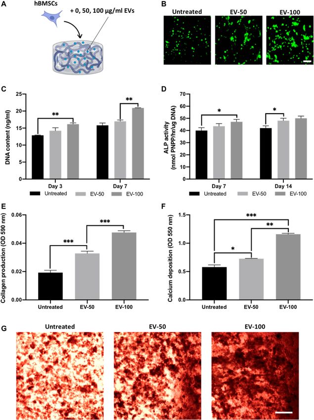
Man et al.                                                                                           ECM-Mimetic Hydrogel Promotes Vesicle-Induced Mineralisation

 FIGURE 6 | The effect of EV-functionalised chitosan-collagen hydrogel on encapsulated hBMSCs osteogenic differentiation. (A) Schematic representation of EV-
 hydrogel groups (0 µg/ml = untreated, 50 µg/ml = EV-50, 100 µg/ml = EV-100). (B) Live/dead staining of encapsulated hBMSCs after 7 days of culture. Scale bar =
 200 µm. (C) DNA content within EV-hydrogels after 7 days of culture. The influence on EV-hydrogels on hBMSCs (D) ALP activity, (E) collagen production, and (F,G)
 calcium deposition during osteogenic culture. Black staining indicates mineral nodules. Scale bar = 200 µm. Data are expressed as mean ± SD (n = 3). *p ≤ 0.05,
 **p ≤ 0.01, and ***p ≤ 0.001.

Frontiers in Bioengineering and Biotechnology | www.frontiersin.org             11                                         March 2022 | Volume 10 | Article 829969
Man et al.                                                                               ECM-Mimetic Hydrogel Promotes Vesicle-Induced Mineralisation

kinetics from these composite hydrogels were investigated using             calcium deposition showed a similar profile, with the pure
the CD63 ELISA (Figure 4D). The release of EVs from these                   chitosan groups exhibiting a significant 1.19, 1.26, and 3.25-
hydrogel formulations was dependant on chitosan/collagen                    fold increase in hBMSCs calcium accumulation compared to
ratios, with an increased quantity of CD63 positive particles               the 65/35, 0/100, and untreated group respectively (p ≤ 0.001)
released from gels containing a greater proportion of chitosan.             (Figures 5E, F).
There was a high average correlation between the release of EVs
and the proportion of collagen within the hydrogel (R2 > 0.94;              3.5 EV-Functionalised Chitosan-Collagen
Supplementary Figure S2). The pure chitosan formulations
(100/0) released a significantly enhanced quantity of CD63
                                                                            Composite Hydrogel Promoted hBMSC
positive particles when compared to the hydrogels containing                Osteogenic Differentiation and
collagen (65/35 and 0/100) at day 1, 3, 5, and 7 (p ≤ 0.001). At day        Mineralisation
1, 3.78 ± 0.76% (100/0), 2.68 ± 0.09% (65/35), and 0.6 ± 0.61% of           The 65/35 composite was utilised as it provides a suitable balance
CD63 positive EVs were released. At day 7, the quantity of CD63             between compressive modulus, osteogenic properties and EV
positive EVs released from the gels increased to 18.91 ± 0.77%              release kinetics. Hydrogels were functionalised with 0, 50, or
(100/0), 8.98 ± 0.66% (65/35), and 1.13 ± 0.18% (0/100). The size           100 ug/ml of EV protein (Figure 6A), and the viability of
distribution of EVs before and after hydrogel incorporation did             hBMSCs was assessed with live/dead staining and DNA
not significantly shift over time between the different hydrogel             quantification. At day 7, live/dead imaging showed an EV
groups (Supplementary Figure S3).                                           dose-dependent increase in the density of viable cells when
                                                                            compared to the untreated control (Figure 6B). There was a
                                                                            significant enhancement in hBMSC proliferation in an EV dose-
3.4 Hydrogel-Released EVs Enhance                                           dependent manner at day 3 (untreated vs. EV-100, p ≤ 0.01) and 7
hBMSC Proliferation and Osteogenic                                          (EV-50 vs. EV-100, p ≤ 0.01) (Figure 6C).
Differentiation                                                                 The influence of EV-functionalised hydrogels on the
The therapeutic efficacy of hydrogel-released EVs on hBMSCs                  mineralisation of encapsulated hBMSCs was assessed following
behaviour was evaluated using the transwell assay. We observed              21 days osteogenic culture. Quantification of ALP activity was
the successful internalisation of hydrogel-released EVs by                  initially conducted to evaluate the osteoinductive capacity of EV-
hBMSCs, with the labelled vesicles primarily located within                 hydrogels. An EV dose-dependent increase in ALP activity was
the cytoplasm of the cell (Figure 5A). There was a significant               observed from the hBMSCs at day 7 (untreated vs. EV-100, p ≤
increase in the proliferation of hBMSCs in all EV-functionalised            0.05) and day 14 (untreated vs. EV-50, p ≤ 0.01) in osteogenic
hydrogel groups when compared to the untreated control (p ≤                 culture (Figure 6D), with the EV-100 group exhibiting a non-
0.001) (Figure 5B). Notably, there was a chitosan-dependent                 significant increase in ALP compared to the EV-50 gels at both
increased in hBMSCs proliferation at day 3 and 7. Cells cultured            time points (p > 0.05). Extracellular matrix production and
with the 100/0 group exhibited a significantly enhanced DNA                  mineralisation were evaluated via quantifying collagen
content when compared to the 0/100 (1.07-fold, p ≤ 0.01) and                production and calcium deposition respectively. Both EV-50
untreated control (1.10-fold, p ≤ 0.001) at day 3. At day 7, a              and EV-100 groups exhibited a significant increase in collagen
similar trend was observed, where the 100/0 group showed a                  production when compared to the untreated control (1.68 and
significantly enhanced hBMSCs DNA content compared to the                    2.47-fold, respectively) (p ≤ 0.001), with the EV-100 group
cells cultured with the 65/35 (1.06-fold, p ≤ 0.01) and 0/100 (1.06-        eliciting a 1.46-fold enhancement compared to the EV-50 gel
fold, p ≤ 0.01) and untreated control (1.12-fold, p ≤ 0.001). The           (p ≤ 0.001) (Figure 6E). A similar trend was observed for calcium
EV functionalised hydrogels significantly promoted hBMSCs                    deposition, with the EV-functionalised gels displaying
migration when compared to the untreated control (>1.48-                    significantly enhanced calcium content compared to the
fold, p ≤ 0.01–0.001) (Figure 5C) (Supplementary Figure S4).                untreated control. The EV-50 and EV-100 gels exhibited a
A chitosan-dependent increase in migration was observed, where              1.28-fold (p ≤ 0.05) and 2.03-fold (p ≤ 0.001) increased in
the 100/0 group exhibited a 1.25 (p ≤ 0.01) and 1.72-fold (p ≤              calcium deposition when compared to the untreated control,
0.001) enhancement in hBMSCs migration compared to 65/35                    with the EV-100 gel displaying a significantly greater degree of
and 0/100, respectively.                                                    calcium deposition and mineral-like nodules when compared to
    The effects of hydrogel-released EVs on hBMSCs osteogenic               the EV-50 group (1.58-fold) (p ≤ 0.01) (Figures 6F, G).
differentiation was evaluated by analysing collagen production
and calcium deposition during osteogenic differentiation. We
observed a significant increase in osteogenic differentiation in all         4 DISCUSSION
EV-functionalised hydrogel groups when compared to the
untreated control following 2 weeks of osteoinductive culture               Harnessing EVs as an acellular tool for bone augmentation
(p ≤ 0.05–0.001). There was a chitosan-dependent                            strategies has gained considerable interest in recent years
enhancement in hBMSCs collagen production, with the 100/0                   (Marolt Presen et al., 2019; Man et al., 2021b). For example,
group exhibited a 1.02 (p > 0.05), 1.05 (p ≤ 0.05), and 1.08-fold           Davies et al. demonstrated that osteoblast-derived EVs enhanced
(p ≤ 0.01) greater collagen content compared to 65/35, 0/100 and            hBMSCs mineralisation when compared to the use of the
untreated control respectively (Figures 5D, F). The effect on               clinically relevant growth factor BMP2 (Davies et al., 2017).

Frontiers in Bioengineering and Biotechnology | www.frontiersin.org    12                                     March 2022 | Volume 10 | Article 829969
Man et al. ECM-Mimetic Hydrogel Promotes Vesicle-Induced Mineralisation
Due to the rapid clearance of systemically administered vesicles microstructure. It has been reported that the introduction of
from the body (Murphy et al., 2019; Elsharkasy et al., 2020), the collagen to chitosan hydrogel increases the porosity of the
logical next step to advance EV-based therapies to the clinical hydrogel (Fernandes et al., 2011). Therefore, cells within
setting, would be the development of an osteoinductive delivery collagen containing hydrogels will likely exhibit increased
system to enhance EVs bioavailability and bioactivity to promote proliferation due to enhanced porosity, consistent with our
bone repair. As cell-derived factors such as EVs are known to findings. Moreover, the increased compressive modulus of
interact with ECM components during mineralisation, the chitosan containing hydrogels is expected to have a positive
delivery of vesicles within an ECM-mimetic biomaterial could influence on osteogenesis due to eliciting increased mechanical
recapitulate the EV function during bone formation. Therefore, stimulation (Ghasemi-Mobarakeh et al., 2015; Sun et al., 2018).
in this study, we investigated the development of a pro-osteogenic As EVs are essentially fingerprints of their parental cells
chitosan-collagen composite hydrogel to control EV release (Kobayashi et al., 2015), the chitosan and collagen substrates
kinetics and synergistically promote EV-induced hBMSCs may differentially influence the osteoinductive efficacy of
osteogenesis. hydrogel-encapsulated cell secreted EVs. We previously
In recent years, a growing number of studies have investigated demonstrated that 3D printed titanium scaffolds exhibiting a
combining EVs with hydrogel systems to promote their release triangular pore conformation significantly promoted osteoblast
kinetics for different therapeutic applications (Riau et al., 2019; differentiation when compared to square pore scaffolds (Man
Man et al., 2020). For instance, Nikravesh et al. reported the et al., 2021c). Importantly, the EVs derived from these triangular
development of a tailorable alginate-based EV release system, by pore scaffolds significantly enhanced the mineralisation of
controlling the physical structuring of the hydrogel during the hBMSCs when compared to osteoblast-derived EVs from
sol-gel transition (Nikravesh et al., 2019). Although the delivery square pore scaffolds. As chitosan containing hydrogels
of these nanoparticles from hydrogels has produced promising promoted mineralisation to a greater degree compared to
results, the administered biomaterial must also provide collagen-containing gels in this study, it is likely the chitosan-
appropriate physical and biological properties to support the laden cells secreted EVs promoted mineralisation within the
defect site and promote de novo bone formation respectively. As hydrogel in an autocrine/paracrine manner, although this
such, we investigated the influence of altering chitosan-collagen would require further investigation. Therefore, the distinct
content on the general material properties of the hydrogel. Our biological effects presented by chitosan or collagen within the
findings showed that the chitosan-collagen formulations hydrogel provides tunability depending on the clinical
exhibited gelation times ranging from 5 to 15 min at 37°C, a application.
clinically reasonable handling time for orthopaedic surgeries Due to issues associated with the scalable/reproducible
(Costa, 2017). Moreover, increasing chitosan content within manufacture of EVs and their rapid clearance in vivo (Gimona
the hydrogel, significantly enhanced the compressive modulus et al., 2017), this further emphasises the importance of enhancing
of the hydrogel, consistent with studies in the literature (Chicatun their bioavailability. There are numerous strategies to tether EVs
et al., 2013; McBane et al., 2013). It has been reported that to biomaterials, which have been extensively reviewed in the
materials which exhibit increased stiffness, promote literature (Brennan et al., 2020; Man et al., 2020). In addition to
osteogenesis through enhanced mechanotransductive selecting pro-osteoinductive materials, chitosan, and collagen
stimulation (Ghasemi-Mobarakeh et al., 2015; Sun et al., were utilised due to their capacity to immobilise EVs via
2018), likely impacting the osteoinductive capacity of these different mechanisms. Chitosan is a cationic polymer, which
gels. Together these findings demonstrate the composite enables the formation of complexes with anionic molecules
hydrogels exhibit suitable in situ gelation times and such as on the surface of EVs. Kumar et al. harnessed the
mechanical properties facilitating its minimally invasive electrostatic interactions of positively charged chitosan with
administration to the defect site. the negative-charged membranes of vesicles to develop a
Although several reports have demonstrated the release simple and robust EV isolation method from a variety of
kinetics of EVs over multiple days from biomaterial systems biofluids (Kumar et al., 2021). Moreover, the amino and
(Mardpour et al., 2019; Man et al., 2020), there have been limited hydroxyl groups have been utilised to deliver numerous drugs
investigations into the role of the delivery device on EV-induced and growth factors (Zhang et al., 2010). Collagen hydrogels have
biological activity. Chitosan and type I collagen were selected due been reported to immobilise EVs via integrin-mediated
to their biocompatible nature and their osteoinductive properties interactions (Buzás et al., 2018). For example, Altei et al.
(Hesse et al., 2010; Liu et al., 2020). As such, we investigated the demonstrated the integral role of EV-associated integrins with
role of altering chitosan-collagen content within the composite binding to collagen. Their findings showed that the introduction
hydrogel to stimulate osteogenic differentiation without EVs. Our of a disintegrin inhibitor (DisBa-01) inhibited the adhesion of
findings showed that hydrogels containing increased chitosan EV-derived from MDA-MB-231 breast cancer cell on collagen
content promoted osteogenic differentiation, while gels with coated tissue culture plastic (Altei et al., 2020). Hao et al. further
greater collagen proportions enhanced proliferation. This demonstrated the importance of integrins in binding EVs to
distinct effect on cellular proliferation and osteogenic matrix proteins. Their findings showed that functionalising an
differentiation has been similarly observed in the literature integrin α4β1 ligand, LLP2A to a material surface enhanced MSC-
(Wang and Stegemann, 2010; Dang et al., 2017), likely due to derived EVs binding and improved vascularisation (Hao et al.,
the respective influence of each biomaterial on the composite 2020). Furthermore, calcium channelling Annexin proteins have
Frontiers in Bioengineering and Biotechnology | www.frontiersin.org 13 March 2022 | Volume 10 | Article 829969Man et al. ECM-Mimetic Hydrogel Promotes Vesicle-Induced Mineralisation
been reported to play a pivotal role in matrix-mineralisation gel significantly enhanced hBMSCs mineralisation when
through collagen binding (von der Mark and Mollenhauer, 1997). compared to the cells cultured with the collagen-released EVs.
Several studies have reported Annexin proteins are upregulated in These findings were consistent with the influence of hydrogel-
mineralising EVs (Eichholz et al., 2020; Man et al., 2021b), with released EVs on proliferation and migration, indicating the
these proteins shown to be critical for the binding of increased quantity of EVs released from chitosan-containing
proteoliposomes to collagen fibrils (Kirsch et al., 2000). Thus, gels promoted hBMSCs osteogenesis. Together, these findings
the chitosan-collagen composite could provide a multi-faceted indicate that the incorporated vesicles retained their biological
mechanism of immobilising EVs to facilitate their controlled potency once released from the hydrogel, demonstrating the
release. To investigate EV discharge from these biomaterials, viability of this composite hydrogel as a suitable delivery vehicle
different chitosan-collagen formulations were tested in regard for these pro-osteogenic nanoparticles.
to their EV release kinetics over 7 days. Our findings showed that For the development of an optimal EV delivery system, it is
an increased proportion of chitosan within the composite important to consider the biomaterials role on EV release and their
enhanced the release rate of EVs from the hydrogel system. functionality, but also to provide a suitable environment that
This may be due to the differential EV affinity and binding to facilitates tissue-specific regeneration. Several studies have
ECM proteins. Studies have shown that the addition of β-GP to demonstrated the osteoinductive capacity of chitosan and
chitosan reduces the cationic nature of the hydrogel, possibly collagen biomaterials (Wang and Stegemann, 2010; Sun et al.,
affecting its capacity to capture EVs (Kolawole et al., 2019). Our 2011), thus providing an appropriate platform to support EV-
finding indicates that collagen exhibited an increased efficacy to induced regeneration. Moreover, to initiate thermo-gelation in
sequester EVs within the composite material compared to chitosan hydrogels, β-GP was incorporated to facilitate its sol-
chitosan. As EVs have been demonstrated to promote cell gel transition at physiological temperatures. It has been previously
recruitment, an important process for endogenous bone repair reported that osteoblast-derived EVs require a phosphate-rich
(Wang et al., 2018), these results suggest the importance of environment to facilitate hBMSCs mineralisation (Davies et al.,
incorporating chitosan in the composite to facilitate the release 2017). Therefore, the incorporation of β-GP provides the sol-gel
of these vesicles from the hydrogel. Together, these findings transition of chitosan and a phosphate source to promote EV-
demonstrate the importance of investigating the release induced mineralisation. Hence, in this present study, we
kinetics of EV-functionalised hydrogels, as this will investigated the influence of EV-functionalised chitosan-collagen
significantly impact the vesicles bioavailability and hydrogel to stimulate encapsulated hBMSCs osteogenic
functionality in situ. differentiation. In this work, the 65/35 composite was utilised as
A potential concern with the delivery of EVs within it provides a suitable balance between compressive modulus,
biomaterials systems, is the possibility that the released osteogenic properties and EV release kinetics. Moreover, several
nanoparticles functionality may be altered, ultimately impacting studies have reported the successful utilisation of chitosan-collagen
their therapeutic efficacy. Moreover, in the bone context, the hydrogels at similar ratios (Wang and Stegemann, 2010; Sun et al.,
recruitment of endogenous cells into the defect site/implanted 2011). We initially showed that the encapsulated hBMSCs
material is essential for successful de novo tissue formation and remained viable within the 65/35 composite hydrogel, consistent
osseointegration (Wang et al., 2018). As EVs have been with findings in the literature (Wang and Stegemann, 2010).
demonstrated to stimulate cellular recruitment (Gholami et al., Moreover, our results showed an EV dose-dependent increase
2021; Zhu et al., 2021), this highlights the importance of in cell proliferation within the hydrogel. This suggest that the
investigating the therapeutic efficacy of hydrogel-released EVs incorporated EVs retained their biological functionality, further
from the composite material. We reported that there were no promoting hBMSCs proliferation in a dose-dependent manner.
significant changes in the size distribution of EVs before and after Importantly, we demonstrated that the EV-functionalised
hydrogel incorporation and between the different formulations. composite gels significantly enhanced hBMSCs osteogenic
This suggests EV incorporation into the material did not cause differentiation and mineralisation when compared to the EV-
substantial damage to the nanoparticle integrity or cause vesicle free groups, consistent with observations in the literature (Man
aggregation. Regarding the functionality of released EVs, we et al., 2021b). Similar to the effects on proliferation, our findings
initially investigated the influence of these nanoparticles on showed an EV dose-dependent increase in mineralisation within
stimulating the migration and proliferation of hBMSCs, a the hydrogel, consistent with the concentration-dependent
critical process for recruitment of endogenous cells into the increase in hBMSCs mineralisation observed within a GelMA
defect site (Perez et al., 2015). Our findings showed a chitosan- nanocomposite hydrogel (Man et al., 2022). These findings
dependent increased in migration and proliferation rate induced by further signify the efficacy of encapsulated EVs in stimulating
EV treatment, consistent with the EV release kinetics observed hBMSCs osteogenesis within this hydrogel system. Taken together,
from these gels. This indicates that the released EVs exhibited a these findings demonstrate the 65/35 composite hydrogel provides
dose-dependent effect on hBMSCs migration and proliferation, a biocompatible environment that facilitates EV-induced stem cell
signifying the EVs retained their biological efficacy in stimulating mineralisation, indicating the therapeutic viability of this acellular
cellular recruitment, consistent with findings in the literature (Mol approach to stimulate bone regeneration.
et al., 2019). In addition to the role of hydrogel-released EVs on In summary, we demonstrated the development of an
cellular recruitment, their capacity to stimulate hBMSCs injectable pro-osteogenic chitosan-collagen composite hydrogel
osteogenesis was investigated. EVs released from the chitosan capable of controlling the release of osteoblast-derived EVs and
Frontiers in Bioengineering and Biotechnology | www.frontiersin.org 14 March 2022 | Volume 10 | Article 829969You can also read