Basement membrane proteins modulate cell migration on bovine pericardium extracellular matrix scaffold
←
→
Page content transcription
If your browser does not render page correctly, please read the page content below
www.nature.com/scientificreports
OPEN Basement membrane proteins
modulate cell migration on bovine
pericardium extracellular matrix
scaffold
Qi Xing, Mojtaba Parvizi, Manuela Lopera Higuita & Leigh G. Griffiths*
Native bovine pericardium (BP) exhibits anisotropy of its surface ECM niches, with the serous surface
(i.e., parietal pericardium) containing basement membrane components (e.g., Laminin, Col IV) and
the fibrous surface (i.e., mediastinal side) being composed primarily of type I collagen (Col I). Native
BP surface ECM niche anisotropy is preserved in antigen removed BP (AR-BP) extracellular matrix
(ECM) scaffolds. By exploiting sideness (serous or fibrous surface) of AR-BP scaffolds, this study aims
to determine the mechanism by which ECM niche influences human mesenchymal stem cells (hMSCs)
migration. Human mesenchymal stem cells (hMSC) seeding on serous surface promoted more rapid
cell migration than fibrous surface seeding. Gene analysis revealed that expression of integrin α3
and α11 were increased in cells cultured on serous surface compared to those on the fibrous side.
Monoclonal antibody blockade of α3β1 (i.e., laminin binding) inhibited early (i.e. ≤ 6 h) hMSC migration
following serous seeding, while having no effect on migration of cells on the fibrous side. Blockade of
α3β1 resulted in decreased expression of integrin α3 by cells on serous surface. Monoclonal antibody
blockade of α11β1 (i.e., Col IV binding) inhibited serous side migration at later time points (i.e., 6–24 h).
These results confirmed the role of integrin α3β1 binding to laminin in mediating early rapid hMSCs
migration and α11β1 binding to Col IV in mediating later hMSCs migration on the serous side of AR-BP,
which has critical implications for rate of cellular monolayer formation and use of AR-BP as blood
contacting material for clinical applications.
Bovine pericardium (BP) derived biomaterials have been widely used in a variety of surgical applications since
its first introduction in clinical practice1. Glutaraldehyde-fixed BP (GFBP) for example is currently widely used
clinically for bioprosthetic heart valve fabrication and arterial patches. GFBP has many advantages compared
to synthetic materials, such as off-shelf availability, easy handling, and reduced suture b leeding2. Additionally,
glutaraldehyde fixation can prevent hyperacute and acute immune response by masking xenoantigens in BP.
However, persistent presence of xenoantigens in GFBP results in chronic immune-mediated degeneration and
subsequent calcification3,4. Additionally, residual glutaraldehyde in GFBP have also been linked with toxic-
ity towards repopulating recipient cells5,6. Consequently, despite its short term benefits GFBP exhibits limited
long term host cell repopulation, tissue integration and biomaterial remodeling. The limitation of GFBP can
be potentially overcome by development of unfixed BP extracellular matrix (ECM) scaffolds which have been
processed to reduce the antigenic content of the biomaterial, rendering it minimally antigenic and compatible
with recipient non-immune cellular repopulation.
An ideal decellularization method should eliminate candidate tissue xenoantigens, while maintaining native
ECM structure–function properties. A variety of decellularization approaches have been explored to remove
antigenic components from native BP, including sodium dodecyl sulfate (SDS), TritonX-100, and t rypsin7. It has
been reported that SDS-decellularization can achieve complete acellularity; however, ECM structure–function
properties of such scaffolds are significantly altered due to the denaturing properties of this ionic denaturing
detergent7–9.. Disruption of the native ECM, especially basement membrane integrity, can negatively impact
cell–matrix interactions altering cell phenotype, proliferation, survival and migration behavior7. Native BP has
two distinct surfaces: (1) the parietal pericardial (i.e., serous) surface consists of an endothelial cell monolayer
and underlying basement membrane; (2) the mediastinal (i.e., fibrous) surface is composed of connective tis-
sue. A stepwise, solubilization based antigen-removal (AR) approach has been shown to significantly reduced
Department of Cardiovascular Diseases, Mayo Clinic, Stabile 4‑58, 200 First Street, Rochester, MN 55905, USA.
*
email: Griffiths.Leigh@mayo.edu
Scientific Reports | (2021) 11:4607 | https://doi.org/10.1038/s41598-021-84161-5 1
Vol.:(0123456789)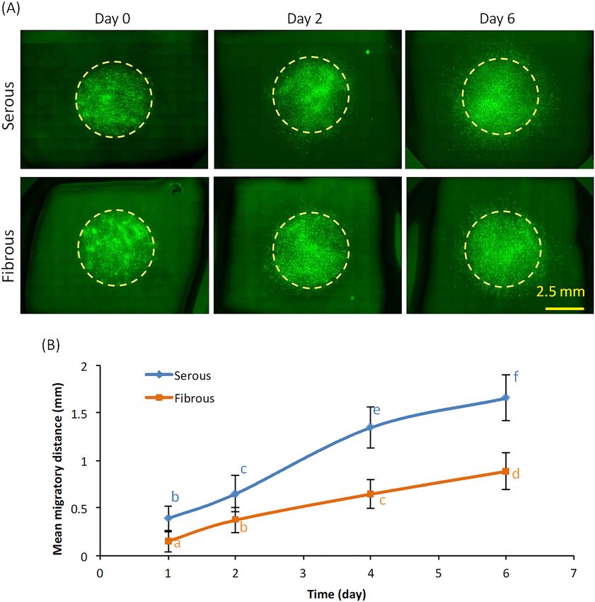
www.nature.com/scientificreports/
Figure 1. Hematoxylin & eosin (HE) and basement membrane proteins staining of serous and fibrous surface
of native BP and AR-BP. (A) HE staining demonstrating that AR scaffolds are completely acellular, while
retaining native BP collagen organization. (B) Collagen IV immunofluoresence staining demonstrating that
AR-BP scaffolds retain native Col IV staining on the serous surface and vascular lumen. Fibrous surface of both
native and AR-BP scaffolds does not contain Col IV. (C) Laminin immunofluoresence staining demonstrating
retention of native tissue laminin content and organization, on the serous surface and vascular lumen of AR-BP
scaffolds.
antigenicity, while maintaining native BP ECM structure–function properties, thereby providing a significant
advance in the field when compared to traditional decellularization m ethods9–11. In particular, the major base-
ment membrane proteins such as laminin and collagen IV (Col IV) are preserved on serous side of BP-AR scaf-
folds, while the fibrous side exhibits a predominantly Col I surface composition. Consequently the surface ECM
niche anisotropy of AR-BP scaffolds provides a unique opportunity to examine the effect of basement membrane
vs. non-basement membrane surface on repopulating cell behavior.
Cell migration on ECM scaffolds is crucial in many biological processes including vascular tissue endotheli-
alization and tissue regeneration. Human mesenchymal stem cells (hMSCs) derived from bone marrow has been
reported to migrate/adhere to area’s of tissue injury or implanted grafts, and contribute to tissue regeneration12.
It is suggested for instance that remodeling of vascular ECM scaffolds is dependent on adhesion, migration,
proliferation and differentiation of such circulating cells. Previous experiments have shown that circulating
progenitor cells, including hMSCs, contribute to endothelialization of the luminal surface of vascular ECM
scaffolds13. The ability of hMSCs to differentiate into vascular cells demonstrates the important role of this cell
population in vascular tissue regeneration14. However, the ECM niche factors which affect hMSC repopulation
rate remain unknown. Determination of the ECM niche factors responsible for modulation of hMSCs migration
on ECM scaffolds is critical to inform which side of such biomaterials should face the vascular lumen to enhance
rate of luminal cellular repopulation.
In this work, hMSCs were employed as model cells to study in vitro cell migration on the serous versus
the fibrous surfaces of BP scaffolds. It was previously shown that the different ECM niches of AR-BP scaffolds
affected hMSCs proliferation and cytokine s ecretion15. We hypothesize that the basement membrane proteins
laminin and collagen IV on the serous surface promote X–Y hMSCs migration across the scaffold surface, while
absence of basement membrane components favors Z-direction migration. We further hypothesize that higher
levels of cell migration is mainly mediated via interaction between integrin α3β1 and laminin. We evaluated the
effect of BP-AR ECM surface niche on hMSC X–Y migration, Z-direction penetration, and proliferation into
AR-BP, and integrin gene expression. To further define the mechanism of ECM niche dependent migration, we
investigated the effect of integrin α3β1 and α11 chain blockade on ECM niche dependent migration and integrin
gene expression.
Results
AR preserves native BP surface morphology and ECM component anisotropy. Effect of AR on
the native ECM niche anisotropy of BP was investigated via histology and immunofluorescence. Hematoxylin
and eosin (H&E) staining of native BP and AR-BP showed that collagen organization of both the serous and
fibrous sides is maintained after AR (Fig. 1A). Immunofluorescence staining for Col IV (Fig. 1B) and Laminin
(Fig. 1C) demonstrated that only serous surface of native BP contained these two basement membrane protein.
Col IV and Laminin content and organization were preserved in BP-AR scaffolds.
Effect of surface ECM niche anisotropy on X–Y planar cell migration and Z‑direction cell pen-
etration. To determine the effect of ECM niche anisotropy on cellular migration, migration of hMCS seeded
onto the serous versus fibrous sides of BP-AR scaffolds was compared. Cell images were taken at specific time
points (Fig. 2A) with image analysis limited to quantification of purely X–Y surface migration rate. Planar X–Y
migration distance was significantly greater for hMSC seeded on the serous surface than on fibrous surface at
all time points (Fig. 2B). By day 6, mean migratory distance on the serous side was 1.66 ± 0.24 mm, compared to
0.89 ± 0.19 mm on the fibrous side (p < 0.001). By day 6, mean migratory distance on serous side was 1.87 times
greater than that of the fibrous side.
Scientific Reports | (2021) 11:4607 | https://doi.org/10.1038/s41598-021-84161-5 2
Vol:.(1234567890)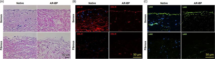
www.nature.com/scientificreports/
Figure 2. Effect of AR-BP sideness on cell migration. (A) Inverted microscopy images of cell migration at
different time points (day 0, 2, 6), demonstrating greater cell migration following serous side seeding than
fibrous side seeding. Scale bar: 2.5 mm; (B) Quantification of cell migration over time in culture, showing
migration rate following serous seeding is greater than that of fibrous seeding. Statistical analysis was performed
using two-way repeated measures ANOVA and Tukey’s multiple comparisons post hoc test. Data points that are
not connected by the same lower case letter are statistically different. (n = 6 scaffolds per group).
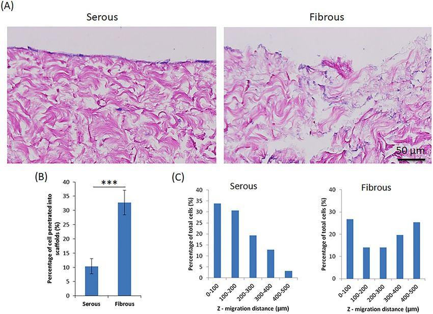
Despite equal seeding densities, a greater number of cells were observed on the serous surface than on
fibrous surface by observing H&E staining at day 6 (Fig. 3A). Cells on serous surface formed a confluent mon-
olayer, whereas those on the fibrous surface did not. However, a significantly higher proportion of cells pen-
etrated in the Z-direction from the fibrous surface (32.79 ± 4.32%), compared to the serous side (10.36 ± 2.67%)
(p < 0.001) (Fig. 3B,C). Furthermore, the mean Z-direction migratory distance for cells migrating from the
fibrous surface (262.68 ± 166.68 µm) was significantly greater than that of cells migrating from the serous sur-
face (180.57 ± 109.69 µm, p < 0.001) (Fig. 3C). Among all the cells penetrated inside the scaffolds, only 35% of
cells migrated deeper than 200 µm from serous side; whereas 59% of cells migrated deeper than 200 µm from
fibrous side.
Effect of surface ECM niche anisotropy on cell proliferation and cell death. Effect of ECM
niche anisotropy on proliferation of hMSC was examined. Early post-seeding cell proliferation, as quantified by
FUCCI staining at 6 h, did not differ between groups (Fig. 4A,B). Despite early cell proliferation identified via
FUCCI, alamar blue intensity did not significantly increase in the first two days regardless of seeding surface. No
significant difference in alamar blue intensity was identified between the serous and fibrous surface on days 1
and 2. Statistical difference was observed from day 3 onward, with cells seeded on the serous surface proliferat-
Scientific Reports | (2021) 11:4607 | https://doi.org/10.1038/s41598-021-84161-5 3
Vol.:(0123456789)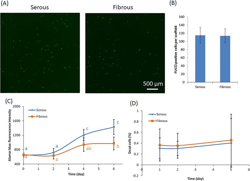
www.nature.com/scientificreports/

                                            Figure 3.  Z-direction cell migration into AR-BP scaffolds following serous or fibrous seeding. (A) HE images
                                            demonstrating cells remain predominantly as a monolayer on the surface following serous seeding. Conversely,
                                            cell penetration into the scaffold is higher following fibrous side seeding. (B) Percentage of cells penetrated into
                                            scaffolds quantified from H&E images. Significantly less cells penetrate into the scaffold following serous seeding
                                            versus fibrous seeding, p < 0.001. (C) Distribution of depth of penetration into the scaffold for those cells which
                                            do migrate in the Z-direction, demonstrating that overall cell penetration depth from serous side is significantly
                                            less than from fibrous side, p < 0.01. Statistical analysis was performed using one-way ANOVA, n = 6 scaffolds
                                            per group.

                                            ing significantly more than those seeded on the fibrous surface (Fig. 4C). As a widely employed method for cell
                                            proliferation quantification, no negative effects of alamar blue on cells or scaffolds were observed. No evidence of
                                            cell death was noted in any group/time point. Percentage of dead cells was less than 0.5% throughout all culture
                                            time points, and not significant different between serous versus fibrous side seeding (p > 0.05) (Fig. 4D).

                                            Effect of surface ECM niche anisotropy on gene expression. To determine which integrins may be
                                            involved in the observed ECM niche mediated differential cell migration on AR-BP scaffolds, hMSCs grown on
                                            either serous or fibrous surface were collected after 6 d of culture for gene analysis. All PCR data from serous or
                                            fibrous surface seeding were normalized to cells seeded on tissue culture plastic. Major integrin genes, including
                                            α1, α2, α3, α4, α5, α6, α11, αv, β1, β3, β7, as well as migration-related genes such as TIMP2 and MMP2 were tested.
                                            Generally speaking, most of the integrin expression was higher in serous-hMSCs than in fibrous-hMSCs (α1, α3,
                                            α4, α5, α6, α11, αv, β1, β3); however, significance was only found for expression of integrins α3, α11, and β7 (Fig. 5).
                                            Although MMP2 (matrix metalloproteinases 2) gene, which is involved in breaking down ECM during cell
                                            migration, did not show statistically difference between serous and fibrous surface (data not shown); TIMP2,
                                            tissue inhibitor of MMP2, was significantly downregulated on serous side relative to fibrous side. These data were
                                            utilized to target candidate integrins for later antibody blocking experiments.

                                            Integrin blocking and gene analysis. In order to show that integrin α3β1 (laminin binding) and integrin
                                            α11β1 (Col IV binding) are one of the potential mechanisms involved in mediating cell migration on basement
                                            membrane (Serous) versus non-basement membrane (Fibrous) ECM environments, integrin blocking experi-
                                            ment were performed. Addition of anti-integrin α3β1 and anti-integrin α11 into cell culture medium significantly
                                            decreased cell migration on the serous side (Fig. 6). Blockade with anti-integrin α3β1 antibody prevented early
                                            migration (i.e., 4 and 6 h) on the serous surface, although migration resumed at later time points (Fig. 6A).
                                            Conversely blockade with anti-integrin α11 antibody inhibited serous side migration after the 6 h time point
                                            (Fig. 6B). By 24 h, mean cell migratory distance on the serous surface decreased by 52.8 ± 10.3% (anti-α3β1) and

          Scientific Reports |   (2021) 11:4607 |                 https://doi.org/10.1038/s41598-021-84161-5                                                      4

Vol:.(1234567890)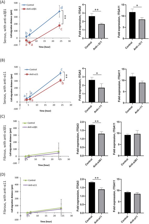
www.nature.com/scientificreports/
Figure 4. Effect of AR-BP sideness on cell proliferation. (A) FUCCI staining and quantification (B) at 6 h
culture, showing no seeding side does not significantly effect number of replicating cells at early time points.
Statistical analysis was performed using one-way ANOVA (n = 6 scaffolds per group). (C) Alarma blue intensity
of cells from 0 to 6 days. At later time points (4 days and 7 days), cell number is greater following serous side
seeding than fibrous side seeding. (D) Dead cell staining showing the dead cell percentage in both culture has
no significant difference at all time points. Statistical analysis for alarma blue intensity and dead cell percentage
was performed using two-way repeated measures ANOVA and Tukey’s multiple comparisons post hoc test. Data
points that are not connected by the same lower case letter are significantly different. (n = 6 scaffolds per group).
24.8 ± 0.8% (anti-α11) respectively, in the presence of integrin blockade. Conversely, addition of anti-integrin
antibodies did not change cell migration significantly on the fibrous surface (Fig. 6C,D).
With addition of anti-integrin α3β1 to cell culture on serous surface, integrin α3 and α11 gene expression
significantly decreased. Blockade with anti-integrin α11β1 to cell culture on serous surface only significantly
reduced integrin α3 expression and had no effect on α11 gene expression. For cells cultured on fibrous surface,
addition of anti-integrin α3β1 or anti-integrin α11β1 significantly decreased integrin α3 expression only, and had
no effect on α11 expression.
Discussion
The present study demonstrates the importance of different ECM niche (i.e., basement membrane versus non-
basement membrane) on hMSCs migration. Specifically, we compared the effect of basement membrane pres-
ence (serous side seeding) versus absence (fibrous side seeding) on hMSC behavior and found that: (1) AR-BP
preserved basement membrane proteins laminin and Col IV on serous surface; (2) hMSCs migrated rate is
increased following seeding on the basement membrane containing surface (i.e., serous side) of AR-BP scaffold
compared to the non-basement membrane surface (i.e., fibrous side); (3) basement membrane presence prevents
Z-direction migration of cells seeded on BP-AR scaffolds; (4) higher expression of integrins α3 and α11 from
cells seeded on the serous surface are two of the major mechanism for enhanced cell migration. (5) Blockade
of integrin α3β1 activity inhibits early post-seeding migration, while blockade of integrin α11β1 inhibits late cell
migration on basement membrane containing surfaces. These findings demonstrate that laminin binding via
integrin α3β1 and Col IV binding via integrin α11β1 are major mechanisms for early and late hMSC migration
respectively, following seeding on basement membrane containing surfaces.
Basement membranes are a critical component in ECM niche of vascular tissue that supports and facilitates
cell growth and function. However, most commonly used decellularization reagents such as sodium dodecyl
sulfate (SDS), sodium deoxycholate, 3-[(3-cholamidopropyl) dimethylammonio]-1-propanesulfonate (CHAPS)
have been shown to disrupt basement membrane structure and composition in ECM scaffolds7. Disruption and/
Scientific Reports | (2021) 11:4607 | https://doi.org/10.1038/s41598-021-84161-5 5
Vol.:(0123456789)www.nature.com/scientificreports/
Figure 5. Gene analysis of cells on serous or fibrous side BP scaffold (Day 6). Expression of ITGA3, ITGA11
and ITGB7 are increased following serous side compared to fibrous side seeding. TIMP 2 expression is
decreased following serous side compared to fibrous side seeding. Statistical analysis was performed using one-
way ANOVA (n = 6 scaffolds per group; *p < 0.05).
or denaturing of native basement membrane structure can negatively impact the cell interaction with ECM scaf-
folds. Indeed, previous studies have demonstrated reduced cell viability when seeded onto SDS-decellularized
bovine pericardium; even after 7 days washout, the toxicity of SDS scaffold remained at 96.5%15. Furthermore,
ECM mediated differential cell behavior is abolished in SDS-decellularized B P15. The current study demonstrates
that AR-BP maintains basement membrane integrity (i.e., laminin and collagen IV organization and content).
Various studies have shown that individual ECM molecules, especially basement membrane components Col
IV and laminin, can promote cell adhesion and migration in vitro. For instance, coating fibronectin, Col IV or
laminin on polycarbonate filters promotes lens epithelial cell migration in a concentration dependent manner16.
Collagen IV coating on cell culture plates has also been shown to increase aortic endothelial cell adhesion and
migration17. Laminin and fibronectin can stimulate directional migration of B16 murine metastatic melanoma
cells in vitro as assessed in modified Boyden c hambers18. However, these studies only investigated the effects of
single component on cell migration. In this work, we used AR-BP scaffold which maintained major basement
membrane components on serous surface, to allow investigation of the impact of the complex ECM niche on cell
migration. We found that hMSCs cultured on serous surface migrated significantly faster than cells on fibrous
surface. Our results are consistent with previous studies that presence of individual basement membrane com-
ponents promote cell migration.
Cell migration is a complex process, and multiple independent mechanism may be involved in cell response
to ECM proteins. Integrins play a critical role in mediating cell adhesion and m igration19. Comparing integrin
gene expression of hMSCs cultured on serous and fibrous surface, we found that serous-seeded hMSCs expressed
significantly higher level of integrin α3 and integrin α11 than fibrous-seeded hMSCs. Integrin dimer α3β1 mainly
binds to laminin, so the presence of laminin on the serous surface may explain the enhanced expression of this
laminin binding integrin. Comparing gene expression of integrin α3 and α11 from cells grown on serous surface
on day 1 (Fig. 6) and day 6 (Fig. 5), the relative expression (normalized to tissue culture plastic) is much higher
on day 1. This observation might indicate that cells were making a lot more integrin at early adhesion and
migration stage in order to move around aggressively, but making less integrin at later time points because they
had already produced sufficient integrin to achieve their matrix mediated migration rate. It was reported that
loss of integrin α3β1 altered actin cytoskeleton dynamics at the leading edge of migrating neurons leading to cell
migration defect20. Conversely, integrin dimer α11β1 is a receptor for collagen, including Col I and Col IV, and
supports cell migration through collagen rich ECM. Previous studies demonstrated that integrin α11β1 mediated
cell adhesion to collagens I and IV and formed focal contacts on collagens21. Blocking integrin α3β1 and α11β1 by
antibodies significantly decreased cell migration on serous surface within 24 h, which confirmed that these two
integrins are involved in hMSCs migration on serous surface. Importantly, the effect of integrin α3β1 blockade
was most prominent at early time points (4 and 6 h) whereas integrin α11β1 blockade prevented migration only
at later time points (24 h). However, blocking integrin α3β1 and α11β1 did not have significant impact on cell
migration on fibrous surface, which were probably due to the lack of laminin and collagen IV on fibrous sur-
face. Addition of anti-integrin α3β1 antibody on serous-hMSCs significantly decreased integrin α3 and α11 gene
expression; addition of anti-integrin α11β1 antibody on serous-hMSCs only significantly decreased α3 expression.
Scientific Reports | (2021) 11:4607 | https://doi.org/10.1038/s41598-021-84161-5 6
Vol:.(1234567890)www.nature.com/scientificreports/
Figure 6. Effect of integrin blocking antibodies on cell migration and gene expression. Anti-α3β1 represents
culture with integrin α3β1 antibody added; anti-α11 represents culture with integrin α11 antibody added; control
represents culture without any antibody addition. (A) Addition of anti-integrin α3β1 (20 µg/mL) on serous side
cell culture, significantly reduces cell migration. Additionally, expression of ITGA3 and ITGA11 are reduced
following anti-integrin α3β1 antibody blockade. (B) Addition of anti-integrin α11 (20 µg/mL) on serous-side cell
culture, significantly reduces cell migration only at later time points (i.e., 24 h). Expression of ITGA3, is reduced
following anti-integrin α11 antibody blockade. (C) Addition of anti-integrin α3β1 (20 µg/mL) on fibrous side cell
culture results in no significant difference in cell migration. Expression of ITGA3 is reduced following anti-
integrin α3β1 antibody blockade. (D) Addition of anti-integrin α11 (20 µg/mL) on fibrous-side cell culture results
in no significant difference in cell migration. However, expression of ITGA3 is reduced following anti-integrin
α11 antibody blockade. Statistical analysis was performed using one-way ANOVA with repeated measures for
time series data (n = 6 scaffolds per group; *p < 0.05; **p < 0.01). Data points that are not connected by the same
lower case letter are significantly different.
Scientific Reports | (2021) 11:4607 | https://doi.org/10.1038/s41598-021-84161-5 7
Vol.:(0123456789)www.nature.com/scientificreports/
These results suggested that integrin α3 is more critical than integrin α11 in directing cell migration on basement
membrane. Previous studies have shown that laminin is the most important basement membrane constituent
igration16. Comparison between laminin and Col IV demonstrated that laminin is more
directing epithelial cell m
effective in promoting cell migration; collagen IV is more effective in promoting cell adhesion16. Another study
found that among various matrix proteins laminin-5 was most potent in promoting adhesion and migration
of different kinds of glioma cells; and integrin α3β1 specifically mediated the interaction with laminin-522. On
the fibrous surface, addition of anti-integrin α3β1 antibody did not block cell migration, although integrin α3
gene expression significantly decreased. Similarly, addition of anti-integrin α11β1 antibody did not block fibrous
side cell migration despite significantly decreasing integrin α3 gene expression. In both case, integrin α11 gene
expression did not change significantly. These results suggested that neither integrin α3 or integrin α11 are critical
regulators of cell migration on the non-basement membrane surface (i.e., Col I) of ECM scaffolds.
Conclusions
This study demonstrates differential hMSCs migration behavior on serous and fibrous surface of AR-BP ECM
scaffolds. AR-BP scaffolds maintained major basement membrane proteins laminin and Col IV on serous surface,
which significantly promoted planar cell migration; whereas, the fibrous surface does not contain laminin and
Col IV and consequently modulates slower hMSCs planar migration. Conversely, Z-direction migration is higher
in the non-basement membrane environment than in the basement membrane containing ECM niche. Further-
more, early hMSCs migration on serous surface is predominantly mediated through integrin α3β1 interaction
with laminin, while later hMSC migration is mediated via integrin α11β1 interaction with Col I. These findings
are valuable for selection of the optimal surface of BP scaffold for clinical applications. Serous side of AR-BP
stimulates faster cell migration and resultant monolayer formation than fibrous side; therefore, it is reasonable to
orient the serous surface toward the vascular lumen when considering utilization of AR-BP as a vascular patch.
Methods
BP scaffold preparation. Bovine pericardial sacs from adult cattle (Spear Products, Coopersburg, PA)
were stripped of epicardial fat and loose connective tissue. Pericardium was cut into square pieces (1 × 1 cm),
and subjected to AR as previously d escribed11. Briefly, BP pieces were subjected to hydrophile solubilization
(10 mM Tris–HCl pH 8, 0.5 mM Pefabloc, 100 mM DTT, 100 mM KCl, 2 mM M gCl2·6H2O, 1% (v/v) antibiotic
antimycotic solution, Sigma) for 48 h, followed by lipophile solubilization (3% (w/v) ASB-16 in hydrophile solu-
bilization buffer) for 48 h at room temperature. Then all samples were treated by nucleic acid digestion (10 mM
Tris–HCl pH 7.6, 150 mM NaCl, 5 mM MgCl2·6H2O, 2.5 Kunitz units/mL DNAse I, 7.5 Kunitz units/mL RNAse
A, 1% (v/v) antibiotic antimycotic solution) for 24 h, followed by washout in 0.5 mM Pefabloc, 10% (v/v) Tris-
Buffered saline, 1% (v/v) antibiotic antimycotic solution for 96 h. All the solutions were changed twice a day.
Cell seeding and culture. hMSC were isolated from fresh, unprocessed human bone marrow from three
healthy donors (Lonza, Allendale, NJ) and transduced with enhanced green fluorescent protein (eGFP) as previ-
ously described15. All hMSCs employed in these studies have been previously qualified for stemness by trilineage
differentiation and surface marker expression studies15,23. For cell seeding experiments, BP-AR scaffolds were
placed in the bottom of 24-well plates with either the serous or fibrous surface face up. 5 mm glass cylinders were
placed on the center of each scaffold and used to confine seeding of eGFP-hMSCs at a density of 500 cells/mm2
in 100 µL hMSC culture media (Dulbecco’s modified Eagle’s medium high glucose (DMEM), 1% (v/v) penicil-
lin–streptomycin (P/S), 1% (v/v) l-glutamine 200 mM (Hyclone Laboratories, South Logan, UT, USA) and 10%
(v/v) fetal bovine serum (FBS, Atlanta Biologicals, Lawrenceville, GA, USA). Glass cylinders were removed after
overnight incubation (Day 0). Seeded scaffolds were cultured at 37 °C and 5% CO2. Culture media was changed
daily.
Quantification of X–Y cell migration distance. Cells were imaged on day 0, 1, 2, 4, and 6 using Nikon
inverted fluorescence microscopy at 4× magnification, with the entire scaffold surface imaged using an auto-
mated stage and stitching software (NIS Elements, Nikon). The cell migration distance was defined as the mean
migratory distance which was obtained by calculating total area covered by cells, converting this area to that of a
circle of equivalent size and employing the radius of this circle as a non-directional estimate of mean migratory
distance at each time point. The increase in mean migratory distance was compared to initial seeding location
(i.e., radius at each time point minus radius at time 0). Six scaffolds were analyzed for each group.
Proliferation. Seeded scaffolds were cultured for 6 days, with alamar blue assay performed on days 0, 2, 4,
and 6. Alamar blue reagent was mixed with cell culture media at the ratio of 1:10. Cells on scaffolds were washed
with fresh media and incubated in 250 µL alamar blue reagent mixture for 1.5 h at 37 °C. 100 µL of supernatant
from each well was transferred in 96-well plate for measurement of fluorescence intensity using a Cytation3
imaging reader (560Ex/590Em). Premo FUCCI cell cycle experiment was performed according to manufac-
turer’s recommendations. Briefly, cells plated on tissue culture flask were transfected with Premo reagent. After
overnight incubation, cells were harvested and seeded on either serous or fibrous AR-BP scaffolds. After 6 h,
the total fluorescent cells on each scaffold were counted. The scaffolds were imaged using Nikon inverted fluo-
rescence microscopy at 4× magnification, with the entire scaffold surface imaged using an automated stage and
stitching software. Six scaffolds were analyzed for each group.
Scientific Reports | (2021) 11:4607 | https://doi.org/10.1038/s41598-021-84161-5 8
Vol:.(1234567890)www.nature.com/scientificreports/
Dead cell assay. Seeded scaffolds were cultured for 5 days, with dead cell assay performed on days 1, 2
and 5. Scaffolds were washed with 1 mL of phosphate-buffer saline (PBS, Sigma) to remove any cell debris and
incubated for 5 min in 1 mL of ethidium homodimer (6 µM, Sigma) in PBS. After the incubation, the ethidium
homodimer solution was removed and replaced with 1 mL of cell media. The scaffolds were imaged using Nikon
inverted fluorescence microscopy at 4× magnification, with the entire scaffold surface imaged using an auto-
mated stage and stitching software. The red fluorescent cells (dead cells) were counted using MATLAB and
expressed as a ratio of green fluorescent live cells number. Six scaffolds were analyzed for each group.
Quantification of cell Z‑direction migratory distance. The H&E histology section of day 6 cell-
seeded BP scaffolds were imaged using Nikon microscopy. The Z-direction migratory distance was defined as
the shortest distance from the cell to the seeding surface. Percentage of cells penetrated into scaffold was defined
as ratio between cells with migratory distance larger than 0 and all cells counted in each image. The penetra-
tion depth distribution only accounted cells with migratory distance larger than 0. The percentage of total cells
(Fig. 3C) was defined as ratio between cells with specific migratory distance and all cells penetrated inside scaf-
folds. Six scaffolds were analyzed for each group and three random power fields were analyzed for each scaffold.
Immunohistochemistry and immunofluorescence staining. Histology sections of BP scaffolds with
or without hMSCs seeding were processed using H&E staining, and immunofluorescence staining. Briefly, scaf-
folds were fixed in 10% buffered formalin, embedded in paraffin, and 4 µm sections created for staining. Immu-
nofluorescence staining for basement membrane proteins laminin and collagen IV was performed using rabbit
anti-laminin (1:50, Invitrogen,) and rabbit anti-collagen IV (1:200, Abcam, Cambridge, MA, USA) primary
antibody. Fluorescent anti-rabbit secondary antibody tagged with Alexa Fluor 647 or Alexa Fluor 488 (1:200,
Abcam) was used for visualization. Nikon Eclipse Ni-E microscopy was used to image H&E slides under 20×
magnification and fluorescence staining slides under 40× magnification.
RT‑PCR. After 6 days of culture, total RNA from seeded scaffolds were extracted using the RNeasy Mini plus
kit (Qiagen, Valencia, CA). Primers specific for target genes (Supplementary Table 1) were purchased from IDT
and GAPDH was used as housekeeping gene. Cells cultured on tissue culture plastic were used as control. RT-
PCR reactions were performed on QuantStudio 7 Flex real-time RT-PCR system (Applied Biosystems, Foster
City, CA, USA), using Tagman PCR Master Mix. The amplification reactions were carried out for up to 40 cycles.
Fold variation in gene expression was quantified using the comparative Ct method: 2 −(ΔCtTreatment−ΔCtControl).
Integrin blocking experiment. Integrin blocking experiments were performed by adding anti-integrin
α3β1 (20 µg/mL) or anti-integrin α11 (20 µg/mL) antibody (Abcam, MA) to cell culture medium. Cells were
cultured up to 24 h and cell migration was imaged at 0, 4, 6 and 24 h. Then total RNA was extracted for gene
analysis.
Statistical analysis. All data are expressed as mean ± standard deviation. One-way ANOVA were per-
formed on gene analysis, FUCCI, and Z-migration experiment. Two-way repeated measures ANOVA and
Tukey’s multiple comparisons post hoc test were performed on average mean migratory distance, alamar blue
proliferation, and dead cell experiments. For all analyses p < 0.05 was considered to be significant.
Received: 28 July 2020; Accepted: 12 February 2021
References
1. Ionescu, M. I., Smith, D. R., Hasan, S. S., Chidambaram, M. & Tandon, A. P. Clinical durability of the pericardial xenograft valve:
Ten years’ experience with mitral replacement. Ann. Thorac. Surg. 34, 265–277. https://doi.org/10.1016/S0003-4975(10)62496-4
(1982).
2. Li, X. et al. Current usage and future directions for the bovine pericardial patch. Ann. Vasc. Surg. 25, 561–568. https://doi.
org/10.1016/j.avsg.2010.11.007 (2011).
3. Golomb, G. et al. The role of glutaraldehyde-induced cross-links in calcification of bovine pericardium used in cardiac valve
bioprostheses. Am. J. Pathol. 127, 122–130 (1987).
4. Schoen, F. J., Levy, R. J. & Piehler, H. R. Pathological considerations in replacement cardiac valves. Cardiovasc. Pathol. 1, 29–52.
https://doi.org/10.1016/1054-8807(92)90006-a (1992).
5. Gendler, E., Gendler, S. & Nimni, M. E. Toxic reactions evoked by glutaraldehyde-fixed pericardium and cardiac valve tissue
bioprosthesis. J. Biomed. Mater. Res. 18, 727–736. https://doi.org/10.1002/jbm.820180703 (1984).
6. Umashankar, P. R., Mohanan, P. V. & Kumari, T. V. Glutaraldehyde treatment elicits toxic response compared to decellularization
in bovine pericardium. Toxicol. Int. 19, 51–58. https://doi.org/10.4103/0971-6580.94513 (2012).
7. Faulk, D. M. et al. The effect of detergents on the basement membrane complex of a biologic scaffold material. Acta Biomater. 10,
183–193. https://doi.org/10.1016/j.actbio.2013.09.006 (2014).
8. Hulsmann, J. et al. Transplantation material bovine pericardium: Biomechanical and immunogenic characteristics after decel-
lularization vs. glutaraldehyde-fixing. Xenotransplantation 19, 286–297. https://doi.org/10.1111/j.1399-3089.2012.00719.x (2012).
9. Wong, M. L., Wong, J. L., Athanasiou, K. A. & Griffiths, L. G. Stepwise solubilization-based antigen removal for xenogeneic scaffold
generation in tissue engineering. Acta Biomater. 9, 6492–6501. https://doi.org/10.1016/j.actbio.2012.12.034 (2013).
10. Wong, M. L., Leach, J. K., Athanasiou, K. A. & Griffiths, L. G. The role of protein solubilization in antigen removal from xenogeneic
tissue for heart valve tissue engineering. Biomaterials 32, 8129–8138 (2011).
Scientific Reports | (2021) 11:4607 | https://doi.org/10.1038/s41598-021-84161-5 9
Vol.:(0123456789)www.nature.com/scientificreports/
11. Dalgliesh, A. J., Parvizi, M., Lopera-Higuita, M., Shklover, J. & Griffiths, L. G. Graft-specific immune tolerance is determined
by residual antigenicity of xenogeneic extracellular matrix scaffolds. Acta Biomater. 79, 253–264. https://doi.org/10.1016/j.actbi
o.2018.08.016 (2018).
12. Ayala-Cuellar, A. P., Kang, J.-H., Jeung, E.-B. & Choi, K.-C. Roles of mesenchymal stem cells in tissue regeneration and immu-
nomodulation. Biomol. Ther. (Seoul) 27, 25–33. https://doi.org/10.4062/biomolther.2017.260 (2019).
13. Talacua, H. et al. In situ tissue engineering of functional small-diameter blood vessels by host circulating cells only. Tissue Eng.
Part A 21, 2583–2594. https://doi.org/10.1089/ten.TEA.2015.0066 (2015).
14. Tsai, T. N. et al. Contribution of stem cells to neointimal formation of decellularized vessel grafts in a novel mouse model. Am. J.
Pathol. 181, 362–373. https://doi.org/10.1016/j.ajpath.2012.03.021 (2012).
15. Liu, Z. Z., Wong, M. L. & Griffiths, L. G. Effect of bovine pericardial extracellular matrix scaffold niche on seeded human mesen-
chymal stem cell function. Sci. Rep. 6, 37089. https://doi.org/10.1038/srep37089 (2016).
16. Olivero, D. K. & Furcht, L. T. Type IV collagen, laminin, and fibronectin promote the adhesion and migration of rabbit lens epi-
thelial cells in vitro. Investig. Ophthalmol. Vis. Sci. 34, 2825–2834 (1993).
17. Herbst, T. J., McCarthy, J. B., Tsilibary, E. C. & Furcht, L. T. Differential effects of laminin, intact type IV collagen, and spe-
cific domains of type IV collagen on endothelial cell adhesion and migration. J. Cell Biol. 106, 1365. https://doi.org/10.1083/
jcb.106.4.1365 (1988).
18. McCarthy, J. B. & Furcht, L. T. Laminin and fibronectin promote the haptotactic migration of B16 mouse melanoma cells in vitro.
J. Cell Biol. 98, 1474. https://doi.org/10.1083/jcb.98.4.1474 (1984).
19. Huttenlocher, A. & Horwitz, A. R. Integrins in cell migration. Cold Spring Harb. Perspect. Biol. 3, a005074–a005074. https://doi.
org/10.1101/cshperspect.a005074 (2011).
20. Schmid, R. S. et al. alpha3beta1 integrin modulates neuronal migration and placement during early stages of cerebral cortical
development. Development 131, 6023–6031. https://doi.org/10.1242/dev.01532 (2004).
21. Tiger, C.-F., Fougerousse, F., Grundström, G., Velling, T. & Gullberg, D. α11β1 integrin is a receptor for interstitial collagens
involved in cell migration and collagen reorganization on mesenchymal nonmuscle cells. Dev. Biol. 237, 116–129. https://doi.
org/10.1006/dbio.2001.0363 (2001).
22. Fukushima, Y., Ohnishi, T., Arita, N., Hayakawa, T. & Sekiguchi, K. Integrin α3β1-mediated interaction with laminin-5 stimulates
adhesion, migration and invasion of malignant glioma cells. Int. J. Cancer 76, 63–72. https: //doi.org/10.1002/(sici)1097-0215(19980
330)76:1%3c63::Aid-ijc11%3e3.0.Co;2-h (1998).
23. Chang, C. W. et al. Mesenchymal stem cell seeding of porcine small intestinal submucosal extracellular matrix for cardiovascular
applications. PLoS ONE 11, e0153412. https://doi.org/10.1371/journal.pone.0153412 (2016).
Acknowledgements
The authors wish to thank Manuela Lopera and Tiffany Griffiths for their kind assistance with cell culture,
immunohistochemistry, and generation of ASB-16 scaffolds. This work was supported by the National Institutes
of Health grant (R01HL121068). Any opinion, findings, and conclusions expressed in this work are those of the
authors and do not necessarily reflect the views of the National Institutes of Health.
Author contributions
Q.X. implemented the experiment, collected data, created the figures, and drafted the manuscript. L.G.G., M.P.,
Q.X. helped edited the final manuscript. L.G.G., M.P., Q.X. contributed to the conception and design of the
experiment; M.P. performed preliminary experiment; M.L.-H. performed dead cell staining experiment.
Competing interests
The authors declare no competing interests.
Additional information
Supplementary Information The online version contains supplementary material available at https://doi.
org/10.1038/s41598-021-84161-5.
Correspondence and requests for materials should be addressed to L.G.G.
Reprints and permissions information is available at www.nature.com/reprints.
Publisher’s note Springer Nature remains neutral with regard to jurisdictional claims in published maps and
institutional affiliations.
Open Access This article is licensed under a Creative Commons Attribution 4.0 International
License, which permits use, sharing, adaptation, distribution and reproduction in any medium or
format, as long as you give appropriate credit to the original author(s) and the source, provide a link to the
Creative Commons licence, and indicate if changes were made. The images or other third party material in this
article are included in the article’s Creative Commons licence, unless indicated otherwise in a credit line to the
material. If material is not included in the article’s Creative Commons licence and your intended use is not
permitted by statutory regulation or exceeds the permitted use, you will need to obtain permission directly from
the copyright holder. To view a copy of this licence, visit http://creativecommons.org/licenses/by/4.0/.
© The Author(s) 2021
Scientific Reports | (2021) 11:4607 | https://doi.org/10.1038/s41598-021-84161-5 10
Vol:.(1234567890)You can also read