BIROn - Birkbeck Institutional Research Online
←
→
Page content transcription
If your browser does not render page correctly, please read the page content below
BIROn - Birkbeck Institutional Research Online

Ahmadi, D. and Thompson, Katherine and Garcia Sakai, V. and Schweins,
R. and Moulin, M. and Haertlein, M. and Strohmeier, G. and Pichler, H.
and Forsyth, V.T. and Barlow, D. and Lawrence, M.J. and Foglia, F. (2022)
Nanoscale structure and dynamics of model membrane lipid raft systems,
studied by neutron scattering methods. Frontiers in Physics 10 , ISSN 2296-
424X.

Downloaded from: https://eprints.bbk.ac.uk/id/eprint/47998/

Usage Guidelines:
Please refer to usage guidelines at https://eprints.bbk.ac.uk/policies.html or alternatively
contact lib-eprints@bbk.ac.uk.
ORIGINAL RESEARCH
                                                                                                                                                 published: 27 April 2022
                                                                                                                                          doi: 10.3389/fphy.2022.864746

                                             Nanoscale Structure and Dynamics of
                                             Model Membrane Lipid Raft Systems,
                                             Studied by Neutron Scattering
                                             Methods
                                             Delaram Ahmadi 1, Katherine C. Thompson 2, Victoria García Sakai 3, Ralf Schweins 4,
                                             Martine Moulin 5,6, Michael Haertlein 5,6, Gernot A. Strohmeier 7,8, Harald Pichler 7,9,
                                             V. Trevor Forsyth 5,6,10,11,12, David J. Barlow 1, M. Jayne Lawrence 1 and Fabrizia Foglia 13*
                                             1
                                               Division of Pharmacy and Optometry, School of Health Sciences, Stopford Building, University of Manchester, Manchester,
                                             United Kingdom, 2Department of Biological Sciences and Institute of Structural and Molecular Biology, Birkbeck University of
                                             London, London, United Kingdom, 3ISIS Neutron and Muon Facility, Science & Technology Facilities Council, Rutherford
                                             Appleton Laboratory, Didcot, United Kingdom, 4Institut Laue-Langevin, Grenoble, France, 5Life Sciences Group, Carl-Ivar
                                             Bra€ndén Building, Institut Laue-Langevin, Grenoble, France, 6Partnership for Structural Biology, Grenoble, France, 7Austrian
                                             Centre of Industrial Biotechnology GmbH, Graz, Austria, 8Institute of Organic Chemistry, NAWI Graz, Graz University of
                                             Technology, Graz, Austria, 9Institute of Molecular Biotechnology, NAWI Graz, BioTechMed Graz, Graz University of Technology,
                                             Graz, Austria, 10Faculty of Natural Sciences, Keele University, Staffordshire, United Kingdom, 11Faculty of Medicine, Lund
                                             University, Lund, Sweden, 12LINXS Institute of Advanced Neutron and X-Ray Science, Lund, Sweden, 13Department of
                          Edited by:         Chemistry, Christopher Ingold Laboratories, University College London, London, United Kingdom
                 Arlette R. C. Baljon,
          San Diego State University,
                        United States        Quasi-elastic neutron scattering (QENS) and small angle neutron scattering (SANS), in
                        Reviewed by:         combination with isotopic contrast variation, have been used to determine the structure
                       Oskar Engberg,
                                             and dynamics of three-component lipid membranes, in the form of vesicles, comprising an
 University Hospital Leipzig, Germany
                     Eneida De Paula,        unsaturated [palmitoyl-oleoyl-phosphatidylcholine (POPC) or dioleoyl-phosphatidylcholine
  State University of Campinas, Brazil       (DOPC)], a saturated phospholipid (dipalmitoyl-phosphatidylcholine (DPPC)), and
                  *Correspondence:           cholesterol, as a function temperature and composition. SANS studies showed vesicle
                        Fabrizia Foglia
                    f.foglia@ucl.ac.uk       membranes composed of a 1:1:1 molar ratio of DPPC:DOPC:cholesterol and a 2:2:1
                                             molar ratio of DPPC:POPC:cholesterol phase separated, forming lipid rafts of ~18 and
                    Specialty section:       ~7 nm diameter respectively, when decreasing temperature from 308 to 297 K. Phase
         This article was submitted to
                   Soft Matter Physics,
                                             separation was reversible upon increasing temperature. The larger rafts observed in
               a section of the journal      systems containing DOPC are attributed to the greater mis-match in lipid alkyl chains
                    Frontiers in Physics
                                             between DOPC and DPPC, than for POPC and DPPC. QENS studies, over the
         Received: 28 January 2022
                                             temperature range 283–323K, showed that the resulting data were best modelled by
          Accepted: 29 March 2022
           Published: 27 April 2022          two Lorentzian functions: a narrow component, describing the “in-plane” lipid diffusion,
                             Citation:       and a broader component, describing the lipid alkyl chain segmental relaxation. The overall
            Ahmadi D, Thompson KC,           “in-plane” diffusion was found to show a significant reduction upon increasing temperature
García Sakai V, Schweins R, Moulin M,
Haertlein M, Strohmeier GA, Pichler H,
                                             due to the vesicle membranes transitioning from one containing rafts to one where the
Forsyth VT, Barlow DJ, Lawrence MJ           component lipids are homogeneously mixed. The use of different isotopic combinations
       and Foglia F (2022) Nanoscale
                                             allowed the measured overall reduction of in-plane diffusion to be understood in terms of
    Structure and Dynamics of Model
       Membrane Lipid Raft Systems,          an increase in diffusion of the saturated DPPC lipid and a corresponding decrease in
                   Studied by Neutron        diffusion of the unsaturated DOPC/POPC lipid. As the rafts are considered to be
                  Scattering Methods.
              Front. Phys. 10:864746.
                                             composed principally of saturated lipid and cholesterol, the breakdown of rafts
      doi: 10.3389/fphy.2022.864746          decreases the exposure of the DPPC to cholesterol whilst increasing the exposure of

Frontiers in Physics | www.frontiersin.org                                           1                                            April 2022 | Volume 10 | Article 864746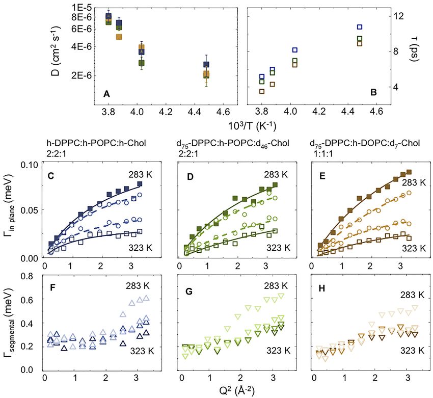
Ahmadi et al.                                                                                                             Neutron Scattering of Lipid Rafts

                                             cholesterol to unsaturated lipid. These results show the sensitivity of lipid diffusion to local
                                             cholesterol concentration, and the importance of considering the local, rather that the
                                             global composition of a membrane when understanding the diffusion processes of lipids
                                             within the membrane. The novel combination of SANS and QENS allows a non-intrusive
                                             approach to characterize the structure and dynamics occurring in phase-separated model
                                             membranes which are designed to mimic the lateral heterogeneity of lipids seen in cellular
                                             membranes–a heterogeneity that can have pathological consequences.
                                             Keywords: QENS, SANS, lipid rafts, lipid, multi-component systems

1 INTRODUCTION                                                                     understanding of lipid raft composition and dynamic behaviour,
                                                                                   could thus offer the potential to facilitate development of future
Biological membranes are critical to the integrity and activity of                 therapies to treat a diverse range of pathological conditions.
cells: they not only provide for a physical compartmentalisation                       Studies that have focused on the structure and physico-
of their internal metabolic processes, but also afford the means by                chemical properties of lipid rafts have generally involved in-
which they are able to regulate the import, export, and exchange                   silico modelling and/or experimental studies utilising model
of substances, and mediate the transduction of chemical signals                    systems [21–31]. Since lipids have different molecular
received extracellularly [1, 2]. In the Singer and Nicholson model                 configurations depending on temperature, structural studies
proposed in 1972, they were viewed as a disordered mosaic in                       have been performed to investigate the formation of rafts in
which bilayer lipids and embedded proteins move freely by lateral                  lipid vesicle membranes as a function of temperature and raft
diffusion, with the lipids considered to play only a passive                       composition [21–23, 30–32]. Despite the temperature
role–providing the building blocks to constitute the barrier                       dependence of lipid mixtures witnessed in in-vitro systems [30,
separating the cell’s internal and external environments.                          32], fundamental questions remain about the physical
    It was later shown that cell membranes also possess regions                    mechanisms that govern the formation, size, and stability of
where the lipids are much more tightly packed, forming highly                      these lo phase lipid “platforms”, especially those formed in
organised micro-domains of around 100 nm in diameter [3].                          vivo, and there has been very little experimental research
These micro- domains–also referred to as lipid rafts–were shown                    performed to probe the dynamic behaviour of lipid raft
to be composed of phospholipid, cholesterol, and sphingolipids                     systems at the molecular level.
[4, 5] and, as a consequence of their lateral segregation and/or de-                   In the work reported here we sought to rectify this deficiency
mixing behaviour, were demonstrated to exist as liquid-ordered                     by employing quasi-elastic neutron scattering (QENS) to explore
(lo) islands of reduced fluidity that co-exist within the “loosely                  the dynamics of lipids on a picosecond timescale following
packed” liquid-disordered (ld phase), frequently unsaturated,                      temperature-induced lipid raft formation in ternary lipid
phospholipids present in the bulk of the membrane [2]. These                       vesicles (widely studied by others [33–38]) composed of
lo phase raft platforms exist by virtue of cholesterol’s propensity to             dipalmitoylphosphatidylcholine (DPPC), cholesterol (Chol)
form condensed molecular complexes with, for example, long                         and either palmitoyloleoyl- or dioleoyl-phosphatidylcholine
chain saturated phospholipids, such that its local concentration                   (POPC and DOPC, respectively). Complementary structural
rises above ~20%, creating zones with an almost binary lipid                       studies of the systems were also performed using small angle
composition [6–8]. For recent reviews covering the history,                        neutron scattering (SANS) and dynamic light scattering (DLS)
composition, and mesoscale organisation of lipid rafts, the                        experiments.
reader is referred to Gori [9], Lu and Fairn [10] and Sezgin                           The novel combination of SANS and QENS methodology
et al [11].                                                                        deployed here is shown to provide an informative and non-
    As regards their functions within biological systems, lipid rafts              intrusive (non-perturbing, reporter-free) way in which to
are known to play major roles in membrane protein trafficking, in                   characterize the structure and dynamics of phase segregated
signal transduction, and in mediating the cellular entry and exit of               model membrane systems at the molecular level. The
pathogens [12, 13]. The dense organisation of their lipids and                     methodology might thus be used to explore the behaviour of
proteins also leads to enhanced protein-protein interaction, and                   model membranes with compositions more closely designed to
can result in accelerated signal transduction and enhanced                         mimic the lateral heterogeneity seen in cellular membranes–a
enzyme activity [12].                                                              heterogeneity that often carries pathological consequences.
    The clustering of rafts has also been implicated in the
pathogenesis of microbial infections [14] and they are thus
relevant to our understanding of the development and spread                        2 MATERIALS AND METHODS
of infectious diseases. Their formation and distribution within
cell membranes are shown to be pertinent to cardiovascular                         2.1 Materials
disease [15], ageing and neurodegenerative diseases [16],                          All protiated components and partially-deuterated phospholipids
inflammation [17] and cancer [18–20]. Further knowledge and                         and cholesterol (e.g. d7-Chol) were purchased from Avanti Polar

Frontiers in Physics | www.frontiersin.org                                     2                                    April 2022 | Volume 10 | Article 864746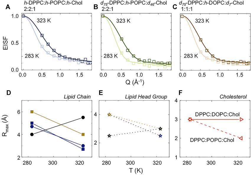
Ahmadi et al.                                                                                                        Neutron Scattering of Lipid Rafts

TABLE 1 | Sample characteristics post preparation.

Lipid composition                             Molar     Apparent hydrodynamic                 Concentration           Solvent             Technique
                                              ratio         diameter (nm):                      (mg/ml)

d75-DPPC:h-DOPC:d7-Chol                       1:1:1           120   ±   15                          30                 D 2O                 QENS
d62-DPPC:h-DOPC:h-Chol                        1:1:1           127   ±   14                          10               D2O:H2O                SANS
d62-DPPC:h-DOPC:h-Chol                       15:60:25         129   ±   14                          10               D2O:H2O                SANS
h-DPPC:h-POPC:h-Chol                          2:2:1           137   ±   16                          30                 D 2O                 QENS
d75-DPPC:h-POPC:d46-Chol                      2:2:1           139   ±   14                          30                 D 2O                 QENS
d62 -DPPC:h-POPC:h-Chol                       2:2:1           132   ±   13                          10               D2O:H2O                SANS

Lipids (Alabaster, AL, United States). Perdeuterated cholesterol             selected to have the same scattering length density (SLD) as
was produced in the Deuteration Laboratory platform of the Life              the lipids comprising the vesicle (assuming the lipids to be
Sciences group [39] at the Institut Laue Langevin (ILL) in                   homogenously mixed at high temperatures). Therefore any
Grenoble, France, using a lipo-engineered cholesterol                        phase separation of the lipids within the vesicle membranes as
producing Pichia pastoris strain CBS7435Δhis4Δku70 Δerg5::                   a result of a reduction in temperature would lead to the formation
pPpGAP-ZeocinTM-[DHCR7]Δerg6::pGAP-G418 [DHCR24]                             of regions of differing SLD’s that are no longer matched to the
[40]. Following adaptation to growth in deuterated minimal                   SLD of the solvent.
medium, cells were grown in a high cell density fermenter                        To prepare the vesicles, the lipids were weighed in accordance
culture. Purification was carried out by HPLC on a                            with the desired molar ratio, correcting for deuteration
NUCLEODUR® 100–10 C18ec column (Macherey-Nagel,                              appropriate, and dissolved in chloroform (spectroscopic grade,
Düren, Germany) using an isocratic mixture consisting of                     Fluka United Kingdom, Ltd., Dorset, United Kingdom). The
acetonitrile/methanol (9:1) at a flow rate of 20 ml/min [41].                 solvent was then evaporated to dryness using a BUCHI 461
Deuterated cholesterol made in this way is now widely used in                rotary evaporator and the resultant dry lipid film hydrated
neutron scattering and related studies [42, 43].                             with either D2O (99.7% D; Aldrich, United Kingdom, Ltd.,
                                                                             Dorset) or a D2O:H2O mixture; where the SLD of the D2O:
2.2 Methods                                                                  H2O dispersion medium was selected to match the “average” SLD
2.1.1 Vesicle Preparation                                                    of the ternary lipid mixture assuming ideal mixing (termed as
Three types of small unilamellar vesicles were prepared: 1) DPPC:            “on-contrast”). For the 1:1:1 molar ratio mixture of d62-DPPC:h-
DOPC:Chol in molar ratio 1:1:1 (raft forming mixture); 2) DPPC:              DOPC:h-Chol, the calculated SLD was 2.29 × 10⁻⁶ Å−2 and so a
DOPC:Chol molar ratio 15:60:25 (non-raft forming mixture) and                mixture of H2O 5.8662 g and D2O 4.5568 g was used. Samples
3) DPPC:POPC:Chol molar ratio 2:2:1 (raft forming mixture).                  were prepared at two different total lipid concentrations, namely
The DPPC:DOPC:Chol 1:1:1 and DPPC:POPC:Chol 2:2:1                            10 and 30 mg ml−1 for SANS and QENS, respectively. Note, that
mixtures were used to study the dynamics in rafts of differing               for our SANS studies the total lipid concentration of 10 mg ml−1
sizes [38]. Different isotopic forms and concentration of the                had been optimised by earlier studies to enable the ready
vesicles were prepared for the SANS and QENS study as                        production of the unilamellar vesicles necessary for the study,
shown in Table 1. Owing to the differences between the                       whilst at the same time yielding sufficient neutron scattering
neutron scattering cross sections of the lipids (as a result of              signal. The higher total lipid concentration used for the QENS
the difference in interactions, coherent and incoherent, between             study is necessary to increase the statistical accuracy of the data.
hydrogen and deuterium: σcohH = 1.76 barn, σcohD = 5.59 barn,                Note that additional broadening due to the diffusion of the centre
σincH = 79.74 barn; σincD = 2.01 barn), selectively deuterated               of mass of whole vesicles can be neglected.
samples were used in order to make ‘invisible’ certain                           To produce unilamellar vesicles of uniform and defined size
components in the mixtures whilist highlighting others. The                  from the highly concentrated lipid suspensions prepared here, the
contrast combination requirements for the two techniques                     lipid suspension was firstly sonicated; the vesicles were then
used, SANS and QENS are slightly different. For QENS the                     extruded under pressure (compressed nitrogen gas at a
use of perdeuterated components within the lipid dispersion                  pressure of 15 bar) through polycarbonate filters of 100 nm
makes it possible to ‘hide’ a molecule’s dynamics and thereby                pore (Nuclepore Track-Etch membrane). To aid the extrusion
highlight the dynamics related to the fully protiated component.             process, the temperature of the extruder was set at 326 K, above
Note here, that due to the limited availability of perdeuterated             the transition temperature of the highest melting point lipid,
cholesterol, experiments were also performed using partially                 namely ~314 ± 1 K for DPPC, to ensure all the lipids were in a
deuterated chol (as in the case of d75-DPPC:h-DOPC:d7-Chol                   fluid state when extruded. The vesicle suspensions were filtered
1:1:1). For the same reason and in order to “hide” the solvent               an odd number of times until the desired mean vesicle diameter
dynamics, the lipid dispersions for QENS were prepared in D2O                size of about 120 nm was achieved, suggesting the formation of a
(if instead H2O were used as the solvent, its signal would                   predominately unilamellar population of vesicles. Table 1 gives
dominate the scattering profile). In the case of the SANS                     the mean apparent hydrodynamic diameter for each type of
experiments, the solvent used to prepare the vesicles was                    vesicle prepared (with a measured polydispersity of ~0.15 in

Frontiers in Physics | www.frontiersin.org                              3                                      April 2022 | Volume 10 | Article 864746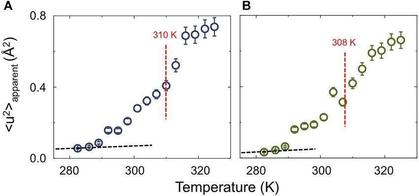
Ahmadi et al.                                                                                                        Neutron Scattering of Lipid Rafts

all cases); the data were measured on a diluted vesicle dispersion        Additional normalization steps, such as removing the signal
(x100) using Dynamic Light Scattering (DLS; NanoBrook Zeta                associated with the empty can and from an appropriately
Potential Analyzer, Brookhaven Instruments Corporation, Long              weighted spectrum of the D2O solvent, as well as full data
Island, NY) over 3 days to ensure that the dispersions were stable        analysis were carried out directly on the S(Q,ω) spectra using
with respect to their size (at ambient temperature), for the length       either Mantid [48] or Origin 2019b. Each Q-slice was analysed
of time required to complete the neutron scattering experiments.          using a built-in least squares algorithm accounting for the
                                                                          instrumental energy resolution along with up to two
2.1.2 Small Angle Neutron Scattering                                      Lorentzian functions.
SANS measurements were performed on the D11 instrument at
the Institut Laue Langevin (ILL) in Grenoble, France [44].                2.1.4 Analysis of QENS Data
Scattering intensities were measured with a 2-D 3He-detector              QENS measurements provide data on the broadening, measured
consisting of 128 × 128 pixels of 7.5 × 7.5 mm2 size.                     as an energy transfer (ħω) that appears around the elastic
Measurements were performed at a wavelength of 5 Å with a                 scattering signal, resulting from dynamical relaxation processes
9% full width at half maximum (FWHM), using 4 and 28 m                    in the system. The latter are mainly due to local motions and/or
sample-detector distances to access scattering vectors (Q = (4π/λ)        diffusional events. More specifically, the scattering function is
sin (θ)) in the range 0.0025–0.2 Å−1. Transmissions were                  composed of both coherent and incoherent scattering terms
measured at 28 m with the attenuated direct beam. Samples                 (owing to the nature of the neutron-nucleus interactions),
were acquired in quartz cuvettes with 1 mm path length and                enabling the computation of the spatio-temporal correlations
measured at either 308 or 279 ± 0.1 K, using a copper sample              between identical nuclei (Sinc) and the static and dynamic
holder for precise temperature control. Absolute intensities and          correlations of distinct nuclei (Scoh) according to [49]:
detector efficiency were determined by using a 1 mm H2O sample
                                                                                         S(Q, ω)  Sinc (Q, ω) + Scoh (Q, ω)                      (1)
as secondary calibration standard, which is cross-calibrated
against polystyrene h-/d- polymer blends. The differential                The elastic signal is modelled using the instrumental resolution
scattering cross section of a 1 mm H2O sample at 5 Å on D11               obtained from the vanadium standard. The broadening is
is 0.929 1/cm. The empty cell and either D2O or the appropriate           modelled using Lorentzian functions, and is analysed by
H2O:D2O solvent mixture were also recorded to properly reduce             examining the correlation of the linewidth (Γ, HWHM) that
the raw data. Data reduction was performed using the facility-            determines the relaxation time (τ) for the process involved. In its
provided LAMP software [45].                                              simplest form, the measured signal, which is typically dominated
    Model-fitting of the SANS profiles was carried out using                by that of hydrogen, provides correlations of single H atoms, and
Sasview [46], employing either a simple power law model or a              can be deconvoluted into vibrational, rotational and translational
model combining a power law and a Broad/Lorentzian                        components:
peak model.
                                                                                    Sinc (Q, ω)  SV (Q, ω) ⊗ SR (Q, ω) ⊗ ST (Q, ω)               (2)
2.1.3 Quasi-Elastic Neutron Scattering                                    In the case of isotropic, harmonic vibrations, the first term can be
QENS measurements were conducted using the IRIS                           written as:
spectrometer [47] at the ISIS Neutron and Muon Facility
                                                                                               Svib (Q, ω)  e−Q (u )3
                                                                                                                 2    2
(Harwell, Oxford, United Kingdom). Samples were loaded into                                                                                       (3)
aluminum annular cans of a neutron path length of 0.5 mm to
                                                                          where < u2> is the mean square displacement.
ensure a sample transmission of 90%. Elastic fixed window scans
                                                                              It is worth noticing that the “experimental scattering function”
(EFWS) were conducted over the temperature range 283–323 ±
                                                                          (e.g. S(Q,ω)measured) contains an implicit “resolution effect”
0.1 K; measurements were taken in increments of 3 ± 0.1 K. Scans
                                                                          (R(ω)), which translates into specific dynamics being “visible
were conducted in both heating and cooling modes to ensure that
                                                                          or not” within the time-scale accessible by a specific instrument.
no hysteresis was present. EFWS were used to locate the phase
                                                                          Note that the “measured scattering function” (e.g. S(Q,ω)measured)
transition temperature range as well as to discriminate the
                                                                          is given by the “real scattering function” (e.g. S(Q,ω)real)
temperature at which the dynamics enters the timescale
                                                                          convoluted with R(Q,ω).
accessible to the spectrometer; this allows specific temperatures
                                                                              Diffusional processes result in a dispersive Γ(Q2) relation
to be selected for further investigation by QENS. Scattering
                                                                          [49–53] from which a diffusion coefficient can be extracted,
profiles were then recorded with an energy resolution of
                                                                          and in general terms can be described with a Lorentzian function:
17.5 μeV, using the PG002 analyser crystal set-up,
investigating dynamics in the picosecond timescale, at                                                     1     Γ
temperatures of 283, 288, 298 and 323 ± 0.1 K. Raw data were                                   S(Q, ω)                                           (4)
                                                                                                           π ω2 + (Γ)2
normalized to incoming neutron flux and corrected for detector
efficiency, by direct comparison with the purely incoherent signal         The dynamics in lipidic systems is a complex mix of processes
scattered from a vanadium standard. The double differential cross         spanning a wide range of time scales. Generally speaking there are
section was then converted into the corresponding dynamic                 two main processes occurring over different timescales, described
structure factor S(Q, ω). Detectors were grouped to provide               as “rattling-in-a-cage”, where a lipid molecule can be thought of
~12 spectra and the energy binned into constant 5 μeV steps.              as moving around within a cage formed by the surrounding lipids,

Frontiers in Physics | www.frontiersin.org                            4                                     April 2022 | Volume 10 | Article 864746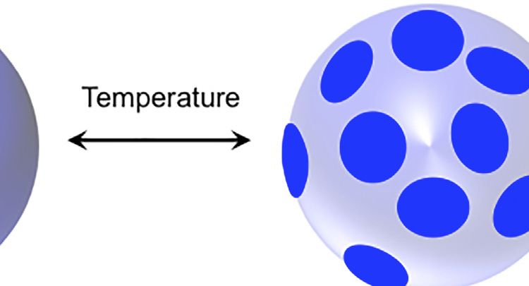
Ahmadi et al.                                                                                                          Neutron Scattering of Lipid Rafts

and “random-walk-like”, where a lipid molecule moves out of its                contribution has been weighted and then summed. For the
cage to occupy a new space within the bilayer and its original                 DPPC:POPC:Chol 2:2:1 mixture, the partially deuterated
location is occupied by a different lipid molecule. However, little            QENS sample was first analysed to model only the POPC
is known about the mechanism of how lipids actually diffuse,                   dynamics. The resulting parameters were then used to
especially in raft-like domains or in real cellular entities.                  constrain the analysis of POPC in the fully protiated sample
   Based on the timescale probed by the QENS instrument used                   and, therefore, to allow the dynamics of the DPPC and cholesterol
here, in the order of picoseconds, we use a jump diffusion model               to be established. A similar approach was used to model the
to best describe the “in-plane” lipid diffusion:                               DPPC:DOPC:Chol 1:1:1 system, where the dynamics of (d75)
                                                                               DPPC was assumed to be almost invisible, allowing d7-Chol to be
                                         D t Q2                                modelled accounting for the dynamics of -CH2 in the chain.
                               ΓT                                   (5)
                                      1 + Q2 Dt τ 0
where Dt is the translational diffusion coefficient and τ0 is a
                                                                               3 RESULT AND DISCUSSION
residence time [49–53]. This model allows us to describe how the
lipid molecule moves out of its cage [53].                                     3.1 Evidence of Raft Formation
    When non-diffusive phenomena are involved, such as in the                  SANS was used to provide evidence of the absence or presence of
case of spatial confinement, Eq. 4 becomes:                                     lipid rafts in small unilamellar vesicles as a function of lipid
                                                      1     ΓT                 composition and temperature. The three lipid mixtures of
   S(Q, ω)  A0 (Q, T)δ(ω) + (1 − A0 (Q, T))                        (6)        differing composition and molar ratios, namely DPPC:DOPC:
                                                      π ω2 + (ΓT )2
                                                                               Chol molar ratio 1:1:1, DPPC:POPC:Chol molar ratio 2:2:1 and
where A0 (Q,T) is the so-called elastic incoherent structural factor           DPPC:DOPC:Chol molar ratio 15:60:25 (Table 1) were
(EISF) and δ(ω) is the delta function representing the elastic peak            investigated, with each sample studied at 279 and 308 ± 0.1 K.
(which accounts for all immobile hydrogen atoms in the system).                These particular compositions were selected on the basis of our
   Scattering profiles were modelled following the approach                     own preliminary studies and those of others reported in the
proposed by Sharma et al. [54–56].                                             literature, which suggest that at ~300 K the lipids within the
                                                                               vesicle membranes laterally phase segregate. In these studies, the
   Sves (Q, ω)  A(Q)Llat (Γlat , ω) + (1 − A(Q)) Ltot (Γtot , ω)    (7)       aqueous medium used for dispersion of the vesicles was prepared
                Sves (Q, ω)  Slat (Q, ω) ⊗ Sint (Q, ω)              (8)       so as to match the mean SLD of the ternary lipid mixture. When
                                                                               the lipids are homogenously dispersed within the vesicle bilayer,
where Sves(Q,ω) represents the scattering function for the entire              i.e., when there is no lateral separation of the lipids (and thus no
system, and contains the contributions from the lateral and                    raft formation), as shown in Figure 1A, there will be negligible
internal motions of the lipid molecules; Ltot (Γtot, ω),                       difference between the SLD of the vesicle lamellae and solvent.
represents a superposition of the lateral and internal motions                 Under these conditions, little or no neutron scattering is expected
of the lipid molecules.                                                        from the sample–a condition referred to as “on-contrast”. When
    Here, the EISF was modelled using a modified version of the                 the lipids in the vesicle bilayer undergo a phase separation or
Volino/Dianoux model [57] originally used to describe “diffusion               “demixing”—a condition typically induced by reducing the
inside a sphere”, as proposed by Carpentier et al. [58]. The model             experimental temperature such that some lipids are in the
describes the lipid as having a linearly varying radius for the                fluid state (here POPC/DOPC) and others (here DPPC) are in
diffusion volumes along the lipid length. This modification is                  the gel phase–spatial differences in lipid composition occur and
based on the consideration that hydrogen atoms along the lipid                 differences in SLD between the dispersion and the medium are
tails could experience mobilities over different spatial extents and,          observed (Figure 1B). Under these conditions an increase in
mathematically, is expressed as:                                               scattering intensity is observed–a condition termed “off-
                                                       2                       contrast”.
                                   1 N 3j1 (QRn )
                    A0 (Q, T)                                    (9)           The SANS profiles for the various vesicle preparations that
                                   N n1 QRn                                   were recorded at the higher experimental temperature of 308 ±
where                                                                          0.1 K showed very little scattering, thereby confirming that the
                                                                               mean SLD of the vesicle lamellae had been well-matched by the
                              n−1
                       Rn        [RN − R1 ] + R1                   (10)       SLD of the H2O/D2O mixture comprising the dispersion
                              N−1
                                                                               medium. The rise in I(Q) that is seen at low Q arises because
N stands for the total number of atoms in the chain to which                   of Porod scattering, due to the small difference in SLD that
hydrogen atoms are bound and the index n starts with the carbon                remains between the lipid acyl chains and the solvent (ca. 2.6 ×
atom the closest to the oxygen of the phosphorus group, which                  10–6 Å−1 vs. ca. 2.3 × 10⁻⁶ Å−1), with the wet/hydrated lipid
connects the lipid chains with the head group; Rn is, therefore, the           phosphocholine headgroups being effectively invisible in the
radius of the diffusion volume for the corresponding hydrogen                  solvent (having an SLD of ca. 2.2 × 10⁻⁶ Å−1). Given the low-
atoms. Eq. 9 has been implemented considering all the unique                   level scattering seen for these systems (being little higher than
atoms in the lipid chain and head groups; and has further                      background), the model-fitting of the SANS profiles measured at
accounted for CH2 and CH3 dynamics in cholesterol. Each                        308 ± 0.1 K [59–61] was performed using a simple power law

Frontiers in Physics | www.frontiersin.org                                 5                                     April 2022 | Volume 10 | Article 864746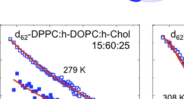
Ahmadi et al. Neutron Scattering of Lipid Rafts
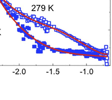
FIGURE 1 | (A,B) Schematic for the lipid raft formation. (C–E) SANS profiles at 279 (open symbols) and 308 K (filled symbols) for: i) d62-DPPC:h-DOPC:h-Chol at
molar ratio of 1:1:1 (C) and 15:60:25 (D) and d62-DPPC:h-POPC:h-Chol at molar ratio of 2:2:1 (E). In red is the best fit for the data using the methodology detailed in the
text. Profiles have been presented in log-log scale to highlight the raft domain, where present.
function–with the fitted exponent, thence providing a description attributed to domain formation [46]—a mixed model
of the interface between the dispersed (lipid lamellae) and the comprising Porod and Lorentzian components was used. The
continuous (aqueous) phases. The values of the fitted power law data were well modeled obtaining Lorentz lengths of 87.2 ± 1.5
(Porod) exponent (n), were obtained as 2.55 ± 0.15, 2.54 ± 0.06, and 34.1 ± 1.9 Å for the 1:1:1 DPPC:DOPC:Chol and 2:2:1 DPPC:
and 1.97 ± 0.15 for the vesicles prepared from 1:1:1 DPPC:DOPC: POPC:Chol vesicles respectively, suggesting the appearance of
Chol, 2:2:1 DPPC:POPC:Chol and 15:60:25 DPPC:DOPC:Chol, domains of ~18 and 7 nm across, with the fitted Porod exponent
respectively. These exponent values (each ca. 2) are characteristic of 2.5 ± 0.1 again indicating a rough interface between the
of lamellae [62] and the values for the two raft-forming systems continuous and disperse phases. It should be noted that the
(being around 2.5) indicate roughened surfaces, while that for the effects of temperature on raft formation are completely
non-raft forming system (being closer to 2) indicates a more reproducible in that repeated heating and cooling cycles cause
smooth surface [62]. the domains to disappear and reform. Furthermore (although not
In two of the compositions studied, namely the 1:1:1 molar shown here) the thickness of the vesicle bilayers, determined by
ratio of DPPC:DOPC:Chol and the 2:2:1 molar ratio of DPPC: SANS studies on vesicles dispersed in 100% D2O at 308 and 279 ±
POPC:Chol molar ratio 2:2:1, decreasing the temperature to 0.1 K, i.e., off-contrast (well modelled assuming a mixture of
279 ± 0.1 K resulted in the appearance of a broad peak in the isolated/single infinite planar lamellar sheets) was identical
scattering curve, indicative of the formation of lipid domains/ irrespective of temperature, showing that the vesicles
rafts (Figures 1C,E). In contrast, in the case of the 15:60:25 molar maintained their structure upon decreasing the temperature.
ratio vesicle suspension of DPPC:DOPC:Chol (Figure 1D), no The size of the domains or rafts determined here are consistent
broad peak was observed, indicative of the absence of lipid raft with the data obtained for similar three-component systems
formation at the experimental temperature. As found when investigated by SANS and MD simulations [24, 31].
modelling the SANS data recorded at the higher temperature Additionally, the differences in the sizes of the rafts with
of 308 ± 0.1 K, it was possible to model the data obtained for the differing lipid composition agree with the findings from
15:60:25 DPPC:DOPC:Chol vesicles at the lower temperature previous studies wherein the sizes of the lipid domains were
using only a power law function with an exponent, n = 1.8 ± 0.05 correlated with a mis-match in the lengths of the lipid
(as compared with the exponent of 1.97 ± 0.15 determined at the hydrocarbon chains [35], as well as differences in line tension
higher temperature of 308 ± 0.1 K). In contrast, in order to model [63]. Furthermore, our results are also in agreement with the
the emergence of the broad peak in the SANS profiles of the other results presented by Zhao et al. [64] where the presence of either
two vesicle preparations–the formation of this peak being POPC or DOPC in a ternary mixture with sphingomyelin and
Frontiers in Physics | www.frontiersin.org 6 April 2022 | Volume 10 | Article 864746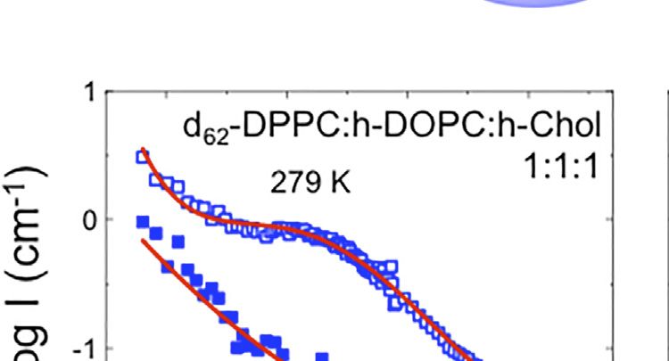
Ahmadi et al.                                                                                                                           Neutron Scattering of Lipid Rafts

  FIGURE 2 | (A) Elastic fixed window scan (EFWS) for fully protiated DPPC:POPC:Chol (molar ratio 2:2:1) (blue triangles) and its partially deuterated analogue (green
  triangles; d75-DPPC:h-POPC:d46-Chol). (B–G) Cartoons depict the phase separation at the two extreme temperatures [323, (B–D), and 283 ± 0.1 K; (E–G)] for fully
  protiated and partially deuterated DPPC:POPC:Chol 2:2:1 (blue and green, respectively) as well as for d75-DPPC:h-DOPC:d7-Chol 1:1:1 (brown). Lipid raft cartoons
  were made using VMD package [75].

cholesterol gave rise to domains of different sizes. The difference                    disentangled, without perturbation. QENS is ideal in this regard
in domain size between lipid mixtures containing POPC vs. those                        as it allows a “serial decoupling” approach (originally introduced
containing DOPC, could relate to the differing interaction of                          by Angell and co-workers for ion-conducting glasses [71, 72], and
cholesterol with the lipids [65, 66]. Microsecond molecular                            now extended to technological membranes [73, 74]).
dynamics simulations indicate that cholesterol interacts more                              In the studies reported here, the mobility of the lipids
favourably with saturated lipid tails and that there is a                              comprising the vesicle bilayers was investigated using IRIS
“competition” between the tighter cholesterol–lipid packing                            (primarily to focus on the lipid lateral diffusion), employing
and the looser lipid–lipid packing as the membrane changes                             isotopic contrast variation to disentangle how the dynamics of
from the ld to lo phase [66].                                                          each lipid component varies as a function of its environment. We
                                                                                       first studied the elastic fixed window scan (EFWS) of the fully
3.2 Lipid Dynamics                                                                     protiated 2:2:1 DPPC:POPC:Chol vesicles and compared this to
The dynamics of lipid membranes includes multiple relaxation                           its perdeuterated analogue, namely d75-DPPC:h-POPC:d46-Chol,
processes on local and global scales [67–70]. These are associated                     to highlight only the effect of raft formation on POPC dynamics
with motions such as diffusion, shape and thickness fluctuations,                       (Figure 2).
membrane undulations, lipid flip-flops, localized rotations, and                             The EFWS data shown in Figure 2A, for DPPC:POPC:Chol 1:
vibrational motions. Such dynamics thus span a broad range of                          1:1 vesicles, reveals that, at temperatures above 283 ± 0.1 K, the
time and spatial scales [65], and in order to study them one thus                      elastic intensity (Intensityelastic) starts to decrease in a linear
needs a technique that will allow the various contributions to be                      manner, pointing to a change in the dynamics of the lipids at

Frontiers in Physics | www.frontiersin.org                                         7                                             April 2022 | Volume 10 | Article 864746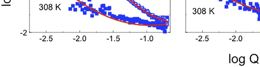
Ahmadi et al.                                                                                                                               Neutron Scattering of Lipid Rafts

  FIGURE 3 | (A) Temperature dependence of the apparent mean-square displacement < u2>apparent for fully protiated DPPC:POPC:Chol molar ratio 2:2:1 (A) and its
  partially deuterated analogue (d75-DPPC:h-POPC:d46-Chol; (B). In the plot is reported the linear fit at low-T (black dashed line) having a slope” d < u2>/dT = 0.23 ± 0.05
  10–3 Å2 K−1. The red dashed lines indicate the mid point as obtained by sigmoidal fit, suggesting a cooperative transition form raft to homogenously dispersed
  composition.

the nanoscale, which would be expected if a phase transition in                           up to now (the data for D2O are reported in Supplementary
the vesicles occurred. Not surprisingly, the gradient of elastic                          Figure S2). Furthermore, we extended our study to vesicles
intensity change seen for the two isotopic forms of DPPC:POPC:                            prepared with a 1:1:1 molar ratio of d75-DPPC:h-DOPC:d7-
Chol 1:1:1 (namely fully protiated DPPC:POPC:Chol and d75-                                Chol, to study the effects of raft dimensions and lipid
DPPC:h-POPC:d46-Chol) is different. The change in slope at                                unsaturation on the dynamics.
around 283 K is associated with an increase in either the entire                              In Figure 4 the scattering profiles of all the isotopic variants of
system (h-DPPC:h-POPC:h-Chol) or just POPC alkyl chain (d75-                              the vesicles investigated by QENS are reported. The D2O
DPPC:h-POPC:d46-Chol) mobility. Furthermore, if we consider                               contribution was weighted and subtracted from the vesicle
that the transition temperatures (Tm) of the individual lipid                             data to enable highlighting of the QENS broadening due to
constituents POPC and DPPC) are ~271 and ~314 K, it is                                    lipid dynamics.
evident that the reduction in elastic intensity cannot be simply                              The QENS data required two Lorentzian components to
associated with a single component lo–ld transition. The                                  obtain a good fit to the experimental data (Supplementary
transition can thus be associated with the structural transition                          Figure S3). Based on the time scale investigated (tens of ps;
from “raft-containing” membranes to those with “homogenously                              Eres = 17.5 μeV), we associate the narrow component to “in-
mixed” lipids (Figures 2B–G).                                                             plane” diffusion, while the broader component is considered to be
   Apparent mean-squared displacements (apparent) can be                              due to the segmental relaxation of the lipid, following the
calculated from the EFWS data and are plotted as a function of                            approach of Sharma et al. [54–56]. The “in-plane” diffusion
temperature in Figure 3. The calculated Debye-Waller factor                               was modelled using the jump diffusion model (Eq. 5, as
(DWF, Eq. 3; Supplementary Figure S1) at low-T, shows a linear                            previously reported [76–79]); the resulting fits are shown in
temperature dependence with slope < u2>/dT = 0.23 ± 0.05 ×                                Figure 4 (see Table 2).
10–3 Å2 K−1. The strong reduction of DWF seen at low-T arises                                 Despite the research reported in the literature, very little is
from the macroscopic stiffening as a consequence of the raft                              known about the mechanism of how lipids diffuse; this is mainly
formation which implies a reorganization of the lipid chain as                            because lipid dynamic behaviour is a complex phenomenon
well as a reduction of the surface movements. From Figure 3 it is                         governed by a hierarchy of fluctuations and movements which
also evident that following the increase in apparent it is                            cover an extremely wide range of correlation times (pico-seconds
possible to follow the transition from raft to homogenously                               to seconds [76]). This explains why different experimental
dispersed composition. The fact that the process is describable                           techniques, and therefore different time scales, as well as
using a sigmoidal function suggests a cooperative process where                           different sample preparations, yield diffusion coefficients that
the presence of rafts favours the formation of more/larger rafts.                         differ by around 2 orders of magnitude [68, 76–83]. For instance,
The sigmoidal function was centred at around 310 K, consistent                            in the case of experiments performed on lipid single-layers on a
with the SANS data which indicates rafts at ~280 K and a                                  solid substrate, the diffusion in the lower leaflet is suppressed by
homogenous distribution of the three-components at ~310 K.                                the presence of the substrate, while the diffusion in the upper
   To gain a detailed insight into the effect of raft formation on                        leaflet may be enhanced by a highly ordered fluid phase of the
the dynamical behaviour of the membrane lipids, QENS                                      lipids [76]. Similarly, in the case of lipid multi-layer systems, by
experiments were performed at a range of temperatures                                     changing sample orientation (i.e., 45° vs. 135°) with respect to the
between 283 and 323 ± 0.1 K for the two samples discussed                                 incident beam, it is possible to probe either “out-of-plane” or “in-

Frontiers in Physics | www.frontiersin.org                                            8                                              April 2022 | Volume 10 | Article 864746
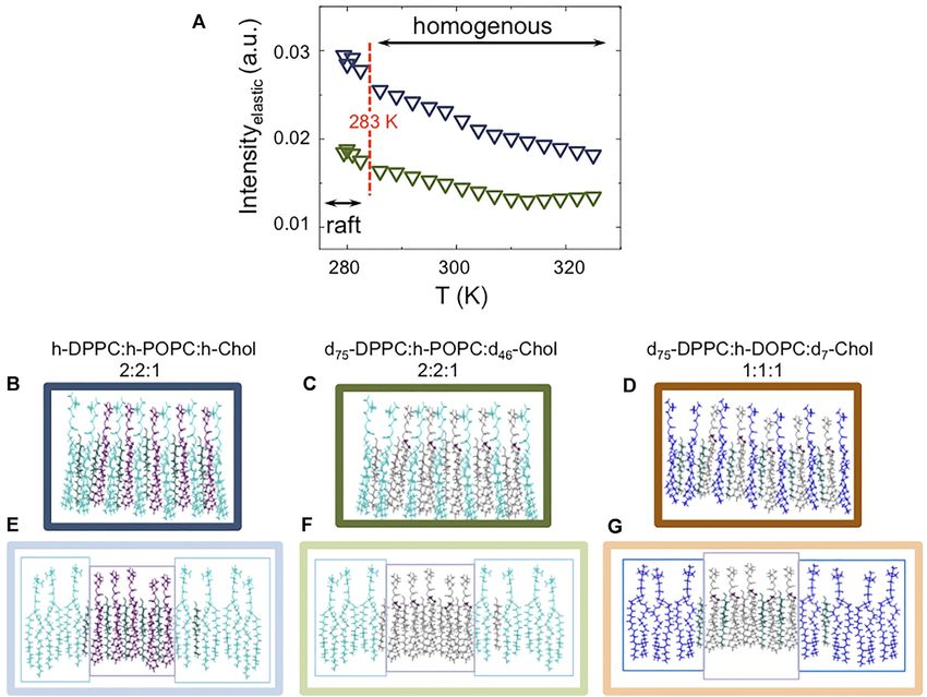
Ahmadi et al.                                                                                                                                 Neutron Scattering of Lipid Rafts

  FIGURE 4 | QENS S (Q,ω) data and model fit at a representative value of Q = 1.25 Å-1, measured on IRIS, with 17.5 μeV resolution, between 283 and 323 K for: 1)
  h-DPPC:h-POPC:h-Chol molar ratio 2:2:1 (in this contrast the dynamics of all three components contributes to the scattering profile); 2) d75-DPPC:h-POPC:d46-Chol
  molar ratio 2:2:1 (in this contrast most of the signal is due to POPC; the other components are virtually “invisible”) and 3) d75-DPPC:h-DOPC:d7-Chol molar ratio 1:1:1 (in
  this contrast most of the signal is due to DOPC; the other components are virtually “invisible”). The central line (grey) due to elastic scattering is modeled by the
  instrumental resolution. The narrow Lorentzian signal (light purple) indicates the lipid diffusion; the broader (dark purple) component represents the lipid segmental
  relaxation. The global fit (red continuous curve) is overlain on the data points (black open squares).

plane” motion, which results in a different (~1 order of                                   this seems at first unexpected, this reduction in diffusion with
magnitude) self-diffusion coefficient [77–79]. Further, a                                   increasing temperature occurs when the vesicular system
variation in fluidity of ~50% was shown by comparing                                        converts from phase separated (containing lipid rafts
unilamellar and multilamellar phases. This variation in fluidity                            platforms; at 283 K) to homogeneous (at 323 K) states. At the
is associated with the enhanced diffusivity of -CH2 in the                                 range of experimental temperatures studied, namely between 283
unilamellar phase, which translates to an increased “in-plane”                             and 323 K, both POPC and DOPC should be in the fluid phase
diffusion [84–86]. In this scenario, and for the data reported here,                       (Tm of ~271 and ~257 K, respectively). However, at the higher
the enhancement in diffusivity (~10−6 cm2 s−1) might arise as a                            temperature of 323 K, the lipids in the vesicular bilayer have
consequence of the sample preparation–involving use of a                                   become homogenously mixed (no rafts present), thus the DOPC/
pressure extruder. Interestingly, it can be seen from the data                             POPC lipids will interact with the cholesterol that is randomly
for the partially deuterated samples, that POPC/DOPC lateral                               distributed throughout the vesicle bilayer at this temperature. As
diffusion reduces by around 70% during the heating. Although                               a consequence of the cholesterol condensation effect, the POPC/

Frontiers in Physics | www.frontiersin.org                                             9                                               April 2022 | Volume 10 | Article 864746
Ahmadi et al.                                                                                                                       Neutron Scattering of Lipid Rafts

  FIGURE 5 | (A–C) EISFs modeled using Eqs 9, 10. (D–F) Comparison between the maximum radii of diffusion used to model the EISF. POPC is reported in blue,
  DOPC in light brown, and DPPC in black. In the bottom panels are reported separately: the comparison between lipid chain (D); the comparison between lipid head
  group (E), and the comparison between cholesterol in the two systems (F).

DOPC dynamics slow down when in the presence of cholesterol.                          Furthermore, despite the interactions between DPPC and
At cooler temperatures, the cholesterol is effectively confined to                     POPC being neither attractive nor repulsive in the lo phase, in
DPPC-rich rafts, and thus interacts little with the DOPC/POPC                         the ld phase these become repulsive [66, 90].
lipids present in the disordered regions. This result is in                               To better investigate this phenomenon, we carefully analyzed
agreement with the earlier findings of Sarangi et al. who                              the EISF (Table 3; Figure 5) to characterize the mobility of the
reported a similar decrease in lipid diffusivity (~75%) upon                          single lipid chains in the entire set of investigated samples. Our
movement from Chol-poor to Chol-rich regions in POPC-                                 results at 283 K indicate that the POPC/DOPC are in their “fluid
Chol lipid bilayers [21].                                                             phase” (low-T and low cholesterol concentration) with an
    In the case of the fully protiated sample a more complicated                      organization in agreement with literature data [90–93]. We
situation must be considered. Upon heating, the lateral diffusion                     further notice that DOPC has a slightly higher (~10%)
is seen to reduce by approximately 40%. This result is a                              mobility than POPC (Figure 5D, squares). This could be due
consequence of two simultaneous phenomena occurring,                                  to a “synchronous” effect of: 1) a difference in Tm between the
namely a decrease in the mobility of the POPC, but an                                 two lipids; as well as, more importantly, 2) the presence of
increase in the mobility of DPPC as the temperature increases.                        cholesterol in the system and 3) its different interaction with
The decrease in the mobility of the POPC occurs because, as                           POPC vs. DOPC. Indeed, the calculations performed by Pandit
previously described, it is exposed to a higher concentration of                      et al. [65], suggest a reduced partial molecular area for
cholesterol when the rafts break down, whereas the mobility of                        cholesterol in the presence of POPC, compared to cholesterol
the DPPC increases as the temperature rises above its Tm                              in the presence of DOPC. These authors indicate that a possible
(~314 K) and as the break down of the rafts, and associated                           reason for such enhanced packing, is a “special arrangement” in
redistribution of the cholesterol in the system effectively lowers                    the POPC bilayer in which the α-face (smooth side) of
the concentration of cholesterol in the local environment of the                      cholesterol packs around the saturated chain, while the β-face
DPPC. This reduction in mobility also agrees with the findings                         (methylated side) packs well around the unsaturated chain,
from previous NMR studies [87]. Our interpretation of the QENS                        resulting in a better packing around cholesterol or
data is in agreement with experimental and MD simulation data,                        neighboring lipids [94]. The data for DPPC suggest a more
indicating that cholesterol in the lo phase mixes ideally with                        compact configuration (~30%), as also clearly demonstrated by
POPC, whilst also having a strong attraction for DPPC. In the ld                      the sharper decrease of the elastic line in the fully protiated
phase, on the other hand, it mixes ideally with DPPC while                            sample (Figure 2A, blue triangles), which is remarkably
exhibiting a significant repulsion for POPC [66, 88, 89].                              independent from the sample composition (Figure 5D, black

Frontiers in Physics | www.frontiersin.org                                       10                                           April 2022 | Volume 10 | Article 864746
Ahmadi et al. Neutron Scattering of Lipid Rafts
TABLE 2 | Dynamical properties extracted from the fits as a function of temperature (from 283 to 323 K) for h-DPPC:h-POPC:h-Chol molar ratio 2:2:1; d75-DPPC:h-POPC:
d46-Chol molar ratio 2:2:1 and d75-DPPC:h-DOPC:d7-Chol molar ratio 1:1:1. In the fully protiated sample the scattering profile is the result of the “sum” of each
component’s dynamics (appropriately weighted). The partially deuterated samples are virtually “invisible”, therefore, used to only highlight dynamics from the h-component
(i.e., POPC and DOPC). In these regards the comparison between 2:2:1 composition (e.g. fully protiated and partially deuterated) would highlight only the dynamics of POPC.
The comparison between 2:2:1 vs. 1:1:1 composition (both partially deuterated) would highlight the difference in dynamics between POPC and DOPC.
Sample Temperature (K) Dtr_jump 10–5 (cm2s−1) τ0 (ps)
d75-DPPC:h-DOPC:d7-Chol 1:1:1 283 0.78 3.5
288 0.50 4.3
298 0.39 6.5
323 0.21 8.9
h-DPPC:h-POPC:h-Chol 2:2:1 283 0.82 5.2
288 0.78 6.0
298 0.58 8.2
323 0.46 10.8
d75-DPPC:h-POPC:d46-Chol 2:2:1 283 0.71 4.6
288 0.63 5.6
298 0.27 7.0
323 0.18 9.5
D2O 283 1.04 1.8
288 1.14 1.2
298 1.70 1.1
323 2.10 1.0
TABLE 3 | Structural parameters obtained from EISF using Eqs 9, 10. Errors are ~10%. Each contribution has been weighted for the fraction of mobile hydrogen atoms in the
system and then added together. The partially deuterated sample was first analysed, to only model POPC, and then these parameters were constrained in the case of the
fully protiated sample to best describe DPPC and cholesterol.
Sample Temperature (K) Lipid Chain 1 (Å) Chain 2 (Å) Head Group (Å)
d75-DPPC:h-DOPC:d7-Chol 283 DOPC 0.2 — 0.2
6.0 — 4.0
DPPC 0.2 — 0.2
4.0 — 2.5
Chol 2.5 — —
3.0 — —
323 DOPC 0.2 — 0.2
4.0 — 3.0
DPPC 0.2 — 0.2
5.5 — 3.0
Chol 2.5 — —
3.0 — —
h-DPPC:h-POPC:h-Chol / d75-DPPC:h-POPC:d46-Chol 283 POPC 0.2 0.2 0.2
5.0 4.7 4.0
DPPC 0.2 — 0.2
4.0 — 2.5
Chol 2.5 — —
3.0 — —
323 POPC 0.2 0.2 0.2
2.7 3.0 2.5
DPPC 0.2 — 0.2
5.5 — 2.5
Chol 2.5 — —
2.0 — —
filled circles). This finding is not surprising as DPPC is an “homogeneous” vesicles leads to a “redistribution” of cholesterol
integral part of the raft platform at 283 K. within the system; this in turn leads to an increase of the DPPC
A different scenario arises when the temperature is increased to mobility and a reduction in the mobility of the POPC and DOPC.
323 K. In this case the conversion from “raft-containing” to Unsurprisingly the mobility of cholesterol at high-T, is dependent
Frontiers in Physics | www.frontiersin.org 11 April 2022 | Volume 10 | Article 864746Ahmadi et al. Neutron Scattering of Lipid Rafts
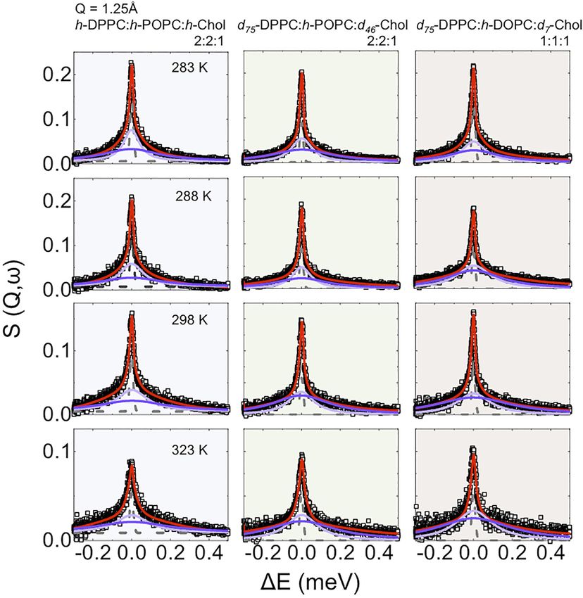
FIGURE 6 | Parameters extracted from the fits as a function of temperature (from 283 to 323 K) for 1) h-DPPC:h-POPC:h-Chol molar ratio 2:2:1 (blue); 2) d75-
DPPC:h-POPC:d46-Chol molar ratio 2:2:1 (green) and 3) d75-DPPC:h-DOPC:d7-Chol molar ratio 1:1:1 (brown). (A,B) Comparison of “in-plane” diffusion coefficient (A)
and relative residence time (B); (C–E) Γ(Q2) relative to the narrow Lorentzian component (lateral diffusion); (F–H) Γ(Q2) relative to the broad Lorentzian component
(segmental relaxation).
upon the vesicle membrane composition (around -30%, comparing homogeneous dispersions absent of any lipid rafts at 308 K, while
molar ratio 1:1:1 vs. 2:2:1; Figure 6F) and agrees with the model at 279 K lipid rafts of ~18 and ~7 nm diameter are formed, in
presented by Pandit et al. [65], who suggested that the presence of agreement with the nanometre-sized structures previously reported
POPC causes a reduction in the partial molecular area for cholesterol in the literature and consistent with the idea that larger rafts are
as a consequence of “more effective” packing. formed when there is a greater mis-match in lipids, i.e. DPPC/DOPC
compared to DPPC/POPC [24, 31]. To learn more about the
dynamics of the lipids within these different phases, we have
4 CONCLUSION used QENS to characterise the “in-plane” diffusion as well as the
lipid segmental relaxations. The elastic incoherent structure factor
The SANS data recorded show that lipid mixtures 1:1:1 DPPC: was used to extract the extent of mobility of the various lipid species
DOPC:cholesterol and 2:2:1 DPPC:POPC:cholesterol form within the mixture and confirms the better packing between Chol-
Frontiers in Physics | www.frontiersin.org 12 April 2022 | Volume 10 | Article 864746Ahmadi et al. Neutron Scattering of Lipid Rafts
PC as well as PC-PC [65, 66, 88–95] at high temperature, as a all data analysis; DJB carried out all SANS data analysis. MM,
consequence of a homogenous distribution of the components in the MH, VTF, GAS, and HP produced the perdeuterated cholesterol
system. The data indicate that upon cooling, the formation of lipid used in the neutron experiments. FF and DA prepared the
rafts rich in DPPC and Chol, is co-operative. Upon warming of the samples and participated in neutron experiments along with
samples the translation diffusion is found to decrease. Analysis of the MJL, DJB, and KCT. VGS and RS enabled access to beamline
different components in the DPPC:POPC:Chol mixtures shows that experiments at both facilities and participated in experiments and
this can be attributed to an increase in mobility of the DPPC as the interpretation of results. All authors contributed to the
temperature increases with an associated decrease in the mobility of preparation of the manuscript.
the POPC. The increase in mobility of the DPPC is attributed not
just to warming the lipid above its Tm, but also to the lowering of the
cholesterol concentration around the DPPC caused by the FUNDING
breakdown of the lipid rafts and consequent redistribution of the
cholesterol in the membrane. In the case of POPC a decrease in FF would like to acknowledge the EPSRC (grant EP/V057863/1)
mobility of around 70% occurs as it goes from being exposed to little for funding. VTF, MH, MJL, and DJB acknowledge the EPSRC for
cholesterol at low temperatures, as the cholesterol is sequestered into grants EP/C015452/1 and GR/R99393/01 that funded the
rafts with the DPPC, to being in a relatively cholesterol rich creation of the Deuteration Laboratory in the Life Sciences
environment when the rafts break down, and the cholesterol Group at the Institut Laue-Langevin/Partnership for Structural
reduces its mobility. Thus, the changes in the diffusion of the Biology. This work benefited from the use of the SasView
system reflect the distribution of the cholesterol, provides application, originally developed under NSF award DMR-
information on how cholesterol changes the diffusion of lipids 0520547. SasView contains code developed with funding from
within bilayers, and highlights the importance of the local the European Union’s Horizon 2020 research and innovation
environment on lipid diffusion. programme under the SINE2020 project, grant agreement No.
We have thus shown here how the temperature-induced phase 654000.
separation in model membrane systems can be explored without
the need to incorporate any labelled reporter molecule (as, of
necessity, required in fluorescence/confocal microscopy studies, ACKNOWLEDGMENTS
for example), using SANS to afford an estimate of the mean sizes
of the lipid domains, and QENS to provide detail on the lipid We thank ISIS (Didcot, United Kingdom) for beam-time on
diffusional behavior. Given that phase segregated model IRIS under proposal number 1720338 (doi:10.5286/ISIS.E.
membranes of the type studied here can be used to mimic the RB1720338) and for financial support for experimental
lipid lateral heterogeneity present in the plasma membranes of consumables (Grant RB 1720338). We are grateful to the
cells, these tools might in future be used to study the lipid Institut Laue Langevin (Grenoble, France) for neutron
dynamics in disease-relevant systems in a controlled and non- beam-time on D11 under proposal number 9-13-722 (doi:
perturbing fashion in vitro. 10.5291/ILL-DATA.9-13-722) as well as for accessing fully
deuterated cholesterol (Grant DL-03-202). FF would like to
acknowledge the EPSRC (grant EP/V057863/1) for funding.
DATA AVAILABILITY STATEMENT VTF, MH, MJL, and DJB acknowledge the EPSRC for grants
EP/C015452/1 and GR/R99393/01 that funded the creation
The datasets presented in this study can be found in online of the Deuteration Laboratory in the Life Sciences Group at
repositories. The names of the repository/repositories and the Institut Laue-Langevin/Partnership for Structural
accession number(s) can be found below: doi:10.5286/ISIS.E. Biology.
RB1720338 doi:10.5291/ILL-DATA.9-13-722.
SUPPLEMENTARY MATERIAL
AUTHOR CONTRIBUTIONS
The Supplementary Material for this article can be found online at:
MJL and KCT initiated the study in close collaboration with DJB. https://www.frontiersin.org/articles/10.3389/fphy.2022.864746/
FF devised the programme of QENS experiments and carried out full#supplementary-material
2. Silvius JR. Role of Cholesterol in Lipid Raft Formation: Lessons from Lipid Model
REFERENCES Systems. Biochim Biophys Acta (Bba) - Biomembranes (2003) 1610(2):174–83. doi:10.
1016/s0005-2736(03)00016-6
1. Shan Y, Wang H. The Structure and Function of Cell Membranes 3. Karnovsky MJ, Kleinfeld AM, Hoover RL, Klausner RD. The Concept of Lipid
Examined by Atomic Force Microscopy and Single-Molecule Force Domains in Membranes. J Cel Biol (1982) 94:1–6. doi:10.1083/jcb.94.1.1
Spectroscopy. Chem Soc Rev (2015) 44(11):3617–38. doi:10.1039/ 4. Brown DA, London E. Functions of Lipid Rafts in Biological Membranes. Annu
c4cs00508b Rev Cel Dev. Biol. (1998) 14(1):111–36. doi:10.1146/annurev.cellbio.14.1.111
Frontiers in Physics | www.frontiersin.org 13 April 2022 | Volume 10 | Article 864746You can also read