Blue light blind spot stimulation upregulates b wave and pattern ERG activity in myopes - Nature
←
→
Page content transcription
If your browser does not render page correctly, please read the page content below
www.nature.com/scientificreports
OPEN Blue light blind‑spot stimulation
upregulates b‑wave and pattern
ERG activity in myopes
Ana Amorim‑de‑Sousa1,5, Tim Schilling2,5, Paulo Fernandes1,5, Yeshwanth Seshadri2,
Hamed Bahmani2,3,4 & José Manuel González‑Méijome1,5*
Upregulation of retinal dopaminergic activity may be a target treatment for myopia progression.
This study aimed to explore the viability of inducing changes in retinal electrical activity with short-
wavelength light targeting melanopsin-expressing retinal ganglion cells (ipRGCs) passing through the
optic nerve head. Fifteen healthy non-myopic or myopic young adults were recruited and underwent
stimulation with blue light using a virtual reality headset device. Amplitudes and implicit times from
photopic 3.0 b-wave and pattern electroretinogram (PERG) were measured at baseline and 10 and
20 min after stimulation. Relative changes were compared between non-myopes and myopes. The
ERG b-wave amplitude was significantly larger 20 min after blind-spot stimulation compared to
baseline (p < 0.001) and 10 min (p < 0.001) post-stimulation. PERG amplitude P50-N95 also showed
a significant main effect for ‘Time after stimulation’ (p < 0.050). Implicit times showed no differences
following blind-spot stimulation. PERG and b-wave changes after blind-spot stimulation were
stronger in myopes than non-myopes. It is possible to induce significant changes in retinal electrical
activity by stimulating ipRGCs axons at the optic nerve head with blue light. The results suggest
that the changes in retinal electrical activity are located at the inner plexiform layer and are likely to
involve the dopaminergic system.
Considering the increasing evidence of a rapid myopic trend in the younger cohorts of the global population,
regulation of myopia progression has become a priority for the scientific community, eye care practitioners and
policy makers. Different optical strategies have been developed and implemented over the past 10 to 15 years in
an attempt to address this evolving global health c oncern1. Beyond the pattern of image formation, it is increas-
ingly evident that the spectral composition and intensity of light are important to consider when attempting
to interfere with myopia p rogression2,3. The contribution of intrinsically photosensitive retinal ganglion cells
(ipRGCs) to different physiological functions has been elucidated in recent years and it is now possible to link
ipRGCs to the modulation of the retinal activity4. Dopamine (DA) has been proposed as one of the neurotrans-
mitters involved in the control of several physiological processes, including eye growth and refractive error
development5. After the early discoveries of Stone et al. relating a decline in retinal dopamine with deprivation
myopia, the potential involvement of DA in myopia development has been the subject of extensive r eview6. A
previous study using electrophysiological techniques suggests that dopaminergic neurons receive excitatory
input from synapses with ON bipolar cells in the inner plexiform layer (IPL)7.
Dopaminergic amacrine cells (DACs) are the main source of retinal dopamine, and are activated by rods,
cones, and ipRGCs in response to light8. The activation of DA receptors, expressed by photoreceptors and
amacrine cells, regulates gap junctions between different retinal elements and affects the amplitude of the elec-
troretinogram (ERG) b-wave9. In fact, DACs can also be upregulated by stimulation of the ON bipolar circuit10.
ON bipolar cells provide excitatory synaptic input to DACs, triggering dopamine release and the regulation of
light responses in the inner retina. Li et al. injected chicken eyes with 6-OHDA which depletes DA from DAC.
Authors showed small changes in dopaminergic pathways as measured with ERGs and oscillatory potentials.
However, such changes were apparently strong enough to block development of deprivation m yopia11. This is
confirmed by the changes observed in the a- and b-waves of ERGs and oscillatory potentials12.
1
Clinical & Experimental Optometry Research Lab (CEORLab), Center of Physics (Optometry), School of Sciences,
University of Minho, Gualtar, 4710‑057 Braga, Portugal. 2Dopavision GmbH, Berlin, Germany. 3Department of
Physiology of Cognitive Processes, Max Planck Institute for Biological Cybernetics, Tübingen, Germany. 4Bernstein
Center for Computational Neuroscience, Tübingen, Germany. 5These authors were equally considered first authors:
Ana Amorim-de-Sousa and Tim Schilling *email: jgmeijome@fisica.uminho.pt
Scientific Reports | (2021) 11:9273 | https://doi.org/10.1038/s41598-021-88459-2 1
Vol.:(0123456789)www.nature.com/scientificreports/
Baseline [ms] 10 min [ms] 20 min [ms]
b-wave n = 10
Mean 31.5 31.3 31.6
Std. error mean 0.276 0.245 0.269
Table 1. Implicit times of photopic ffERG in myopes.
Figure 1. Mean and standard error of the mean (SEM) of implicit time of b-wave in ms for baseline and 10 and
20 min after blue light stimulation of the blind-spot in 10 myopes.
The effect of DA regulation on pattern electroretinogram (PERG) responses has also been observed in humans
with Parkinson’s d isease13–15. The progressive loss of retinal dopaminergic neurons and the consequent impact
on dopamine regulation was strongly correlated with a latency delay and amplitude reduction observed in visual
evoked potentials (VEPs) and PERGs of Parkinson’s disease patients when compared with healthy controls16. This
decrease in PERG responses observed in Parkinson’s disease patients can be reversed with levodopa therapy17.
Even patients in the early stages of Parkinson’s disease show the bioelectrical dysfunctions of the retina related
to dopamine deficiency. This was evident by a reduction of mean amplitudes of several ERG tests, including a
reduction in the photopic b-wave. In those patients, the application of dopamine antagonists induces a prolonga-
tion of the ERG a- and b-wave latency and a diminution in the b-wave a mplitude18.
In clinical and experimental studies of human retinal function, the ERG b-wave is a measure that is com-
monly evaluated in research19. An important component of the ERG curve, the b-wave primarily reflects the
post-synaptic retinal cells of the photoreceptors, namely the bipolar cells. Blocking the neurotransmission from
photoreceptors to bipolar cells eliminates the b-wave20,21. In photopic ERG recordings, the b-wave is shaped by
depolarizing ON-bipolar cells in the ascending phase and hyperpolarizing OFF-bipolar and horizontal cells in
the descending phase, which pull the depolarization towards b aseline19. The general function of retinal ganglion
cells can be assessed using PERG recordings, where P50 and N95 peaks are of particular interest to researchers.
In transient PERG, ON and OFF pathways contribute equally to the waveform. However, while P50 originates
from both firing and non-firing of the pathways, N95 is suggested to be mainly related with firing activity22.
Melanopsin-expressing ipRGCs are a potential target system for a physiological enhancement of DA levels in
the myopic eye, as they have been shown to project via their axon collaterals onto DACs23 and thereby modulate
DA levels in response to light24,25.
The axons of ipRGCs pass through the optic nerve head, which corresponds to the blind-spot, and express
melanopsin, as shown in rodents and h umans26–28. Therefore, stimulation of the blind-spot with blue light over-
lapping with the sensitivity of melanopsin (around 480 nm29) could generate a retrograde effect of upregulation
of DA secretion by DACs in the IPL30. Synaptic contact with bipolar cells at the same level might also be observed.
This effect can be indirectly evaluated by analyzing the b-wave31.
Therefore, it was hypothesized that direct stimulation of the blind-spot with short-wavelength light would
induce a retrograde effect on retinal ganglion cells, measurable by PERG, and that their interaction with bipolar
cells in the IPL could be evaluated through the b-wave of full-field ERG (ffERG). We further hypothesized that
such changes would be stronger in the myopic retina where a lack of DA could make them more sensitive to
DA released in response to short-wavelength light32, and thus result in changes to the ERG response following
blue light stimulation.
Results
First experiment: effect of blue light blind‑spot stimulation in myopic eyes (b‑wave
light‑adapted 3.0 ffERG). Statistical or clinically relevant differences were assessed in implicit time for
all participants after blind-spot stimulation. Repeated measures ANOVA did not show a significant difference
(p = 0.157). The results are shown as mean and standard error of mean (SEM) values in Table 1 and Fig. 1.
Scientific Reports | (2021) 11:9273 | https://doi.org/10.1038/s41598-021-88459-2 2
Vol:.(1234567890)www.nature.com/scientificreports/
Figure 2. Mean and standard error of the mean (SEM) of b-wave in µV for baseline and 10 and 20 min after
blue light stimulation of the blind-spot in 10 myopes.
Figure 3. Mean and standard error of the mean (SEM) of PERG P50-N95 in µV of baseline and 10 and 20 min
after blue light stimulation of the blind-spot in 5 myopes.
Baseline [ms] 10 min [ms] 20 min [ms]
P50 n = 5
Mean 49.1 48.3 45.8
Std. error mean 0.697 0.628 1.17
N95 n = 5
Mean 94.3 93.1 95.3
Std. error mean 1.50 2.43 3.11
Table 2. Implicit times of PERG in myopes.
When assessing the changes in amplitude of the b-wave after stimulation, a repeated measures ANOVA
revealed a significant main effect for ‘Time after stimulation’ (p < 0.001). Tukey-corrected post-hoc tests showed
that the b-wave was significantly larger after 20 min compared to baseline (p < 0.001) and 10 min (p < 0.001)
after stimulation of the blind-spot with blue light (see Fig. 2). No significant difference was observed between
baseline and the 10 min condition (p = 0.990).
Second experiment: is the previous effect observed in PERG? In PERG P50-N95, a repeated meas-
ures ANOVA revealed a significant main effect for ‘Time after stimulation’ (p < 0.050). Post-hoc tests corrected
with Tukey showed that P50-N95 was significantly larger 20 min after blind-spot stimulation compared to base-
line (p < 0.050) and the 10 min condition (p < 0.050), such as previously observed in the b-wave amplitude (see
Fig. 3).
Once again, implicit time did not show any significant differences after blind-spot stimulation in a repeated
ANOVA for P50 (p = 0.109) and N95 (p = 0.741). See mean and SEM in Table 2 and Fig. 4A,B (P50 and N95
implicit time, respectively).
Scientific Reports | (2021) 11:9273 | https://doi.org/10.1038/s41598-021-88459-2 3
Vol.:(0123456789)www.nature.com/scientificreports/
Figure 4. Mean and standard error of the mean (SEM) of implicit time of PERG P50 (A) and N95 (B) in ms of
baseline and 10 and 20 min after blue light stimulation of the blind-spot in 5 myopes.
Figure 5. Mean and standard error of the mean (SEM) of change in ERG amplitude relative to baseline in 5
myopes and 5 non-myopes for b-wave and PERG P50-N95 at 10 min (left) and 20 min (right) after stimulation.
Third experiment: comparison between myopes and non‑myopes. After blind-spot stimulation
an increase in both the PERG and b-wave was observed in myopes but not in non-myopes. The independ-
ent one-sided t-test showed a significantly larger change in amplitude in myopes compared to non-myopes
in the PERG P50-N95 (p < 0.050) and in the b-wave (p < 0.010), 20 min after blind-spot stimulation with blue
light (Fig. 5). No significant difference was found after 10 min of blind-spot stimulation in the PERG P50-N95
(p = 0.510) or in the b-wave (p = 0.680).
Discussion
To the best of our knowledge, the present study is the first evaluating the effect of stimulating the blind-spot
with blue light using a virtual reality (VR) system on the electrophysiological response of the human retina.
The results of this study have revealed two important points. First, that the level of retinal activity measured by
photopic 3.0 of ffERG and PERG, potentially related to the dopaminergic path, can be increased in myopic eyes
after blue light stimulation of the blind-spot. This may reflect a retrograde feedback effect of the ipRGCs at the
level of the IPL where they connect with amacrine and bipolar cells as shown by the PERG and b-wave response.
Second, the effect we have shown was observed in myopes and not in non-myopes, and therefore this stimulation
technology may have major implications for future myopia control.
The amplitude of both the b-wave and PERG responses only increased in myopic eyes 20 min after the blind-
spot was stimulated with blue light.
Although statistical analyses did not indicate a significant change in the implicit time following blind-spot
stimulation, the small, but apparent, acceleration of the P50 peak should not be overlooked as it suggests that blue
light stimulation may induce a faster response of ON and OFF pathways. In fact, these non-significant trends in
implicit time are worth investigating in future studies with a greater number of participants and can be used as
informative average of expected values for similar studies.
Myopic eyes have been shown to have lower levels of dopaminergic activity within the retina and susceptibility
to form deprivation myopia in animal models was higher when the DOPAC/ DA ratio was lower33. This sug-
gests that lower metabolic activity involving DA is associated with a stronger predisposition to develop higher
degrees of m yopia33.
Based on the simultaneous upregulation of the PERG and b-wave observed in the present study in myopic
eyes, we can advance with a proposed mechanism that confirms the hypothesis raised in this study. Connectivity
Scientific Reports | (2021) 11:9273 | https://doi.org/10.1038/s41598-021-88459-2 4
Vol:.(1234567890)www.nature.com/scientificreports/
between ipRGCs and DACs has been established at the level of the I PL34. Zhou et al. have proposed a mechanism
in which bright flickering light that stimulates the ON pathway and ipRGCs can alter DA synthesis and release6.
In the present study a flickering blue light within the maximum range of sensitivity of melanopsin-containing
ipRGCs has been used. A relationship between ipRGCs and DACs has been established as both are driven by
ON bipolar cells stratifying in the outermost IPL10. Therefore, it is plausible that the activation of ipRGCs, as
measured by the PERG, could be responsible for the observed b-wave changes. Such changes may reflect the
upregulation of DA as a result of the link between ipRGCs and DACs at the level of the IPL. The protective role
of the activation of the ON channel in myopia has been documented in several animal models35. The present
results open the door to the use of this intervention approach in humans.
Additional research has found that exposure to bright light during outdoor activities is effective at delaying the
onset of myopia and that this effect may be related to higher DA activity36,37. Therefore, the results of the current
study point to the potential of blind-spot stimulation with short-wavelength light to elicit a retrograde effect in
the IPL. This retrograde effect may be reflected in the increased PERG amplitude after blind-spot stimulation
that is mirrored in the b-wave amplitude of myopes, independent of the degree of change created by the blue
light stimulus at each retinal layer. Similar b-wave behavior for different levels of change in PERG activity could
indicate a binary gate. In such a case the increase in b-wave activity does not increase linearly with the degree of
PERG amplitude change. Rather, the b-wave increases to a similar level irrespective of the PERG response once
the PERG is upregulated. This is true even at different levels across individuals, as the b-wave upregulation is
similar for different participants.
An intriguing question from the present study arises from the fact that unlike myopes, non-myopes showed
fairly stable PERG and b-wave activity over the course of the first experiment. A previous study with guinea
pigs showed that apomorphine, a non-selective DA receptor agonist, did not change eye growth in guinea pigs
with normal v ision38. A possible explanation would be that non-myopes already have high levels of DA activity,
which could limit the improvement of retinal activity and eye growth, whereas DA receptors in myopes may
show a higher affinity to DA production/release by exogenous factors. Feldkaemper and Schaeffel also suggested
that DA receptors may be involved in some regulatory mechanisms, although there is no agreement between
studies on the t opic39. Another interesting question to address in the future is if the induced effect influences the
whole retina, or some specific areas preferentially. Stimulation of the blind-spot with blue light might activate
melanopsin-expressing ganglion cells. The blind-spot corresponds to the optic nerve head where the axons of
ipRGCs pass through and express melanopsin, as shown in rats26. The dendrites of these cells were found to
co-stratify23 and signal with DACs (peak density at 2.5–3 mm eccentricity)40 in a retrograde manner41,42, sug-
gesting that photosensitive retinal ganglion cells might play a role in retinal DA regulation. However, Munteanu
et. al. found a light-dependent development and functionality of DACs and DA levels with no influence of pho-
tosensitive ganglion cells in mice. Alternatively, a major contribution of the rod-pathway to the DA regulatory
mechanism was s uggested43.
In this study the blind-spot, which corresponds to the optic nerve head, was stimulated with blue-light via VR
system. However, the optic nerve head can be stimulated also via silent substitution technology44. Eye movements
could be recorded during stimulation in future experiments as soon as the technical limitations of an eye-tracker
in the VR system are solved and allow a precise fixation determination.
A potential limitation of the present study is that trials have been done in young adults aged 18 to 25 years,
while the target population for myopia control treatments is y ounger45. Typically, children would be between
the ages of 6 and 12 years, and continue their treatments until 18 years or older46,47. The need for information
concerning the levels of DA in children’s retinas, particularly in those at risk of developing myopia, has been
raised by Zhou et al.6. Several ERG studies investigating retinal function point to a reduction in the retinal
activity of myopes compared to emmetropes48,49. In a previous study with myopic children from the ATOM 2
study, no association was found between the ffERG amplitude and axial length. Rather, an association between
retinal sensitivity and baseline axial length was identified, suggesting a loss of retinal sensitivity prior to the typi-
cal amplitude reduction found in myopic a dults50. Li et al. observed changes over one year in refraction, axial
elongation, and retinal activity (global-flash mfERG) in emmetropic children 6–9 years old. After one year they
registered an axial elongation around 0.37 mm with a mean spherical refraction of − 0.55 D in most children.
They also noted that children who became myopes already presented a subclinical reduction of central inner
retinal function prior to myopia development compared to those who remained emmetropes51.
Another potential limitation of the present study is that measurements were not taken at the same time of the
day for all participants. However, for each participant, PERG and b-wave recordings were obtained within the
same period of the day in order to avoid the effect of circadian changes when comparing the retinal response4.
We conducted a brief analysis to verify whether participants measured during the morning differed from those
measured during the afternoon (data not shown in this study). Despite differences in absolute values, an increase
in retinal activity after 20 min was observed independently of the time of the day.
The assumption that IPL is potentially mediated by DA release after blind-spot stimulation is consistent with
the structure of the ON and OFF pathway circuits in the mammalian retina at the level of the IPL and could
be mediated by ON bipolar cells synapsing with ipRGCs and amacrine cells4. Therefore, based on the present
results we can hypothesize that the upregulation of retinal electrical activity could be related to an upregulation
of DA release. Interestingly, this effect is quite selective of myopic eyes, rather than emmetropic eyes, which
showed consistent activity before and after stimulation. This suggests that the blue light stimulus only produced
an improvement in myopes, where retinal activity may be compromised due to some anatomical or physiologi-
cal changes, while normal eyes did not experience any effect of the treatment. In a similar way, previous studies
reported improvements in the retinal activity of diabetic animals after administrating treatment, while in normal
eyes no influence of the treatment was observed52–54.
Scientific Reports | (2021) 11:9273 | https://doi.org/10.1038/s41598-021-88459-2 5
Vol.:(0123456789)www.nature.com/scientificreports/
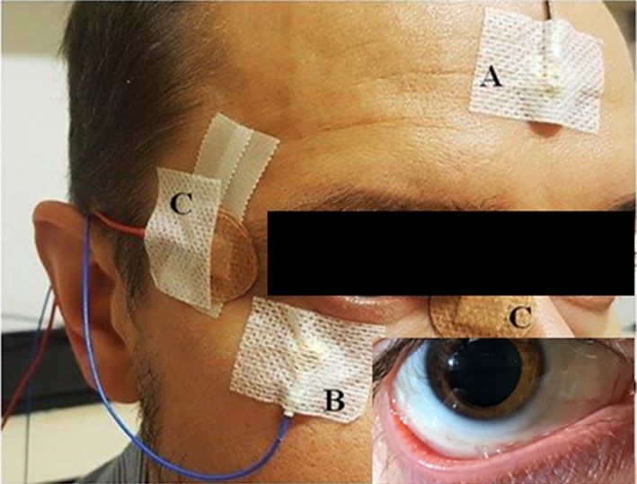
Figure 6. Illustrative position of the electrodes for ERG measurements. (A) Gold-cup ground electrode; (B)
God-cup reference electrode; (C) DTL active electrode. For illustration purpose, small picture insert shows the
position of the DTL electrode in the conjunctival area.
This raises the possibility to interfere with the development of refractive error by stimulating DA release in
myopic patients. The approach presented in this study demonstrates that this can be achieved without direct
exposure of the retina to short-wavelength light, by instead stimulating the system through the blind-spot.
Another question raised from this study is if treatments that intend to stimulate DA release in the retina might
be effective at delaying myopia onset, slowing its progression, or if both prophylactic and therapeutic effects
might be possible. This is certainly an important question to answer in future clinical trials.
In conclusion, this study showed that following blind-spot stimulation with blue light the amplitudes of the
PERG’s P50-N95 and ERG’s b-wave are increased after 20 min in myopes. These changes in amplitude are larger in
myopes than in non-myopes. Therefore, the results presented in this study confirm and reinforce the hypothesis
that stimulation of the blind-spot with short-wavelength light matching the sensitivity of melanopsin in ipRGCs
elicits increased activity in retinal ganglion cells as measured with the PERG technique. This also activates what
seems to be a retrograde effect in the IPL that might involve an upregulation of DA release, by direct contact with
DACs, the increased ON bipolar circuit observed in the b-wave, or through both mechanisms.
Methods
This was an exploratory study conducted by the Clinical and Experimental Optometry Research Laboratory at
the University of Minho (Braga, Portugal). The study was divided into three experiments. The first experiment
aimed to observe the effect of stimulating the blind-spot with blue light on the b-wave recorded with the light-
adapted 3.0 protocol of the ffERG in a myopic population. The purpose of the second experiment was to verify if
the previous effect was also present in the ganglionic layer of myopic participants by recording PERG responses.
The third experiment tested the hypothesis that after blue light stimulation in the blind-spot myopes would show
a larger change in amplitude than non-myopes.
Participants. For the purpose of the study, young and healthy participants without any history of ocular or
systemic disease were recruited based on the following inclusive criteria: age between 18 and 30 years, manifest
refractions between + 0.50 Diopters (D) and − 4.00 D, astigmatism below 1.00 D, and visual acuity of at least
0.0 logMAR with habitual correction. This was to avoid any influences on the ERG response due to structural
changes in the retina caused by axial elongation55,56.
In the first experiment, ten myopic participants (− 1.90 ± 1.19 D) with mean age of 25.0 ± 4.6 years were
recruited for light-adapted 3.0 b-wave ERG measurements. Thereafter, five of those myopes were randomly
selected (mean age of 24.8 ± 5.8 years; mean spherical equivalent of − 1.90 ± 1.40 D) for PERG recordings (second
experiment), considering that the b-wave effect of the first experiment was observed in all participants. For the
third experiment, the b-wave and PERG recording of the same five myopic participants were compared with 5
non-myopic participants (mean age of 25.2 ± 3.7; mean spherical equivalent of 0.08 ± 0.23D).
Protocol. The protocol of the three experiments followed the principles of the Declaration of Helsinki and
was approved by the Ethics Committee for Health and Life Sciences of the University of Minho. All partici-
pants provided written informed consent. Measurements for all experiments were taken between 10:00 a.m.
and 5:00 p.m. to avoid the influence of DA on the circadian cycle, and participants had no caffeine or nicotine
consumption at least 5 h before each measurement. The full protocol included VR calibration of each indi-
vidual’s blind-spot position before ERG electrode placement; baseline ERG measurement followed by blind-spot
stimulation44,57,58; and further ERG acquisitions thereafter. The b-wave and PERG protocols for each participant
were performed on different days, but during the same period (morning or afternoon).
The RETI-port/scan21 (Roland Consult, Wiesbaden, Germany) was used for the electrophysiological record-
ings following the ISCEV guidelines59,60. Before placing the electrodes, the skin was cleansed with an abrasive
gel. Next, gold-cup reference and ground electrodes and active DTL-plus electrodes (Dawson-Trick-Litzkow)
were placed (Fig. 6). Impedance was checked before each measurement and recordings only taken when it was
smaller than 5 kOhm. All ERG measurements were recorded binocularly. To maintain a consistent degree of
Scientific Reports | (2021) 11:9273 | https://doi.org/10.1038/s41598-021-88459-2 6
Vol:.(1234567890)www.nature.com/scientificreports/
Figure 7. Illustrative diagram of the basic ERG output of the light-adapted 3.0 ffERG with its 2 main curves:
a-wave and b-wave. The arrow indicates the stimulus flash.
adaptability between participants and measurements prior to each b-wave recording participants underwent
10 min of light adaptation in the ERG Ganzfeld at a constant background luminous intensity of 30 cd/m2, meas-
ured with a luminance meter (LS-150, Konica Minolta, Osaka). Before each PERG measurement, participants had
a 5-min break. Both ERG protocols took place in an isolated room with a Faraday cage, under ambient lighting
of approximately 400 lx, measured using an illuminance meter (T-10A, Konica Minolta, Osaka). To ensure the
environment conditions, before each protocol we repeatedly measure the illuminance at the point of examina-
tion (at 1 m from the display at eye’s level in PERG, and at the measure position of Ganzfeld in b-wave). ERG
responses were recorded before (baseline), and 10 and 20 min after 1-min stimulus exposure in order to assess
the possible differences on the effect of blind-spot stimulation with blue light.
Although ISCEV ERG protocols require a fully dilated and stable pupil size it is unknown to what extent
topical mydriatics might influence the response of ipRGCs to blue light stimulation. According to Mojumder
and Wensel, topical administration of atropine and phenylephrine together, but not separately, leads to a slow,
dramatic enhancement of a- and b-waves by an unknown mechanism independent of pupil d ilation61. To account
for this finding and to work under normal physiological conditions of pupil size (a critical factor in determin-
ing the effectiveness of the treatment) measures were recorded under non-dilated conditions. The pupil size is
obtained 3 times per patient to obtain an average value. Each measurement session takes several seconds, which
allow the pupil to fluctuate within the physiological rhythm. Therefore, the value obtained should be understood
not only as an instantaneous measurement but an average value within the normal fluctuations of the pupil.
Pupil size was checked with an infrared pupilometer (VIP-200, NeurOptics, California) before and after all ERG
measurements to ensure that luminance conditions were stable, with minimal fluctuations within physiological
terms, without adverse impact for the consistency of the measurements.
ERG tests. In this study, the light-adapted 3.0 ERG test of ffERG was recorded in all participants and the
PERG was recorded only in those participants included in the second and third experiments. The two ERG
methodologies assess the electrophysiological response of different cellular groups in the retina (cones and bipo-
lar cells59, and ganglion cells60, respectively) of both eyes.
For the light-adapted 3.0 ERG test we used a sequence of five single-flashes of white light (3.0 cd.s/m2) gener-
ated in a Ganzfeld stimulator against a light white background (30 cd/m2), with a stimulus rate of 0.625 Hz and
a recording bandpass filter of 1–300 Hz. The resulting wave response was a photopic single-flash cone response,
reproducing discernible a- and b-waves59, similar to the one represented in Fig. 7.
For the PERG evaluation, the transient PERG protocol was used. This protocol detects the retinal activity in
response to the reversal of black and white squares of a checkerboard stimulus (Fig. 8A). In this study, the pattern
stimulus was generated on an LCD monitor (ProLite B1980SD, iiyama) with a frame rate of 60 Hz, covering a field
size of 15º at an observation distance of 1 m. The black and white reversing checkerboard was presented with a
check size of 0.8º for a transient reversal of 1.53 rev/s, with a mean illuminance of 152.64 ± 0.64 lx (Illuminance
meter T-10A, Konica Minolta, Osaka). The mean luminance of the black and white checks was 1.47 ± 0.06 cd/
m2 and 220.32 ± 1.23 cd/m2, respectively (Luminance meter LS-150, Konica Minolta, Osaka). Participants were
asked to maintain their fixation on a red X located at the corner of the four central squares. The signals were
amplified and filtered (first-order bandpass 5–50 Hz). Sweep length was 180 ms (sample freq. 2.84 Hz) and 200
sweeps used for averaging. PERG recordings result in a wave response similar to the one represented in Fig. 8B.
In general, the positive peak (P50) is produced by the retinal ganglion cells and other inner cells, while N95 is
generated almost exclusively by retinal ganglion c ells60.
Blind‑spot stimulation. The stimulation of the optic nerve head with blue light was performed with a pro-
prietary application (VR stimulation application, Dopavision GmbH, Germany) for the Android smartphone
Samsung Galaxy S7 (Samsung, Seoul, South Korea) with a Super AMOLED display (5.1 in., 1440 × 2560 pixels)
inserted in a VR system (Trust International B.V., Dordrecht, Netherlands) called VR box. Participants used this
app by placing the smartphone inside the VR box. For setup calibration and to ensure that the blue light stimulus
fell onto the blind-spot, participants were instructed to use the built-in controls to adjust the position of a red
disc until it was no longer visible while looking at an ABC fixation target, which is a combination of bullseye and
crosshair that has been shown to have high fixation stability62. Participants had to match the stimulus size of less
than 3° on the smartphone display for each eye separately until they did not see the blind-spot stimulus, thereby
Scientific Reports | (2021) 11:9273 | https://doi.org/10.1038/s41598-021-88459-2 7
Vol.:(0123456789)www.nature.com/scientificreports/
Figure 8. (A) Stimulus used for recording ganglion cell electrical activity with PERG methodology. The black
and white checks alternate from black-to-white and white-to-black at a regular frequency, with a nearly 100%
luminance contrast between checks. (B) Typical PERG wave response (example from one of the participants).
ensuring that the stimulus fell on the optic nerve head while looking to the fixation target. The stimulus presen-
tation consisted of a blue stimulus (450 nm peak wavelength, which is within the maximum relative sensitivity
of melanopsin) at the calibrated blind-spot, with a flicker frequency of 15 Hz and luminance of 14.74 ± 1.11 cd/
m2, and a black background. Flicker at 15 Hz was used because low temporal frequencies should be avoided for
its negative effects on myopia development, as demonstrated in guinea pigs and chickens35,63. Furthermore, it
has been shown that 1 h of either 2 or 20 Hz flickering illumination results in a reduction of DOPA, whereas in
between temporal frequencies such as 10 Hz showed the o pposite64. In the present study the exposure time to
the stimulus was 1 min. Participants were instructed to carefully keep fixation on the fixation target and not to
move their eyes during the stimulation period.
Data analysis. Data analyses were conducted on the implicit time and amplitude of the b-waves of the light-
adapted curve ERG cone response and of P50-N95 of the PERG. For the comparison between myopes and non-
myopes, ERG amplitudes were normalized to the baseline. Although both ERG measurements and blind-spot
stimulation was performed binocularly, only ERG responses of one eye for each participant were considered.
The eye was selected based on the blind-spot calibration. The eye with the calibration closest to the average cali-
bration position was selected in order to reduce potentially large individual differences in blind-spot position.
This also had the advantage of reducing the impact of any calibrated positions that were unintentionally inexact.
Statistical analysis. The sample sizes of the three experiments ensure 80% power for statistical analysis.
Repeated measures ANOVAs for the factor ‘Time after stimulation’ with Tukey-corrected post-hoc tests were
conducted65,66 for amplitude and corresponding implicit times separately. To test the hypothesis, an independent
one-sided t-test was performed on change in amplitude. For statistical analysis JAMOVI (1.1.9.0, jamovi project,
2019)67 was used.
Data availability
The datasets generated during and/or analysed during the current study are available from the corresponding
author on reasonable request.
Received: 8 November 2020; Accepted: 5 April 2021
References
1. González-Méijome, J. et al. Strategies to regulate myopia progression with contact lenses: A review. Eye Contact Lens 42, 24–34
(2016).
2. Torii, H. et al. Violet light exposure can be a preventive strategy against myopia progression. EBioMedicine 15, 210–219 (2017).
3. González-Méijome, J. M. New research routes to fight myopia. EBioMedicine 16, 24–25 (2017).
4. Chakraborty, R. et al. Circadian rhythms, refractive development, and myopia. Ophthalmic Physiol. Opt. 38, 217–245 (2018).
5. Witkovsky, P. Dopamine and retinal function. Doc. Ophthalmol. 108, 17–40 (2004).
6. Zhou, X., Pardue, M., Iuvone, P. & Qu, J. Dopamine signaling and myopia development: What are the key challenges. Prog. Retin.
Eye Res 61, 60–71 (2017).
7. Wang, M., Schaeffel, F., Jiang, B. & Feldkaemper, M. Effects of light of different spectral composition on refractive development
and retinal dopamine in chicks. Investig. Ophthalmol. Vis. Sci. 59, 4413–4424 (2018).
8. Zhang, D., Zhou, T. & McMahon, D. Functional heterogeneity of retinal dopaminergic neurons underlying their multiple roles in
vision. J. Neurosci. Off. J. Soc. Neurosci. 27, 692–699 (2007).
9. Popova, E. & Kupenova, P. Effects of dopamine receptor blockade on the intensity response function of ERG b- and d-waves in
dark adapted eyes. Vis. Res 88, 22–29 (2013).
10. Dumitrescu, O., Pucci, F., Wong, K. & Berson, D. Ectopic retinal ON bipolar cell synapses in the OFF inner plexiform layer:
Contacts with dopaminergic amacrine cells and melanopsin ganglion cells. J. Comp. Neurol. 517, 226–244 (2009).
Scientific Reports | (2021) 11:9273 | https://doi.org/10.1038/s41598-021-88459-2 8
Vol:.(1234567890)www.nature.com/scientificreports/
11. Li, X., Schaeffel, F., Kohler, K. & Zrenner, E. Dose-dependent effects of 6-hydroxy dopamine on deprivation myopia, electroreti-
nograms, and dopaminergic amacrine cells in chickens. Vis. Neurosci. 9, 483–492 (1992).
12. Tian, N., Xu, H. & Wang, P. Dopamine D2 receptors preferentially regulate the development of light responses of the inner retina.
J. Neurosci. 41, 17–30 (2015).
13. Langheinrich, T. et al. Visual contrast response functions in Parkinson’s disease: Evidence from electroretinograms, visually evoked
potentials and psychophysics. Clin. Neurophysiol. 111, 66–74 (2000).
14. Tebartz van Elst, L., Greenlee, M., Foley, J. & Lücking, C. Contrast detection, discrimination and adaptation in patients with
Parkinson’s disease and multiple system atrophy. Brain J. Neurol. 120, 2219–2228 (1997).
15. Ikeda, H., Heda, G. & Ellis, C. Electrophysiological signs of retinal dopamine deficiency in recently diagnosed Parkinson’s disease
and a follow up study. Vis. Res. 34, 2629–2638 (1994).
16. Garcia-Martin, E. et al. Electrophysiology and optical coherence tomography to evaluate Parkinson disease severity. Investig.
Ophthalmol. Vis. Sci. 55, 696–705 (2014).
17. Peppe, A. et al. Does pattern electroretinogram spatial tuning alteration in Parkinson’s disease depend on motor disturbances or
retinal dopaminergic loss?. Electroencephalogr. Clin. Neurophysiol. 106, 374–382 (1998).
18. Nowacka, B., Lubiński, W., Honczarenko, K., Potemkowski, A. & Safranow, K. Bioelectrical function and structural assessment of
the retina in patients with early stages of Parkinson’s disease (PD). Doc. Ophthalmol. 131, 95–104 (2015).
19. Lam, B. L. Electrophysiology of Vision: Clinical Testing and Applications (Taylor & Francis Group, 2005).
20. Pepperberg, D., Brown, P., Lurie, M. & Dowling, J. Visual pigment and photoreceptor sensitivity in the isolated skate retina. J. Gen.
Physiol. 71, 369–396 (1978).
21. Goodman, G. & Bornschein, H. Comparative electroretinographic studies in congenital night blindness and total color blindness.
AMA Arch. Ophthalmol. 58, 174–182 (1957).
22. Luo, X. & Frishman, L. Retinal pathway origins of the pattern electroretinogram (PERG). Investig. Ophthalmol. Vis. Sci. 52,
8571–8584 (2011).
23. Vugler, A. et al. Dopamine neurones form a discrete plexus with melanopsin cells in normal and degenerating retina. J. Exp. Neurol.
205, 26–35 (2007).
24. Ashby, R., Ohlendorf, A. & Schaeffel, F. The effect of ambient illuminance on the development of deprivation myopia in chicks.
Investig. Ophthalmol. Vis. Sci. 50, 5348–5354 (2009).
25. McCarthy, C., Megaw, P., Devadas, M. & Morgan, I. Dopaminergic agents affect the ability of brief periods of normal vision to
prevent form-deprivation myopia. Exp. Eye Res. 84, 100–107 (2007).
26. Hattar, S., Liao, H., Takao, M., Berson, D. & Yau, K. Melanopsin-containing retinal ganglion cells: Architecture, projections, and
intrinsic photosensitivity. Science 295, 1065–1070 (2002).
27. Hannibal, J., Christiansen, A. T., Heegaard, S., Fahrenkrug, J. & Kiilgaard, J. F. Melanopsin expressing human retinal ganglion cells:
Subtypes, distribution, and intraretinal connectivity. J. Comp. Neurol. 525, 1934–1961 (2017).
28. Mure, L. S., Vinberg, F., Hanneken, A. & Panda, S. Functional diversity of human intrinsically photosensitive retinal ganglion cells.
Science 366, 1251–1255 (2019).
29. Berson, D., Dunn, F. & Takao, M. Phototransduction by retinal ganglion cells that set the circadian clock. Science 295, 1070–1073
(2002).
30. Schilling, T., Soltanlou, M., Seshadri, Y., Nuerk, H. C. & H. B. Blue Light and Melanopsin contribution to the pupil constriction
in the blind-spot, parafovea and periphery. In Proceedings of the 13th International Joint Conference on Biomedical Engineering
Systems and Technologies–Volume 5 HEALTHINF: HEALTHINF 8 (2020). https://doi.org/10.5220/0008972404820489.
31. Wu, Z., Hadoux, X., Hui, F., Sarossy, M. & Crowston, J. Photopic negative response obtained using a handheld electroretinogram
device: Determining the optimal measure and repeatability. Transl. Vis. Sci. Technol. 5, 8 (2016).
32. Strickland, R., Landis, E. & Pardue, M. Short-wavelength (violet) light protects mice from myopia through cone signaling. Investig.
Ophthalmol. Vis. Sci. 61, 13 (2020).
33. Chakraborty, R. & Pardue, M. Molecular and biochemical aspects of the retina on refraction. Prog. Mol. Biol. Transl. Sci. 134,
249–267 (2015).
34. Prigge, C. et al. M1 ipRGCs influence visual function through retrograde signaling in the retina. J. Neurosci. 36, 7184–7197 (2016).
35. Crewther, S., Barutchu, A., Murphy, M. & Crewther, D. Low frequency temporal modulation of light promotes a myopic shift in
refractive compensation to all spectacle lenses. Exp. Eye Res. 83, 322–328 (2006).
36. Read, A., Collins, M. & Vincent, S. Light exposure and physical activity in myopic and emmetropic children. Optom. Vis. Sci. 91,
330–341 (2014).
37. Ashby, R. S. & Schaeffel, F. The effect of bright light on lens-compensation in chicks. Investig. Ophthalmol. Vis. Sci. https://doi.org/
10.1167/iovs.09-4689 (2010).
38. Dong, F. et al. Inhibition of experimental myopia by a dopamine agonist: Different effectiveness between form deprivation and
hyperopic defocus in guinea pigs. Mol. Vis. 17, 2824–2834 (2011).
39. Feldkaemper, M. & Schaeffel, F. An updated view on the role of dopamine in myopia. Exp. Eye Res. 114, 106–119 (2013).
40. Kolb, H., Fernandez, E. & Nelson, R. Webvision The Organization of the Retina and Visual System (2016).
41. Zhang, D. et al. Intraretinal signaling by ganglion cell photoreceptors to dopaminergic amacrine neurons. PNAS 105, 14181–14186
(2008).
42. Zhang, D., Belenky, M., Sollars, P., Pickard, G. & McMahon, D. Melanopsin mediates retrograde visual signaling in the retina.
PLoS ONE 7, e42647 (2012).
43. Munteanu, T. et al. Light-dependent pathways for dopaminergic amacrine cell development and function. Elife 7, e39866 (2018).
44. Klee, S., Link, D., Bessler, P. & Haueisen, J. Optoelectrophysiological stimulation of the human eye using fundus-controlled silent
substitution technique. J. Biomed. Opt. 16, 015002 (2011).
45. Morgan, I. et al. The epidemics of myopia: Aetiology and prevention. Prog. Retin. Eye Res. 62, 134–149 (2018).
46. Sankaridurg, P. et al. Myopia control with novel central and peripheral plus contact lenses and extended depth of focus contact
lenses: 2 year results from a randomised clinical trial. Ophthalmic Physiol. Opt 39, 294–307 (2019).
47. Chamberlain, P. et al. A 3-year randomized clinical trial of misight lenses for myopia control. Optom. Vis. Sci. 96, 556–567 (2019).
48. Luu, C., Lau, A. & Lee, S.-Y. multifocal electroretinogram in adults and children with myopia. Arch. Ophthalmol. 124, 328–334
(2006).
49. Ho, W.-C., Kee, C.-S. & Chan, H.H.-L. Myopic children have central reduction in high contrast multifocal ERG response, while
adults have paracentral reduction in low contrast response. Investig. Ophthalmol. Vis Sci. 53, 3695–3702 (2012).
50. Chia, A., Li, W., Tan, D. & Luu, C. Full-field electroretinogram findings in children in the atropine treatment for myopia (ATOM2)
study. Doc. Ophthalmol. 126, 177–186 (2013).
51. Li, S.Z.-C. et al. Subclinical decrease in central inner retinal activity is associated with myopia development in children. Investig.
Ophthalmol. Vis. Sci. 58, 4399–4406 (2017).
52. Aung, M. et al. Dopamine deficiency contributes to early visual dysfunction in a rodent model of type 1 diabetes. J. Neurosci. 34,
726–736 (2014).
53. Kim, M. et al. Dopamine deficiency mediates early rod-driven inner retinal dysfunction in diabetic mice. Investig. Ophthalmol.
Vis. Sci. 59, 572–581 (2018).
Scientific Reports | (2021) 11:9273 | https://doi.org/10.1038/s41598-021-88459-2 9
Vol.:(0123456789)www.nature.com/scientificreports/
54. Motz, C. T., Chesler, K. C., Allen, R. S., Bales, K. L., Mees, L. M., Feola, A. J., ... & Pardue, M. T. Novel detection and restorative
levodopa treatment for preclinical diabetic retinopathy. Diabetes. 69(7), 1518-1527 (2020).
55. Matsumura, S., Kuo, A. & Saw, S. An update of eye shape and myopia. Eye Contact Lens 45, 279–285 (2019).
56. Read, S., Fuss, J., Vincent, S., Collins, M. & Alonso-Caneiro, D. Choroidal changes in human myopia: Insights from optical coher-
ence tomography imaging. Clin. Exp. Optom. 102, 270–285 (2019).
57. Bahmani, H., Seshadri, Y. & Schilling, T. MyopiaX: invisible myopia treatment using visible light. In 17th International Myopia
Conference (IMC) (2019).
58. Bahmani, H., Seshadri, Y. & Schilling, T. Blind-spot stimulation with blue light changes pupil size and enhances contrast sensitivity.
Investig. Ophthalmol. Vis. Sci. 60, 5267–5267 (2019).
59. Mcculloch, D. L., Marmor, M. F., Brigell, M. G. & Bach, M. ISCEV STANDARDS ISCEV Standard for full-field clinical electrore-
tinography (2015 update ). 1–12 (2015) https://doi.org/10.1007/s10633-014-9473-7.
60. Bach, M., Brigell, M. G., Hawlina, M., Meigen, T. & Viswanathan, S. ISCEV STANDARDS ISCEV standard for clinical pattern
electroretinography ( PERG ): 2012 update. 1–7 (2013) https://doi.org/10.1007/s10633-012-9353-y.
61. Mojumder, D. & Wensel, T. Topical mydriatics affect light-evoked retinal responses in anesthetized mice. Investig. Ophthalmol.
Vis. Sci. 51, 567–576 (2010).
62. Thaler, L., Schütz, A., Goodale, M. & Gegenfurtner, K. What is the best fixation target? The effect of target shape on stability of
fixational eye movements. Vis. Res. 76, 31–42 (2013).
63. Luo, X. et al. Myopia induced by flickering light in guinea pig eyes is associated with increased rather than decreased dopamine
release. Mol. Vis. 23, 666 (2017).
64. Rohrer, B., Iuvone, P. & Stell, W. Stimulation of dopaminergic amacrine cells by stroboscopic illumination or fibroblast growth
factor (bFGF, FGF-2) injections: Possible roles in prevention of form-deprivation myopia in the chick. Brain Res. 686, 169–181
(1995).
65. Singmann, H. afex: Analysis of Factorial Experiments. [R package]. R Core Team, R: A Language and envionment for statistical
computing. [Computer software] (2018).
66. Lenth, R. emmeans: Estimated Marginal Means, aka Least-Squares Means. [R package]. R Core Team, R: A Language and envion-
ment for statistical computing. [Computer software] (2018).
67. Project, T. jamovi. jamovi (version 1.1). [Computer Software] (2019).
Acknowledgements
This work was supported by the Federal Ministry of Education and Research, Industrie-in-Klinik-Plattform
Program BMBF, Germany (FKZ: 13GW0256). This project was also supported by the Portuguese Foundation
for Science and Technology (FCT) PTDC/FIS-OPT/0677/2014, the Strategic Funding UID/FIS/04650/2013 at
Center of Physics, UMinho, and predoctoral grant SFRH/ BD/136684/2018 to AAS. In addition, we would like
to thank Nikita Wong for some literature search and language proofreading.
Author contributions
All authors conceived the experiments. A.A.S. and P.F. conducted the experiments and T.S. analyzed the results.
A.A.S., P.F., T.S. and J.M.G.M. equally contributed to the introduction and the discussion of the results. All
authors reviewed the manuscript.
Competing interests
AAS, PF and JMGM do not have any conflict of interests with the products or brands used during the study. TS,
YS, and HB are supported by and have financial interest in Dopavision GmbH.
Additional information
Correspondence and requests for materials should be addressed to J.M.G.-M.
Reprints and permissions information is available at www.nature.com/reprints.
Publisher’s note Springer Nature remains neutral with regard to jurisdictional claims in published maps and
institutional affiliations.
Open Access This article is licensed under a Creative Commons Attribution 4.0 International
License, which permits use, sharing, adaptation, distribution and reproduction in any medium or
format, as long as you give appropriate credit to the original author(s) and the source, provide a link to the
Creative Commons licence, and indicate if changes were made. The images or other third party material in this
article are included in the article’s Creative Commons licence, unless indicated otherwise in a credit line to the
material. If material is not included in the article’s Creative Commons licence and your intended use is not
permitted by statutory regulation or exceeds the permitted use, you will need to obtain permission directly from
the copyright holder. To view a copy of this licence, visit http://creativecommons.org/licenses/by/4.0/.
© The Author(s) 2021
Scientific Reports | (2021) 11:9273 | https://doi.org/10.1038/s41598-021-88459-2 10
Vol:.(1234567890)You can also read