BUTYRYL/CAPROYL-COA:ACETATE COA-TRANSFERASE: CLONING, EXPRESSION AND CHARACTERIZATION OF THE KEY ENZYME INVOLVED IN MEDIUM-CHAIN FATTY ACID ...
←
→
Page content transcription
If your browser does not render page correctly, please read the page content below
Bioscience Reports (2021) 41 BSR20211135
https://doi.org/10.1042/BSR20211135

Research Article

Butyryl/Caproyl-CoA:Acetate CoA-transferase:
cloning, expression and characterization of the key
enzyme involved in medium-chain fatty acid
biosynthesis

                                                                                                                                                                                           Downloaded from http://portlandpress.com/bioscirep/article-pdf/41/8/BSR20211135/918824/bsr-2021-1135.pdf by guest on 22 December 2021
  Qingzhuoma Yang1,2,3 , Shengtao Guo2,3,4 , Qi Lu1,2 ,                                           Yong Tao1,5 , Decong Zheng1,2 , Qinmao Zhou1,2 and
Jun Liu5
1 Key Laboratory of Environmental and Applied Microbiology, Environmental Microbiology Key Laboratory of Sichuan Province, Chengdu Institute of Biology, Chinese Academy of
Science, Chengdu 610041, China; 2 College of Life Sciences, University of Chinese Academy of Sciences, Beijing 100049, China; 3 Key Laboratory of Bio-Resource and
Eco-Environment of Ministry of Education, College of Life Sciences, Sichuan University, Chengdu, Sichuan 610065, China; 4 BGI Education Center, University of Chinese Academy
of Sciences, Shenzhen 518083, China; 5 Faculty of Bioengineering, Sichuan University of Science and Engineering, Zigong 643000, China
Correspondence: Yong Tao (taoyong@cib.ac.cn)

                                           Coenzyme A transferases (CoATs) are important enzymes involved in carbon chain
                                           elongation, contributing to medium-chain fatty acid (MCFA) biosynthesis. For example,
                                           butyryl-CoA:acetate CoA transferase (BCoAT) is responsible for the final step of bu-
                                           tyrate synthesis from butyryl-CoA. However, little is known about caproyl-CoA:acetate
                                           CoA-transferase (CCoAT), which is responsible for the final step of caproate synthesis from
                                           caproyl-CoA. In the present study, two CoAT genes from Ruminococcaceae bacterium
                                           CPB6 and Clostridium tyrobutyricum BEY8 were identified by gene cloning and expres-
                                           sion analysis. Enzyme assays and kinetic studies were carried out using butyryl-CoA or
                                           caproyl-CoA as the substrate. CPB6-CoAT can catalyze the conversion of both butyryl-CoA
                                           into butyrate and caproyl-CoA into caproate, but its catalytic efficiency with caproyl-CoA
                                           as the substrate was 3.8-times higher than that with butyryl-CoA. In contrast, BEY8-CoAT
                                           had only BCoAT activity, not CCoAT activity. This demonstrated the existence of a specific
                                           CCoAT involved in chain elongation via the reverse β-oxidation pathway. Comparative bioin-
                                           formatics analysis showed the presence of a highly conserved motif (GGQXDFXXGAXX)
                                           in CoATs, which is predicted to be the active center. Single point mutations in the con-
                                           served motif of CPB6-CoAT (Asp346 and Ala351 ) led to marked decreases in the activity for
                                           butyryl-CoA and caproyl-CoA, indicating that the conserved motif is the active center of
                                           CPB6-CoAT and that Asp346 and Ala351 have a significant impact on the enzymatic activ-
                                           ity. This work provides insight into the function of CCoAT in caproic acid biosynthesis and
                                           improves understanding of the chain elongation pathway for MCFA production.

                                         Introduction
Received: 09 May 2021
                                         Medium-chain fatty acids (MCFAs, C6 –C12 ) are widely utilized in agriculture and industry. For example,
Revised: 07 July 2021                    n-caproic acid (C6 ) is used as a precursor for the production of fragrances [20], antimicrobial agents [11],
Accepted: 30 July 2021                   and drop-in biofuels [19]. Recent studies have shown that MCFAs produced from renewable feedstock by
                                         anaerobic fermentation hold promise for replacing fossil resources and botanical oils, such as palm kernel
Accepted Manuscript online:
02 August 2021                           oil, to meet the requirements for sustainable development [26]. A few microorganisms, such as Megas-
Version of Record published:             phaera elsdenii [32], Ruminococcaceae bacterium CPB6 [50], Acinetobacter spp. [21], and Clostridium
12 August 2021                           kluyveri [46], have been reported to be able to synthesize MCFAs from renewable feedstock via the carbon

© 2021 The Author(s). This is an open access article published by Portland Press Limited on behalf of the Biochemical Society and distributed under the Creative Commons Attribution   1
License 4.0 (CC BY).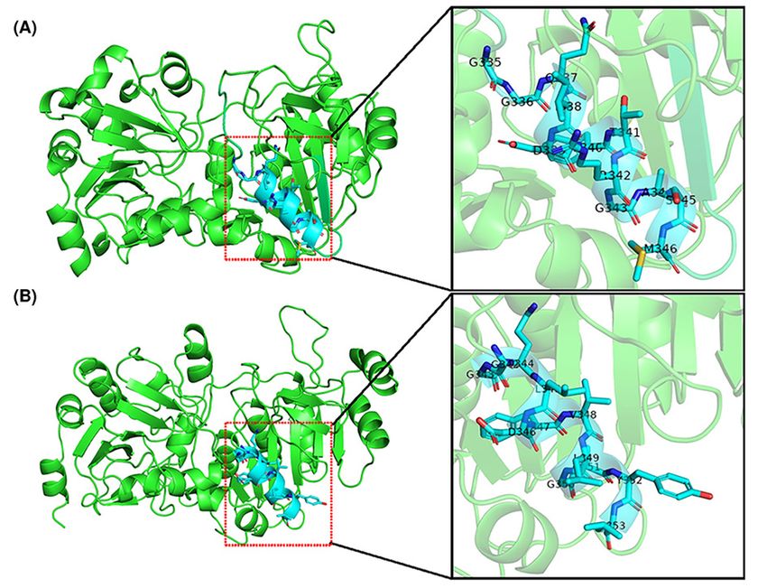
Bioscience Reports (2021) 41 BSR20211135
                                                                                                                                         https://doi.org/10.1042/BSR20211135

           chain elongation pathway [1]. In the process of chain elongation, intermediates of acidogenesis, such as acetate (C2 )
           and n-butyrate (C4 ), act as substrates and are elongated to caproic acid (C6 ) and octanoic acid (C8 ) by addition of
           acetyl-CoA in reverse β-oxidation cycles [40,34]. C2 or C4 , transformed to acetyl-CoA or butyryl-CoA, respectively,
           represents the initial substrate for elongation via reverse β-oxidation. This pathway has been identified as a key
           metabolic process in MCFA biosynthesis [36].
              The production of high concentrations of butyrate (>10 mM) in vitro has been reported in some anaerobes, such as
           Roseburia [13] and Faecalibacterium [27]. Butyrate is normally generated from two molecules of acetyl-CoA, yield-
           ing acetoacetyl-CoA, which is then converted into butyryl-CoA [47]. In the latter reaction, butyryl-CoA is exchanged
           with exogenously derived acetate to yield acetyl-CoA and butyrate [35]. The enzymes responsible for butyrate produc-
           tion in the reverse β-oxidation pathway are acetyl-CoA acetyltransferase (AtoB, EC 2.3.1.9), 3-hydroxybutyryl-CoA
           dehydrogenase (Hbd, EC 1.1.1.157), enoyl-CoA hydratase (Crt, EC 4.2.1.17), butyryl-CoA dehydrogenase (Bcd, EC
           1.3.2.1), and butyryl-CoA:acetate CoA transferase (BCoAT, EC 2.8.3.8) [18]. Among them, BCoAT is a well-known

                                                                                                                                                                                           Downloaded from http://portlandpress.com/bioscirep/article-pdf/41/8/BSR20211135/918824/bsr-2021-1135.pdf by guest on 22 December 2021
           coenzyme A transferase (CoAT) responsible for the final step of butyric acid synthesis, transforming the CoA moi-
           ety from butyryl-CoA to an exogenous acetate molecule, which results in the formation of butyrate and acetyl-CoA
           [17,8]. CoATs are abundant in anaerobic fermenting bacteria that cope with low ATP yields, but they are also found
           in aerobic bacteria and in the mitochondria of humans and other mammals [44]. The synthesis pathway and key
           genes associated with butyric acid in MCFA biosynthesis via reverse β-oxidation are well understood. However, little
           is known about key genes involved in the conversion of butyric acid (C4 ) into caproic acid (C6 ). Although most genes
           responsible for butyric acid production are suggested to function in further chain elongation of MCFAs [18], the fact
           that many butyrate-producing bacteria, such as Clostridium tyrobutyricum, produce only butyric acid instead of
           caproic acid via the reverse β-oxidation pathway suggests that there may be different functional genes involved in the
           production of caproic acid.
              Recently, our study showed that Ruminococcaceae bacterium CPB6 is a caproic acid-producing bacterium with
           a highly prolific ability to perform chain elongation and can produce caproic acid (C6 ) from lactate (as an electron
           donor) with C2 –C4 carboxylic acids and heptanoic acid (C7 ) with C3 –C5 carboxylic acids as electron acceptors (EAs)
           [51,45]. Moreover, a set of genes correlated with chain elongation were identified by sequencing and annotating the
           entire genome of the CPB6 strain [48]. However, very little information is available on enzymes involved in the con-
           version of C4 into C6 , especially the gene responsible for the conversion of caproyl-CoA into caproic acid.
              In the present study, we cloned a predicted caproyl-CoA:acetate CoA-transferase (CCoAT) gene from the caproic
           acid-producing strain CPB6 [51] and a BCoAT gene from the butyric acid-producing C. tyrobutyricum BEY8 and
           expressed the two proteins in Escherichia coli BL21 (DE3) using pET28a. The aims of the present study were to: (i)
           compare differences in sequence, structure, enzymatic activity, and substrate specificity between CCoAT and BCoAT;
           (ii) identify the active center of CCoAT and its effects on the activities of enzymes with different structures; and (iii)
           verify the existence of CCoAT in the caproic acid biosynthesis pathway.

           Materials and methods
           Strain growth conditions
           E. coli DH5α (TsingKe, Chengdu, China) and E. coli BL21 (DE3) (Transgene, Beijing, China) were cultured in
           Luria broth (LB) medium supplemented with 50 μg/ml kanamycin (Sangon Biotech, Shanghai, China) at 37◦ C.
           Ruminococcaceae bacterium CPB6 was grown anaerobically at 37◦ C in modified reinforced Clostridium medium
           (Binder, Qingdao, China). C. tyrobutyricum BEY8 was grown anaerobically at 37◦ C in TGY medium (30 g/l tryptone,
           20 g/l glucose, 10 g/l yeast extract, and 1 g/l l-cysteine hydrochloric acid; pH 7).

           Gene cloning and plasmid construction
           The CoAT genes were amplified from the genomic DNA of Ruminococcaceae bacterium CPB6 [48] or C. tyrobu-
           tyricum BEY8 [22] through PCR using the primers listed in Table 1. During amplification, the following conditions
           were used: initial denaturation (5 min at 98◦ C); followed by 30 cycles of denaturation (10 s at 98◦ C), annealing (30 s
           at 52◦ C), and elongation (1 min at 72◦ C); and a final extension (5 min at 72◦ C). The PCR products were verified by
           agarose gel electrophoresis, recovered using a PCR purification kit (Fuji, Chengdu, China), and seamlessly inserted
           into pET28a double digested with Not I and Sal I (Thermo, Waltham, U.S.A.) to construct the recombinant plasmid
           using a seamless cloning kit (Biomed, Beijing, China). The recombinant plasmids were verified by Sanger sequencing
           and then used to transform E. coli BL21 (DE3) cells.

2   © 2021 The Author(s). This is an open access article published by Portland Press Limited on behalf of the Biochemical Society and distributed under the Creative Commons Attribution
    License 4.0 (CC BY).
Bioscience Reports (2021) 41 BSR20211135
https://doi.org/10.1042/BSR20211135

Table 1 Bacterial strains, plasmids, and primers used in the present study
                              Description                                                                                                        Reference or source

  Strains
    CPB6                      Ruminococcaceae bacterium CPB6                                                                                     Zhu et al.
    BEY8                      C. tyrobutyricum BEY8                                                                                              Hu et al.
  E. coli DH5α                TreliefTtMm 5α chemically competent cells                                                                          TsingKe
  E. coli BL21 (DE3)          Expression chemically competent cells                                                                              Transgene
  Plasmids
  pET28a                      E. coli expression vector (Kan, T7 promoter)                                                                       The present study
  pET28a-CoAT-CPB6            pET28a carrying the CoAT gene from CPB6 fused with a His tag at the N-terminus                                     The present study
  pET28a-CoAT-BEY8            pET28a carrying the CoAT gene from BEY8 fused with a His tag at the N-terminus                                     The present study

                                                                                                                                                                                           Downloaded from http://portlandpress.com/bioscirep/article-pdf/41/8/BSR20211135/918824/bsr-2021-1135.pdf by guest on 22 December 2021
  D346H-mutant                pET28a-CoAT-CPB6 with the Asp346 aa mutation in CoAT                                                               The present study
  A351P-mutant                pET28a-CoAT-CPB6 with the Ala351 aa mutation in CoAT                                                               The present study
  Primers                     Sequence (5 –3 )
  YT43-Salι-fw                TAATACGACTCACTATAGGG                                                                                               The present study
  YT44-Notι-rv                GCTAGTTATTGCTCAGCGG                                                                                                The present study
  YT50-CPB6-fw1               GTGGTGCTCGAGTGCATGAGTTTTCAAGAAGAATATGCACAAAAACTGAC                                                                 The present study
  YT51-CPB6-rv1               CGAATTCGAGCTCCGTTAAATTTTATTGCTTCTGCGCCAGATGC                                                                       The present study
  YT52-BEY8-fw1               CGAATTCGAGCTCCGTCGACATGAGTTTTGAGGAATTGTATAAGAGTAAAGTTGTTAGT                                                        The present study
  YT53-BEY8-rv1               GTGGTGCTCGAGTGCGGCCGCTTTTATAAGTTCTCTAGCTCTTTGTTTTAATGTCTTACCTCTAAG                                                 The present study
  YT60-A351P-fw2              AGCTGGATTTTGTTCTGGGTCCCTATCTGAGCCACGGT                                                                             The present study
  YT61-A351P-rv2              GACCCAGAACAAAATCCAGCTGACCGGCAC                                                                                     The present study
  YT62-D346H-fw2              AGCGGTGCCGGTGGTCAGCTGCATTTTGTTCT                                                                                   The present study
  YT63-D346H-rv2              GCAGCTGACCACCGGCACCGCTAATCTGACGAAA                                                                                 The present study

  1 Underscored   letters match the sequence of vectors for seamless cloning.
  2 The   sequences corresponding to the mutated codons are written in bold.

Expression and purification of the CoA-transferases
E. coli BL21 (DE3) were transformed with the recombinant plasmids pET28-CoAT-CPB6 (pET28-CCoAT) and
pET28-CoAT-BEY8 (pET28-BCoAT). The transformed cells were cultured in LB medium containing 50 μg/ml
kanamycin at 37 ◦ C until the OD600 reached 0.5 and then further cultured at 22 ◦ C for 12 h with 0.4 mM IPTG.
The cultured cells were harvested by centrifugation (8000×g, 10 min) at 4◦ C, and the cell pellet was resuspended in
50 mM potassium phosphate (pH 8.0). The cells were then disrupted with an ultrasonicator (Huxi, Shanghai, China)
for 30 min (200 W, 4 s, interval 6 s) and centrifuged at 8000×g for 30 min to remove the insoluble material. Then,
the enzyme was purified with Ni-NTA Sepharose (Genscript, Nanjing, China) and eluted with 50 mM sodium phos-
phate (pH 8) containing 300 mM NaCl and 250 mM imidazole. Finally, the purity and MW of the enzyme were
assessed using SDS/PAGE analysis. Moreover, the enzyme was analyzed via Western blotting with anti-6× His rabbit
polyclonal antibody (Sangon Biotech, Shanghai, China) to determine whether the target protein was obtained. The
protein concentrations were determined using a BCA protein assay kit (Solarbio, Beijing, China).

Enzymatic characterization
The CoAT activity in crude enzyme extracts and of purified recombinant proteins was measured by determining
the concentration of acetyl-CoA, a reaction byproduct, using a citrate synthase assay described in previous studies
with minor modifications [37,25]. In brief, the reaction was initiated by the addition of enzyme (up to 20 ng/ml)
and was performed in a total volume of 1 ml at 25◦ C: 100 mM potassium phosphate buffer (pH 7), 200 mM sodium
acetate, 1 mM 5,5 -dithiobis(2-nitrobenzoate), 1 mM oxaloacetate, 8.4 nkat citrate synthase (Sigma, St. Louis, U.S.A.),
and 0.5 mM CoA derivatives (Sigma, St. Louis, U.S.A.). The released CoA, corresponding to the formed amount of
acetyl-CoA, was detected by measuring the absorbance at 412 nm. One unit of activity is defined as the amount of
enzyme that converts 1 μmol of acetyl-CoA per min under these conditions.
  The kinetic parameters of the recombinant protein were also calculated by using a coupled spectrophotometric
enzyme assay through citrate synthesis [35]. The reaction mixture was the same as that mentioned above, and the
concentrations of butyryl-CoA or caproyl-CoA were varied from 0.5 to 5 mM. The kinetic parameters were computed
using the Lineweaver–Burk transformation of the Michaelis–Menten equation, in which velocity is a function of the

© 2021 The Author(s). This is an open access article published by Portland Press Limited on behalf of the Biochemical Society and distributed under the Creative Commons Attribution   3
License 4.0 (CC BY).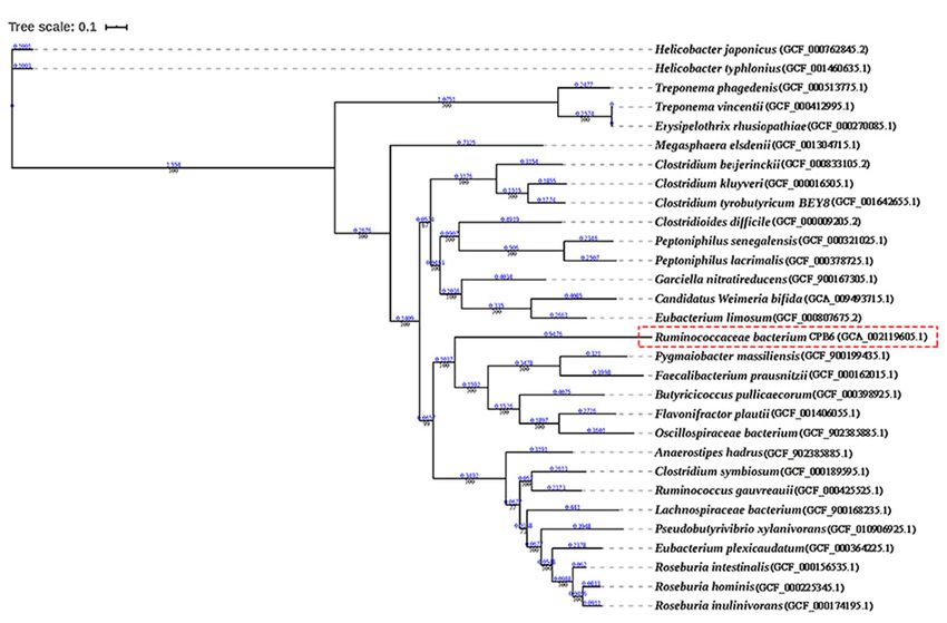
Bioscience Reports (2021) 41 BSR20211135
                                                                                                                                         https://doi.org/10.1042/BSR20211135

           substrate [5,6]. The catalytic constant (k cat ) was defined as the number of CoAT molecules formed by one molecule
           of enzyme in a single second. All measurements were performed in triplicate for each biological replication.

           Sequence alignment and phylogenetic reconstruction
           Multiple alignment of CoAT amino acid sequences was performed using ESPript [9]. With the Akaike information
           criterion (AIC), the amino acid substitutions were predicted using ProtTest (version 3.4.2). We constructed a phyloge-
           netic tree of the whole genomes of strains containing CoAT [20,12,3]. The construction followed the general approach
           of [15,49] and employed sequences downloaded from the online database NCBI (https://www.ncbi.nlm.nih.gov/).
           MEGA-X software was used to construct the whole genome phylogenetic tree [43]. The phylogenetic relationships of
           CoATs from different species were obtained by using OrthoFinder [15], and MUSCLE (version 3.8.31) was used to
           calibrate the 119 shared single-copy genes [14]. The phylogenomic tree was derived from a supermatrix comprising

                                                                                                                                                                                           Downloaded from http://portlandpress.com/bioscirep/article-pdf/41/8/BSR20211135/918824/bsr-2021-1135.pdf by guest on 22 December 2021
           these shared single-copy genes with 41213 unambiguously aligned amino acids using the maximum likelihood (ML)
           method in RAxML (version 8.2.10) [41] under the PROTGAMMAAUTO model, with 100 bootstrap replicates.

           Prediction of tertiary structures of CoAT proteins
           The online modeling tool ScanProsite was used for protein homology comparisons, and SWISS-MODEL [2] was
           used to predict the active sites, tertiary structures and corresponding functions of CoAT proteins in multiple strains
           (Ruminococcaceae bacterium CPB6, Clostridium kluyveri, Megasphaera elsdenii, Clostridium tyrobutyricum
           BEY8, Lachnospiraceae bacterium, and Anaerostipes hadrus) to confirm possible variations in the tertiary struc-
           tures. The targeted sequence was uploaded to search for the best matched template on the basis of data coverage and
           identity. We subsequently conducted model–template alignment for structural comparisons. Finally, the predicted
           CoAT protein tertiary structures were embellished and labeled using PyMOL (version 2.3.3) [31] and were adjusted
           in a similar pattern to identify variations.

           Identification of putative positively selected sites
           According to the above analysis, relatively conserved regions were identified, and the differences in properties
           and structures among different amino acids were compared with find two amino acids that might be key sites for
           site-directed mutagenesis. The point mutation vectors were constructed with the Fast Mutagenesis System (Trans-
           gene, Beijing, China). The QuikChange PCR method using pfu DNA polymerase was performed to generate the
           D346H mutant and A351P mutant. The recombinant plasmid (pET28a-CoAT-CPB6) was used as template DNA, and
           the complementary mutagenic oligonucleotides used as primers are shown in Table 1. After PCR amplification, the
           mixture was digested with restriction enzymes using DpnI to remove methylated template DNA and then sequenced
           (TsingKe, Chengdu, China) to verify site mutagenesis before being used to transform E. coli BL21 (DE3) (Transgene,
           Beijing, China). After purification, the enzymatic activities in the presence of butyryl-CoA and caproyl-CoA were
           measured following the method described above for the wildtype.

           Results
           Cloning, expression, and purification of CoA-transferase
           According to the genome sequences of strains CPB6 and C. tyrobutyricum BEY8, specific primers targeting CoAT
           genes were designed and synthesized (Table 1). Agarose gel electrophoresis showed that the size of the PCR products
           and the double-digestion products was approximately 1300 bp, consistent with the expected sizes of the CPB6-CoAT
           (1344 bp) (Supplementary Figure S1) and the BEY8-CoAT (1233 bp) genes (Supplementary Figure S2). Sequence
           analysis of the recombinant CoAT plasmids showed that the cloned genes shared 100% similarity with the pre-
           dicted CoAT genes of strains CPB6 (a CCoAT) and BEY8 (a BCoAT). This finding indicated that the recombinant E.
           coli/pET28a-CCoAT and E. coli/pET28a-BCoAT were successfully constructed.
              To characterize the functions of CoAT proteins, two recombinant plasmids (pET28a-CCoAT and pET28a-BCoAT)
           were expressed in E. coli BL21 (DE3). Single bands of the purified proteins were detected on SDS/polyacrylamide
           gels after affinity chromatography (Figure 1A). As shown in Figure 1A, there was no obvious protein band ap-
           proximately the size of the target protein in E. coli/pET28a (control), while a single band was observed in E.
           coli/pET28a-BCoAT (lane 2) and E. coli/pET28a-CCoAT (lane 3), and the band sizes were consistent with the ex-
           pected sizes of BEY8-CoAT (46 kDa) and CPB6-CoAT (49 kDa). Furthermore, Western blotting analysis with a His
           antibody (Figure 1B) also demonstrated that the observed bands were consistent with the expected molecular mass
           of BEY8-CoAT and CPB6-CoAT (approximately 46–49 kDa).

4   © 2021 The Author(s). This is an open access article published by Portland Press Limited on behalf of the Biochemical Society and distributed under the Creative Commons Attribution
    License 4.0 (CC BY).
Bioscience Reports (2021) 41 BSR20211135
https://doi.org/10.1042/BSR20211135
Downloaded from http://portlandpress.com/bioscirep/article-pdf/41/8/BSR20211135/918824/bsr-2021-1135.pdf by guest on 22 December 2021
Figure 1. Purification and Western blot analysis of CPB6-CoAT (a CCoAT) and BEY8-CoAT (a BCoAT)
Analysis of purified CCoAT and BCoAT via SDS/PAGE (A). Analysis of purified CCoAT and BCoAT via Western blotting with
an anti-His-tag antibody (B). M, molecular mass marker. Lanes: 1, pET28a; 2, BCoAT; 3, CCoAT; 4, CCoAT-D346H mutant; 5,
CCoAT-A351P mutant. Samples (∼2 μg) were visualized by Coomassie Brilliant Blue staining after electrophoresis. Molecular mass
positions are shown by markers (kDa).
Table 2 Purification and specific activities of the CoATs1
Enzyme Total protein, mg Total activity, U Specific activity, U/mg of protein Purification fold
Butyryl-CoA Caproyl-CoA
CPB6-CoAT
Crude extract 87.04 180.2 2.07 +
− 0.06 5.11 +
− 0.08 1
Purified protein2 22.61 244.2 10.8 +
− 0.02 27.6 +
− 0.15 5.4
BEY8-CoAT
Crude extract 63.10 436.0 6.91 +
− 0.12 ND1 1
Purified protein 2
17.66 462.7 26.2 +
− 0.09 ND1 3.8
1 The purification data in the table were obtained from 300 ml of culture medium. Abbreviations: ND, not detectable; U, μmol/min.
2 Protein was purified via affinity chromatography.
Enzyme assay
The CoAT activity of crude enzyme extracts was determined by measuring the production of acetyl-CoA from
butyryl-CoA or caproyl-CoA [37]. Previously, the key reactions for butyrate and caproate production have been
reported to be (1) butyryl-CoA + acetate → butyrate + acetyl-CoA and (2) caproyl-CoA + acetate → caproate +
acetyl-CoA [40,16,51]. As shown in Table 2, the crude and purified BEY8-CoAT activities with butyryl-CoA and
sodium acetate as substrates were 6.91 + − 0.12 and 26.2 + − 0.09 U/mg of protein, respectively. However, this enzyme
showed no activity for caproyl-CoA. This result suggests that BEY8-CoAT is a BCoAT, similar to the CoAT from
Clostridium acetobutylicum ATCC 824 that is able to produce butyrate instead of caproate, and its purified enzyme
activity was 29.1 U/mg of protein [10]. Moreover, the butyrate-producing bacterium Coprococcus sp. strain L2-50
from the human large intestine showed very high BCoAT activity (118.39 + − 5.02 U/mg of protein) but no CCoAT
activity [13]. This result indicates that the BCoAT probably has substrate specificity for butyryl-CoA. In contrast, the
activities of crude and purified CPB6-CoAT with butyryl-CoA and sodium acetate as substrates were 2.07 + − 0.06 and
10.8 +
− 0.02 U/mg of protein, and the activities with caproyl-CoA and sodium acetate as substrates were 5.11 +
− 0.08
and 27.6 + − 0.15 U/mg of protein, respectively (Table 2), indicating that CPB6-CoAT can catalyze the conversion of
both butyryl-CoA into butyrate and caproyl-CoA into caproate. Notably, the crude and purified CPB6-CoAT activity
for caproyl-CoA was 2.5–2.6-times higher (5.11 vs 2.07, 27.56 vs 10.28 U/mg of protein) than that for butyryl-CoA,
suggesting that CPB6-CoAT specifically prefers caproyl-CoA as a substrate instead of butyryl-CoA.
© 2021 The Author(s). This is an open access article published by Portland Press Limited on behalf of the Biochemical Society and distributed under the Creative Commons Attribution 5
License 4.0 (CC BY).Bioscience Reports (2021) 41 BSR20211135
https://doi.org/10.1042/BSR20211135
Table 3 Kinetic parameters for the CoATs
Enzyme Butyryl-CoA Caproyl-CoA Reference
kcat /K m kcat /K m
K m (μM) kcat (min−1 ) (mM−1 .min−1 ) K m (μM) kcat (min−1 ) (mM−1 .min−1 )
BEY8-CoAT 370 +
− 4.1 13.9 +
− 0.7 37.7 +
− 0.2 ND3 ND3 ND3 The present study
CPB6-CoAT 537 +
− 10 5.81 +
− 1.5 10.8 +
− 0.2 359 +
− 5.3 14.7 +
− 0.9 41.1 +
− 0.2 The present study
CPB6-CoAT-D346H-mutant 747 +− 2.8 1.30 +
− 0.2 1.73 +
− 0.1 748 +
− 7.9 4.24 +
− 0.2 5.66 +
− 0.1 The present study
CPB6-CoAT-A351P-mutant 623 +
− 4.4 2.99 +
− 0.9 4.80 +
− 0.2 532 +
− 2.5 6.78 +
− 1.2 12.8 +
− 0.5 The present study
PGN 07251 520 +
− 10 9.33 +
− 0.7 17.95 +
− 0.1 NR3 NR3 NR3 [35]
CoA transferase2 21.0 +
− 0.1 NR3 NR3 NR3 NR3 NR3 [10]
1 Butyryl-CoA:acetate CoA transferase from Porphyromonas gingivalis.
Downloaded from http://portlandpress.com/bioscirep/article-pdf/41/8/BSR20211135/918824/bsr-2021-1135.pdf by guest on 22 December 2021
2 Butyryl-CoA:acetate CoA transferase from Clostridium acetobutylicum ATCC 824.
3 ND is defined as not determined. Abbreviation: NR, not reported.
Kinetics of CoA-transferases
The kinetic parameters of the recombinant proteins were investigated using a colorimetric assay according to a previ-
ous study [35]. Initial velocities were determined at fixed sodium acetate concentrations with different butyryl-CoA
or caproyl-CoA concentrations. K m and V m values were estimated from secondary plots (‘Materials and meth-
ods’ section). Additionally, k cat values were calculated from enzyme concentrations in the reaction mixtures. The
double-reciprocal enzyme kinetics plot showed that the reactions of the two CoATs follow a ternary-complex mech-
anism (Supplementary Figure S4).
As k cat /K m can be used to compare the catalytic efficiency of different substrates catalyzed by the same enzyme
[24], a lower K m value indicates that the enzyme has a higher affinity for the substrate, and vice versa [30]. In this
study, the K m , k cat and k cat /K m values for CPB6-CoAT with caproyl-CoA were 359 + − 5.3 μM, 14.7 +
−1
− 0.9 min and
41.1 − 0.2 mM .min , respectively, and those with butyryl-CoA were 537 − 10 μM, 5.81 − 1.5 min and 10.8 +
+ −1 −1 + + −1
−
0.2 mM−1 .min−1 , respectively (Table 3). The catalytic efficiency of CPB6-CoAT for caproyl-CoA was 3.8-times (41.1
+
− 0.2 vs 10.8 +
−1 −1
− 0.2 mM .min ) higher than that for butyryl-CoA, consistent with our previous result showing that
the CCoAT activity is predominantly higher than the BCoAT activity [51]. The K m of CPB6-CoAT for caproyl-CoA
was significantly lower than that for butyryl-CoA (359 + − 5.3 vs 537 +− 10 μM), illustrating the higher affinity of this
enzyme for caproyl-CoA relative to butyryl-CoA. These results also partly explain why caproate instead of butyrate
is always the predominant product in the fermentation broth of strain CPB6 [48,45]. BEY8-CoAT had only BCoAT
activity, with K m , k cat and k cat /K m values of 370 +
− 4.1 μM, 13.9 +
− 0.7 min and 37.7 +
−1 −1 −1
− 0.2 mM .min , respectively,
and there was no detectable CCoAT activity (Tables 2 and 3), supporting our previous results showing that strain BEY8
produces only butyric acid as the predominant product. Statistical gap analysis of the above enzymatic experimental
data showed P-values that were less than 0.01, indicating a significant difference between them. Similar to the results
of Lee et al. [22], the CoAT from C. tyrobutyricum only catalyzes the conversion of butyryl-CoA into butyrate and
is not responsible for chain elongation of larger or higher carbon-numbered (>C5 ) fatty acids.
Phylogenetics of the whole genome and multiple amino acid sequence
alignment
A phylogenetic tree of CoATs from different strains was constructed, as shown in Supplementary Figure S5. The
whole-genome phylogenetic tree was constructed based on 119 single-copy genes (including CoATs) that were com-
mon among 29 strains (Figure 2). These strains have a wide range of butyrate metabolic pathways [28], for example,
Roseburia sp., Faecalibacterium prausnitzii, and Coprococcus sp. from the human gut exhibit BCoAT activity val-
ues of 38.95, 18.64, and 118.39 U/mg of protein (crude extracts), respectively [13]. The two species closest to strain
CPB6 were Pygmaiobacter massiliensis [4] and F. prausnitzii [39], which are also butyric acid-producing bacteria
in human feces. Interestingly, the species closest to C. tyrobutyricum BEY8 was C. kluyveri, which is a well-known
caproic acid-producing bacterium. This close relationship may be because they belong to the same genus, Clostrid-
ium.
Based on the alignment results generated from CoAT protein sequences from six different species, 14 amino acids
(GXGGQXDFXXGAXX, positions 340–353) of the CoATs in all the microbes were highly conserved (except in M.
elsdenii), and their secondary structures consisted of 17 α-helices and 21 β-sheets (Figure 3). The sequence similar-
ities between CPB6-CoAT and the analyzed CoATs were as follows: C. kluyveri (37.67%), M. elsdenii (10.27%), C.
6 © 2021 The Author(s). This is an open access article published by Portland Press Limited on behalf of the Biochemical Society and distributed under the Creative Commons Attribution
License 4.0 (CC BY).Bioscience Reports (2021) 41 BSR20211135
https://doi.org/10.1042/BSR20211135
Downloaded from http://portlandpress.com/bioscirep/article-pdf/41/8/BSR20211135/918824/bsr-2021-1135.pdf by guest on 22 December 2021
Figure 2. Phylogenetic tree of the whole genomes of 29 strains containing the CoA-transferase
Numbers at the nodes indicate the levels of bootstrap values. The scale bar for the tree represents a distance of 0.1 substitutions
per site.
tyrobutyricum BEY8 (38.04%), Lachnospiraceae bacterium (60.59%), and Anaerostipes hadrus (58.52%). Among
the six bacteria, strains CPB6, C. kluyveri, and M. elsdenii are caproic acid-producing bacteria, while C. tyrobu-
tyricum BEY8, Lachnospiraceae bacterium, and A. hadrus are butyric acid-producing bacteria. The alignment re-
sults showed that CPB6-CoAT shared lower similarity (10.27–37.67%) with the CoATs of C. kluyveri and M. elsdenii
and higher similarity (58.52–60.59%) with the CoATs of Lachnospiraceae bacterium and A. hadrus. This may be
because strain CPB6 belongs to the family Ruminococcaceae, which is closer to Lachnospiraceae and Anaerostipes
at the taxonomic phylogeny level than to Megasphaera and Clostridium.
Prediction and comparison of the three-dimensional structure and active
site
As shown in Figure 4A, the three-dimensional (3D) structure of CPB6-CoAT has one subunit that may consist of
two main domains, resulting in a characteristic two-domain fold in a homotetrameric structure. A comparison of
the 3D structures of the six CoAT proteins (Figure 4) showed that these CoATs shared similar conformations of
their structural elements (α-helices and β-strands) with slight structural modifications in the loop regions and active
centers, with the exception of the CoAT from M. elsdenii (Figure 4C). The 3D structure of the M. elsdenii protein
was obviously different from that of other CoATs, and the divergences were located not only in the structural elements
of α-helices and β-strands but also in the loops. This may be attributed to the distant genetic relationship between M.
elsdenii and the other five bacteria. Although M. elsdenii produces caproic acid via acetyl-CoA and succinate [23],
the functions of the CoATs may differ between strain CPB6 and M. elsdenii. The 3D structures of CoATs among C.
tyrobutyricum BEY8, Lachnospiraceae bacterium, and A. hadrus shared almost the same conformation of α-helices
and β-strands except for some slight variation in the loops (Figure 4D–F). The protein structure and active center
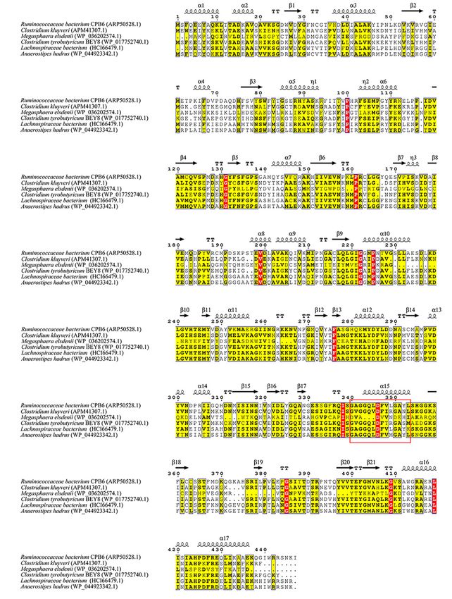
structures were further compared between CPB6-CoAT and BEY8-CoAT, as shown in Figure 5, and both showed
similar 3D structures except for the location and structure of the active center. The predicted active sites of the six
CoATs are shown in Table 4. The predicted active center of the CPB6-CoAT protein was located between amino
© 2021 The Author(s). This is an open access article published by Portland Press Limited on behalf of the Biochemical Society and distributed under the Creative Commons Attribution 7
License 4.0 (CC BY).Bioscience Reports (2021) 41 BSR20211135
https://doi.org/10.1042/BSR20211135
Downloaded from http://portlandpress.com/bioscirep/article-pdf/41/8/BSR20211135/918824/bsr-2021-1135.pdf by guest on 22 December 2021
Figure 3. Multiple amino acid sequence alignment for CoATs
The structure contained 17 α-helices and 21 β-pleated sheets, which are represented with symbols. Nonconserved, 60% con-
served, and 100% conserved residues are marked with white, yellow, and red font, respectively. Conserved motifs are boxed with
a red frame.
8 © 2021 The Author(s). This is an open access article published by Portland Press Limited on behalf of the Biochemical Society and distributed under the Creative Commons Attribution
License 4.0 (CC BY).Bioscience Reports (2021) 41 BSR20211135
https://doi.org/10.1042/BSR20211135
Downloaded from http://portlandpress.com/bioscirep/article-pdf/41/8/BSR20211135/918824/bsr-2021-1135.pdf by guest on 22 December 2021
Figure 4. Predicted 3D structures of representative CoAT proteins
3D structures of CoATs from Ruminococcaceae bacterium CPB6 (A), C. kluyveri (B), M. elsdenii (C), C. tyrobutyricum BEY8 (D),
Lachnospiraceae bacterium (E), and A. hadrus (F). Helices of the catalytic domains, β-pleated sheets, loop regions, and active
centers are colored sky blue, red, purple, and green, respectively.
Figure 5. Predicted 3D structures of the CoAT proteins (left) and the active center (right)
(A) BEY8-CoAT; (B) CPB6-CoAT.
© 2021 The Author(s). This is an open access article published by Portland Press Limited on behalf of the Biochemical Society and distributed under the Creative Commons Attribution 9
License 4.0 (CC BY).Bioscience Reports (2021) 41 BSR20211135
https://doi.org/10.1042/BSR20211135
Table 4 Prediction of the active sites of CoATs in different strains
Strain Location of active site Sequence of active site
Ruminococcaceae bacterium CPB6 (ARP50528.1) 342–353 GGQLDFVLGAYL
C. kluyveri (APM41307.1) 335–346 GGQVDFIRGANL
M. elsdenii (WP 036202574.1) 356–367 ADSYTYKKAPTL
C. tyrobutyricum BEY8 (WP 017752740.1) 335–346 GGQIDFTRGASM
Lachnospiraceae bacterium (HCI66479.1) 340–351 GGQLDFVLGAYK
A. hadrus (WP 044923342.1) 342–353 GGQLDFVMGAYL
acids 342 and 353 (GGQLDFVLGAYL), while the active center of the BEY8-CoAT protein (GGQIDFTRGASM) was
Downloaded from http://portlandpress.com/bioscirep/article-pdf/41/8/BSR20211135/918824/bsr-2021-1135.pdf by guest on 22 December 2021
located at amino acids 335–346, and both active sites contained phenylalanine and tyrosine (Figure 5).
Site-directed mutagenesis
Site-directed mutagenesis was used to verify the active site of the proteins [38]. According to the predicted active
center of CPB6-CoAT (GGQLDFVLGAYL, 342–353 aa) and compared with others (Table 4), site-directed mutage-
nesis targeting Asp346 and Ala351 was carried out to identify the effects of the two residues on the catalytic activity of
CPB6-CoAT. Specifically, Asp346 was replaced by His and Ala351 was replaced by Pro via site-directed mutagenesis.
The nucleotide substitutions were confirmed by Sanger sequencing of the DNA (Supplementary Figure S6). Enzyme
assays showed that compared with wildtype CPB6-CoAT, the Asp346 substitution led to an approximately 76% loss
of BCoAT activity and 72% loss of CCoAT activity, while the Ala351 substitution resulted in an almost 50% loss of
BCoAT activity and 55% loss of CCoAT activity (Supplementary Figure S7). Statistical gap analysis of the above en-
zymatic experimental data (P-values C5 ) fatty acids [22]. Moreover, the K m of BEY8-CoAT
for butyryl-CoA (370 + − 4.1 μM) was obviously greater than that of CPB6-CoAT (537 + − 10 μM), indicating that
BEY8-CoAT had a higher enzymatic affinity for butyryl-CoA than CPB6-CoAT. Similarly, the CoAT (PGN 0725)
from Porphyromonas gingivalis [35,47] and CoAT from C. acetobutylicum ATCC 824 [10] both catalyze the con-
version of butyryl-CoA into butyrate, with K m values of 520 + − 10 and 21.0 +
− 0.1 μM, respectively. This indicates
that BCoAT generally has higher affinity and catalytic activity for butyryl-CoA than CCoAT, while no BCoAT from
butyric acid bacteria displayed affinity and catalytic activity for caproyl-CoA. These results suggest that BCoAT is
only involved in chain elongation of C2 –C4 , not in that of C4 to C6 or C8 .
Our previous study showed that the rate of caproate production with caproyl-CoA as the substrate in strain CPB6
was 3.5-times higher than that observed with butyryl-CoA as the substrate and suggested the existence of a CCoAT
that specifically prefers caproyl-CoA instead of butyryl-CoA as the substrate [51]. In this study, CPB6-CoAT was
confirmed for the first time to be a CCoAT responsible for the final step of caproate formation, although it has low
BCoAT activity for butyryl-CoA. These data demonstrated the existence of a specific CCoAT involved in the chain
elongation of MCFAs, which is significantly different from the function of BCoAT. The CPB6-CoAT protein cat-
alyzed transferase reactions via a ternary-complex kinetic mechanism, whereas some other CoA transferases from
Acidaminococcus fermentans [6], C. acetobutylicum ATCC 824 [10] and Clostridium propionicum [38], which
10 © 2021 The Author(s). This is an open access article published by Portland Press Limited on behalf of the Biochemical Society and distributed under the Creative Commons Attribution
License 4.0 (CC BY).Bioscience Reports (2021) 41 BSR20211135
https://doi.org/10.1042/BSR20211135
belong to family I transferases, were reported to catalyze a transferase reaction via a ping-pong bi–bi mechanism.
Thus, CPB6-CoAT was different from them in terms of substrate specificity and kinetic mechanism. The detailed
mechanism underlying this functional difference needs to be further studied.
The structure of proteins plays an important role in their functional properties and catalytic efficiency [52]; for
example, succinyl CoA:3-ketoate CoA transferase from pig heart [42] and 4-hydroxybutyrate CoA-transferase from
Clostridium aminobutyricum [29] showed unexpected changes in protein modification and specific activity when
their crystal structures changed. In the present study, a comparison of the 3D and active center structures showed
similarities and differences between CPB6-CoAT and other CoATs (Figure 4 and Supplementary Figure S5), which
may have affected enzyme catalytic function and activity. On the basis of these results, studying the functional dif-
ferences caused by the structural changes in CoATs is of great significance. The exact structure and function of the
active center of the CPB6-CoAT protein remains to be determined through subsequent comprehensive experiments
and analysis. Additionally, site-directed mutagenesis showed that two residues (Asp346 and Ala351 ) in the conserved
Downloaded from http://portlandpress.com/bioscirep/article-pdf/41/8/BSR20211135/918824/bsr-2021-1135.pdf by guest on 22 December 2021
motif (GGQLDFVLGAYL, 342–353 aa) had significant effects on the enzymatic activity of CPB6-CoAT, but the ef-
fect of Ala351 on the enzyme activity was lower than that of Asp346 . Generally, the exchange of Asp (an acidic amino
acid) to His led to loss of a carboxy group and the introduction of two amidogens, while the replacement of Ala with
Pro led to loss of an amidogen and the introduction of a carboxy group. Ala lacks a bulky side chain and therefore
would likely not have any steric or electrostatic effects, and this change would not destroy the conformation of the
main chain [7]. Differences in structures and properties among the sequences may be the reason for the differences in
CoAT activity [33]. These results demonstrate that the conserved motif of CPB6-CoAT is directly linked to enzymatic
activity. However, the effects of other residues on enzyme activity require further study to elucidate the function of
the conserved motif in CPB6-CoAT. Clarification of the precise enzymatic mechanisms underlying enzyme binding
of the butyryl-CoA or caproyl-CoA substrates might require crystallographic analyses.
In conclusion, these results confirmed the existence of a CCoAT involved in the production of caproic acid, and
the enzyme is apparently different from the BCoAT responsible for the production of butyric acid. The present study
improves our understanding of the metabolic reactions underlying chain elongation via the reverse β-oxidation path-
way. However, determination of the detailed CCoAT structure and its function in MCFA biosynthesis require further
study through protein crystallization and X-ray crystal structure analyses.
Supporting Information
The Supporting Information is available free of charge on the Publications website.
• PCR of the CPB6-CoAT gene (encoding a CCoAT) and identification of the recombinant plasmid are shown in
Supplementary Figure S1.
• PCR of the BEY8-CoAT gene (encoding a BCoAT) and identification of the recombinant plasmid are shown in
Supplementary Figure S2.
• Western blot analysis of CPB6-CoAT (a CCoAT) and BEY8-CoAT (a BCoAT) is shown in Supplementary Figure
S3.
• The double-reciprocal enzyme kinetics (Lineweaver–Burk) plot is shown in Supplementary Figure S4.
• The phylogenetic tree of CoATs from different strains is shown in Supplementary Figure S5.
• The sequencing peak diagram of site-directed mutant and wildtype CPB6-CoAT is shown in Supplementary
Figure S6.
• The comparison of CoA-transferase activities (Mutant 1, D346H-mutant; Mutant 2, A351P-mutant) is shown in
Supplementary Figure S7.
• The initial velocity of the reaction in different samples at different substrate concentrations is shown in Supple-
mentary Figure S8.
Data Availability
All data generated or analyzed during the present study are included in this article and its supporting information.
Competing Interests
The authors declare that there are no competing interests associated with the manuscript.
© 2021 The Author(s). This is an open access article published by Portland Press Limited on behalf of the Biochemical Society and distributed under the Creative Commons 11
Attribution License 4.0 (CC BY).Bioscience Reports (2021) 41 BSR20211135
https://doi.org/10.1042/BSR20211135
Funding
This work was supported by the Natural Science Foundation of China [grant number 31770090]; the Sichuan Science and Tech-
nology Support Program [grant number 2021YJ0022] and the Open-Foundation Project of CAS Key Laboratory of Environmental
and Applied Microbiology [grant number KLCAS-2017-01].
CRediT Author Contribution
Qingzhuoma Yang: Conceptualization, Resources, Data curation, Software, Formal analysis, Methodology, Writing—original
draft, Writing—review and editing. Shengtao Guo: Conceptualization, Software, Formal analysis, Validation, Investigation. Qi Lu:
Resources, Data curation, Validation, Visualization, Methodology. Yong Tao: Conceptualization, Data curation, Formal analysis,
Funding acquisition, Project administration, Writing—review and editing. Decong Zheng: Conceptualization, Software, Visualiza-
tion. Qinmao Zhou: Software, Investigation, Methodology. Jun Liu: Resources, Supervision, Methodology.
Downloaded from http://portlandpress.com/bioscirep/article-pdf/41/8/BSR20211135/918824/bsr-2021-1135.pdf by guest on 22 December 2021
Acknowledgements
We would like to thank Dr. Su Dan for her advice and assistance with the present study.
Abbreviations
BCoAT, butyryl-CoA:acetate CoA transferase; CCoAT, caproyl-CoA:acetate CoA transferase; CoAT, coenzyme A transferase;
k cat , catalytic constant; LB, Luria broth; MCFA, medium-chain fatty acid; 3D, three-dimensional.
References
1 Angenent, L.T., Richter, H., Buckel, W., Spirito, C.M., Steinbusch, K.J., Plugge, C.M. et al. (2016) Chain elongation with reactor microbiomes:
open-culture biotechnology to produce biochemicals. Environ. Sci. Technol. 50, 2796–2810, https://doi.org/10.1021/acs.est.5b04847
2 Arnold, K., Bordoli, L., Kopp, J. and Schwede, T. (2006) The SWISS-MODEL workspace: a web-based environment for protein structure homology
modelling. Bioinformatics 22, 195–201, https://doi.org/10.1093/bioinformatics/bti770
3 Bajaj, J.S., Ridlon, J.M., Hylemon, P.B., Thacker, L.R., Heuman, D.M., Smith, S. et al. (2012) Linkage of gut microbiome with cognition in hepatic
encephalopathy. Am. J. Physiol. Gastrointest. Liver Physiol. 302, G168–G175, https://doi.org/10.1152/ajpgi.00190.2011
4 Bilen, M., Mbogning, M.D., Cadoret, F., Dubourg, G., Daoud, Z., Fournier, P.E. et al. (2017) ‘Pygmaiobacter massiliensis’ sp. nov., a new bacterium
isolated from the human gut of a Pygmy woman. New Microbes New Infect. 16, 37–38, https://doi.org/10.1016/j.nmni.2016.12.015
5 Bloess, S., Beuel, T., Kruger, T., Sewald, N., Dierks, T. and Fischer von Mollard, G. (2019) Expression, characterization, and site-specific covalent
immobilization of an L-amino acid oxidase from the fungus Hebeloma cylindrosporum. Appl. Microbiol. Biotechnol. 103, 2229–2241,
https://doi.org/10.1007/s00253-018-09609-7
6 Buckel, W., Dorn, U. and Semmler, R. (1981) Glutaconate CoA-Transferase from Acidaminococcus fermentans. Eur. J. Biochem. 118, 315–321,
https://doi.org/10.1111/j.1432-1033.1981.tb06404.x
7 Cao, J., Dang, G., Li, H., Li, T., Yue, Z., Li, N. et al. (2015) Identification and characterization of lipase activity and immunogenicity of LipL from
Mycobacterium tuberculosis. PLoS ONE 10, e0138151, https://doi.org/10.1371/journal.pone.0138151
8 Charrier, C., Duncan, G.J., Reid, M.D., Rucklidge, G.J., Henderson, D., Young, P. et al. (2006) A novel class of CoA-transferase involved in short-chain
fatty acid metabolism in butyrate-producing human colonic bacteria. Microbiology 152, 179–185, https://doi.org/10.1099/mic.0.28412-0
9 Cuff, J.A. and Barton, G.J. (2000) Application of multiple sequence alignment profiles to improve protein secondary structure prediction. Proteins 40,
502–511, https://doi.org/10.1002/1097-0134(20000815)40:3%3c502::AID-PROT170%3e3.0.CO;2-Q
10 Wiesenborn, D.P., Rudolph, F.B. and Papoutsakis, E.T. (1989) Coenzyme A transferase from Clostridium acetobutylicum ATCC 824 and its role in the
uptake of acids. Appl. Environ. Microbiol. 55, 323–329, https://doi.org/10.1128/aem.55.2.323-329.1989
11 Desbois, A.P. (2012) Potential applications of antimicrobial fatty acids in medicine, agriculture and other industries. Recent Pat. Anti Infect. Drug Discov.
7, 111–122, https://doi.org/10.2174/157489112801619728
12 Dowd, S.E., Callaway, T.R., Wolcott, R.D., Sun, Y., McKeehan, T., Hagevoort, R.G. et al. (2008) Evaluation of the bacterial diversity in the feces of cattle
using 16S rDNA bacterial tag-encoded FLX amplicon pyrosequencing (bTEFAP). BMC Microbiol. 8, 1–8, https://doi.org/10.1186/1471-2180-8-125
13 Duncan, S.H., Barcenilla, A., Stewart, C.S., Pryde, S.E. and Flint, H.J. (2002) Acetate utilization and butyryl coenzyme A (CoA):acetate-CoA transferase
in butyrate-producing bacteria from the human large intestine. Appl. Environ. Microbiol. 68, 5186–5190,
https://doi.org/10.1128/AEM.68.10.5186-5190.2002
14 Edgar, R.C. (2004) MUSCLE: multiple sequence alignment with high accuracy and high throughput. Nucleic Acids Res. 32, 1792–1797,
https://doi.org/10.1093/nar/gkh340
15 Emms, D.M. and Kelly, S. (2015) OrthoFinder: solving fundamental biases in whole genome comparisons dramatically improves orthogroup inference
accuracy. Genome Biol. 16, 1–14, https://doi.org/10.1186/s13059-015-0721-2
16 González-Cabaleiro, R., Lema, J.M., Rodrı́guez, J. and Kleerebezem, R. (2013) Linking thermodynamics and kinetics to assess pathway reversibility in
anaerobic bioprocesses. Energy Environ. Sci. 6, 3780–3789, https://doi.org/10.1039/c3ee42754d
17 Heider, J. (2001) A new family of CoA-transferases. FEBS 509, 345–349, https://doi.org/10.1016/S0014-5793(01)03178-7
12 © 2021 The Author(s). This is an open access article published by Portland Press Limited on behalf of the Biochemical Society and distributed under the Creative Commons
Attribution License 4.0 (CC BY).Bioscience Reports (2021) 41 BSR20211135
https://doi.org/10.1042/BSR20211135
18 Seedorf, H., Fricke, W.F., Veith, B., Bruggemann, H., Liesegang, H., Strittmatter, A. et al. (2008) The genome of Clostridium kluyveri, a strict anaerobe
with unique metabolic features. Proc. Natl. Acad. Sci. U.S.A. 105, 2128–2133, https://doi.org/10.1073/pnas.0711093105
19 Herman, N.A., Li, J., Bedi, R., Turchi, B., Liu, X., Miller, M.J. et al. (2017) Development of a high-efficiency transformation method and implementation
of rational metabolic engineering for the industrial butanol hyperproducer Clostridium saccharoperbutylacetonicum Strain N1-4. Appl. Environ.
Microbiol. 83, 02942–02916, https://doi.org/10.1128/AEM.02942-16
20 Kenealy, W.R., Cao, Y. and Weimer, P.J. (1995) Production of caproic acid by cocultures of ruminal cellulolytic bacteria and Clostridium kluyveri grown
on cellulose and ethanol. Appl. Microbiol. Biotechnol. 44, 507–513, https://doi.org/10.1007/BF00169952
21 Kucek, L.A., Nguyen, M. and Angenent, L.T. (2016) Conversion of L-lactate into n-caproate by a continuously fed reactor microbiome. Water Res. 93,
163–171, https://doi.org/10.1016/j.watres.2016.02.018
22 Lee, J., Jang, Y.S., Han, M.J., Kim, J.Y. and Lee, S.Y. (2016) Deciphering Clostridium tyrobutyricum metabolism based on the whole-genome sequence
and proteome analyses. mBio 7, 1–12, https://doi.org/10.1128/mBio.00743-16
23 Lee, N.R., Lee, C.H., Lee, D.Y. and Park, J.B. (2020) Genome-scale metabolic network reconstruction and in silico analysis of hexanoic acid producing
Megasphaera elsdenii. Microorganisms 8, 1–12, https://doi.org/10.3390/microorganisms8040539
Downloaded from http://portlandpress.com/bioscirep/article-pdf/41/8/BSR20211135/918824/bsr-2021-1135.pdf by guest on 22 December 2021
24 Li, T.B., Zhao, F.J., Liu, Z., Jin, Y., Liu, Y., Pei, X.Q. et al. (2019) Structure-guided engineering of ChKRED20 from Chryseobacterium sp. CA49 for
asymmetric reduction of aryl ketoesters. Enzyme Microb. Technol. 125, 29–36, https://doi.org/10.1016/j.enzmictec.2019.03.001
25 Lindenkamp, N., Schurmann, M. and Steinbuchel, A. (2013) A propionate CoA-transferase of Ralstonia eutropha H16 with broad substrate specificity
catalyzing the CoA thioester formation of various carboxylic acids. Appl. Microbiol. Biotechnol. 97, 7699–7709,
https://doi.org/10.1007/s00253-012-4624-9
26 Liu, B., Kleinsteuber, S., Centler, F., Harms, H. and Strauber, H. (2020) Competition between butyrate fermenters and chain-elongating bacteria limits
the efficiency of medium-chain carboxylate production. Front. Microbiol. 11, 1–13, https://doi.org/10.3389/fmicb.2020.00336
27 Louis, P. and Flint, H.J. (2009) Diversity, metabolism and microbial ecology of butyrate-producing bacteria from the human large intestine. FEMS
Microbiol. Lett. 294, 1–8, https://doi.org/10.1111/j.1574-6968.2009.01514.x
28 Louis, P., Young, P., Holtrop, G. and Flint, H.J. (2010) Diversity of human colonic butyrate-producing bacteria revealed by analysis of the
butyryl-CoA:acetate CoA-transferase gene. Environ. Microbiol. 12, 304–314, https://doi.org/10.1111/j.1462-2920.2009.02066.x
29 Macieira, S., Zhang, J., Velarde, M., Buckel, W. and Messerschmidt, A. (2009) Crystal structure of 4-hydroxybutyrate CoA-transferase from Clostridium
aminobutyricum. Biol. Chem. 390, 1251–1263, https://doi.org/10.1515/BC.2009.147
30 Makabe, K., Hirota, R., Shiono, Y., Tanaka, Y. and Koseki, T. (2020) Biochemical and structural investigation of rutinosidase from Aspergillus oryzae.
Appl. Environ. Microbiol. 87, 1–27, https://doi.org/10.1128/AEM.02438-20
31 Marchler-Bauer, A., Anderson, J.B., Chitsaz, F., Derbyshire, M.K., DeWeese-Scott, C., Fong, J.H. et al. (2009) CDD: specific functional annotation with
the Conserved Domain Database. Nucleic Acids Res. 37, D205–D210, https://doi.org/10.1093/nar/gkn845
32 Marounek, M., Fliegrova, K. and Bartos, S. (1989) Metabolism and some characteristics of ruminal strains of Megasphaera elsdenii. Appl. Environ.
Microbiol. 55, 1570–1573, https://doi.org/10.1128/aem.55.6.1570-1573.1989
33 Park, S.H., Kim, S.J., Park, S. and Kim, H.K. (2019) Characterization of organic solvent-tolerant lipolytic enzyme from Marinobacter lipolyticus isolated
from the Antarctic Ocean. Appl. Biochem. Biotechnol. 187, 1046–1060, https://doi.org/10.1007/s12010-018-2865-5
34 San-Valero, P., Abubackar, H.N., Veiga, M.C. and Kennes, C. (2020) Effect of pH, yeast extract and inorganic carbon on chain elongation for hexanoic
acid production. Bioresour. Technol. 300, 1–7, https://doi.org/10.1016/j.biortech.2019.122659
35 Sato, M., Yoshida, Y., Nagano, K., Hasegawa, Y., Takebe, J. and Yoshimura, F. (2016) Three CoA transferases involved in the production of short chain
fatty acids in Porphyromonas gingivalis. Front. Microbiol. 7, 1–13, https://doi.org/10.3389/fmicb.2016.01146
36 Scarborough, M.J., Myers, K.S., Donohue, T.J. and Noguera, D.R. (2020) Medium-chain fatty acid synthesis by “Candidatus Weimeria bifida” gen. nov.,
sp. nov., and “Candidatus Pseudoramibacter fermentans” sp. nov. Appl. Environ. Microbiol. 86, 1–19, https://doi.org/10.1128/AEM.02242-19
37 Scherf, U. and Buckel, W. (1991) Purification and properties of4-hydroxybutyrate coenzyme a transferase from Clostridium aminobutyricum. Appl.
Environ. Microbiol. 57, 2699–2702, https://doi.org/10.1128/aem.57.9.2699-2702.1991
38 Selmer, T., Willanzheimer, A. and Hetzel, M. (2002) Propionate CoA-transferase from Clostridium propionicum: cloning of the gene and identification of
glutamate 324 at the active site. Eur. J. Biochem. 269, 372–380, https://doi.org/10.1046/j.0014-2956.2001.02659.x
39 Sitkin, S. and Pokrotnieks, J. (2019) Clinical potential of anti-inflammatory effects of Faecalibacterium prausnitzii and butyrate in inflammatory bowel
disease. Inflamm. Bowel Dis. 25, e40–e41, https://doi.org/10.1093/ibd/izy258
40 Spirito, C.M., Richter, H., Rabaey, K., Stams, A.J. and Angenent, L.T. (2014) Chain elongation in anaerobic reactor microbiomes to recover resources
from waste. Curr. Opin. Biotechnol. 27, 115–122, https://doi.org/10.1016/j.copbio.2014.01.003
41 Stamatakis, A. (2014) RAxML version 8: a tool for phylogenetic analysis and post-analysis of large phylogenies. Bioinformatics 30, 1312–1313,
https://doi.org/10.1093/bioinformatics/btu033
42 Tammam, S.D., Rochet, J.-C. and Fraser, M.E. (2007) Identification of the cysteine residue exposed by the conformational change in pig heart
succinyl-CoA:3-ketoacid coenzyme A transferase on binding coenzyme A. Biochemistry 46, 10852–10863, https://doi.org/10.1021/bi700828h
43 Tamura, K., Peterson, D., Peterson, N., Stecher, G., Nei, M. and Kumar, S. (2011) MEGA5: molecular evolutionary genetics analysis using maximum
likelihood, evolutionary distance, and maximum parsimony methods. Mol. Biol. Evol. 28, 2731–2739, https://doi.org/10.1093/molbev/msr121
44 Jacob, U., Mack, M., Clausen, T., Huber, R., Buckel, W. and Messerschmidt, A. (1996) Glutaconate CoA-transferase from Acidaminococcus fermentans
the crystal structure reveals homology with other CoA-transferases. Structure 5, 415–426, https://doi.org/10.1016/S0969-2126(97)00198-6
45 Wang, H., Li, X., Wang, Y., Tao, Y., Lu, S., Zhu, X. et al. (2018) Improvement of n-caproic acid production with Ruminococcaceae bacterium CPB6:
selection of electron acceptors and carbon sources and optimization of the culture medium. Microb. Cell Fact. 17, 99–108,
https://doi.org/10.1186/s12934-018-0946-3
© 2021 The Author(s). This is an open access article published by Portland Press Limited on behalf of the Biochemical Society and distributed under the Creative Commons 13
Attribution License 4.0 (CC BY).Bioscience Reports (2021) 41 BSR20211135
https://doi.org/10.1042/BSR20211135
46 Wang, Y., Li, B., Dong, H., Huang, X., Chen, R., Chen, X. et al. (2018) Complete genome sequence of Clostridium kluyveri JZZ applied in Chinese
strong-flavor liquor production. Curr. Microbiol. 75, 1429–1433, https://doi.org/10.1007/s00284-018-1539-4
47 Yoshida, Y., Sato, M., Nagano, K. and Hasegawa, Y. (2015) Production of 4-hydroxybutyrate from succinate semialdehyde in butyrate biosynthesis in
Porphyromonas gingivalis. Biochim. Biophys. Acta Gen. Subj. 1850, 2582–2591, https://doi.org/10.1016/j.bbagen.2015.09.019
48 Tao, Y., Zhu, X., Wang, H., Wang, Y., Li, X., Jin, H. et al. (2017) Complete genome sequence of Ruminococcaceae bacterium CPB6: a newly isolated
culture for efficient n-caproic acid production from lactate. J. Biotechnol. 259, 91–94, https://doi.org/10.1016/j.jbiotec.2017.07.036
49 Zhang, B., Bowman, C. and Hackmann, T.J. (2021) A new pathway for forming acetate and synthesizing ATP during fermentation in bacteria. Appl.
Environ. Microbiol. 87, 02959–20, https://doi.org/10.1101/2020.04.13.039867
50 Zhu, X., Tao, Y., Liang, C., Li, X., Wei, N., Zhang, W. et al. (2015) The synthesis of n-caproate from lactate: a new efficient process for medium-chain
carboxylates production. Sci. Rep. 5, 14360–14369, https://doi.org/10.1038/srep14360
51 Zhu, X., Zhou, Y., Wang, Y., Wu, T., Li, X., Li, D. et al. (2017) Production of high-concentration n-caproic acid from lactate through fermentation using a
newly isolated Ruminococcaceae bacterium CPB6. Biotechnol. Biofuels 10, 102–114, https://doi.org/10.1186/s13068-017-0788-y
52 Zoraghi, R., See, R.H., Gong, H., Lian, T., Swayze, R., Finlay, B.B. et al. (2010) Functional analysis, overexpression, and kinetic characterization of
Downloaded from http://portlandpress.com/bioscirep/article-pdf/41/8/BSR20211135/918824/bsr-2021-1135.pdf by guest on 22 December 2021
pyruvate kinase from methicillin-resistant Staphylococcus aureus. Biochemistry 49, 7733–7747, https://doi.org/10.1021/bi100780t
14 © 2021 The Author(s). This is an open access article published by Portland Press Limited on behalf of the Biochemical Society and distributed under the Creative Commons Attribution
License 4.0 (CC BY).You can also read