Characterization and protective effects of lytic bacteriophage pAh6.2TG against a pathogenic multidrug-resistant Aeromonas hydrophila in Nile ...

Page created by Chester Kennedy
 
CONTINUE READING
Characterization and protective effects of lytic bacteriophage pAh6.2TG against a pathogenic multidrug-resistant Aeromonas hydrophila in Nile ...
Posted on Authorea 14 Jun 2021 | The copyright holder is the author/funder. All rights reserved. No reuse without permission. | https://doi.org/10.22541/au.162366557.74509707/v1 | This a preprint and has not been peer reviewed. Data may be preliminary.

                                                                                                                                                                                                                                                               Characterization and protective effects of lytic bacteriophage
                                                                                                                                                                                                                                                               pAh6.2TG against a pathogenic multidrug-resistant Aeromonas
                                                                                                                                                                                                                                                               hydrophila in Nile tilapia ( Oreochromis niloticus )
                                                                                                                                                                                                                                                               Le Thanh Dien1 , Le Buu Ky2 , Bui The Huy2 , Muhammad Fadhlullah Mursalim1 ,
                                                                                                                                                                                                                                                               Pattanapon Kayansamruaj3 , Saengchan Senapin4 , Channarong Rodkhum1 , and Ha Dong5
                                                                                                                                                                                                                                                               1
                                                                                                                                                                                                                                                                 Chulalongkorn University Faculty of Veterinary Science
                                                                                                                                                                                                                                                               2
                                                                                                                                                                                                                                                                 Truong Dai hoc Tien Giang
                                                                                                                                                                                                                                                               3
                                                                                                                                                                                                                                                                 Kasetsart University Faculty of Fisheries
                                                                                                                                                                                                                                                               4
                                                                                                                                                                                                                                                                 National Center for Genetic Engineering and Biotechnology
                                                                                                                                                                                                                                                               5
                                                                                                                                                                                                                                                                 Suan Sunandha Rajabhat University Faculty of Science and Technology

                                                                                                                                                                                                                                                               June 14, 2021

                                                                                                                                                                                                                                                               Abstract
                                                                                                                                                                                                                                                               Bacteriophage is considered an alternative to antibiotics and environmentally friendly approach to tackle antimicrobial resis-
                                                                                                                                                                                                                                                               tance (AMR) in aquaculture. Here, we reported isolation, morphology and genomic characterizations of a newly isolated lytic
                                                                                                                                                                                                                                                               bacteriophage, designated pAh6.2TG. Host range and stability of pAh6.2TG in different environmental conditions, and protec-
                                                                                                                                                                                                                                                               tive efficacy against a pathogenic multidrug-resistant (MDR) Aeromonas hydrophila in Nile tilapia were subsequently evaluated.
                                                                                                                                                                                                                                                               The results showed that pAh6.2TG is a member of the family Myoviridae which has genome size of 51,780 bp, encoding 65
                                                                                                                                                                                                                                                               putative open reading frames (ORFs), and is most closely related to Aeromonas phage PVN02 (99.33% nucleotide identity).
                                                                                                                                                                                                                                                               The pAh6.2TG was highly specific to A. hydrophila and infected 83.3% tested strains of MDR A. hydrophila (10 out of 12)
                                                                                                                                                                                                                                                               with relative stability at pH 7 9, temperature 0 40 ° C and salinity 0 40 ppt. In experimental challenge, pAh6.2TG treatments
                                                                                                                                                                                                                                                               significantly improved survivability of Nile tilapia exposed to a lethal dose of the pathogenic MDR A. hydrophila, with relative
                                                                                                                                                                                                                                                               percent survival (RPS) of 73.3% and 50% for phage multiplicity of infection (MOI) 1.0 and 0.1, respectively. Significant reduc-
                                                                                                                                                                                                                                                               tion of bacterial counts in rearing water at 3 h (6.7 ± 0.5 to 18.1 ± 6.98 folds) and in fish liver at 48 h post-treatment (2.7 ±
                                                                                                                                                                                                                                                               0.24 to 34.08 ± 26.4 folds) was observed in phage treatment groups while opposite pattern for bacterial counts was observed
                                                                                                                                                                                                                                                               in untreated control. Interestingly, the surviving fish provoked specific antibody (IgM) against the challenged A. hydrophila.
                                                                                                                                                                                                                                                               These results might explain the higher survival in phage treatment groups. In summary, the findings suggested that the lytic
                                                                                                                                                                                                                                                               bacteriophage pAh6.2TG is an effective alternative to antibiotics to control MDR A. hydrophila in tilapia and possibly other
                                                                                                                                                                                                                                                               freshwater fish.

                                                                                                                                                                                                                                                               Characterization and protective effects of lytic bacteriophage pAh6.2TG against a pathogenic
                                                                                                                                                                                                                                                               multidrug-resistant Aeromonas hydrophila in Nile tilapia (Oreochromis niloticus)
                                                                                                                                                                                                                                                               Le Thanh Dien1,2,3 , Le Buu Ky3 , Bui The Huy3 , Muhammad Fadhlullah Mursalim1,2,4 , Pattanapon
                                                                                                                                                                                                                                                               Kayansamruaj5,6 , Saengchan Senapin7,8 , Channarong Rodkhum1* , Ha Thanh Dong9,10*
                                                                                                                                                                                                                                                               1
                                                                                                                                                                                                                                                                Fish Infectious Diseases Research Unit (FID RU), Department of Veterinary Microbiology, Faculty of Ve-
                                                                                                                                                                                                                                                               terinary Science, Chulalongkorn University, Bangkok, Thailand
                                                                                                                                                                                                                                                               2
                                                                                                                                                                                                                                                                The International Graduate Program of Veterinary Science and Technology (VST), Faculty of Veterinary
                                                                                                                                                                                                                                                               Science, Chulalongkorn University, Bangkok, Thailand

                                                                                                                                                                                                                                                                                                                              1
Characterization and protective effects of lytic bacteriophage pAh6.2TG against a pathogenic multidrug-resistant Aeromonas hydrophila in Nile ...
3
                                                                                                                                                                                                                                                                Department of Biotechnology and Plant Protection, Faculty of Agriculture and Food Technology, Tien
                                                                                                                                                                                                                                                               Giang University, Tien Giang, Vietnam
Posted on Authorea 14 Jun 2021 | The copyright holder is the author/funder. All rights reserved. No reuse without permission. | https://doi.org/10.22541/au.162366557.74509707/v1 | This a preprint and has not been peer reviewed. Data may be preliminary.

                                                                                                                                                                                                                                                               4
                                                                                                                                                                                                                                                                   Veterinary Study Program, Faculty of Medicine, Hasanuddin University, Makassar, Indonesia
                                                                                                                                                                                                                                                               5
                                                                                                                                                                                                                                                                Center of Excellence in Aquatic Animal Health Management, Faculty of Fisheries, Kasetsart University,
                                                                                                                                                                                                                                                               Bangkok, Thailand
                                                                                                                                                                                                                                                               6
                                                                                                                                                                                                                                                                   Department of Aquaculture, Faculty of Fisheries, Kasetsart University, Bangkok, Thailand
                                                                                                                                                                                                                                                               7
                                                                                                                                                                                                                                                                National Center for Genetic Engineering and Biotechnology (BIOTEC), National Science and Technology
                                                                                                                                                                                                                                                               Development Agency (NSTDA), Pathum Thani, Thailand
                                                                                                                                                                                                                                                               8
                                                                                                                                                                                                                                                                Fish Health Platform, Center of Excellence for Shrimp Molecular Biology and Biotechnology (Centex
                                                                                                                                                                                                                                                               Shrimp), Faculty of Science, Mahidol University, Bangkok, Thailand
                                                                                                                                                                                                                                                               9
                                                                                                                                                                                                                                                                   Faculty of Science and Technology, Suan Sunandha Rajabhat University, Bangkok, Thailand
                                                                                                                                                                                                                                                               10
                                                                                                                                                                                                                                                                 School of Environment, Resources and Development, Asian Institute of Technology, Pathum Thani, Thai-
                                                                                                                                                                                                                                                               land
                                                                                                                                                                                                                                                               Running head : Lytic phage against MDR Aeromonas hydrophila
                                                                                                                                                                                                                                                               Corresponding authors:
                                                                                                                                                                                                                                                               H. T. Dong (hadongntu@gmail.com)
                                                                                                                                                                                                                                                               C. Rodkhum (channarong.r@chula.ac.th)
                                                                                                                                                                                                                                                               Highlights
                                                                                                                                                                                                                                                                     • A lytic phage pAh6.2TG specific to multidrug-resistant (MDR)Aeromonas hydrophila isolates was
                                                                                                                                                                                                                                                                       isolated, identified and characterized in this study.
                                                                                                                                                                                                                                                                     • pAh6.2TG was classified as a member of the family Myoviridaewhich has genome size of 51,780 bp,
                                                                                                                                                                                                                                                                       encoding 65 putative open reading frames (ORFs)
                                                                                                                                                                                                                                                                     • pAh6.2TG was highly stable at pH = 7 9, temperature from 4 to 40o C, and salinity from 0 to 40 ppt.
                                                                                                                                                                                                                                                                     • Phage pAh6.2TG significantly improved survivability of Nile tilapia challenged with the pathogenic
                                                                                                                                                                                                                                                                       MDR A. hydrophila with RPS of 50 73.3%
                                                                                                                                                                                                                                                               Abstract
                                                                                                                                                                                                                                                               Bacteriophage is considered an alternative to antibiotics and environmentally friendly approach to tackle
                                                                                                                                                                                                                                                               antimicrobial resistance (AMR) in aquaculture. Here, we reported isolation, morphology and genomic cha-
                                                                                                                                                                                                                                                               racterizations of a newly isolated lytic bacteriophage, designated pAh6.2TG. Host range and stability of
                                                                                                                                                                                                                                                               pAh6.2TG in different environmental conditions, and protective efficacy against a pathogenic multidrug-
                                                                                                                                                                                                                                                               resistant (MDR) Aeromonas hydrophila in Nile tilapia were subsequently evaluated. The results showed that
                                                                                                                                                                                                                                                               pAh6.2TG is a member of the family Myoviridae which has genome size of 51,780 bp, encoding 65 putative
                                                                                                                                                                                                                                                               open reading frames (ORFs), and is most closely related to Aeromonas phage PVN02 (99.33% nucleotide
                                                                                                                                                                                                                                                               identity). The pAh6.2TG was highly specific to A. hydrophila and infected 83.3% tested strains of MDR
                                                                                                                                                                                                                                                               A. hydrophila (10 out of 12) with relative stability at pH 7 9, temperature 0 40 ° C and salinity 0 40 ppt.
                                                                                                                                                                                                                                                               In experimental challenge, pAh6.2TG treatments significantly improved survivability of Nile tilapia exposed
                                                                                                                                                                                                                                                               to a lethal dose of the pathogenic MDR A. hydrophila , with relative percent survival (RPS) of 73.3% and
                                                                                                                                                                                                                                                               50% for phage multiplicity of infection (MOI) 1.0 and 0.1, respectively. Significant reduction of bacterial
                                                                                                                                                                                                                                                               counts in rearing water at 3 h (6.7 ± 0.5 to 18.1 ± 6.98 folds) and in fish liver at 48 h post-treatment (2.7
                                                                                                                                                                                                                                                               ± 0.24 to 34.08 ± 26.4 folds) was observed in phage treatment groups while opposite pattern for bacterial
                                                                                                                                                                                                                                                               counts was observed in untreated control. Interestingly, the surviving fish provoked specific antibody (IgM)
                                                                                                                                                                                                                                                               against the challenged A. hydrophila . These results might explain the higher survival in phage treatment
                                                                                                                                                                                                                                                               groups. In summary, the findings suggested that the lytic bacteriophage pAh6.2TG is an effective alternative
                                                                                                                                                                                                                                                               to antibiotics to control MDR A. hydrophila in tilapia and possibly other freshwater fish.

                                                                                                                                                                                                                                                                                                                     2
Characterization and protective effects of lytic bacteriophage pAh6.2TG against a pathogenic multidrug-resistant Aeromonas hydrophila in Nile ...
Keywords: Aeromonas hydrophila , alternative to antibiotics, antimicrobial resistance, aquaculture, bacte-
                                                                                                                                                                                                                                                               riophage, multidrug resistance
Posted on Authorea 14 Jun 2021 | The copyright holder is the author/funder. All rights reserved. No reuse without permission. | https://doi.org/10.22541/au.162366557.74509707/v1 | This a preprint and has not been peer reviewed. Data may be preliminary.

                                                                                                                                                                                                                                                               INTRODUCTION
                                                                                                                                                                                                                                                               The farming of carps, tilapias, and catfishes accounts for 35.84% of world aquaculture production with revenue
                                                                                                                                                                                                                                                               of 83 billion dollars in 2018. They contribute not only great economic value but also food and global nutrition
                                                                                                                                                                                                                                                               security (FAO, 2020; Naylor et al., 2021). One of the challenges for sustainable aquaculture is production loss
                                                                                                                                                                                                                                                               due to infectious diseases (Stentiford et al., 2020; Stentiford et al., 2017).Aeromonas hydrophila infection is
                                                                                                                                                                                                                                                               considered one of the most important bacterial diseases responsible for the loss of millions of dollars in the
                                                                                                                                                                                                                                                               global freshwater aquaculture industry (da Silva et al., 2012; Hossain et al., 2014; Peterman & Posadas, 2019;
                                                                                                                                                                                                                                                               Pridgeon & Klesius, 2012). The control of this disease still heavily relies on antibiotics, especially in low-
                                                                                                                                                                                                                                                               middle income countries (LMICs). Consequently, a global issue of concern of multidrug-resistant (MDR)A.
                                                                                                                                                                                                                                                               hydrophila is becoming increasingly ubiquitous (Guz & Kozinska, 2004; Patil et al., 2016; Stratev & Odeyemi,
                                                                                                                                                                                                                                                               2016). Non-antibiotic approaches can minimize the requirement for antimicrobials to tackle infectious diseases
                                                                                                                                                                                                                                                               in both animals and human health (Hoelzer et al., 2018). In the battle to combat A. hydrophila infection
                                                                                                                                                                                                                                                               in aquaculture system, bacteriophage is one of the environmentally friendly approaches which replace or
                                                                                                                                                                                                                                                               complement chemotherapy to reduce the hazard of bacterial disease and antimicrobial resistance in aquatic
                                                                                                                                                                                                                                                               animals.
                                                                                                                                                                                                                                                               Lytic bacteriophages (also called phages) are unique viruses that can infect and kill bacterial cells (Ku-
                                                                                                                                                                                                                                                               tateladze & Adamia, 2010). Phage therapy is a viable option to control bacterial infections due to their
                                                                                                                                                                                                                                                               unique advantages, including high host specificity, rapid self-proliferation, and low intrinsic toxicity (Cao
                                                                                                                                                                                                                                                               et al., 2021). For instance, Luo et al. (2018) injected phage HN48 with multiplicity of infection (MOI) = 1
                                                                                                                                                                                                                                                               (MOI represents the ratio of the numbers of virus particles to the numbers of the host cells) against Strep-
                                                                                                                                                                                                                                                               tococcus agalactiae infection in Nile tilapia (Oreochromis niloticus ) with relative percent survival (RPS) of
                                                                                                                                                                                                                                                               60%. Feeding phage cocktails of PVHp5 and PVHp8 showed protective effectiveness in turbot (Scophthalmus
                                                                                                                                                                                                                                                               maximus ) against Vibrio harveyi infection with RPS from 38.6 to 79.5% (Cui et al., 2021). In addition,
                                                                                                                                                                                                                                                               intraperitoneal injection of phages FpV4 and FPSV-D22 showed protection of rainbow trout (Oncorhynchus
                                                                                                                                                                                                                                                               mykiss ) to Flavobacterium psychrophilum with RPS of 53.8%, while feed-based and bath administrations
                                                                                                                                                                                                                                                               were not effective (Donati et al., 2021). Previous studies have demonstrated that phages can be applied
                                                                                                                                                                                                                                                               in aquaculture to combat A. hydrophila infection (Anand et al., 2016; Cao et al., 2020; Dang et al., 2021;
                                                                                                                                                                                                                                                               Jun et al., 2013; Le et al., 2018). Hence, strategy using phages for biocontrol of A. hydrophila has become
                                                                                                                                                                                                                                                               increasingly attractive. The earlier studies have analyzed phenotypic and genotypic characterization, and
                                                                                                                                                                                                                                                               evaluated protective effect of phages against A. hydrophila , including Myoviridae pAh1-C and pAh6-C (Jun
                                                                                                                                                                                                                                                               et al., 2013); Podoviridae Ahp1 (Wang et al., 2016); Myoviridae pAh-1 (Easwaran et al., 2017);Myoviridae
                                                                                                                                                                                                                                                               CT45P and TG25P (Hoang et al., 2019);Podoviridae MJG (Cao et al., 2020), Myoviridae AHP-1 (Chandra-
                                                                                                                                                                                                                                                               rathna et al., 2020); Siphoviridae Akh-2 (Akmal et al., 2020), Podoviridae LAh1-LAh6, Siphoviridae LAh7,
                                                                                                                                                                                                                                                               andMyoviridae LAh10 (Kabwe et al., 2020); Myoviridae PVN-02 (Tu et al., 2020); Myoviridae AhyVDH1
                                                                                                                                                                                                                                                               (Cheng et al., 2021). In this study, we isolated and characterized specific an A. hydrophila phage from water
                                                                                                                                                                                                                                                               sources in Mekong Delta, Vietnam. Subsequently, we evaluated its protective effects for juvenile Nile tilapia
                                                                                                                                                                                                                                                               challenged with a pathogenic MDR A. hydrophila .
                                                                                                                                                                                                                                                               MATERIALS AND METHODS
                                                                                                                                                                                                                                                               Bacterial isolates
                                                                                                                                                                                                                                                               All bacterial strains used in this study are listed in Table 1. The isolates of Aeromonas , Streptococcus ,
                                                                                                                                                                                                                                                               andEdwardsiella were cultured in Tryptic Soy Broth (TSB; Becton Dickerson, USA) at 28 ° C while Lac-
                                                                                                                                                                                                                                                               tobacillusisolates were cultured in De man, Rogosa, and Sharpe (MRS, HiMedia, India) broth at 37 o C.
                                                                                                                                                                                                                                                               All laboratory isolates ofAeromonas were previously isolated from diseased fish using selective medium,
                                                                                                                                                                                                                                                               Rimler-Shotts agar (RS, HiMedia, India) supplemented with Novobiocin (Oxoid, UK), identified by PCR
                                                                                                                                                                                                                                                               and sequencing ofgyr B housekeeping gene (Navarro & Martı́nez-Murcia, 2018). Multidrug-resistant strains
                                                                                                                                                                                                                                                               of A. hydrophila (Table S1) were identified based on the method proposed by Magiorakos et al. (2012).

                                                                                                                                                                                                                                                                                                                     3
Characterization and protective effects of lytic bacteriophage pAh6.2TG against a pathogenic multidrug-resistant Aeromonas hydrophila in Nile ...
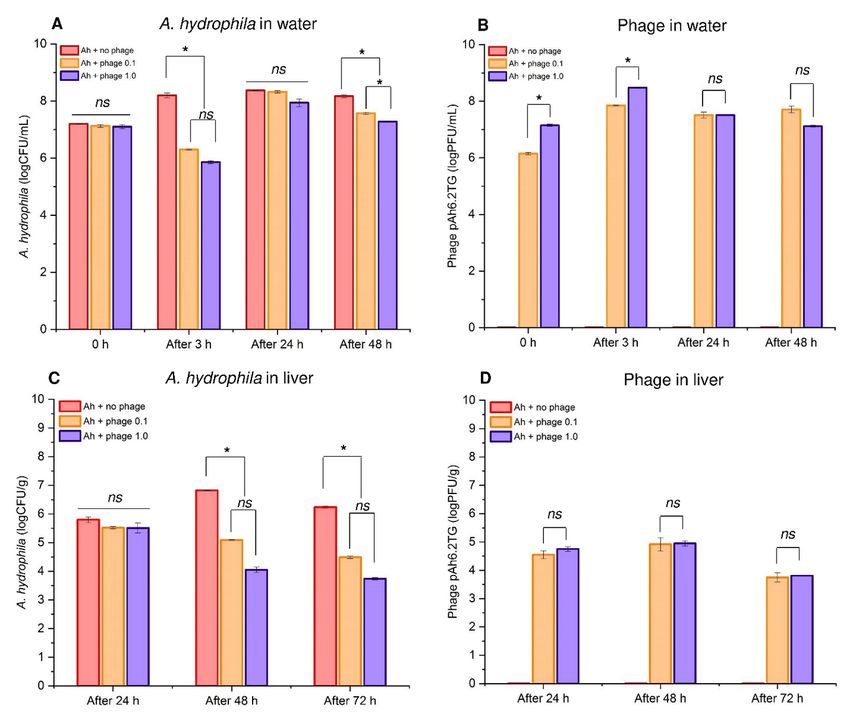
Phage isolation and morphology
Posted on Authorea 14 Jun 2021 — The copyright holder is the author/funder. All rights reserved. No reuse without permission. — https://doi.org/10.22541/au.162366557.74509707/v1 — This a preprint and has not been peer reviewed. Data may be preliminary.

                                                                                                                                                                                                                                                               Preparation of host strain
                                                                                                                                                                                                                                                               The MDR A. hydrophila BT09 (Tables 1 and S1) was chosen as a bacterial host for phage isolation. Prior to
                                                                                                                                                                                                                                                               phage isolation, prophage induction using Mitomycin C (Sigma-Aldrich, USA) was carried out as described
                                                                                                                                                                                                                                                               by Walker et al. (2009) to ensure that the host cells do not contain prophage. Briefly, 100 μL of bacterial
                                                                                                                                                                                                                                                               cells suspended in normal saline solution (OD600 = 0.6) was added into each of 10 mL of TSB supplemented
                                                                                                                                                                                                                                                               with 250, 500, and 1,000 ng/mL of Mitomycin C. All cultures were incubated at 28 °C for 8 h. The induced
                                                                                                                                                                                                                                                               phage production using Mitomycin C was evaluated by the Plaque Drop Assay (Adams, 1959).
                                                                                                                                                                                                                                                               Phage isolation
                                                                                                                                                                                                                                                               Water samples were collected from striped catfish culture ponds in Tien Giang Province, Vietnam. The
                                                                                                                                                                                                                                                               samples were enriched to increase phage concentration according to Van Twest and Kropinski (2009) and
                                                                                                                                                                                                                                                               isolated by Plaque Assay method described by Jun et al. (2013). Briefly, the samples were centrifuged at
                                                                                                                                                                                                                                                               4,500 x g , 4 o C for 30 min, and the supernatant was filtered through a 0.2 μm filter (Merck Millipore, USA)
                                                                                                                                                                                                                                                               to remove residual bacteria cells. Then, 10 mL filtrate was mixed with 10 mL of A. hydrophila BT09 in TSB
                                                                                                                                                                                                                                                               supplemented with 1.0 mM CaCl2 and 0.5 mM MgSO4 (MTSB). The mixture was cultured at 28 °C for 24
                                                                                                                                                                                                                                                               h with 50 rpm shaking. The mixture was then centrifuged at 10,000 x g , 4o C for 15 min, and the collected
                                                                                                                                                                                                                                                               supernatant was serially diluted (10-1 to 10-4 ). A volume of 100 μL of each dilution was transferred to a
                                                                                                                                                                                                                                                               tube containing 3.0 mL of TSA 0.5% agar supplemented with 1.0 mM CaCl2 and 0.5 mM MgSO4 (MTSA),
                                                                                                                                                                                                                                                               together with 100 μL ofA. hydrophila . The mixture was vortexed lightly and poured onto a plate of TSA
                                                                                                                                                                                                                                                               1.5% agar. The plates were incubated at 28o C for 16 h and the growth of phages was observed (clear plaque
                                                                                                                                                                                                                                                               on the plate). The individual clear plaque was picked and aseptically transferred to 200 μL of SM buffer (100
                                                                                                                                                                                                                                                               mM NaCl, 10 mM MgSO4 , 50 mM Tris-HCl, pH 7.5). The mixture was vortexed vigorously and kept in 4
                                                                                                                                                                                                                                                               o
                                                                                                                                                                                                                                                                 C refrigerator overnight. The phages in SM buffer were obtained by filtering the supernatant through a 0.2
                                                                                                                                                                                                                                                               μm filter after centrifugation at 10,000 x g for 10 min. The filtrate was propagated four times continuously
                                                                                                                                                                                                                                                               using the same protocol mentioned above for purification of the obtained phages. The isolated phages were
                                                                                                                                                                                                                                                               stored in SM buffer supplemented with 30% glycerol at -80 o C until used.
                                                                                                                                                                                                                                                               Examination of phage morphology
                                                                                                                                                                                                                                                               The structure and size of the phage were determined by Transmission Electron Microscope (TEM). The
                                                                                                                                                                                                                                                               specific procedure was as follows; the phage solution (3 mL) was centrifuged twice at 200,000 x g for 90 min.
                                                                                                                                                                                                                                                               The pellets were resuspended in sterile distilled water. A volume of 50 μL of 1% glutaraldehyde (g/vol) was
                                                                                                                                                                                                                                                               then added to immobilize the sample and rinsed with 0.1 M of cacodylate before proceeding with the dye.
                                                                                                                                                                                                                                                               The samples were coated with 0.1% Poly-Lysine solution onto the surface of the 200-mesh carbon-coated
                                                                                                                                                                                                                                                               grids to increase the adhesion of phages on the mesh. A volume of 10 μL of the phages was added to the
                                                                                                                                                                                                                                                               grid and allowed to dry naturally for 5 min. The samples were dyed with 1% uranyl acetate sterilized with
                                                                                                                                                                                                                                                               a 0.2 μm filter. The samples were washed with distilled water, allowed to dry for 5 min and imaged with a
                                                                                                                                                                                                                                                               TEM-JEOL 1010 (Japan) with light projected through the grid for about 5 s at 80 kV. Phage morphology
                                                                                                                                                                                                                                                               was classified according to the guideline of International Committee on Taxonomy of Viruses (ICTV) and
                                                                                                                                                                                                                                                               Ackermann (2007).
                                                                                                                                                                                                                                                               2.3. Host range and specificity
                                                                                                                                                                                                                                                               The host range of phage pAh6.2TG isolated in this study was conducted on the collection of 17 A. hydrophila
                                                                                                                                                                                                                                                               isolates from diseased fish (Tables 1 and S1). In this study, the Plaque Drop Assay was performed as described
                                                                                                                                                                                                                                                               by Adams (1959) with minor modifications. Briefly, double-layer agar plates containing tested bacterial cells
                                                                                                                                                                                                                                                               were prepared. Then, 5 μL of phages (108 PFU/mL) was dropped on the surface of each plate, kept without
                                                                                                                                                                                                                                                               moving for 30 min and incubated at 28 o C for 16 h. Normal saline solution was used as negative control.
                                                                                                                                                                                                                                                               Phage susceptibility was indicated by a clear zone appearing at the location of the drops while no clear
                                                                                                                                                                                                                                                               zone indicated unsusceptible host. Specificity test of phage pAh6.2TG to other common aquatic pathogens
                                                                                                                                                                                                                                                               (Aeromonas veronii ,Aeromonas schubertii , Edwardsiella ictaluri ,Streptococcus agalactiae ) and probiotic

                                                                                                                                                                                                                                                                                                                     4
Characterization and protective effects of lytic bacteriophage pAh6.2TG against a pathogenic multidrug-resistant Aeromonas hydrophila in Nile ...
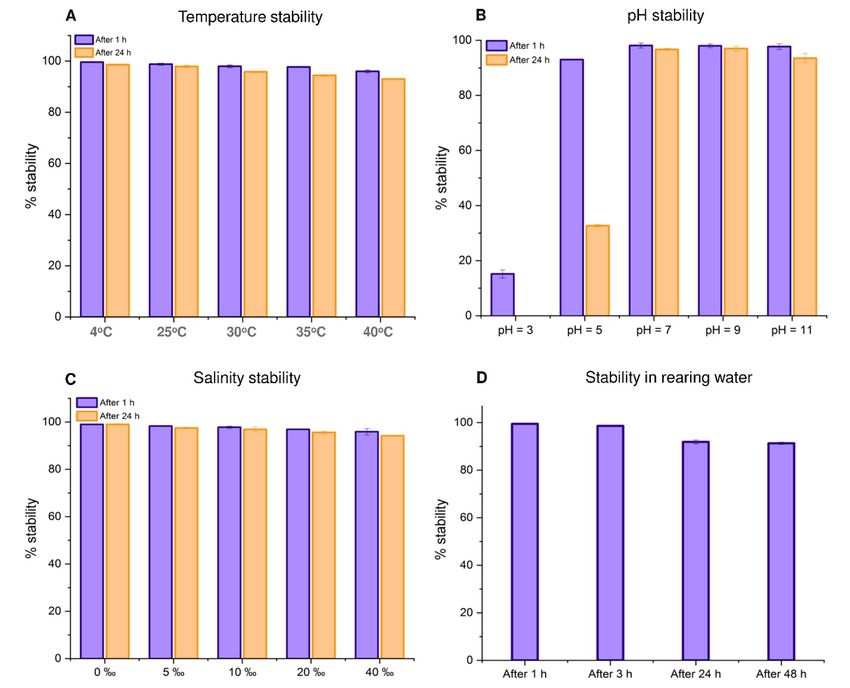
bacteria (Lactobacillus fermentum , Lactobacillus plantarum ) (Table 1) was done in the same manner. All
Posted on Authorea 14 Jun 2021 — The copyright holder is the author/funder. All rights reserved. No reuse without permission. — https://doi.org/10.22541/au.162366557.74509707/v1 — This a preprint and has not been peer reviewed. Data may be preliminary.

                                                                                                                                                                                                                                                               tests for host range and specificity were done in triplicates.
                                                                                                                                                                                                                                                               2.4. Phage stability in different environmental conditions
                                                                                                                                                                                                                                                               Stability of phage pAh6.2TG at different temperature (4, 25, 30, 35, and 40o C), pH (3, 5, 7, 9 and 11),
                                                                                                                                                                                                                                                               salinity (0, 5, 10, 20, 40These tests were carried out by incubating 100 μL of phage culture (approx. 109
                                                                                                                                                                                                                                                               PFU/mL) at the respective temperatures, pH, and salinity for 1 and 24 h in 10 mL of SM buffer. All
                                                                                                                                                                                                                                                               the experiments were conducted in triplicates. The stability of phages in rearing water was performed in
                                                                                                                                                                                                                                                               duplicates by adding 2 mL of phage pAh6.2TG (approx. 8.5 × 10¹⁰ PFU/mL) into 50 L of water (pH =
                                                                                                                                                                                                                                                               7.0 ± 1.0, 0% NaCl) containing 20 of Nile tilapia and maintained at 30 ± 1.0 o C. The concentration of
                                                                                                                                                                                                                                                               viable phages was enumerated by plaque assay (Jun et al., 2013). Phage concentration (logPFU/mL) before
                                                                                                                                                                                                                                                               incubation in different conditions was set to be 100%.
                                                                                                                                                                                                                                                               2.5. Genome characterization
                                                                                                                                                                                                                                                               Phage genome extraction and next-generation sequencing
                                                                                                                                                                                                                                                               The phage particles prepared by liquid propagation in TSBM were desalted using Millipore Amicon ultra-
                                                                                                                                                                                                                                                               centrifugal filter 10,000 NMWL (Merck, United States) at 10,000 x g , 4 o C for 15 min and concentrated by
                                                                                                                                                                                                                                                               ultracentrifugation at 300,000 x g , 4o C for 3 h (Beckman Coulter, German). The pellets were resuspended
                                                                                                                                                                                                                                                               in SM buffer. Phage genomic DNA was extracted using Phage DNA Extraction Kit (Cat. 46800, Norgen
                                                                                                                                                                                                                                                               Biotek, Canada) following the manufacturer’s protocol. Quality and concentration of DNA were measured
                                                                                                                                                                                                                                                               by Nanodrop (Colibri, German) and Quibit 4.0 (Thermo Scientific, United States). Purified genomic DNA
                                                                                                                                                                                                                                                               (3.15 ng/μL) was subjected to library preparation and sequencing using Next Generation Sequencing System
                                                                                                                                                                                                                                                               with Illumina Novaseq 6000 platform (Pair-end, 150; library construction size, 350 bp; data output, 1.0 GB,
                                                                                                                                                                                                                                                               data quality, Q30 > 80) at KTEST company, Vietnam.
                                                                                                                                                                                                                                                               Phage genome assembly and annotation
                                                                                                                                                                                                                                                               Raw reads were filtered using Fastp v 0.20.1 with the qualified phred score [?] Q25 and 8 bases trimming
                                                                                                                                                                                                                                                               from 5’/3’ end (Chen et al., 2018). Host associated sequences were filtered out by mapping trimmed reads to
                                                                                                                                                                                                                                                               the genome of A. hydrophila type strain (accession no. NZ CP016990.1) using Bowtie2 v 2.3.4.3 (Langmead
                                                                                                                                                                                                                                                               & Salzberg, 2012). Only unaligned reads were subjected to genome assembly using Unicycler v 0.4.8 (Wick
                                                                                                                                                                                                                                                               et al., 2017) on the Galaxy web platform at usegalaxy.org (Afgan et al., 2016). Potential phage sequence was
                                                                                                                                                                                                                                                               identified by submitting the assembled contigs to PHASTER web server (Arndt et al., 2016). The predicted
                                                                                                                                                                                                                                                               phage sequence (assigned as ‘pAh6.2TG’ in this study) was annotated using Prokka v 1.14.6 with Viruses
                                                                                                                                                                                                                                                               annotation mode (Seemann, 2014). The annotated phage genome was visualized using DNAplotter (Carver
                                                                                                                                                                                                                                                               et al., 2009).
                                                                                                                                                                                                                                                               Phage taxonomic identification and phylogenetic reconstruction
                                                                                                                                                                                                                                                               Identification of phage species was carried using VICTOR web service (Meier-Kolthoff & Göker, 2017). VIC-
                                                                                                                                                                                                                                                               TOR is a tool that perform pairwise genome comparison of prokaryotic viruses and automatically constructs
                                                                                                                                                                                                                                                               phylogenomic trees using Genome-BLAST Distance Phylogeny method (GBDP) with the formula D0. This
                                                                                                                                                                                                                                                               tool also classifies the virus at the species, genus and family level with the taxon boundaries estimating
                                                                                                                                                                                                                                                               by OPTSIL program (Göker et al., 2009). Herein, only the genomes of the viruses belonging to the fami-
                                                                                                                                                                                                                                                               ly Myoviridae (n = 91) were included in this genome comparison since pAh6.2TG was predicted as an
                                                                                                                                                                                                                                                               unknownMyoviridae by PHASTER tool described in the above section.
                                                                                                                                                                                                                                                               In addition to genome comparison, the phylogenetic analyses based on the terminase large subunit (terL)
                                                                                                                                                                                                                                                               and major capsid protein (MCP) amino acid sequences of pAh6.2TG and other related species (predicted
                                                                                                                                                                                                                                                               by VICTOR) were also performed via PhyloSuite v1.2.2 (Zhang et al., 2020). Amino acid sequences were
                                                                                                                                                                                                                                                               aligned using MAFFT (Katoh & Standley, 2013) and the maximum-likelihood trees were constructed using
                                                                                                                                                                                                                                                               IQ-TREE (Nguyen et al., 2015) with 5,000 ultrafast bootstraps and best-fit model (LG+G4) estimated by
                                                                                                                                                                                                                                                               ModelFinder. Phylogenomic tree, terL- and MCP-based trees were visualized using Phandango (Hadfield

                                                                                                                                                                                                                                                                                                                    5
Characterization and protective effects of lytic bacteriophage pAh6.2TG against a pathogenic multidrug-resistant Aeromonas hydrophila in Nile ...
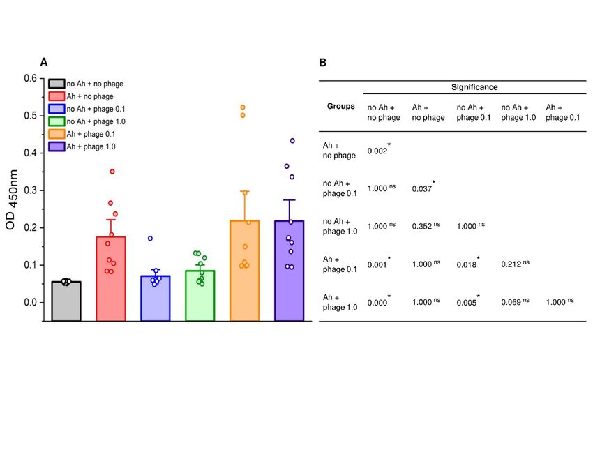
et al., 2018) and iTOL web tools (Letunic & Bork, 2019). Lastly, the protein sequence similarities between
Posted on Authorea 14 Jun 2021 — The copyright holder is the author/funder. All rights reserved. No reuse without permission. — https://doi.org/10.22541/au.162366557.74509707/v1 — This a preprint and has not been peer reviewed. Data may be preliminary.

                                                                                                                                                                                                                                                               pAh6.2TG and the closest viral taxa were determined using CoreGenes3.5 web server with Blastp threshold
                                                                                                                                                                                                                                                               score at 75 (Turner et al., 2013).
                                                                                                                                                                                                                                                               Effect of phage on Nile tilapia challenged with MDR A. hydrophila
                                                                                                                                                                                                                                                               Experimental fish
                                                                                                                                                                                                                                                               Healthy Nile tilapia (10.5 ± 4.7 g) obtained from a commercial tilapia hatchery in Thailand were acclimated
                                                                                                                                                                                                                                                               for 2 weeks in dechlorinated tap water with aeration at 28 ± 1.0 o C before the experiments. The fish were
                                                                                                                                                                                                                                                               fed with commercial tilapia feed (crude-protein 30%) at rate of about 3% of fish weight twice daily. Before
                                                                                                                                                                                                                                                               starting the experiments, ten fish were randomly selected for bacterial isolation and found to be free of A.
                                                                                                                                                                                                                                                               hydrophila . The experimental animal protocols were approved by Chulalongkorn University (Approval no.
                                                                                                                                                                                                                                                               CU-IACUC 2031006).
                                                                                                                                                                                                                                                               Fish survivability and sample collection
                                                                                                                                                                                                                                                               This experiment aimed to investigate whether lytic phage treatment improves survivability of Nile tilapia
                                                                                                                                                                                                                                                               challenged with a pathogenic MDRA. hydrophila BT14. A total of 258 fish were randomly divided into six
                                                                                                                                                                                                                                                               groups with 2 replicate tanks per each group (Figure S1): Group 1 was exposed to culture medium without
                                                                                                                                                                                                                                                               phage (no Ah + no phage); Group 2 was exposed to bacteria without phage (Ah + no phage); Group 3 was
                                                                                                                                                                                                                                                               exposed to culture medium and phage pAh6.2TG at multiplicity of infection (MOI) = 0.1 (no Ah + phage
                                                                                                                                                                                                                                                               0.1); Group 4 was exposed to culture medium and treated with phage at MOI = 1.0 (no Ah + phage 1.0);
                                                                                                                                                                                                                                                               Group 5 was challenged with A. hydrophila and treated with phage at MOI = 0.1 (Ah + phage 0.1); Group
                                                                                                                                                                                                                                                               6 was challenged with A. hydrophila and treated with phage at MOI = 1.0 (Ah + phage 1.0).
                                                                                                                                                                                                                                                               In bacterial challenge groups (2, 5 and 6), 1 L of MDR A. hydrophila BT14 (approx. 8 × 10⁸ CFU/mL) was
                                                                                                                                                                                                                                                               added to 50 L water to reach a final concentration of approx. 2 × 10⁷ CFU/mL. Groups 5 and 6 tanks had 2
                                                                                                                                                                                                                                                               and 20 mL of phage pAh6.2TG (approx. 8.5 × 10¹⁰ PFU/mL) added to reach a final concentration of approx.
                                                                                                                                                                                                                                                               2 × 10⁶ and 2 × 10⁷ PFU/mL, respectively. Group 2 tank had 20 mL of SM buffer without phage added.
                                                                                                                                                                                                                                                               The mixtures in groups 2, 5 and 6 were maintained at 29 ± 1.0 ° C with aeration for 3 h. In culture medium
                                                                                                                                                                                                                                                               exposure groups (1, 3 and 4), 1 L of TSB was added to 50 L water. Groups 3 and 4 tanks had 2 and 20 mL
                                                                                                                                                                                                                                                               of phage pAh6.2TG (approx. 8.5 × 10¹⁰ PFU/mL) added, respectively. After 3 h, the fish were transferred
                                                                                                                                                                                                                                                               to all groups, maintained at 29 ± 1° C with aeration for 14 days. In order to investigate the effect of phage
                                                                                                                                                                                                                                                               on the concentration of A. hydrophila in rearing water, a volume of 25 mL water from groups 2, 5 and 6
                                                                                                                                                                                                                                                               were sampled at 3, 24 and 48 h after exposure with phage. A volume of 1 mL water was centrifuged at 4 o C,
                                                                                                                                                                                                                                                               10.000 x g , for 5 min. The supernatant were collected and diluted in SM buffer to measure concentration of
                                                                                                                                                                                                                                                               phage by Plaque Assay method (Jun et al., 2013). The pellet was washed 1 time and re-suspended in 1 mL
                                                                                                                                                                                                                                                               of PBS buffer. Bacterial concentration was then enumerated by conventional plate count method using RS
                                                                                                                                                                                                                                                               supplemented with Novobiocin (Harrigan & McCance, 2014). In order to investigate the effect of phage on
                                                                                                                                                                                                                                                               the concentration of A. hydrophila in liver, two fish from groups 2, 5 and 6 were sampled at 24, 48 and 72 h
                                                                                                                                                                                                                                                               after exposure with phage. The fish were necropsied, and 0.1 g of live tissue was collected and homogenized
                                                                                                                                                                                                                                                               in a microtube containing 900 μL of SM buffer. The samples were then centrifuged at 10.000 x g , for 5 min.
                                                                                                                                                                                                                                                               The supernatant and pellet were used for respective phage and bacterial enumeration same as above.
                                                                                                                                                                                                                                                               The remaining fish were observed daily for 14 days, and mortality was recorded. Representative moribund or
                                                                                                                                                                                                                                                               freshly dead fish were collected for bacterial re-isolation using RS supplemented with Novobiocin as described
                                                                                                                                                                                                                                                               above. The RPS was calculated according to the formula described by Ellis (1988): RPS = (1 - % mortality
                                                                                                                                                                                                                                                               in challenge / % mortality in control) * 100.
                                                                                                                                                                                                                                                               Determination of serum antibody by the enzyme-linked immunosorbent assay (ELISA)
                                                                                                                                                                                                                                                               For the comparison of specific antibody (IgM) levels against A. hydrophila between experimental groups,
                                                                                                                                                                                                                                                               blood samples of 5 surviving fish in each tank (10 fish/group) were collected at the end of the experiment
                                                                                                                                                                                                                                                               (day 14). Sera were collected after centrifugation at 8,000 x g for 15 min, stored at -20 o C until used. ELISA
                                                                                                                                                                                                                                                               assay was carried out following the protocol described by Dien et al. (2021).

                                                                                                                                                                                                                                                                                                                     6
Characterization and protective effects of lytic bacteriophage pAh6.2TG against a pathogenic multidrug-resistant Aeromonas hydrophila in Nile ...
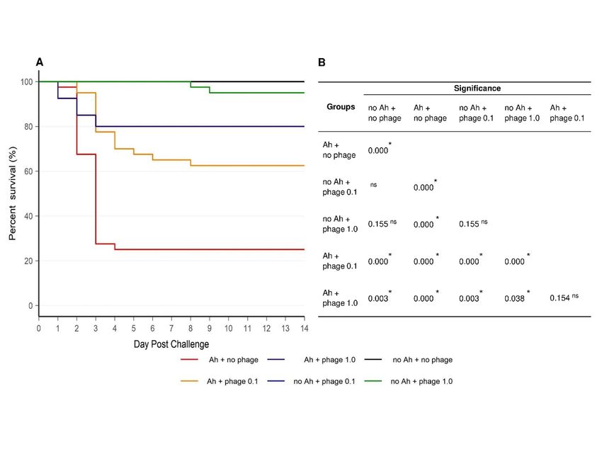
Statistical Analysis
Posted on Authorea 14 Jun 2021 — The copyright holder is the author/funder. All rights reserved. No reuse without permission. — https://doi.org/10.22541/au.162366557.74509707/v1 — This a preprint and has not been peer reviewed. Data may be preliminary.

                                                                                                                                                                                                                                                               Percent survival data from the challenge experiments was analyzed by the Kaplan-Meier method and dif-
                                                                                                                                                                                                                                                               ferences among groups were tested using a log-rank test, p -values of 0.05 or less were considered to be
                                                                                                                                                                                                                                                               statistically significant. Enumeration of A. hydrophilaconcentration and phage titer in rearing water and
                                                                                                                                                                                                                                                               fish liver samples was analyzed by ANOVA. Dunnett post-hoc test was used to measure specific differences
                                                                                                                                                                                                                                                               between pairs of mean. The OD450nm readings from the indirect ELISA assay were analyzed using a Kruskal-
                                                                                                                                                                                                                                                               Wallis test. Multiple comparison analyses were performed by Bonferroni test. All statistical analyses were
                                                                                                                                                                                                                                                               performed using SPSS Software ver22.0 (IBM Corp., USA).
                                                                                                                                                                                                                                                               RESULTS
                                                                                                                                                                                                                                                               Prophage induction, phage isolation, and morphology
                                                                                                                                                                                                                                                               Although three doses of Mitomycin C (250, 500, and 1,000 ng/mL) were used for prophage induction, no
                                                                                                                                                                                                                                                               plaque was detected, indicating thatA. hydrophila BT09 did not contain prophage and was suitable as a
                                                                                                                                                                                                                                                               bacterial host for lytic phage isolation. Subsequently, a phage, designated pAh6.2TG, was isolated from a
                                                                                                                                                                                                                                                               freshwater sample. Phage pAh6.2TG produced medium, clear, and round plaques with diameter of 1.3 1.8
                                                                                                                                                                                                                                                               mm (Figure 1A-B) after 16 h of incubation. TEM morphology examination showed that the phage had an
                                                                                                                                                                                                                                                               icosahedral head with 59.6 ± 2.5 nm diameter (n = 3) and a contractile tail which was 137 ± 10.2 nm in
                                                                                                                                                                                                                                                               length and 20.2 ± 2.7 nm in diameter (n = 3) (Figure 1C-D). Based on the morphological features, phage
                                                                                                                                                                                                                                                               pAh6.2TG was initially classified to the Myoviridae family.
                                                                                                                                                                                                                                                               Host range and specificity of phage pAh6.2TG
                                                                                                                                                                                                                                                               Among all bacterial isolates tested, pAh6.2TG showed lytic activity against 10/17 A. hydrophila isolates
                                                                                                                                                                                                                                                               (Table 1) of which 8 isolates were MDR (Table S1). In contrast, no lytic activity was observed against other
                                                                                                                                                                                                                                                               fish bacterial pathogens including A. veronii , A. schubertii, E. ictaluri , S. agalactiae as well as two probiotic
                                                                                                                                                                                                                                                               bacteria L. fermentum , and L. plantarum (Table 1).
                                                                                                                                                                                                                                                               Stability of phage pAh6.2TG at different environmental conditions
                                                                                                                                                                                                                                                               Stability of pAh6.2TG at different temperatures (4 to 40o C) is shown in Figure 2A. Similar percentages of
                                                                                                                                                                                                                                                               viable phage were detected after 1 h (96 ± 0.55 – 99.6 ± 0.08%) and 24 h (93 ± 0.23 – 98.6 ± 0.17%) of
                                                                                                                                                                                                                                                               incubation, indicating that pAh6.2TG is a relatively thermostable phage.
                                                                                                                                                                                                                                                               Phage pAh6.2TG was stable (93.5 ± 1.69 – 97 ± 0.87%) at pH 7, 9 and 11 (Figure 2B). However, the phage
                                                                                                                                                                                                                                                               pAh6.2TG was not stable at low pH. At pH 5, 93 ± 0.24% phage remained viable after 1h, and decreased
                                                                                                                                                                                                                                                               sharply to 32.7 ± 0.44% (from 7.88 to 2.58 ± 0.06 logPFU/mL) after 24 h. At pH 3, only 15.2 ± 1.47% (1.19
                                                                                                                                                                                                                                                               ± 0.2 logPFU/mL) of phage was still viable after 1 h and reduced to undetectable level at 24 h (Figure 2B).
                                                                                                                                                                                                                                                               Phage pAh6.2TG was relatively stable at a wide range of salinity (0 – 40 viable after 1 and 24 h, respectively
                                                                                                                                                                                                                                                               (Figure 2C).
                                                                                                                                                                                                                                                               In fish-rearing water (30 +- 1 o C, pH 6.9, 0% NaCl) spiked with phages, percentage of stability at 1 and 3
                                                                                                                                                                                                                                                               h were 99.5 +- 0.15% and 98.6 +- 0.11%, respectively. After 24 and 48 h, phage titer decreased slightly to
                                                                                                                                                                                                                                                               91.9 +- 0.85% and 91.3 +- 0.5%, equivalent to 6.52 +- 0.07 and 6.47 +- 0.03 logPFU/mL, respectively.
                                                                                                                                                                                                                                                               Genome characterization of pAh6.2TG phage
                                                                                                                                                                                                                                                               Based on the assembly graph generated by Unicycler software, pAh6.2TG was predicted to contain a circular
                                                                                                                                                                                                                                                               genome with a length of 51,780 bp, a GC content of 52.48%, encoding 65 putative open reading frames
                                                                                                                                                                                                                                                               (ORFs) (Table S2) without tRNA genes (Figure 3). According to bioinformatics prediction, pAh6.2TG
                                                                                                                                                                                                                                                               genome consists of three main functional modules: i) phage structure and DNA packaging (major capsid
                                                                                                                                                                                                                                                               protein, baseplate protein, tail fiber protein, and terminase subunit), ii) DNA metabolism and replication
                                                                                                                                                                                                                                                               (RNA polymerase, DNA polymerase, DNA helicase, 5’-3’ exonuclease, DNA ligase, and Ribonucleoside-
                                                                                                                                                                                                                                                               diphosphate reductase large subunit), and iii) host lysis (cell wall hydrolase).

                                                                                                                                                                                                                                                                                                                       7
The closest phage taxonomic classification of pAh6.2TG toward other 91Myoviridae phages in the public
Posted on Authorea 14 Jun 2021 — The copyright holder is the author/funder. All rights reserved. No reuse without permission. — https://doi.org/10.22541/au.162366557.74509707/v1 — This a preprint and has not been peer reviewed. Data may be preliminary.

                                                                                                                                                                                                                                                               database revealed thatAeromonas phage pAh6.2TG and PVN02 (accession no. LR813619) were classified
                                                                                                                                                                                                                                                               as the identical species with 99.33% identity. The result also showed total 64/65 ORFs were homologous
                                                                                                                                                                                                                                                               between pAh6.2TG and PVN02 (97.3 - 100 % nt. identity), except for ORF03 that showed the highest
                                                                                                                                                                                                                                                               homology (70%) to another Aeromonas phage pAh6-C (Table S2). Phylogenetic analysis based on whole
                                                                                                                                                                                                                                                               genome (Figure 4A-B), major capsid protein sequence (Figure 4C), and terminase large subunit sequence
                                                                                                                                                                                                                                                               (Figure 4D) confirmed high homology of phage pAh6.2TG and phage PVN02. In addition, pAh6.2TG
                                                                                                                                                                                                                                                               was closely related to the Aeromonas phage pAh6-C (accession no. KJ858521), Shewanella phage Spp001
                                                                                                                                                                                                                                                               (accession no. NC023594), and Shewanella phage SppYZU05 (accession no. NC047824) (Figure 4).
                                                                                                                                                                                                                                                               In vivo challenge results
                                                                                                                                                                                                                                                               Phage pAh6.2TG improved survivability of Nile tilapia challenged with the MDR A. hydrophila
                                                                                                                                                                                                                                                               In vivo experiment showed that 100% fish in negative control group (no Ah + no phage) survived after
                                                                                                                                                                                                                                                               14 days, while only 25% survival was recorded in positive control group (Ah + no phage) (Figure 5).
                                                                                                                                                                                                                                                               Interestingly, there was 62.5% and 80% survival in groups treated with pAh6.2TG with MOI = 0.1 (Ah
                                                                                                                                                                                                                                                               + phage 0.1) and MOI = 1.0 (Ah + phage 1.0), respectively. These differences in percentage of survival
                                                                                                                                                                                                                                                               of 2 phage treated groups were not statistically significant (p = 0.154) but statistically significant with
                                                                                                                                                                                                                                                               positive control group (p = 0.000). The remaining two groups treated with phage without bacteria had 95
                                                                                                                                                                                                                                                               – 100% survival. The relative percent survival (RPS) of two treatments groups were 50% (MOI = 0.1) and
                                                                                                                                                                                                                                                               73.3% (MOI = 1), respectively. The moribund fish in challenge groups showed behavioral abnormalities
                                                                                                                                                                                                                                                               (lethargy, loss of appetite, and surface swimming) and pale liver. Using selective medium, pure colonies with
                                                                                                                                                                                                                                                               morphological characteristics of A. hydrophila were successfully isolated from representative dead fish (n=3).
                                                                                                                                                                                                                                                               Phage pAh6.2TG suppressed bacterial concentration in water and fish tissue
                                                                                                                                                                                                                                                               Fluctuation of bacterial concentration and phage titer in water and fish liver are shown in Figure 6 and
                                                                                                                                                                                                                                                               Table S3. In rearing water, after 3 h of bacteria and phages exposure, bacterial concentration reduced 6.7
                                                                                                                                                                                                                                                               +- 0.5 folds in group treated with phage MOI = 0.1, and 18.1 +- 6.98 folds in group treated with phage
                                                                                                                                                                                                                                                               MOI = 1.0 (Figure 6A). The calculation of fold changes is displayed in Table S3. In contrast, after 3 h,
                                                                                                                                                                                                                                                               bacterial concentration increased 10.2 +- 3.15 folds in Ah + no phage group. Simultaneously, phage titer in
                                                                                                                                                                                                                                                               groups treated with phage MOI = 0.1 and 1.0 after 3 h increased 51.04 +- 5.16 folds, and 20.98 +- 1.03 folds,
                                                                                                                                                                                                                                                               respectively (Figure 6B). Phage was absent in Ah + no phage control group. At 24 h post-challenge, bacterial
                                                                                                                                                                                                                                                               concentration in three groups was increased, while phage concentration in water slightly decreased. Besides,
                                                                                                                                                                                                                                                               slight reduction of bacterial and phage concentration was observed in all groups at 48 h post-treatment
                                                                                                                                                                                                                                                               (Figure 6A – B).
                                                                                                                                                                                                                                                               Moreover, in fish liver, bacterial concentrations of 5.8 +- 0.14, 5.52 +- 0.06, 5.51 +- 0.24 logCFU/g were
                                                                                                                                                                                                                                                               recorded in Ah + no phage, Ah + phage 0.1, and Ah + phage 1.0 groups, respectively (Figure 6C). In
                                                                                                                                                                                                                                                               Ah + phage 0.1 and Ah + phage1.0 groups, phage titers were 4.55 +- 0.2 and 4.75 +- 0.12 logPFU/g,
                                                                                                                                                                                                                                                               respectively (Figure 6D). Similar pattern of phage concentration in rearing water was observed at 48 h
                                                                                                                                                                                                                                                               post-challenge, while bacterial load decreased in all groups. In fish liver, compared to 24 h post-treatment,
                                                                                                                                                                                                                                                               bacterial concentration in Ah + no phage groups increased 10.69 +- 3.85 folds, while in Ah + phage 0.1 and
                                                                                                                                                                                                                                                               Ah + phage 1.0 groups, bacteria decreased 2.7 +- 0.24 and 34.08 +- 26.4 folds, respectively (Table S3).
                                                                                                                                                                                                                                                               The bacterial load in fish liver of Ah + no phage group decreased 3.8 +- 0.64 folds, from 6.58 x 10⁶ +- 3.18
                                                                                                                                                                                                                                                               x 105 at 24 h post-challenge to 1.75 x 10⁶ +- 2.12 x 10⁵ CFU/g at 72 h post-challenge (Table S3). The
                                                                                                                                                                                                                                                               same pattern was recorded in Ah+ phage 0.1 and Ah + phage 1.0 groups with 4.03 +- 0.83 and 2.18 +- 0.96
                                                                                                                                                                                                                                                               fold-reduction, respectively (Table S3). At 72 h post-challenge, phage titer in fish liver decreased 15.13 +-
                                                                                                                                                                                                                                                               3.35 and 13.96 +- 3.95 folds in groups treated with phage 0.1 and 1.0 at 24 h post-challenge, respectively
                                                                                                                                                                                                                                                               (Table S3).
                                                                                                                                                                                                                                                               Surviving fish developed specific IgM against MDRA. hydrophila
                                                                                                                                                                                                                                                               All surviving fish in three groups challenged with MDR A. hydrophila had significantly higher levels of

                                                                                                                                                                                                                                                                                                                     8
specific antibody (IgM) compared to the three unchallenged groups (p < 0.05) as measured by indirect
Posted on Authorea 14 Jun 2021 — The copyright holder is the author/funder. All rights reserved. No reuse without permission. — https://doi.org/10.22541/au.162366557.74509707/v1 — This a preprint and has not been peer reviewed. Data may be preliminary.

                                                                                                                                                                                                                                                               ELISA, Kruskal - Wallis test: H (5) = 35.218,p = 0.000 (Figure 7). The serum from fish in the Ah + no
                                                                                                                                                                                                                                                               phage, Ah + phage 0.1, and Ah + phage 1.0 groups had OD readings of 0.18 +- 0.09, 0.22 +- 0.17, and
                                                                                                                                                                                                                                                               0.22 +- 0.12, respectively. The IgM level was slightly higher in 2 phage treated groups but not statistically
                                                                                                                                                                                                                                                               significant difference. In contrast, the low level of OD450 readings were recorded in the remaining groups
                                                                                                                                                                                                                                                               (0.06 +- 0.003 to 0.08 +- 0.03) (Figure 7).
                                                                                                                                                                                                                                                               DISCUSSION
                                                                                                                                                                                                                                                               The Myoviridae phages specific to A. hydrophila are highly diverse in nature (Chandrarathna et al., 2020;
                                                                                                                                                                                                                                                               Cheng et al., 2021; Jun et al., 2013). The lytic pAh6.2TG isolated in this study had genome characteristics
                                                                                                                                                                                                                                                               most closely related to phage PVN02 (99.33% nt. identity) in the GenBank database, previously isolated
                                                                                                                                                                                                                                                               from Vietnam (Tu et al., 2020). The origins of two phages from the closed geographical area of Mekong basin,
                                                                                                                                                                                                                                                               although from different rivers, may explain the high genomic similarity of pAh6.2TG and PVN02. Compared
                                                                                                                                                                                                                                                               to previously reported A. hydrophila -specific phages, pAh6.2TG (51,780 bp) had similar genome size with
                                                                                                                                                                                                                                                               the phage PVN02 (51,668 bp) from Vietnam (Tu et al., 2020), and pAh6-C (53,744 bp) from Korea (Jun et
                                                                                                                                                                                                                                                               al., 2015), but is larger than phage AhyVDH1 (39,175 bp) from China (Cheng et al., 2021), and smaller than
                                                                                                                                                                                                                                                               phage LAh10 (260,310 bp) from Australia. The latter is the largest known phage infecting A. hydrophila
                                                                                                                                                                                                                                                               (Kabwe et al., 2020). Genome analysis indicated that pAh6.2TG does not contain potential virulent genes
                                                                                                                                                                                                                                                               or antimicrobial resistant genes, suggesting it is highly relevant as a biocontrol agent in aquaculture systems
                                                                                                                                                                                                                                                               without concern of antimicrobial-resistant gene transmission.
                                                                                                                                                                                                                                                               Climate change has affected aquaculture environments by perturbing chemical and physical properties of
                                                                                                                                                                                                                                                               water, particularly in the increase of water temperature and salinization (Maulu et al., 2021; Seggel &
                                                                                                                                                                                                                                                               De Young, 2016). The stability of pAh6.2TG under a wide range of temperatures (4 – 40 deg C) and
                                                                                                                                                                                                                                                               salinity (0 – 40 ppt) might be important characteristics for its wider application in diverse aquaculture
                                                                                                                                                                                                                                                               environments. Relatively high stability of pAh6.2TG in fish-rearing water suggests that immersion route is
                                                                                                                                                                                                                                                               practical. However, low viability of pAh6.2TG at pH 3 – 5 suggests that oral administration might not be
                                                                                                                                                                                                                                                               applicable due to the low pH in gastrointestinal tract of aquatic animals, e.g. pH in Nile tilapia stomach
                                                                                                                                                                                                                                                               range from 1.4 – 2.0 (Moriarty, 1973).
                                                                                                                                                                                                                                                               One of the major limitations of phage application is its narrow host range and geographical specificity
                                                                                                                                                                                                                                                               (Culot et al., 2019; Perez-Sanchez et al., 2018; Ross et al., 2016). Although the newly isolated phage
                                                                                                                                                                                                                                                               pAh6.2TG could lyse multiple isolates of MDR A. hydrophila from Vietnam, however, it does not lyse the
                                                                                                                                                                                                                                                               isolates from Thailand and other bacterial species from the same or different genera. Therefore, to expand
                                                                                                                                                                                                                                                               wider application of phage in aquaculture, a cocktail of multiple phage strains from different geographical
                                                                                                                                                                                                                                                               locations might be the better approach to tackle not only AMR A. hydrophila but also other important
                                                                                                                                                                                                                                                               bacterial pathogens in freshwater fishes. In addition, the specific infection of pAh6.2TG to A. hydrophila
                                                                                                                                                                                                                                                               and not probiotic bacteria suggest the potential combination of phage therapy and probiotics to combat
                                                                                                                                                                                                                                                               MDR A. hydrophila infection in aquaculture.
                                                                                                                                                                                                                                                               Carps, tilapias, and catfishes are crucial inland freshwater fish that play a vital role for food system trans-
                                                                                                                                                                                                                                                               formation to tackle micronutrient deficiencies in LMICs (FAO, 2020; Hicks et al., 2019). A. hydrophila
                                                                                                                                                                                                                                                               infection is one of the most important bacterial diseases responsible for the loss of millions of dollars in the
                                                                                                                                                                                                                                                               global freshwater aquaculture industry (Hossain et al., 2014; Peterman & Posadas, 2019; Pridgeon & Klesius,
                                                                                                                                                                                                                                                               2012). Increasing prevalence of pathogenic MDRA. hydrophila in aquaculture poses the high risk for serious
                                                                                                                                                                                                                                                               uncontrollable disease outbreaks and public health concern due to spread of AMR. Non-antibiotic approach
                                                                                                                                                                                                                                                               using lytic phages, therefore, was explored to control disease caused by MDR A. hydrophila in aquaculture
                                                                                                                                                                                                                                                               systems. In this study, we provided in vivo evidences for the efficacy of phage application in rearing water
                                                                                                                                                                                                                                                               which is effective at suppressing bacterial concentration in water as well as reducing the bacterial load in
                                                                                                                                                                                                                                                               fish liver. The presence of phages in the fish liver also suggests that immersion administration could deliver
                                                                                                                                                                                                                                                               considerably large number of phages into the fish tissue. These factors may contribute to improvement of
                                                                                                                                                                                                                                                               survivability (RPS = 50 – 75%) of tilapia. Importantly, not only was there higher survival in phage treated
                                                                                                                                                                                                                                                               groups, but all surviving fish also developed specific IgM against A. hydrophila . This suggests that phages

                                                                                                                                                                                                                                                                                                                      9
possibly weakened the bacteria which allowed the fish immune system to respond more effectively and saved
Posted on Authorea 14 Jun 2021 — The copyright holder is the author/funder. All rights reserved. No reuse without permission. — https://doi.org/10.22541/au.162366557.74509707/v1 — This a preprint and has not been peer reviewed. Data may be preliminary.

                                                                                                                                                                                                                                                               the fish from death. Similarly, there were several studies using phages as therapeutic agent to control A.
                                                                                                                                                                                                                                                               hydrophila infection. Le et al. (2018) used phage cocktails (F2 and F5) with MOI = 0.01, 1.0, and 100 to
                                                                                                                                                                                                                                                               control A. hydrophila infection in striped catfish (Pangasianodon hypophthalmus ) by injection administra-
                                                                                                                                                                                                                                                               tion and obtained RPS of 16.33%, 44.9%, and 100%, respectively. Immersion treatment of 1 x 10⁸ PFU/mL
                                                                                                                                                                                                                                                               phage Akh-2 improved survivability of Nile tilapia with RPS of 41.1% (Akmal et al., 2020). Cao et al. (2020)
                                                                                                                                                                                                                                                               applied phage MJG by injection, immersion, and oral administration to control a pathogenic A. hydrophila
                                                                                                                                                                                                                                                               in rainbow trout and the fish gained RPS of 100%, 66.7%, and 50%, respectively. Dang et al. (2021) showed
                                                                                                                                                                                                                                                               protective efficacy of phage PVN02-sprayed feed against A. hydrophila 4.4T in striped catfish with RPS from
                                                                                                                                                                                                                                                               75.6 – 87.8%.
                                                                                                                                                                                                                                                               The findings in this study suggest a potential approach using phage as prophylactic agent that was effective
                                                                                                                                                                                                                                                               in protecting Nile tilapia from a MDR A. hydrophila . This approach provided comparable RPS to other
                                                                                                                                                                                                                                                               promising alternatives to antibiotics, such as probiotic-based or plant-based products (Dawood et al., 2020;
                                                                                                                                                                                                                                                               Kuebutornye et al., 2020; Naliato et al., 2021; Neamat-Allah et al., 2021). Apart from tilapia, pAh6.2TG has
                                                                                                                                                                                                                                                               great potential to be applied in catfish aquaculture industry due to the lytic activity of pAh6.2TG against
                                                                                                                                                                                                                                                               multiple MDRA. hydrophila strains isolated from diseased striped catfish.
                                                                                                                                                                                                                                                               In summary, this study reported a newly isolated lytic phage pAh6.2TG that infects several isolates of MDR
                                                                                                                                                                                                                                                               A. hydrophila . The phage was classified as a member of Myoviridae based on a combination of morphol-
                                                                                                                                                                                                                                                               ogy and genomic characterization. In vitro tests showed that pAh6.2TG was relatively stable at different
                                                                                                                                                                                                                                                               environmental conditions. Using this phage as prophylactic agent was successful at reducing mortality in
                                                                                                                                                                                                                                                               Nile tilapia. Phage pAh6.2TG application in rearing water not only suppressed MDR A. hydrophila loads
                                                                                                                                                                                                                                                               in the rearing water and colonization of the bacteria in fish liver, but also improved fish survivability. These
                                                                                                                                                                                                                                                               findings supported that pAh6.2TG could be used in rearing water for biocontrol of MDR A. hydrophila
                                                                                                                                                                                                                                                               infection towards sustainable aquaculture.
                                                                                                                                                                                                                                                               NUCLEOTIDE SEQUENCE DATA
                                                                                                                                                                                                                                                               Phage pAh6.2TG sequence data has been submitted to the GeneBank databases under accession number
                                                                                                                                                                                                                                                               MZ336020.
                                                                                                                                                                                                                                                               ACKNOWLEDGEMENTS
                                                                                                                                                                                                                                                               Le Thanh Dien received the scholarships of Chulalongkorn University for ASEAN and NON-ASEAN coun-
                                                                                                                                                                                                                                                               tries, and the 90th Anniversary of Chulalongkorn University Scholarship. Channarong Rodkhum was sup-
                                                                                                                                                                                                                                                               ported by Thailand Science Research and Innovation (TSRI) Fund, Chulalongkorn University FRB640001 -
                                                                                                                                                                                                                                                               01 31 6. Ha Thanh Dong was supported by UK Government – Department of Health and Social Care
                                                                                                                                                                                                                                                               (DHSC), Global AMR Innovation fund (GAMRIF) and the International Development Research Center
                                                                                                                                                                                                                                                               (IDRC), Ottawa, Canada. We would like to acknowledge Kamphaengsaen Fisheries Research Station, Fac-
                                                                                                                                                                                                                                                               ulty of Fisheries, Kasetsart University, Thailand for providing experimental fish.
                                                                                                                                                                                                                                                               CONFLICT OF INTEREST
                                                                                                                                                                                                                                                               The authors declare that there are no conflicts of interest.
                                                                                                                                                                                                                                                               AUTHOR CONTRIBUTION STATEMENT
                                                                                                                                                                                                                                                               Le Thanh Dien : Conceptualization, Methodology, Investigation, Formal analysis, Writing - original draft.
                                                                                                                                                                                                                                                               Le Buu Ky : Investigation. Bui The Huy : Investigation. Pattanapon Kayansamruaj: Methodology,
                                                                                                                                                                                                                                                               Data analysis. Mohammad Fadhlullah Mursalim: Investigation. Saengchan Senapin : Data curation,
                                                                                                                                                                                                                                                               Review & Editing. Channarong Rodkhum : Supervision, Validation, Review & Editing. Ha Thanh
                                                                                                                                                                                                                                                               Dong : Conceptualization, Data curation, Writing Review & Editing, Supervision, and Validation.
                                                                                                                                                                                                                                                               ETHICAL APPROVAL
                                                                                                                                                                                                                                                               The authors confirm that the ethical policies of the journal, as noted on the journal’s author guidelines

                                                                                                                                                                                                                                                                                                                     10
page, have been adhered to and the appropriate ethical review committee approval has been received. This
Posted on Authorea 14 Jun 2021 — The copyright holder is the author/funder. All rights reserved. No reuse without permission. — https://doi.org/10.22541/au.162366557.74509707/v1 — This a preprint and has not been peer reviewed. Data may be preliminary.

                                                                                                                                                                                                                                                               project has been reviewed and approved by the Institutional Animal Care and Use Committee (IACUC)
                                                                                                                                                                                                                                                               in accordance with university regulations and policies governing the care and use of laboratory animals.
                                                                                                                                                                                                                                                               The review has followed guidelines documented in Ethical Principles and Guidelines for Use of Animals for
                                                                                                                                                                                                                                                               Scientific Purposes edited by the National Research Council of Thailand.
                                                                                                                                                                                                                                                               DATA AVAIBILITY STATEMENT
                                                                                                                                                                                                                                                               The data that support the findings of this study are available on request.
                                                                                                                                                                                                                                                               References
                                                                                                                                                                                                                                                               Ackermann, H.-W. (2007). 5500 Phages examined in the electron microscope. Archives of Virology, 152 (2),
                                                                                                                                                                                                                                                               227-243. https://doi.org/10.1007/s00705-006-0849-1
                                                                                                                                                                                                                                                               Adams, M. H. (1959). Bacteriophages. Bacteriophages (pp. 443-522): Wiley Intersciences, New York
                                                                                                                                                                                                                                                               Afgan, E., Baker, D., Van den Beek, M., Blankenberg, D., Bouvier, D., Čech, M., . . . Eberhard, C. (2016).
                                                                                                                                                                                                                                                               The Galaxy platform for accessible, reproducible and collaborative biomedical analyses: 2016 update.Nucleic
                                                                                                                                                                                                                                                               Acids Research, 44 (W1), W3-W10. https://doi.org/10.1093/nar/gkw343
                                                                                                                                                                                                                                                               Akmal, M., Rahimi-Midani, A., Hafeez-ur-Rehman, M., Hussain, A., & Choi, T.-J. (2020). Isolation, charac-
                                                                                                                                                                                                                                                               terization, and application of a bacteriophage infecting the fish pathogen Aeromonas hydrophila .Pathogens,
                                                                                                                                                                                                                                                               9 (3), 215. https://doi.org/10.3390/pathogens9030215
                                                                                                                                                                                                                                                               Anand, T., Vaid, R. K., Bera, B. C., Singh, J., Barua, S., Virmani, N., . . . Tripathi, B. (2016). Isolation
                                                                                                                                                                                                                                                               of a lytic bacteriophage against virulent Aeromonas hydrophila from an organized equine farm.Journal of
                                                                                                                                                                                                                                                               Basic Microbiology, 56 (4), 432-437. https://doi.org/10.1002/jobm.201500318
                                                                                                                                                                                                                                                               Arndt, D., Grant, J. R., Marcu, A., Sajed, T., Pon, A., Liang, Y., & Wishart, D. S. (2016). PHASTER:
                                                                                                                                                                                                                                                               a better, faster version of the PHAST phage search tool. Nucleic Acids Research, 44 (W1), W16-W21.
                                                                                                                                                                                                                                                               https://doi.org/10.1093/nar/gkw387
                                                                                                                                                                                                                                                               Cao, Y., Li, S., Han, S., Wang, D., Zhao, J., Xu, L., . . . Lu, T. (2020). Characterization and application
                                                                                                                                                                                                                                                               of a novel Aeromonasbacteriophage as treatment for pathogenic Aeromonas hydrophilainfection in rainbow
                                                                                                                                                                                                                                                               trout. Aquaculture, 523 , 735193. https://doi.org/10.1016/j.aquaculture.2020.735193
                                                                                                                                                                                                                                                               Cao, Y., Zhang, Y., Lan, W., & Sun, X. (2021). Characterization of vB VpaP MGD2, a newly isolated
                                                                                                                                                                                                                                                               bacteriophage with biocontrol potential against multidrug-resistant Vibrio parahaemolyticus .Archives of
                                                                                                                                                                                                                                                               Virology, 166 (2), 413-426. https://doi.org/10.1007/s00705-020-04887-x
                                                                                                                                                                                                                                                               Carver, T., Thomson, N., Bleasby, A., Berriman, M., & Parkhill, J. (2009).          DNAPlot-
                                                                                                                                                                                                                                                               ter:   circular and linear interactive genome visualization. Bioinformatics, 25 (1), 119-120.
                                                                                                                                                                                                                                                               https://doi.org/10.1093/bioinformatics/btn578
                                                                                                                                                                                                                                                               Chandrarathna, H., Nikapitiya, C., Dananjaya, S., De Silva, B., Heo, G.-J., De Zoysa, M., &
                                                                                                                                                                                                                                                               Lee, J. (2020). Isolation and characterization of phage AHP-1 and its combined effect with chlo-
                                                                                                                                                                                                                                                               ramphenicol to controlAeromonas hydrophila . Brazilian Journal of Microbiology, 51 (1), 409-416.
                                                                                                                                                                                                                                                               https://doi.org/10.1007/s42770-019-00178-z
                                                                                                                                                                                                                                                               Chen, S., Zhou, Y., Chen, Y., & Gu, J. (2018). fastp: an ultra-fast all-in-one FASTQ preprocessor. Bioin-
                                                                                                                                                                                                                                                               formatics, 34 (17), i884-i890. https://doi.org/10.1093/bioinformatics/bty560
                                                                                                                                                                                                                                                               Cheng, Y., Gao, D., Xia, Y., Wang, Z., Bai, M., Luo, K., . . . Xiao, W. (2021). Characterization of novel
                                                                                                                                                                                                                                                               bacteriophage AhyVDH1 and its lytic activity against Aeromonas hydrophila . Current Microbiology, 78
                                                                                                                                                                                                                                                               (1), 329-337. https://doi.org/10.1007/s00284-020-02279-7
                                                                                                                                                                                                                                                               Cui, H., Cong, C., Wang, L., Li, X., Li, J., Yang, H., . . . Xu, Y. (2021). Protective effectiveness of feeding
                                                                                                                                                                                                                                                               phage cocktails in controlling Vibrio harveyi infection of turbot Scophthalmus maximus . Aquaculture, 535

                                                                                                                                                                                                                                                                                                                     11
, 736390. https://doi.org/10.1016/j.aquaculture.2021.736390
Posted on Authorea 14 Jun 2021 — The copyright holder is the author/funder. All rights reserved. No reuse without permission. — https://doi.org/10.22541/au.162366557.74509707/v1 — This a preprint and has not been peer reviewed. Data may be preliminary.

                                                                                                                                                                                                                                                               Culot, A., Grosset, N., & Gautier, M. (2019). Overcoming the challenges of phage therapy for industrial
                                                                                                                                                                                                                                                               aquaculture: A review.Aquaculture, 513 , 734423. https://doi.org/10.1016/j.aquaculture.2019.734423
                                                                                                                                                                                                                                                               da Silva, B. C., Mouriño, J. L. P., Vieira, F. N., Jatobá, A., Seiffert, W. Q., & Martins, M. L. (2012). Hae-
                                                                                                                                                                                                                                                               morrhagic septicaemia in the hybrid surubim (Pseudoplatystoma corruscans × Pseudoplatystoma fasciatum
                                                                                                                                                                                                                                                               ) caused by Aeromonas hydrophila . Aquaculture Research, 43 (6), 908-916. https://doi.org/10.1111/j.1365-
                                                                                                                                                                                                                                                               2109.2011.02905.x
                                                                                                                                                                                                                                                               Dang, T. H. O., Xuan, T. T. T., Duyen, L. T. M., Le, N. P., & Hoang, H. A. (2021). Protective efficacy of
                                                                                                                                                                                                                                                               phage PVN02 against haemorrhagic septicaemia in striped catfish Pangasianodon hypophthalmus via oral
                                                                                                                                                                                                                                                               administration. Journal of Fish Diseases, n/a (n/a). https://doi.org/10.1111/jfd.13387
                                                                                                                                                                                                                                                               Dawood, M. A., Moustafa, E. M., Elbialy, Z. I., Farrag, F., Lolo, E. E., Abdel-Daim, H. A., . . . Van Doan, H.
                                                                                                                                                                                                                                                               (2020). Lactobacillus plantarum L-137 and/or β-glucan impacted the histopathological, antioxidant, immune-
                                                                                                                                                                                                                                                               related genes and resistance of Nile tilapia (Oreochromis niloticus ) against Aeromonas hydrophila .Research
                                                                                                                                                                                                                                                               in Veterinary Science, 130 , 212-221. https://doi.org/10.1016/j.rvsc.2020.03.019
                                                                                                                                                                                                                                                               Dien, L. T., Vu Linh, N., Sangpo, P., Senapin, S., St-Hilaire, S., Rodkhum, C., & Dong, H. T. (2021).
                                                                                                                                                                                                                                                               Ozone nanobubble treatments improve survivability of Nile tilapia (Oreochromis niloticus ) challenged with a
                                                                                                                                                                                                                                                               pathogenic multidrug-resistant Aeromonas hydrophila .Journal of Fish Diseases, n/a (n/a). https://doi.org/
                                                                                                                                                                                                                                                               10.1111/jfd.13451
                                                                                                                                                                                                                                                               Donati, V. L., Dalsgaard, I., Sundell, K., Castillo, D., Er-Rafik, M., Clark, J., . . . Madsen, L. (2021).
                                                                                                                                                                                                                                                               Phage-mediated control ofFlavobacterium psychrophilum in Aquaculture: In vivoexperiments to compare
                                                                                                                                                                                                                                                               delivery methods. Frontiers in Microbiology, 12 , 438. https://doi.org/10.3389/fmicb.2021.628309
                                                                                                                                                                                                                                                               Dong, H., Techatanakitarnan, C., Jindakittikul, P., Thaiprayoon, A., Taengphu, S., Charoensapsri, W., . . .
                                                                                                                                                                                                                                                               Senapin, S. (2017).Aeromonas jandaei and Aeromonas veronii caused disease and mortality in Nile tilapia,
                                                                                                                                                                                                                                                               Oreochromis niloticus (L.).Journal of Fish Diseases, 40 (10), 1395-1403. https://doi.org/10.1111/jfd.12617
                                                                                                                                                                                                                                                               Dong, H. T., Nguyen, V. V., Le, H. D., Sangsuriya, P., Jitrakorn, S., Saksmerprome, V., . .
                                                                                                                                                                                                                                                               . Rodkhum, C. (2015). Naturally concurrent infections of bacterial and viral pathogens in dis-
                                                                                                                                                                                                                                                               ease outbreaks in cultured Nile tilapia (Oreochromis niloticus ) farms.Aquaculture, 448 , 427-435.
                                                                                                                                                                                                                                                               https://doi.org/10.1016/j.aquaculture.2015.06.027
                                                                                                                                                                                                                                                               Dong, H. T., Nguyen, V. V., Phiwsaiya, K., Gangnonngiw, W., Withyachumnarnkul, B., Rodkhum,
                                                                                                                                                                                                                                                               C., & Senapin, S. (2015). Concurrent infections of Flavobacterium columnare and Edwardsiella ic-
                                                                                                                                                                                                                                                               taluri in striped catfish, Pangasianodon hypophthalmus in Thailand. Aquaculture, 448 , 142-150.
                                                                                                                                                                                                                                                               https://doi.org/10.1016/j.aquaculture.2015.05.046
                                                                                                                                                                                                                                                               Easwaran, M., Dananjaya, S., Park, S., Lee, J., Shin, H., & De Zoysa, M. (2017). Characterization of
                                                                                                                                                                                                                                                               bacteriophage pAh-1 and its protective effects on experimental infection of Aeromonas hydrophila in Zebrafish
                                                                                                                                                                                                                                                               (Danio rerio ). Journal Fish Diseases, 40 , 841-846. https://doi.org/10.1111/jfd.12536
                                                                                                                                                                                                                                                               Ellis, A. (1988). General principals of fish vaccination. Fish vaccination (pp. 1-19): Academic Press, New
                                                                                                                                                                                                                                                               York
                                                                                                                                                                                                                                                               FAO. (2020). The State of World Fisheries and Aquaculture 2020 - Meeting the sustainable development
                                                                                                                                                                                                                                                               goals. Fisheries and Aquaculture Department, Food and Agriculture Organization of the United Nations,
                                                                                                                                                                                                                                                               Rome. https://doi.org/10.4060/ca9229en
                                                                                                                                                                                                                                                               Göker, M., Garcı́a-Blázquez, G., Voglmayr, H., Tellerı́a, M. T., & Martı́n, M. P. (2009). Molecu-
                                                                                                                                                                                                                                                               lar taxonomy of phytopathogenic fungi: a case study in Peronospora . PLoS One, 4 (7), e6319. htt-
                                                                                                                                                                                                                                                               ps://dx.doi.org/10.1371%2Fjournal.pone.0006319

                                                                                                                                                                                                                                                                                                                     12
Guz, L., & Kozinska, A. (2004). Antibiotic susceptibility ofAeromonas hydrophila and A. sobria isolated
Posted on Authorea 14 Jun 2021 — The copyright holder is the author/funder. All rights reserved. No reuse without permission. — https://doi.org/10.22541/au.162366557.74509707/v1 — This a preprint and has not been peer reviewed. Data may be preliminary.

                                                                                                                                                                                                                                                               from farmed carp (Cyprinus carpio L.). Bulletin of the Veterinary Institute in Pulawy, 48 , 391-395.
                                                                                                                                                                                                                                                               Hadfield, J., Croucher, N. J., Goater, R. J., Abudahab, K., Aanensen, D. M., & Harris, S. R. (2018).
                                                                                                                                                                                                                                                               Phandango: an interactive viewer for bacterial population genomics. Bioinformatics, 34 (2), 292-293.
                                                                                                                                                                                                                                                               http://dx.doi.org/10.1093/bioinformatics/btx610
                                                                                                                                                                                                                                                               Harrigan, W. F., & McCance, M. E. (2014). Laboratory Methods in Microbiology : Elsevier Science.
                                                                                                                                                                                                                                                               Hicks, C. C., Cohen, P. J., Graham, N. A., Nash, K. L., Allison, E. H., D’Lima, C., . . . Thorne-Lyman,
                                                                                                                                                                                                                                                               A. L. (2019). Harnessing global fisheries to tackle micronutrient deficiencies. Nature, 574 (7776), 95-98.
                                                                                                                                                                                                                                                               https://doi.org/10.1038/s41586-019-1592-6
                                                                                                                                                                                                                                                               Hoang, H. A., Xuan, T. T. T., Nga, L. R., & Oanh, D. T. H. (2019). Selection of phages to control
                                                                                                                                                                                                                                                               Aeromonas hydrophila - An infectious agent in striped catfish. Biocontrol Science, 24 (1), 23-28. htt-
                                                                                                                                                                                                                                                               ps://doi.org/10.4265/bio.24.23
                                                                                                                                                                                                                                                               Hoelzer, K., Bielke, L., Blake, D. P., Cox, E., Cutting, S. M., Devriendt, B., . . . Lemiere, S. (2018). Vaccines
                                                                                                                                                                                                                                                               as alternatives to antibiotics for food producing animals. Part 1: challenges and needs.Veterinary Research,
                                                                                                                                                                                                                                                               49 (1), 64. https://doi.org/10.1186/s13567-018-0560-8
                                                                                                                                                                                                                                                               Hossain, M. J., Sun, D., McGarey, D. J., Wrenn, S., Alexander, L. M., Martino, M. E., . . . Liles, M. R. (2014).
                                                                                                                                                                                                                                                               An Asian origin of virulentAeromonas hydrophila responsible for disease epidemics in United States-farmed
                                                                                                                                                                                                                                                               catfish. MBio, 5 (3), e00848-00814. https://doi.org/10.1128/mBio.00848-14
                                                                                                                                                                                                                                                               Jhunkeaw, C., Khongcharoen, N., Rungrueng, N., Sangpo, P., Panphut, W., Thapinta, A., . . .
                                                                                                                                                                                                                                                               Dong, H. T. (2021). Ozone nanobubble treatment in freshwater effectively reduced pathogenic fish
                                                                                                                                                                                                                                                               bacteria and is safe for Nile tilapia (Oreochromis niloticus ). Aquaculture, 534 , 736286. htt-
                                                                                                                                                                                                                                                               ps://doi.org/10.1016/j.aquaculture.2020.736286
                                                                                                                                                                                                                                                               Jun, J. W., Kim, H. J., Yun, S. K., Chai, J. Y., & Park, S. C. (2015). Genomic structure of the Aeromo-
                                                                                                                                                                                                                                                               nas bacteriophage pAh6-C and its comparative genomic analysis. Archives of Virology, 160 (2), 561-564.
                                                                                                                                                                                                                                                               https://doi.org/10.1007/s00705-014-2221-1
                                                                                                                                                                                                                                                               Jun, J. W., Kim, J. H., Shin, S. P., Han, J. E., Chai, J. Y., & Park, S. C. (2013). Protecti-
                                                                                                                                                                                                                                                               ve effects of the Aeromonas phages pAh1-C and pAh6-C against mass mortality of the cyprinid
                                                                                                                                                                                                                                                               loach (Misgurnus anguillicaudatus ) caused by Aeromonas hydrophila .Aquaculture, 416 , 289-295. htt-
                                                                                                                                                                                                                                                               ps://doi.org/10.1016/j.aquaculture.2013.09.045
                                                                                                                                                                                                                                                               Kabwe, M., Brown, T., Speirs, L., Ku, H., Leach, M., Chan, H. T., . . . Tucci, J. (2020). Novel bacteriophages
                                                                                                                                                                                                                                                               capable of disrupting biofilms from clinical strains of Aeromonas hydrophila . Frontiers in Microbiology, 11
                                                                                                                                                                                                                                                               , 194. https://doi.org/10.3389/fmicb.2020.00194
                                                                                                                                                                                                                                                               Katoh, K., & Standley, D. M. (2013). MAFFT multiple sequence alignment software version 7:
                                                                                                                                                                                                                                                               improvements in performance and usability.Molecular Biology and Evolution, 30 (4), 772-780. htt-
                                                                                                                                                                                                                                                               ps://doi.org/10.1093/molbev/mst010
                                                                                                                                                                                                                                                               Kuebutornye, F. K., Wang, Z., Lu, Y., Abarike, E. D., Sakyi, M. E., Li, Y., . . . Hlordzi, V. (2020). Effects
                                                                                                                                                                                                                                                               of three host-associatedBacillus species on mucosal immunity and gut health of Nile tilapia, Oreochromis
                                                                                                                                                                                                                                                               niloticus and its resistance againstAeromonas hydrophila infection. Fish & Shellfish Immunology, 97 , 83-95.
                                                                                                                                                                                                                                                               https://doi.org/10.1016/j.fsi.2019.12.046
                                                                                                                                                                                                                                                               Kutateladze, M., & Adamia, R. (2010). Bacteriophages as potential new therapeutics to replace or supple-
                                                                                                                                                                                                                                                               ment antibiotics. Trends in Biotechnology, 28 (12), 591-595. https://doi.org/10.1016/j.tibtech.2010.08.001
                                                                                                                                                                                                                                                               Langmead, B., & Salzberg, S. L. (2012). Fast gapped-read alignment with Bowtie 2. Nature Methods, 9 (4),
                                                                                                                                                                                                                                                               357. https://doi.org/10.1038/nmeth.1923

                                                                                                                                                                                                                                                                                                                      13
Le, T. S., Nguyen, T. H., Vo, H. P., Doan, V. C., Nguyen, H. L., Tran, M. T., . . . Kurtboke, D. I. (2018). Pro-
Posted on Authorea 14 Jun 2021 — The copyright holder is the author/funder. All rights reserved. No reuse without permission. — https://doi.org/10.22541/au.162366557.74509707/v1 — This a preprint and has not been peer reviewed. Data may be preliminary.

                                                                                                                                                                                                                                                               tective fffects of bacteriophages against Aeromonas hydrophila species Causing Motile Aeromonas Septicemia
                                                                                                                                                                                                                                                               (MAS) in striped catfish. Antibiotics (Basel), 7 (1). doi:10.3390/antibiotics7010016
                                                                                                                                                                                                                                                               Letunic, I., & Bork, P. (2019). Interactive Tree Of Life (iTOL) v4: recent updates and new developments.
                                                                                                                                                                                                                                                               Nucleic Acids Research, 47 (W1), W256-W259. https://doi.org/10.1093/nar/gkz239
                                                                                                                                                                                                                                                               Luo, X., Liao, G., Liu, C., Jiang, X., Lin, M., Zhao, C., . . . Huang, Z. (2018). Characterization of bacterio-
                                                                                                                                                                                                                                                               phage HN 48 and its protective effects in Nile tilapia Oreochromis niloticus againstStreptococcus agalactiae
                                                                                                                                                                                                                                                               infections. Journal of Fish Diseases, 41 (10), 1477-1484. https://doi.org/10.1111/jfd.12838
                                                                                                                                                                                                                                                               Magiorakos, A.-P., Srinivasan, A., Carey, R., Carmeli, Y., Falagas, M., Giske, C., . . . Olsson-Liljequist,
                                                                                                                                                                                                                                                               B. (2012). Multidrug-resistant, extensively drug-resistant and pandrug-resistant bacteria: an international
                                                                                                                                                                                                                                                               expert proposal for interim standard definitions for acquired resistance. Clinical Microbiology and Infection,
                                                                                                                                                                                                                                                               18 (3), 268-281. https://doi.org/10.1111/j.1469-0691.2011.03570.x
                                                                                                                                                                                                                                                               Maulu, S., Hasimuna, O. J., Haambiya, L. H., Monde, C., Musuka, C. G., Makorwa, T. H., . . . Nsekanabo,
                                                                                                                                                                                                                                                               J. D. (2021). Climate change effects on aquaculture production: sustainability implications, mitigation, and
                                                                                                                                                                                                                                                               adaptations. Frontiers in Sustainable Food Systems, 5 , 70. https://doi.org/10.3389/fsufs.2021.609097
                                                                                                                                                                                                                                                               Meier-Kolthoff, J. P., & Göker, M. (2017). VICTOR: genome-based phylogeny and classification of proka-
                                                                                                                                                                                                                                                               ryotic viruses.Bioinformatics, 33 (21), 3396-3404. https://doi.org/10.1093/bioinformatics/btx440
                                                                                                                                                                                                                                                               Moriarty, D. (1973). The physiology of digestion of blue-green algae in the cichlid fish, Tilapia nilotica .
                                                                                                                                                                                                                                                               Journal of Zoology, 171 (1), 25-39. https://doi.org/10.1111/j.1469-7998.1973.tb07514.x
                                                                                                                                                                                                                                                               Naliato, R. F., Carvalho, P. L. P. F., Vicente, I. S. T., Xavier, W. d. S., Guimaraes, M. G., Rodrigues, E.
                                                                                                                                                                                                                                                               J. D., . . . Orsi, R. d. O. (2021). Ginger (Zingiber officinale ) powder improves growth performance and
                                                                                                                                                                                                                                                               immune response but shows limited antioxidant capacity for Nile tilapia infected with Aeromonas hydrophila
                                                                                                                                                                                                                                                               . Aquaculture Nutrition . https://doi.org/10.1111/anu.13229
                                                                                                                                                                                                                                                               Navarro, A., & Martinez-Murcia, A. (2018). Phylogenetic analyses of the genus Aeromonas based on house-
                                                                                                                                                                                                                                                               keeping gene sequencing and its influence on systematics. Journal of Applied Microbiology, 125 (3), 622-631.
                                                                                                                                                                                                                                                               https://doi.org/10.1111/jam.13887
                                                                                                                                                                                                                                                               Naylor, R. L., Hardy, R. W., Buschmann, A. H., Bush, S. R., Cao, L., Klinger, D. H., . . .
                                                                                                                                                                                                                                                               Troell, M. (2021). A 20-year retrospective review of global aquaculture. Nature, 591 (7851), 551-563.
                                                                                                                                                                                                                                                               https://doi.org/10.1038/s41586-021-03308-6
                                                                                                                                                                                                                                                               Neamat-Allah, A. N., Mahmoud, E. A., & Mahsoub, Y. (2021).          Effects of dietary white mul-
                                                                                                                                                                                                                                                               berry leaves on hemato-biochemical alterations, immunosuppression and oxidative stress induced by
                                                                                                                                                                                                                                                               Aeromonas hydrophila in Oreochromis niloticus .     Fish & Shellfish Immunology, 108 , 147-156.
                                                                                                                                                                                                                                                               https://doi.org/10.1016/j.fsi.2020.11.028
                                                                                                                                                                                                                                                               Nguyen, L.-T., Schmidt, H. A., Von Haeseler, A., & Minh, B. Q. (2015). IQ-TREE: a fast and effective
                                                                                                                                                                                                                                                               stochastic algorithm for estimating maximum-likelihood phylogenies. Molecular Biology and Evolution, 32
                                                                                                                                                                                                                                                               (1), 268-274. https://doi.org/10.1093/molbev/msu300
                                                                                                                                                                                                                                                               Patil, H. J., Benet-Perelberg, A., Naor, A., Smirnov, M., Ofek, T., Nasser, A., . . . Cytryn,
                                                                                                                                                                                                                                                               E. (2016).     Evidence of increased antibiotic resistance in phylogenetically-diverse Aeromonas iso-
                                                                                                                                                                                                                                                               lates from semi-intensive fish ponds treated with antibiotics. Frontiers in Microbiology, 7 , 1875.
                                                                                                                                                                                                                                                               https://doi.org/10.3389/fmicb.2016.01875
                                                                                                                                                                                                                                                               Perez-Sanchez, T., Mora-Sanchez, B., & Balcazar, J. L. (2018). Biological approaches for disease con-
                                                                                                                                                                                                                                                               trol in aquaculture: advantages, limitations and challenges. Trends in Microbiology, 26 (11), 896-903.
                                                                                                                                                                                                                                                               https://doi.org/10.1016/j.tim.2018.05.002

                                                                                                                                                                                                                                                                                                                     14
You can also read