Convergent Evolution of Broadband Reflectors Underlies Metallic Coloration in Butterflies
←
→
Page content transcription
If your browser does not render page correctly, please read the page content below
ORIGINAL RESEARCH
                                                                                                                                                published: 30 June 2020
                                                                                                                                          doi: 10.3389/fevo.2020.00206

                                            Convergent Evolution of Broadband
                                            Reflectors Underlies Metallic
                                            Coloration in Butterflies
                                            Anna Ren 1† , Christopher R. Day 1,2† , Joseph J. Hanly 1 , Brian A. Counterman 3 ,
                                            Nathan I. Morehouse 4 and Arnaud Martin 1*
                                            1
                                              Department of Biological Sciences, The George Washington University, Washington, DC, United States, 2 Epigenetics
                                            and Stem Cell Biology Laboratory, National Institute of Environmental Health Sciences, National Institutes of Health, Durham,
                                            NC, United States, 3 Department of Biological Sciences, Mississippi State University, Starkville, MS, United States,
                                            4
                                              Department of Biological Sciences, University of Cincinnati, Cincinnati, OH, United States

                                            Butterfly wings often display structural colors, which are the result of light reflection from
                                            chitinous nanostructures that adorn the wing scales. Amongst these structural colors
                                            are broadband metallic reflections, which have been previously linked to an ultrathin
                           Edited by:       broadband reflector in the nymphalid butterfly Argyrophorus argenteus. To test if similar
                   Marcus Kronforst,
                                            optical modes of broadband, specular reflectance have evolved in other butterfly taxa,
            The University of Chicago,
                         United States      we characterized the reflective scales of eight species from five Papilionoidea families
                        Reviewed by:        using microspectrophotometry (MSP), light microscopy in reflected and transmitted
                    Matthew Shawkey,        modes, and scanning electron microscopy (SEM). In Nymphalidae, Pieridae, and
            Ghent University, Belgium
                           Bodo Wilts,      Hesperidae, and Lycaenidae, we find that broadband specularity is due to spatial mixing
   Université de Fribourg, Switzerland      of densely juxtaposed colorful reflectances that change across microscale distances
                         Zhengzhi Mu,
                                            (e.g., 1–3 µm). These seemingly convergent silver scales are unpigmented, show a
                 Jilin University, China
                                            continuous upper lamina with reduced windows, and consist of an air-cuticle sandwich
                   *Correspondence:
                        Arnaud Martin       of variable thickness, forming an undulatory thin-film. Strikingly, Hypochrysops apelles
                     arnaud@gwu.edu         (Lycaenidae) shows a novel mode of silver reflectance with spatial color mixing occurring
     † These   authors have contributed     across the entire proximo-distal length of the scale (>100 µm), transitioning from blue
                    equally to this work
                                            to red hues between the stem and the tip of the scales. Unlike the undulatory type,
                    Specialty section:      this reflector shows flat thin-films which also includes a multilayered lower lamina,
          This article was submitted to
                                            responsible for selective color iridescence in other lycaenids or in sunset moths. Finally,
  Evolutionary Developmental Biology,
                a section of the journal    the gold scales of Anteros formosus (Riodinidae) show mixed reflectance in the green-
    Frontiers in Ecology and Evolution      to-red range, seemingly produced by a thin film in the lower lamina. Our comparative
           Received: 15 March 2020          study suggests that evolution of metallic broadband reflectance repeatedly involved
            Accepted: 04 June 2020
           Published: 30 June 2020
                                            spatial color mixing and unperforated upper laminae, and is accomplished using at
                             Citation:
                                            least three types of ultrastructural modifications. Undulatory thin-film systems, based
             Ren A, Day CR, Hanly JJ,       on geometric adjustments of the transverse profile of the upper lamina and scale lumen,
 Counterman BA, Morehouse NI and
                                            are widespread and may have evolved repeatedly from more generic colorless scale
Martin A (2020) Convergent Evolution
  of Broadband Reflectors Underlies         morphologies, while lycaenid and riodinid broadband reflectors may be elaborations of
     Metallic Coloration in Butterflies.    pre-existing iridescent states.
              Front. Ecol. Evol. 8:206.
       doi: 10.3389/fevo.2020.00206         Keywords: structural colors, specular reflectance, Lepidoptera, convergent evolution, cuticular ultrastructure

Frontiers in Ecology and Evolution | www.frontiersin.org                           1                                                  June 2020 | Volume 8 | Article 206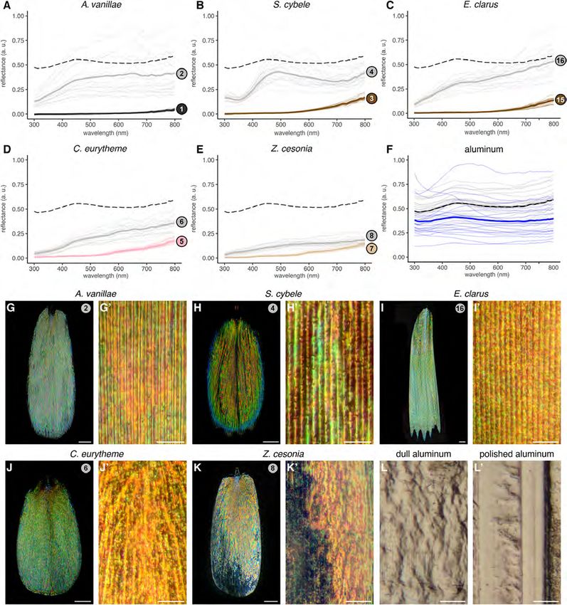
Ren et al.                                                                                                           Evolution of Butterfly Silver Scales

INTRODUCTION                                                                  of these scales, where spatial mixing results in an overall
                                                                              broadband reflectance (Vukusic et al., 2008; Stavenga et al., 2012;
Biological mirrors and tissues with metallic appearances have                 Wilts et al., 2013).
evolved in many forms across the tree of life (Land, 1972) –                      Metallic reflectances are widespread across the Lepidoptera,
from fish skin (McKenzie et al., 1995; Levy-Lior et al., 2008;                suggesting that the morphologies underlying them may be
Jordan et al., 2012), to scarab beetles (Agez et al., 2017),                  relatively “accessible” in the evolutionary sense (e.g., achievable
squid eyes (Holt et al., 2011; Ghoshal et al., 2013), and even                by simple changes to scale architecture). There are a number of
begonia leaves (Lee, 2009; Zhang et al., 2009). Rather than                   ways that such transitions to metallic coloration might occur.
relying on pigments per se, these mirror-like tissue reflectances             The current literature provides a limited view of the broader
result from microscopic optical structures that backscatter a                 evolutionary patterns of metallic coloration in butterflies. Prior
wide spectrum of continuous wavelengths. For instance, dermal                 studies indicate that silvery states may have evolved at least
layers of guanine crystals of random thickness and distribution               three times, in Hesperiidae (Ge et al., 2017), Nymphalidae
form a disordered stack termed a chaotic reflector and generate               (Vukusic et al., 2008), and Lycaenidae (Wilts et al., 2013),
the broadband reflectance that gives herring and sardines their               presumably via relatively simple departures from the traditional
silvery aspect (Denton and Land, 1971; Levy-Lior et al., 2008;                scale architecture to achieve the mechanism described above
Jordan et al., 2012). Metallic reflections are also common among              (Ghiradella, 2010). Those cases invariably display a highly
arthropods, including the elytra of many species of beetles and               reflective configuration under the form of ectopic lamination,
the pupal casing of some butterflies (Neville, 1977; Steinbrecht              i.e., an apparent covering of the micropores (sometimes called
et al., 1985; Berthier, 2007; Kinoshita, 2008; Biro and Vigneron,             windows or pepper-pots) that perforate the upper lamina of
2011; Agez et al., 2017). These examples of insect metallic                   less reflective scales. This kind of simplification of the upper
colors rely on broadband reflectance from multiple layers of                  lamina to a film-like sheet has been consistently observed in
chitin with differing thicknesses, often termed chirped stacks                scanning electron micrographs of silver-metallic scales, including
(Kinoshita and Yoshioka, 2005; Biro and Vigneron, 2011). As in                from nymphalid species of the Heliconiinae and Satyrinae sub-
the chaotic reflector, those chirped mirrors are often hundreds               families (Simonsen, 2007; Giraldo, 2008; Vukusic et al., 2008;
of microns thick because they rely on the stacking of bilayers                García-Barros and Meneguz, 2012; Dinwiddie et al., 2014), in
of dielectric materials with distinctive refractive indices (Deparis          the lycaenid Curetis acuta (Wilts et al., 2013; Liu et al., 2019),
et al., 2006; Cook and Amir, 2016; Chiadini et al., 2017), typically          and in the hesperid Carystoides escalantei (Ge et al., 2017). In
from air or cytoplasm (low-index 1 < nL < 1.33), and chitin                   the polymorphic species Argynnis niobe, scales from flat white
(high-index 1.53 < nH < 1.56), melanized chitin (high-index                   spots show more perforation of the upper lamina compared to
1.56 < nH < 1.8), or guanine platelets (1.46 < nH < 1.85)                     scales from individuals with brighter, recognizably silver spots
(Leertouwer et al., 2011; Jordan et al., 2012; Stavenga et al., 2015a).       (Simonsen, 2007). Thus, silver scales could be a more reflective
Reducing the thickness of a chirped mirror theoretically requires             elaboration of white scales, perhaps by reduction of the upper
increasing the number and refractive index of the dielectric                  lamina fenestration.
materials. In particular, the difference in refractive index between              Alternatively, broad-spectrum reflectance could evolve from
layers determines how much light is reflected at each interface.              increased disorder in iridescent scales that are already highly
The smaller this difference, the less light is reflected, implying            reflective, i.e., iridescent scale types with a more narrow color
that chirped stacks relying on natural materials can only achieve             profile. Referring to a classification of mechanisms of iridescence
reasonable reflectances using multilayers of 10 thin films or more            in butterflies (Ghiradella, 1989; Vukusic et al., 2000; Mouchet
(Land, 1972; Cook and Amir, 2016).                                            and Vukusic, 2018), silver scales could derive from “Type II”
    The wing scales of butterflies and moths (order: Lepidoptera)             body-lamellae scales, especially the Type IIa scales (or Urania-
are typically 1–2 µm thick. In contrast with thick chirped                    type) that are the most common mode of iridescence in
and chaotic reflectors, they have evolved ultrathin broadband                 Lycaenidae (Lippert and Gentil, 1959; Schmidt and Paulus, 1970;
reflectors only requiring sub-micron thickness (Vukusic et al.,               Tilley et al., 2002; Biró et al., 2007). Unlike silver scales, Type
2008; Stavenga et al., 2012; Wilts et al., 2013), often by the                IIa scales generate iridescent colors by internal multilayering
mixing of colors across a reflective surface rather than producing            (Ghiradella, 1989; Wilts et al., 2008; Yoshioka et al., 2013),
broadband reflectance at all points on the surface as in chirped              and show a level of reflectance that is linearly proportional
stacks. In the examples described to date, broad spectrum                     to the extent of upper lamina filling (Wilts et al., 2008).
reflectance of the wing scales results from the spatial mixing of             Whether this configuration can yield the spatial color mixing
parallel strips of colors that run alongside the periodic ridges              of heterogeneous colors and achieve broad reflectance spectra
of the reflective scale side (Vukusic et al., 2008), and derive               remains an open question. Finally, none of the previously
from thin-film interference between air and two chitin layers,                profiled silver scale architectures shows “Type I” ridge-lamellae
called the lower and upper laminae. The color heterogeneity                   scales emblematic of blue Morpho butterflies, or “Type III”
is produced by an undulatory thin-film interface, where the                   body-scattering structures that involve a porous upper lamina
sawtooth-shaped cross-sectional profile of the scale and the                  opening into crystal-like inclusions such as pigment granules
varying thicknesses of the air layer result in the reflection                 or gyroid structures (Vukusic et al., 2000; Dolan et al., 2015;
of a wide range of colors across a single scale. Such color                   Singer et al., 2016; Wilts et al., 2017; Mouchet and Vukusic,
variation disappears from the far field scattering appearance                 2018). We shall note, however, that there are no reported studies

Frontiers in Ecology and Evolution | www.frontiersin.org                  2                                         June 2020 | Volume 8 | Article 206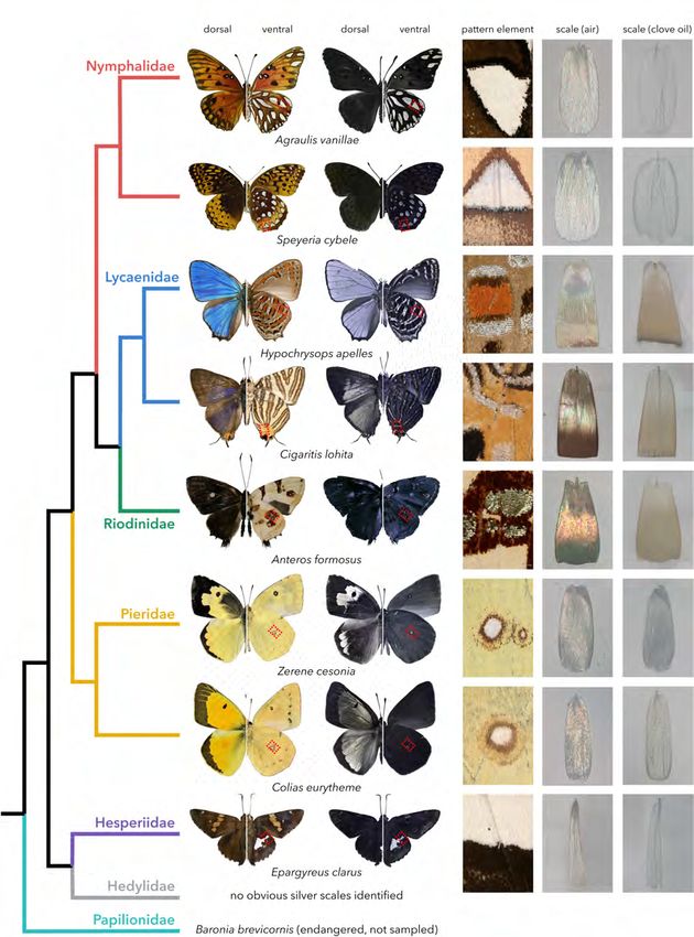
Ren et al.                                                                                                       Evolution of Butterfly Silver Scales

of metallic coloration and its underlying structural causes in            Metallic Scales Are Unpigmented,
either the Pieridae or the Riodinidae, which is surprising in the         Except in Lycaenidae and Riodinidae
latter case given the family’s vernacular name of “metalmarks.”
                                                                          We isolated single scales from metallic elements of the
The mechanisms of broadband reflectance in those clades thus
                                                                          eight sampled species for further observations, with all
remain unexplored.
                                                                          subsequent images and analyses focused on silver elements
    In this study we sought to expand our comparative knowledge
                                                                          from the hindwings (Figure 1C). In order to assess the
of the ultrastructures underlying specular (mirror-like), broad-
                                                                          relative contribution of pigmentation and structural thin-film
spectrum reflectance in butterflies, or in other words, to explore
                                                                          interference to silver scale coloration, we used reflected-light
the diversity of scales showing metallic coloration. Using a
                                                                          microscopy in air (refractive index n = 1) and transmitted-
combination of transmitted and reflected light microscopy,
                                                                          light microscopy under clove oil immersion (n = 1.53), a
electron microscopy, and microspectrophotometry, we describe
                                                                          medium that matches the refractive index of chitin and thus
the modalities of specular, broad-spectrum reflectance and
                                                                          substantially reduces light diffraction (Mayor, 1806; Stavenga
derive insights into the convergent evolution of metallic colors
                                                                          et al., 2015b; Thayer et al., 2020). When immersed in clove oil,
across butterflies.
                                                                          silver scales from species belonging to Nymphalidae, Pieridae,
                                                                          and Hesperiidae turned transparent, highlighting a lack of
                                                                          pigment (Figures 1D,E). In contrast, the metallic scales of the
RESULTS                                                                   three lycaenid and riodinid species showed a small amount of
                                                                          light-brown pigmentation, likely melanin, which increases the
Silver Coloration Exists in Five Out of                                   refractive index of chitin and may thus change the reflectance of
                                                                          the scale (Land, 1972).
Seven Butterfly Families
To gain insights into the diversity of structures underlying
                                                                          Metallic Scales Show Consistent
metallic broadband reflectance in the wing scales of
butterflies (superfamily Papilionoidea), we selected one or               Modifications of Their Upper Surfaces
two representatives from five of the seven families in this lineage       We examined the exterior features of silver scales in scanning
(Figure 1A): Nymphalidae (Agraulis vanillae and Speyeria                  electron micrographs, and systematically compared them to
cybele, sub-family: Heliconiinae), Lycaenidae (Hypochrysops               adjacent non-reflective scale types. As in previous studies
apelles, sub-family: Theclinae and Cigaritis lohita, sub-family:          (see section “Introduction”), metallic scales showed extensive
Aphnaeinae), Riodinidae (Anteros formosus, sub-family:                    lamination, where the spaces between crossribs are filled rather
Riodininae), Pieridae (Colias eurytheme and Zerene cesonia,               than left empty, as found in less reflective scales (Figure 2). With
sub-family: Coliadinae), and Hesperidae (Epargyreus clarus, sub-          the exception of the closely related Z. cesonia and C. eurytheme,
family: Eudaminae). We found no clear case of silver reflectance          all silver scales also showed an increase in inter-ridge distance
in the Hedylidae family, which includes only one extant genus.            (Figures 3A,C). Increasing the surface of light-reflecting upper
We also omitted Papilionidae, though it is noteworthy that                laminae could be of functional importance, particularly in the
certain morphs of the endangered species Baronia brevicornis              two species where inter-ridge spacings are the most differentiated:
brevicornis display silver patches with bright reflectance                silver scale ridge intervals are 1.5× larger than in adjacent scales
(Vazquez, 1987), and that broad-spectrum light-scattering scales          in A. vanillae (silver, mean = 2.29 µm; black, mean = 1.45 µm),
with a diffuse white appearance have been described in Graphium           and 2.4× larger in H. apelles (silver, mean = 2.73 µm; orange,
sarpedon (Stavenga et al., 2010, 2012).                                   mean = 1.16 µm). Finally, all silver scales were wider than
    While the gold/silver metallic color elements of our specimens        adjacent scales (Figures 3B,D), indicating that silver scales may
were always on the ventral side, this may be a trend rather than          be tuned to provide dense coverage on the wing and maximize
a rule, as illustrated by the nymphalid Argyrophorus argenteus            the surface area of light reflectance. Thus, compared to light-
that displays a completely silver dorsal side (Vukusic et al.,            absorbing, pigment-colored scales, metallic scales achieve higher
2008). Overall, this phylogenetic sampling may represent at               reflectance by maximizing the opportunity for backscattering of
least four cases of homoplasy, with at least one origin in each           incident light via three concomitant mechanisms: expansion of
of the nymphalid, lycaenid and riodinid, pierid, and hesperid             lamination/decrease in fenestration of upper wing scale surface,
lineages, or more independent acquisitions of the specular,               increase in ridge distance, and increase in overall scale width. In
broad-spectrum reflectance. In contrast, we can assume a likely           the next section, we investigate possible mechanisms underlying
homology between silver scales found in the ventral hindwing              the broad spectrum reflectance of these specialized scales.
silver blotches from “fritillary butterflies” of the Heliconiinae
sub-family (here, A. vanillae and S. cybele), or in the ventral           Undulatory Thin-Film Broadband
discal ocelli of Coliadinae (C. eurytheme and Z. cesonia); we             Reflectors Are Widespread
thus sampled two species in each lineage to get insights on the           In order to substantiate the broad reflectance of silver scales that
range of divergence within silver scales of shared origin and             can be inferred from direct observation and UV-photography,
context. The reflectance of all silver elements extended into the         we used a microspectrophotometer to compare the reflectance
ultraviolet range, as shown by UV-photography excluding visible           spectra of metallic scales vs. adjacent, non-metallic scales from
light wavelengths above 400 nm (Figure 1B).                               each species (Figures 4A–E). We focus on nymphalid, pierid, and

Frontiers in Ecology and Evolution | www.frontiersin.org              3                                          June 2020 | Volume 8 | Article 206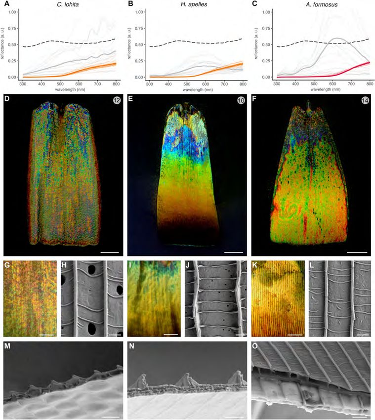
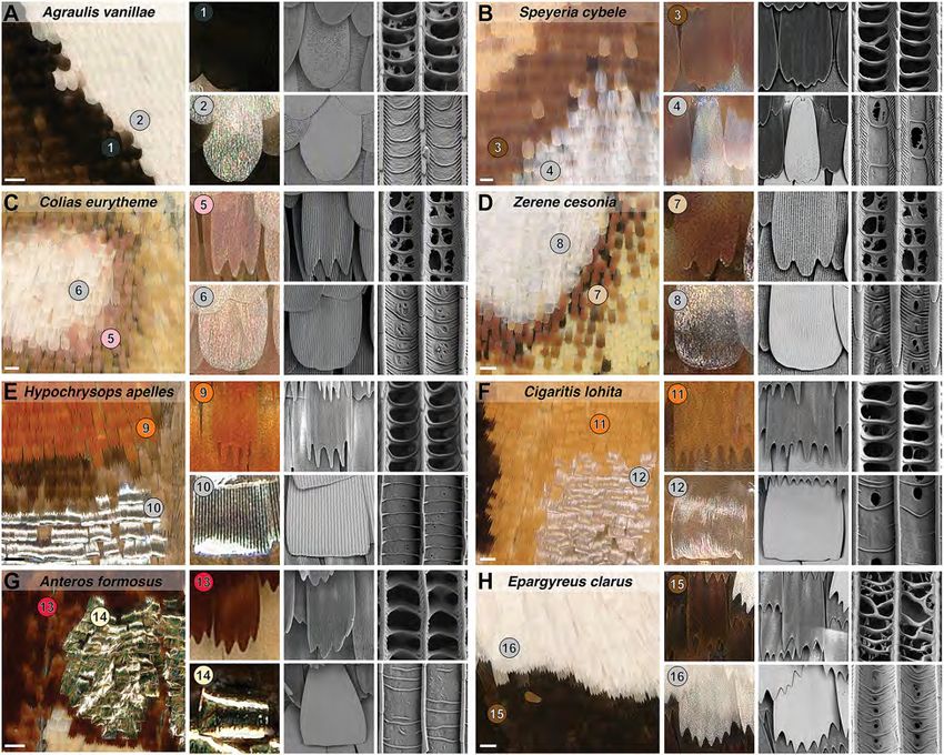
Ren et al.                                                                                                                               Evolution of Butterfly Silver Scales

  FIGURE 1 | Repeated occurrence of metallic scales in butterflies. (A) Phylogenetic relationships between the eight species sampled in this study (Espeland et al.,
  2018; Wiemers et al., 2019). (B) Ultraviolet (UV-B) photographs reveal reflectance in the non-visible range. (C) Magnified views of the reflective patterns obtained by
  a digital microscope, corresponding to red square insets in panels (A,B). (D,E) Reflected-light microscopy of single silver or gold scales in air (n = 1) and
  transmitted-light in clove oil (n = 1.53).

Frontiers in Ecology and Evolution | www.frontiersin.org                             4                                                  June 2020 | Volume 8 | Article 206
Ren et al. Evolution of Butterfly Silver Scales
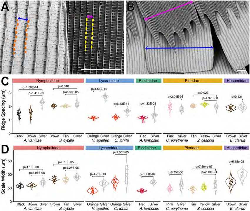
FIGURE 2 | Modified morphologies of metallic scales in five butterfly families. (A,B) Nymphalidae. (C,D) Pieridae. (E,F) Lycaenidae. (G) Riodinidae. (H) Hesperiidae.
Each panel shows a silver pattern imaged by digital light microscopy (left insets, scale bars = 100 µm), and close-up views of non-metallic (upper rows) vs. metallic
(lower rows) scales under light and electron microscopy. The right columns show the ultrastructural details of the upper lamina, highlighting the fenestration of
non-metallic scales vs. the smooth surface of reflective scales across two inter-ridge intervals. Numbered bullets mark scale types and species that are referred to in
further figures.
hesperid samples first, and will address the more unusual optical Next, we used reflected-light microscopy to test if those
morphologies observed in the lycaenid/riodinid clade samples in unpigmented silver scales would reproduce the pattern of
subsequent sections. spatial color mixing previously described in A. argenteus (sub-
The unpigmented silver scales of A. vanillae and S. cybele family: Satyrinae). In this species, color strips run parallel
(sub-family: Heliconiinae) produced a relatively flat reflectance to the scale ridges as the sawtooth profile of the upper
spectra across the UV and visible range (300–700 nm) and lamina imposes variation in the thickness of the subjacent air
extending into the infrared range (700–800 nm), consistent with lumen (Vukusic et al., 2008; Mouchet and Vukusic, 2018).
previous results (Briscoe et al., 2010). Hesperid (E. clarus) and This ultrathin configuration produces dense rods of colors,
pierid (C. eurytheme, Z. cesonia) unpigmented scales showed a each less than 1–3 µm thick, that alternate across the scale
similar broad reflectance. The discal spot scales of Z. cesonia width. We found that similar stripes can be discerned in
reflected less than half of the light reflected by laboratory-grade A. vanillae and S. cybele (Figures 4G,H), while in E. clarus,
aluminum foil (Figures 4E,F), which is used here as a familiar Z. cesonia, and C. eurytheme, individual colors are more speckled
point of comparison. Finally, all measurements of reflective and pointillist (Figures 4I–L). Thus, all five species from
surfaces showed more variation across measurements compared the Nymphalidae, Pieridae, and Hesperiidae achieve broad-
to adjacent pigmented scales, due to surface irregularities and the spectrum reflectance via dense spatial color mixing spread across
angle-dependency of light reflectance in the silver samples. the scale surface.
Frontiers in Ecology and Evolution | www.frontiersin.org 5 June 2020 | Volume 8 | Article 206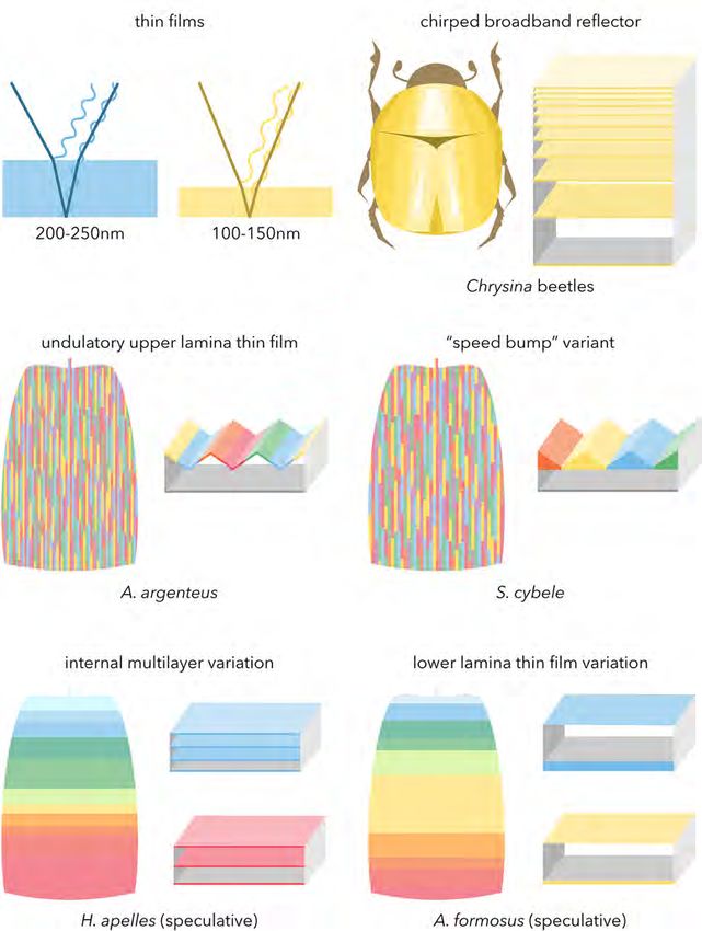
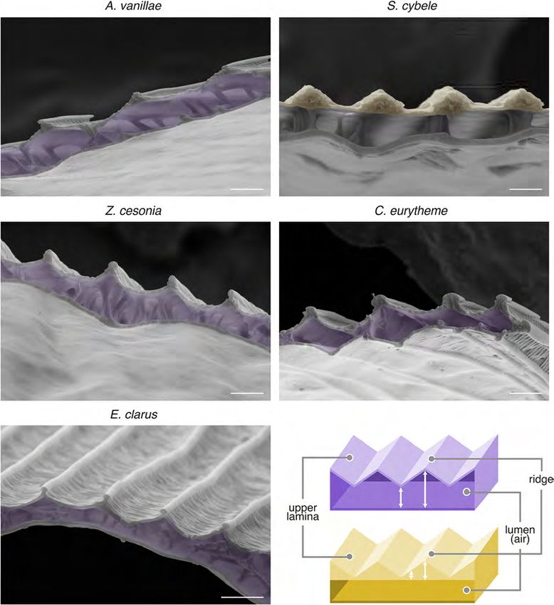
Ren et al.                                                                                                                              Evolution of Butterfly Silver Scales

  FIGURE 3 | Reflective scales tend to have larger ridge intervals and wider surfaces. (A) Measurement of ridge distance in the metallic (14) vs. orange scales (13) of
  H. apelles. (B) Measurement of maximal scale width in the metallic (14) vs. orange scales (13) of H. apelles. (C) Ridge distances across metallic vs. adjacent scale
  types from eight species (N = 25 scales per scale type). Each measurement corresponds to the Fourier mode, or average ridge interval per scale, extracted from the
  transversal pixel intensity across a single scale SEM. Pairwise differences show significant p-values following Mann-Whitney U tests. Pierid silver scales are the only
  metallic scales with smaller ridge intervals compared to adjacent scale types. (D) Maximal scale width distances across metallic vs. adjacent scale types from eight
  species (N = 25 scales per scale type). All metallic scales are enlarged compared to adjacent scales types (p-values, Mann-Whitney U tests). Scale bars,
  (A,B) = 10 µm. SEM image datasets are accessible on the Dryad online repository (Day et al., 2020).

   Based on those observations, we hypothesized that these                              (Figure 5H’), while in other nymphalids such as A. vanillae
metallic scales would resemble the bilayered internal anatomy                           (Figure 5G’) and A. argenteus (Vukusic et al., 2008), multiple
of A. argenteus scales, and examined the SEM profile of those                           color transitions can be observed within a single ridge interval
scale samples after transversal cryofracture (Figure 5). We found                       of comparable dimensions (Figure 3B). Thus, the S. cybele
that A. vanillae, E. clarus, C. eurytheme, and Z. cesonia share the                     mode of undulation is structurally distinct from other examples
serrated profile of A. argenteus, characterized by two laminae                          (Figure 5F), and achieves similar broadband reflectance albeit
of relatively constant thickness and an internal air layer of                           through a coarser mode of spatial color mixing.
varying thickness (“undulatory lumen”). S. cybele likewise has                              Nonetheless, the similarities between these morphologies
an undulating profile in its upper section, but with a flat                             suggest that the optical principles of an undulatory thin-
lumen and an upper lamina of varied thickness that resembles                            film reflector are at play across these butterfly lineages, with
“speed bumps,” due to local chitin thickening of the ridge area                         wavelength-specific variation in light reflectance across the scale
(Figure 5B). Of note, under near-field observation, S. cybele                           surface due to the periodic profiles of either the upper chitin
shows a single strip of color per ridge interval of 2–2.3 µm                            layer or the scale lumen. We suggest the previous biophysical

Frontiers in Ecology and Evolution | www.frontiersin.org                            6                                                   June 2020 | Volume 8 | Article 206
Ren et al.                                                                                                                                Evolution of Butterfly Silver Scales

  FIGURE 4 | Silver broadband reflectance resulting from densely intermixed colored reflections. (A–E) Reflectance spectra of silver pattern elements (gray lines),
  compared to adjacent arrays of non-reflective scales (black, brown, pink and beige lines). Faded lines represent individual measurements taken within the same wing
  surface. Dotted lines indicate the averaged spectra of polished aluminum foil for comparison. (F) Reflectance spectra of the polished side (gray) and dull side (blue) of
  aluminum foil. (G–K’) High-magnification reflected-light microscopy of single silver scales using a MPlanFLN 50× lens (G–K), and an LMPlan Achromatic 100× lens
  (G’–K’). (L,L’) High magnification reflected light microscopy of dull and polished sides of aluminum foil under the 100× lens. Scale bars: (G–K) = 20 µm;
  (G’–K’,L,L’) = 10 µm.

characterization of a butterfly undulatory thin-film reflector                           in Nymphalidae, Hesperiidae, and Pieridae. Such reflectors may
(Vukusic et al., 2008) applies here, and infer that this general                         have evolved repeatedly by tuning pre-existing scale elements of
mechanism accounts for multiple cases of metallic iridescence                            the scale groundplan (Ghiradella, 2010) to create a undulatory

Frontiers in Ecology and Evolution | www.frontiersin.org                             7                                                   June 2020 | Volume 8 | Article 206
Ren et al.                                                                                                                                  Evolution of Butterfly Silver Scales

  FIGURE 5 | Internal scale anatomy of nymphalid, pierid, and hesperid undulatory thin-film reflectors. (A–E) SEM of transversal sections of the metallic scales of
  A. vanillae, S. cybele, Z. cesonia, C. eurytheme, and E. clarus reveal the undulatory profile of the specular thin-film. False colors highlight the layer with the most
  consistent fluctuation in thickness, i.e., the air lumen (purple) in most cases, or for S. cybele, the thickened ridge regions (yellow). (F) Schematic representation of the
  two types of thin-film thickness undulation as observed in panels (A,C–E) (top, purple) and (B) (bottom, yellow). Scale bars: (A–E) = 1 µm.

thin-film system with a single inner air layer between the upper                           as well as in the specular scales of Curetis acuta, another lycaenid
and lower laminae.                                                                         (Wilts et al., 2013; Liu et al., 2019). In particular the reflectance
                                                                                           spectra, color speckling at microscopic levels, upper lamina
                                                                                           external views, and transversal inner anatomies all resembled the
Two Modes of Broadband Reflectance in                                                      hesperid and pierid samples (Figures 6A,D,G,H,M). Thus, the
Lycaenidae                                                                                 C. lohita reflector follows an undulatory thin-film architecture,
We also examined the features of silver scales from C. lohita                              providing yet another evolutionary occurrence of this category of
and H. apelles, two lycaenid species, and from the gold scales of                          mechanism in a fourth butterfly family.
A. formosus, a species from the riodinid sister lineage (Figure 6).                           In contrast, the silver scales of H. apelles showed a distinctive
All three types of metallic scales differed from other sub-families.                       spectrum and a strikingly divergent mode of color mixing, with
The silver scales of C. lohita showed optical and structural                               a continuous color transition, from blue to red, stemming from
features reminiscent of the undulatory thin-film configuration                             the base to the distal region of the scale (Figures 6B,E,I). This
found in Nymphalidae, Hesperiidae, and Pieridae in this study,                             longitudinal gradient represents a novel, previously undescribed

Frontiers in Ecology and Evolution | www.frontiersin.org                               8                                                    June 2020 | Volume 8 | Article 206
Ren et al. Evolution of Butterfly Silver Scales FIGURE 6 | Three distinct optical mechanisms drive broadband reflectance in the Lycaenidae + Riodinidae clade. (A–C) Averaged reflectance spectra of metallic pattern elements (gray lines), compared to an adjacent array of non-specular scales (orange and red lines). Faded lines represent individual measurements within the same wing surface. Dotted lines indicate the averaged spectra of polished aluminum foil for comparison. (D–G,I,K) High-magnification reflected-light microscopy of single metallic scales using a MPlanFLN 50× lens (D–F), and an LMPlan Achromatic 100× lens (G,I,K). (H,J,L) SEM top views of metallic scales. (M–O) SEM of transversal sections. In A. formosus (O), more proximal regions of the scale (dark gray, upper) have thicker lower laminae than distal regions (light gray, lower). Scale bars, (D–F) = 20 µm; (G,I,K) = 10 µm, (H,J,L,M–O) = 1 µm. mode of additive color mixing. Some of this color gradient wing surface, and thus likely to measure the apex and orange-red is hinted at in our spectral measurements, which exhibited a portion of the scale. pronounced peak in the long wavelengths due to the fact that the The external aspect of the H. apelles upper surface is also light paths of the MSP instrument are coaxial and normal to the peculiar, with large inter-ridge distances (Figure 3B) and periodic Frontiers in Ecology and Evolution | www.frontiersin.org 9 June 2020 | Volume 8 | Article 206
Ren et al.                                                                                                           Evolution of Butterfly Silver Scales

crossribs that are perfectly perpendicular to the ridges, indicative          a relatively simple reflector with a flat upper lamina and lumen,
of the complete flatness of the upper lamina (Figure 6J).                     and a variable lower lamina. In contrast with the nymphalid
The internal anatomy of cross-sectioned scales also revealed                  butterflies that use lower laminae to reflect homogenous color
a divergent morphology, lacking the marks of transversal                      spectra (Stavenga et al., 2014; Wasik et al., 2014; Thayer et al.,
periodicity observed in undulatory thin-film types. Instead, the              2020), we infer that local heterogeneities in those structures may
upper lamina and its underlying air layer are flat, and the ridges            underlie the spectral spread and spatial mixing of reflected colors
appear as thin vertical walls that are unlikely to provide sufficient         in the green-to-red range.
light diffraction for producing dense arrays of colors. Another
anatomical feature that varies in the longitudinal axis must thus
explain the proximo-distal color gradient. Interestingly, the lower           DISCUSSION
lamina shows a double-layering reminiscent of the Type-IIa
multilayered thin films observed in other iridescent lycaenids                Metallic Coloration in Lepidoptera
(Lippert and Gentil, 1959; Schmidt and Paulus, 1970; Tilley et al.,           We compared optical and ultrastructural structural aspects of
2002; Biró et al., 2007) and in sunset moths (Prum et al., 2006;              metallic scales in five out of seven families of Papilionoidea
Yoshioka and Kinoshita, 2007; Yoshioka et al., 2008). To suggest              (sensu Espeland et al., 2018), and identified several types of
an optical mixing for the observed color mixing, we sought to                 broadband reflectors that use distinct chitinous modifications to
examine if thickness and periodicity parameters of this system                achieve additive color mixing, including two modes of metallic
varied in the longitudinal direction. Unfortunately, the fragility            coloration that remain speculative before further biophysical
of those scales prevented us from properly cryo-fracturing those              characterization (Figure 7). While our phylogenetic sampling
scales and imaging them in the sagittal plane with SEM. Thus,                 is currently too coarse for ancestral state reconstruction or
further characterization of this newly discovered broadband                   detailed retracing of the evolution of metallic colorations
reflector will require alternative methods such as transmission               on the butterfly phylogeny, we are starting to uncover
electron microscopy (TEM), identification of the parameters that              general patterns of phenotypic convergence where sometimes
vary in the proximo-distal axis, and proper biophysical modeling.             similar, and sometimes novel ultrastructures achieve specular,
                                                                              broad-spectrum reflectance. Beyond butterflies, there is also
                                                                              a tremendous diversity of analogous metallic colorations in
A Simple Lower Lamina Thickness Likely                                        moths, including in Hepialidae (e.g., Korscheltellus lupulinus),
Drives Gold Reflectance in a Riodinid                                         Tortricidae (e.g., Pelochrista ridingsana), Crambidae (e.g., Ramila
We further examined the gold scales of a riodinid, A. formosus.               ruficostalis), Nolidae (e.g., the Mirror Moth Titulcia confictella),
Microspectrophotometry shows remarkable brightness in the                     Cosmopterigidae (e.g., Cosmopterix montisella), and Saturniidae
500–800 nm green-to-infrared range (Figure 6C), and a lower                   (e.g., Attacus atlas). In this section, we provide an overview
reflectance in the UV-to-blue range (< 500nm), consistent with                of the different types of broadband reflectors that have
the gold rather than silver sheen of this reflector. Reflected                evolved in Lepidoptera.
microscopy reveals longitudinal stripes of green, yellow, and red
colors, but unlike in the typical undulatory thin-films, we were              Undulatory Thin-Film Broadband
unable to resolve such transitions across distance of 1–3 µm with             Reflectors
a 100× lens (Figures 6F,K). The A. formosus gold scale upper                  The undulatory thin-film type described in A. argenteus is
lamina and inner air layer, as observed in top and cross-sectional            characterized by a continuous upper lamina, and an inner air
views (Figures 6L,O), also contradicts the role of an undulatory              layer of periodic thickness (Vukusic et al., 2008). This sawtoothed
thin-film as defined above. In the absence of any other obvious               thin-film results in a mode of spatial color mixing where reflected
optically relevant structures, the lower lamina of those scales is            wavelengths alternate across a 1–3 µm interval in the transversal
likely responsible for their bright reflectance. This is supported            axis. We recovered similar features in two other Nymphalidae,
by the fact that in comparison, the subjacent scale fractured at a            A. vanillae and S. cybele, with the nuance that the S. cybele
location ∼10 µm closer to the distal edge displays a significantly            thin-film showed dome shaped thickenings of the chitin layer
thinner lower lamina. Although this change in thickness appears               at the level of ridges, rather than a sawtooth shaped lumen as
to occur over a small proximo-distal distance, note that the                  observed in A. vanillae and our other samples (Figures 7C,D).
transition from blue-violet to yellow-green color is similarly                This reinforces the previously made inference that silver patterns
rapid (Figure 6O). Indeed, according to thin-film models that                 are a homoplasy within different “fritillary” butterflies of the
incorporate the refractive index of chitin, thicker laminae reflect           subfamily Heliconiinae (Simonsen, 2007), specifically between
shorter wavelengths (Stavenga et al., 2014). We thus expect the               tribe Argynnini such as S. cybele, and basal members of tribe
violet/blue base of the A. formosus scale to be thicker than the              Heliconiini such as A. vanillae. Second, Argynnini displays a
rest of the scale, which only shows green-yellow-red colors under             variety of reflective scales that vary from dull white to shiny
the reflected light microscope (Figures 6F,K), and the green-to-              silver and that are similar in external ultrastructures (Simonsen,
infrared shifts responsible for the spectrally broad gold likely arise        2007), but likely vary slightly in their transversal geometry.
from microvariations in lower lamina thickness that we could not              A similar architecture and mode of additive color mixing has
detect with SEM. Overall, these results suggest that the bright               been well-described in the noctuid moth Eudocima materna,
gold iridescence of the riodinid A. formosus scales derives from              where apposition of mirror scales with pigmented scales yields

Frontiers in Ecology and Evolution | www.frontiersin.org                 10                                          June 2020 | Volume 8 | Article 206
Ren et al. Evolution of Butterfly Silver Scales FIGURE 7 | Broadband reflectors underlying metallic iridescence in butterflies. Boxed schematics feature high-refractive index (filled) and low-refractive index (empty) materials. (A) Basic configuration of light reflection in simple chitinous thin-films. (B) Thick chirped stack reflector as observed in metallic beetles and butterfly pupae (after Neville, 1977). (C,D) Undulatory thin-films coupling a sawtoothed upper surface with an inner air layer, including the newly described variant from fritillary butterflies. Color array depicts the resulting rod-like mode of additive color mixing as observed under epi-illumination (Figures 4G–H’; see also Vukusic et al., 2008). (E,F) Speculative modes of proximo-distal additive color mixing as observed in a silvery lycaenid and golden riodinid (see main text for details). an angle-dependent reflective effect (Kelley et al., 2019). In this thin-films, notably from scales in a full white state where the framework, specularity may be achieved by efficient angular upper lamina is unperforated. If this holds true in Heliconiinae, reflections in the inter-ridge intervals and spatial color mixing, then this mode of convergence in silver iridescence may extend while more dull states could result from profiles that scatter to Satyrinae (Vukusic et al., 2008; García-Barros and Meneguz, more light. The evolution of undulatory broadband reflectors 2012), and other butterfly families as we documented here in may proceed by increases in the amplitude of light-scattering Hesperiidae, Pieridae, and one out of our two Lycaenidae samples Frontiers in Ecology and Evolution | www.frontiersin.org 11 June 2020 | Volume 8 | Article 206
Ren et al.                                                                                                        Evolution of Butterfly Silver Scales

(C. lohita). In any case, the hollow lepidopteran bauplan scale            showed an original configuration. These were the brightest
type (Ghiradella, 2010) that is necessary to yield the undulatory          among all the species sampled here, but their ultrastructure
thin-film seemingly extends to some early diverging branches of            simply consisted of a flat, mostly featureless upper lamina, a flat
the lepidopteran phylogeny (Simonsen, 2001). We predict that               air lumen, and a similarly flat lower lamina. We propose that
the most distant case of convergent undulatory thin-film may               a rapid and pronounced reduction in lower lamina thickness
have occurred in silver-reflective Hepialidae such as the silver-          is responsible for the abrupt shift from blue-violet to yellow-
spotted ghost moth (Leto venus), since reflective white scales             green in the most proximal third of the scale. This thick (200–
in this family show a bilayered architecture reminiscent of the            250 nm) region appearing blue is consistent with models and
butterflies and skippers studied here (Simonsen, 2002).                    existing examples of thin-films (Stavenga et al., 2014; Thayer
                                                                           et al., 2020), and likewise a drop to 100–150 nm produces
Proximo-Distal Color Mixing in the                                         red and yellow (Thayer et al., 2020). We extrapolate then that
                                                                           microvariations in lower lamina thickness are likely responsible
Multilayered Scales of Lycaenids
                                                                           for the reds, yellows, and greens in the majority of the scale
We discovered a new mode of broad-reflectance in H. apelles
                                                                           (Figure 7F), but we were unable to generate interpretable sagittal
where the visible color spectrum is selectively reflected across
                                                                           sections by cryofracture due to scale fragility. Most intriguing,
the proximo-distal axis of the scale, from violet and blue colors
                                                                           it remains unclear how A. formosus achieved brightness levels
at the base, to red at the tip (Figures 6E,I). This rainbow
                                                                           that exceeded the reflectance of our specular reflectance standard
transition in the near field, likely coupled to scale stacking,
                                                                           (measurements > 1.00 in Figure 6C), a level of gold iridescence
produces broadband reflectance in the far field. What can
                                                                           that is only achieved in other insects with much thicker stacks
we infer about the ultrastructural basis of this proximodistal
                                                                           of chitin (Steinbrecht et al., 1985; Seago et al., 2009; Biro and
color mixing? In H. apelles, the multilayered lower lamina
                                                                           Vigneron, 2011). While we have described here the “metalmark”
(Figure 6N) is reminiscent of body-lamellae scale iridescence
                                                                           (riodinid) system superficially, a better understanding of how
that are widespread in Lycaenidae (Lippert and Gentil, 1959;
                                                                           such seemingly flat, simple, and ultrathin sandwich of chitin
Schmidt and Paulus, 1970; Tilley et al., 2002; Biró et al., 2007;
                                                                           and air can yield an intensely specular output will be of
Wilts et al., 2008). In those butterflies, additional chitin layers
                                                                           particular interest.
increase reflectance, meaning that the multilayered lower lamina
contributes to the brightness of the scales (Wilts et al., 2008).
                                                                           Broadband Microrib Gratings
Sunset moths also show analogously multilayered structures, and
                                                                           A fourth type of metallic reflectance mechanism has been
changes in the thickness and spacing of their internal layers on
                                                                           described in the literature, but was not observed in this study.
the order of 10–50 nm are responsible for shifts as drastic as
                                                                           We dub “microrib grating” a type of 2D diffraction grating
pale blue to orange (Yoshioka and Kinoshita, 2007; Yoshioka
                                                                           made of dense herringbone crossrib arrays, and where upper
et al., 2008, 2013; Imafuku et al., 2012). In addition, melanin
                                                                           and lower laminae are apposed (“fused scales”), forming a single
is present in the distal portion of the scale and absent at the
                                                                           thin film without a lumen. It has been linked to silver/gold
base, perhaps increasing chitin refractive index or even forming
                                                                           iridescence in the skipper butterfly Carystoides escalantei (Ge
a gradient that could contribute to spectral spread (Land, 1972).
                                                                           et al., 2017). Outside of the butterfly/skipper lineage, it has been
While we could not resolve here the mechanisms responsible
                                                                           best characterized in the Micropterigidae Micropterix calthella
for the continuous color transition from blue to red hues, we
                                                                           (Kilchoer et al., 2019), where the fused lamina acts as a
extrapolate that microvariations in layering and refractivity drive
                                                                           bronze/gold thin-film reflector whose specularity is enhanced
this shift along single scales (Figure 7E).
                                                                           by the diffractive scattering of the overlaying microribs (D’Alba
   From an evolutionary perspective, one may postulate that the
                                                                           et al., 2019). Other likely examples include the Noctuidae
multilayered state of lycaenid and other body-lamellae (“Type
                                                                           moths Diachrysia (Plusia) balluca and Trichoplusia orichalcea
IIa”) color iridescent scales (Ghiradella, 1989; Vukusic et al.,
                                                                           (Ghiradella, 1991; Brink et al., 1995). Scales from Adela
2000; Mouchet and Vukusic, 2018) can prime the evolution of
                                                                           reaumurella (Adelidae) and Stigmella malella (Nepticulidae)
broadband reflectance. In Hypochrysops spp., the iridescent blue
                                                                           show fused laminae and herringbone microribs that are
scales widespread across the dorsal surface are composed of a
                                                                           consistent with the metallic sheens of these moths (van Eldijk
lower lamina of 7–8 layers and a perforated upper lamina (Ingram
                                                                           et al., 2018). The white reflective scales of Paysandisia archon
and Parker, 2008) while we have observed three layers in the
                                                                           (Castniidae) display this arrangement (Stavenga et al., 2018).
ventral silver scales. Transition to the silver state would then
                                                                           In Papilionidae, the reflective component of the glass scales of
require a filling of the perforations in the upper lamina, and
                                                                           G. sarpedon is also due to a microrib grating, with membranal
merging of internal layers to produce a variety of chitin layer
                                                                           bilin pigments restricting the reflectance spectrum to a blue-
thicknesses and spacings along the proximo-distal axis.
                                                                           green hue (Stavenga et al., 2010, 2012). In the basal Papilionidae
                                                                           Baronia brevicornis, an electron micrograph also suggests a
Tuning of Lower Lamina in the Gold                                         microrib grating arrangement that is indicative of a reflective
Scales of a Riodinid                                                       type (Simonsen et al., 2011), but it is unclear if the image was
The optical and structural basis of color mixing in the specular           taken from a silver morph of this polymorphic species (Vazquez,
scales of metalmarks butterflies (Riodinidae) has remained                 1987). Herringbone patterns and fused laminae are characteristic
undescribed, and our data from the gold scales of A. formosus              of early diverging lepidopteran lineages (Kristensen, 1970;

Frontiers in Ecology and Evolution | www.frontiersin.org              12                                          June 2020 | Volume 8 | Article 206
Ren et al. Evolution of Butterfly Silver Scales
Simonsen, 2001; Deparis et al., 2006) and in accordance with lens at 50× magnification and a VH-Z100T lens at the
these authors, we extrapolate that the microrib diffraction grating 300× magnification. UV-photography was performed under
could be a sophistication of the lepidopteran scale ancestral state, the illumination of GE BlackLight 13-Watt T3 Spiral Light
but it is also noteworthy that a convergent scale morphology and Bulbs, using a full-spectrum converted Panasonic G3 camera,
microrib grating mechanism have been linked to gold iridescence mounted with a Kyoei-Kuribayashi 35mm F3.5 lens on a
in Collembola, suggesting the optical trick may extend across helicoid focusing adapter, and stacked Hoya U-330 and Schott
many hexapod lineages that also bear cuticular scales (D’Alba BG39 1.5 mm glass filters eliminating the visible and infrared
et al., 2019). In any case, we gather from the literature that wavelengths above 400 nm.
diffraction gratings consisting of dense microribs and apposed
laminae are likely common and ancient enhancers of metallic
coloration in Lepidoptera. Single-Scale Light Microscopy
Individual scales were removed from spread butterflies and
positioned on a glass slide with an eyelash tool, and stitch-
CONCLUSION imaged with a Keyence VHX-5000 microscope in reflected light
mode and a VH-Z100R lens at 1000× magnification. All scales
Metallic appearances require spectral spread that result were imaged in full coaxial lighting except for H. apelles, which
from increased reflectance variance in the geometry of the required additional ring lighting for best balance of resolution
scale thin-films. The literature has pinpointed 2D diffraction and color features. The same scales were then immersed in
gratings, characterized by dense herringbone microribs and clove oil, mounted with a coverslip, and imaged in transmission
apposed lower laminae, as a common theme for metallic mode. For reflective microscopy, single scales were imaged
reflections. Our study indicates that in butterflies and beyond, with an Olympus BX53M microscope in Reflected Bright-Field
a bright output requires an unperforated upper lamina, mode, mounted with an Olympus MPlanFLN 50×/NA 0.80
and that broadband reflectance is also often reached by objective and an SC50 color camera, before focus-stacking
transversal undulations of the upper chitinous section and with the Olympus Stream software, and XY-stitching in Adobe
air lumen (Figures 7D–G). This configuration may be a Photoshop. For reflective microscopy at higher-magnifications,
simple derivation from traditional scale types including the same scales were imaged on a trinocular AmScope ME580-2L
depigmented, white light-scattering scales. Proximo-distal metallurgical microscope mounted with a Nikon D5300 camera,
thickness gradients in the lower section of the scale (simple a Varimag II camera adapter at 3.5× magnification setting, and
or multilayered lower laminas) appear as another mode of an LMPlan Achromatic 100×/NA 0.8 long working distance
broadband reflectance as alluded in Lycaenidae and Riodinidae, objective. Optimal balance of resolution and color features
a phenomenon that will require further comparative studies and were obtained under polarized light with a field diaphragm at
biophysical characterization. maximal shutting position, and an aperture diaphragm on 60–
80% shut position.
MATERIALS AND METHODS
Spectral Measurements
Butterflies Reflectance spectra were measured from a 250 µm2 region of the
A. vanillae larvae were obtained from Shady Oak Farms surface of intact wing sections, and thus represent the combined
(Florida, United States) and reared on Passiflora suberosa reflectance of cover scales, underlying ground scales, and wing
or Passiflora incarnata until adult emergence. Specimens of membrane (Stavenga et al., 2014). Reflectance measurements
S. cybele, C. eurytheme, Z. cesonia, and E. clarus were were taken using a custom-built microspectrophotometer
collected from wild populations in the vicinity of Silver (20/20PV, CRAIC Technologies, Inc., San Dimas, CA,
Spring (Maryland, United States), Mason Neck (Virginia, United States) equipped with a 5× UV-vis objective (LMU-
United States), Starkville (Mississippi, United States), and 5X-NUV, Thorlabs, Inc., Newton, NJ, United States). Samples
Washington (District of Columbia, United States), respectively. were illuminated with a xenon arc lamp (XBO 75 W/2, OSRAM
Specimens of H. apelles (orig. Papua – New Guinea), C. GmbH, Munich, Germany), with the light path oriented
(Spindasis) lohita (orig. Bali, Indonesia), and A. formosus (orig. normal to the wing surface and coaxial with the axis of light
Peru) were imported from online retailers with appropriate collection. Reflectance spectra were calculated in relation to a
collection and transit permits. high-reflectivity specular reflectance standard (STAN-SSH-NIST,
Ocean Optics, Inc., Dunedin, FL, United States). For each
Color and UV Macro-Photography scale type, individual spectra were the average of 25 spectra
Pinned specimens were imaged in the visible range using a measured consecutively (integration time: 200–350 ms, total
Nikon D5300 camera mounted with a Micro-Nikkor 105mm measurement time per average spectra: 5–8.75 s). In order to
f/2.8G lens on a StackShot rail and focused-stacked using provide a familiar point of comparison, measurements were also
the Helicon Remote and Helicon Focus software. A Keyence taken on the polished and dull side of aluminum foil (0.018 mm,
VHX-5000 digital microscope was used to generate stitched Fisherbrand), and resulted in spectra consistent with other
high-resolution images of wing patterns using a VH-Z00T studies (Vukusic et al., 2008; Pozzobon et al., 2020).
Frontiers in Ecology and Evolution | www.frontiersin.org 13 June 2020 | Volume 8 | Article 206Ren et al. Evolution of Butterfly Silver Scales
Scanning Electron Microscopy (SEM) species, using a custom semi-automated R pipeline that derives
For surface imaging of scales of distinct identities, wing patterns ultrastructural parameters from large SEM images (Day et al.,
of interest were excised and mounted on SEM stubs with double- 2019). Briefly, ridge spacing was assessed by Fourier transforming
sided carbon tape, and color imaged under the Keyence VHX- intensity traces of the ridges acquired from the FIJI software
5000 microscope for registration of scale type. Samples were (Schindelin et al., 2012). Scale width was directly measured in FIJI
sputter-coated with two 12.5 nm layers of gold for improving by manually tracing a line, orthogonal to the ridges, at the section
sample conductivity, with the second layer applied after tilting of maximal width.
the stub by 45◦ . SEM images were acquired on a FEI Teneo LV
SEM, using secondary electrons (SE) and an Everhart-Thornley
detector (ETD) using a beam energy of 2.00 kV, beam current of DATA AVAILABILITY STATEMENT
25 pA, and a 10 µs dwell time. Individual images were stitched
using the Maps 3.10 software (Thermo Fisher Scientific). The datasets generated for this study are available on request to
To minimize charging for high magnification views of scale the corresponding author.
surface morphology, individual scales were collected by brushing
the surface of the wing with an eyelash tool, then dusted onto
an SEM stub with double-sided carbon tape. Stubs were sputter- AUTHOR CONTRIBUTIONS
coated with one 12.5 nm layer of gold, and imaged at 2.00 kV/25
AR and CD equally performed all the imaging and experiments,
pA with a 10 µs dwell time. One sample per species was imaged,
and contributed to the figures and data analysis. JH, BC, and
and all the SEM images used for morphometric analysis are
NM contributed to the data analysis and intellectual support. AM
accessible on the Dryad online repository (Day et al., 2020).
supervised the project and wrote the manuscript with input from
For documenting internal scale anatomy, silver pattern
all co-authors. All authors contributed to the article and approved
elements were excised and cryo-fractured following previous
the submitted version.
recommendations (Wasik et al., 2014; Matsuoka and Monteiro,
2018; Thayer et al., 2020). Briefly, wing sections were submerged
in liquid nitrogen, immediately placed silver-side down onto a
FUNDING
silicon wafer, and cut with a fresh ceramic-coated microtome
blade. Alternatively, excised wing sections were placed silver-side This research was funded by the National Science Foundation
down onto a silicon wafer and secured with foam board, glassine, award IOS-1755329 to AM and BC, and a Masters student
and a binder clip before submersion in liquid nitrogen and fellowship allocated to AR by the GWU Minor in Sustainability.
cutting as previously described. After allowing to dry, individual
cut scales were placed using an eyelash tool on copper tape, such
that the cut edges were approximately parallel to and overhanging ACKNOWLEDGMENTS
the tape edge. The copper tape was bent to 90◦ and placed on a
stub so that the scales’ cut edges faced upwards, i.e., normal to the We thank Huimin Chen for advice and support with image
stub surface, and secured with additional copper tape. The stubs analysis, Rod Eastwood for assisting with butterfly identification,
were sputter-coated with a 10–12.5 nm layer of gold, and imaged and Christine Brantner and Anastas Popratiloff for their technical
at 5.00 kV/6.3 pA and a 10 µs dwell time. assistance at the GW Nanofabrication and Imaging Center.
Additionally we thank Amruta Tendolkar for providing eyelashes
Morphometric Analysis for manipulations of single scales, Rachel Thayer and Marshall
Morphometric measurements of scale widths and ridge distances Nakatani for insights on cryo-fracturing techniques, and John Lill
were carried out on 25 scales of each color from eight for providing the S. cybele specimen from his personal collection.
REFERENCES visual pigment coincides with wing pigment evolution in heliconius butterflies.
Proc. Natl. Acad. Sci. U.S.A. 107, 3628–3633. doi: 10.1073/pnas.09100
Agez, G., Bayon, C., and Mitov, M. (2017). Multiwavelength micromirrors in the 85107
cuticle of scarab beetle Chrysina gloriosa. Acta Biomater. 48, 357–367. doi: Chiadini, F., Fiumara, V., and Scaglione, A. (2017). Design of bioinspired
10.1016/j.actbio.2016.11.033 chirped reflectors using a genetic algorithm. Bioinspirat. Biomimet. Bioreplicat.
Berthier, S. (2007). Iridescences: The Physical Colors Of Insects. Berlin: Springer. 2017:101620T.
Biró, L. P., Kertész, K., Vértesy, Z., Márk, G. I., Bálint, Z., Lousse, V., et al. (2007). Cook, C. Q., and Amir, A. (2016). Theory of chirped photonic crystals in biological
Living photonic crystals: butterfly scales—nanostructure and optical properties. broadband reflectors. Optica 3, 1436–1439.
Mater. Sci. Eng. C 27, 941–946. doi: 10.1016/j.msec.2006.09.043 D’Alba, L., Wang, B., Vanthournout, B., and Shawkey, M. D. (2019). The golden
Biro, L. P., and Vigneron, J.-P. (2011). Photonic nanoarchitectures in butterflies age of arthropods: ancient mechanisms of colour production in body scales.
and beetles: valuable sources for bioinspiration. Laser Photon. Rev. 5, 27–51. J. R. Soc. Interf. 16:20190366.
doi: 10.1002/lpor.200900018 Day, C. R., Hanly, J. J., Ren, A., and Martin, A. (2019). Sub-micrometer insights into
Brink, D. J., Smit, J. E., Lee, M. E., and Möller, A. (1995). Optical diffraction by the the cytoskeletal dynamics and ultrastructural diversity of butterfly wing scales.
microstructure of the wing of a moth. Appl. Opt. 34, 6049–6057. Dev. Dyn. 248, 657–670. doi: 10.1002/dvdy.63
Briscoe, A. D., Bybee, S. M., Bernard, G. D., Yuan, F., Sison-Mangus, M. P., Day, C. R., Hanly, J. J., Ren, A., and Martin, A. (2020). Data from:
Reed, R. D., et al. (2010). Positive selection of a duplicated UV-sensitive Convergent Evolution of Broadband Reflectors Underlies Metallic Colorations
Frontiers in Ecology and Evolution | www.frontiersin.org 14 June 2020 | Volume 8 | Article 206Ren et al. Evolution of Butterfly Silver Scales in Butterflies, v3, Dryad, Dataset. Available online at: https://doi.org/10.5061/ Lee, D. W. (2009). “Plant tissue optics: micro-and nanostructures,” in Proceedings dryad.2fqz612mc of the Biomimetics and Bioinspiration, San Diego, CA. Denton, E. J., and Land, M. F. (1971). Mechanism of reflexion in silvery layers Leertouwer, H. L., Wilts, B. D., and Stavenga, D. G. (2011). Refractive index of fish and cephalopods. Proc. R. Soc. Lond. Ser. B Biol. Sci. 178, 43–61. doi: and dispersion of butterfly chitin and bird keratin measured by polarizing 10.1098/rspb.1971.0051 interference microscopy. Opt. Exp. 19, 24061–24066. Deparis, O., Vandenbem, C., Rassart, M., Welch, V. L., and Vigneron, J.-P. Levy-Lior, A., Pokroy, B., Levavi-Sivan, B., Leiserowitz, L., Weiner, S., and Addadi, (2006). Color-selecting reflectors inspired from biological periodic multilayer L. (2008). Biogenic guanine crystals from the skin of fish may be designed structures. Optics Exp. 14, 3547–3555. to enhance light reflectance. Cryst. Growth Design 8, 507–511. doi: 10.1021/ Dinwiddie, A., Null, R., Pizzano, M., Chuong, L., Krup, A. L., Tan, H. E., et al. cg0704753 (2014). Dynamics of F-actin prefigure the structure of butterfly wing scales. Dev. Lippert, W., and Gentil, K. (1959). Über lamellare feinstrukturen bei Biol. 392, 404–418. doi: 10.1016/j.ydbio.2014.06.005 den schillerschuppen der schmetterlinge vom urania-und morpho- Dolan, J. A., Wilts, B. D., Vignolini, S., Baumberg, J. J., Steiner, U., and Wilkinson, typ. Zeitschrift Morphol. Ökol. Tiere 48, 115–122. doi: 10.1007/bf0040 T. D. (2015). Optical properties of gyroid structured materials: from photonic 7836 crystals to metamaterials. Adv. Opt. Mater. 3, 12–32. doi: 10.1002/adom. Liu, X., Wang, D., Yang, Z., Zhou, H., Zhao, Q., and Fan, T. (2019). Bright silver 201400333 brilliancy from irregular microstructures in butterfly Curetis acuta moore. Adv. Espeland, M., Breinholt, J., Willmott, K. R., Warren, A. D., Vila, R., Toussaint, E. F., Opt. Mater. 7:1900687. doi: 10.1002/adom.201900687 et al. (2018). A comprehensive and dated phylogenomic analysis of butterflies. Matsuoka, Y., and Monteiro, A. (2018). Melanin pathway genes regulate color and Curr. Biol. 28, 770–778. morphology of butterfly wing scales. Cell Rep. 24, 56–65. doi: 10.1016/j.celrep. García-Barros, E., and Meneguz, M. (2012). Estructura de las escamas ventrales de 2018.05.092 las alas de Coenonympha Hübner,[1819], con especial referencia a C. pamphilus Mayor, A. G. (1806). The Development of the Wing Scales and Their Pigment in (L., 1758) y su morfotipo lyllus (Esper, 1805)(Lepidoptera: Nymphalidae). Butterflies and Moths. Cambridge, MA: Harvard College. SHILAP Rev. Lepidopterol. 40, 279–293. McKenzie, D. R., Yin, Y., and McFall, W. D. (1995). Silvery fish skin as an example Ge, D., Wu, G., Yang, L., Kim, H.-N., Hallwachs, W., Burns, J. M., et al. (2017). of a chaotic reflector. Proc. R. Soc. Lond. Ser. A Math. Phys. Sci. 451, 579–584. Varying and unchanging whiteness on the wings of dusk-active and shade- doi: 10.1098/rspa.1995.0144 inhabiting Carystoides escalantei butterflies. Proc. Natl. Acad. Sci. U.S.A. 114, Mouchet, S. R., and Vukusic, P. (2018). Structural colours in lepidopteran scales. 7379–7384. doi: 10.1073/pnas.1701017114 Adv. Insect Physiol. 54, 1–53. doi: 10.1016/bs.aiip.2017.11.002 Ghiradella, H. (1989). Structure and development of iridescent butterfly scales: Neville, A. C. (1977). Metallic gold and silver colours in some insect cuticles. lattices and laminae. J. Morphol. 202, 69–88. doi: 10.1002/jmor.1052020106 J. Insect Physiol. 23, 1267–1274. doi: 10.1016/0022-1910(77)90069-5 Ghiradella, H. (1991). Light and color on the wing: structural colors in butterflies Pozzobon, V., Levasseur, W., Do, K.-V., Palpant, B., and Perré, P. (2020). and moths. Appl. Opt. 30, 3492–3500. Household aluminum foil matte and bright side reflectivity measurements: Ghiradella, H. (2010). Insect cuticular surface modifications: scales and other application to a photobioreactor light concentrator design. Biotechnol. Rep. structural formations. Adv. Insect Physiol. 38, 135–180. doi: 10.1016/s0065- 25:e00399. doi: 10.1016/j.btre.2019.e00399 2806(10)38006-4 Prum, R. O., Quinn, T., and Torres, R. H. (2006). Anatomically diverse butterfly Ghoshal, A., DeMartini, D. G., Eck, E., and Morse, D. E. (2013). Optical parameters scales all produce structural colours by coherent scattering. J. Exp. Biol. 209, of the tunable Bragg reflectors in squid. J. R. Soc. Interf. 10:20130386. doi: 748–765. doi: 10.1242/jeb.02051 10.1098/rsif.2013.0386 Schindelin, J., Arganda-Carreras, I., Frise, E., Kaynig, V., Longair, M., Pietzsch, T., Giraldo, M. A. (2008). Butterfly Wing Scales: Pigmentation and Structural et al. (2012). Fiji: an open-source platform for biological-image analysis. Nat. Properties. Thesis, University of Groningen. doi: 10.1098/rsif.2013.0386 Methods 9:676. doi: 10.1038/nmeth.2019 Holt, A. L., Sweeney, A. M., Johnsen, S., and Morse, D. E. (2011). A highly Schmidt, K., and Paulus, H. (1970). Die feinstruktur der flüigelschuppen einiger distributed Bragg stack with unique geometry provides effective camouflage for Lycaeniden (Insecta, Lepidoptera). Zeitschrift Morphol. Tiere 66, 224–241. doi: Loliginid squid eyes. J. R. Soc. Interf. 8, 1386–1399. doi: 10.1098/rsif.2010.0702 10.1007/bf00280735 Imafuku, M., Kubota, H. Y., and Inouye, K. (2012). Wing colors based on Seago, A. E., Brady, P., Vigneron, J.-P., and Schultz, T. D. (2009). Gold bugs and arrangement of the multilayer structure of wing scales in lycaenid butterflies beyond: a review of iridescence and structural colour mechanisms in beetles (Insecta: Lepidoptera). Entomol. Sci. 15, 400–407. doi: 10.1111/j.1479-8298. (Coleoptera). J. R. Soc. Interf. 6, S165–S184. 2012.00525.x Simonsen, T. J. (2001). The wing vestiture of the non-ditrysian Lepidoptera Ingram, A. L., and Parker, A. R. (2008). A review of the diversity and evolution of (Insecta). Comparative morphology and phylogenetic implications. Acta Zool. photonic structures in butterflies, incorporating the work of John Huxley (The 82, 275–298. doi: 10.1046/j.1463-6395.2001.00089.x Natural History Museum, London from 1961 to 1990). Philos. Trans. R. Soc. B Simonsen, T. J. (2002). Wing scale covering supports close relationship between Biol. Sci. 363, 2465–2480. doi: 10.1098/rstb.2007.2258 Callipielus and Dalaca, austral South American ghost moths (Lepidoptera: Jordan, T. M., Partridge, J. C., and Roberts, N. W. (2012). Non-polarizing Hepialidae). Stud. Neotrop. Fauna Environ. 37, 65–69. doi: 10.1076/snfe.37.1. broadband multilayer reflectors in fish. Nat. Photon. 6:759. doi: 10.1038/ 65.2117 nphoton.2012.260 Simonsen, T. J. (2007). Comparative morphology and evolutionary aspects of Kelley, J. L., Tatarnic, N. J., Schröder-Turk, G. E., Endler, J. A., and Wilts, B. D. the reflective under wing scale-pattern in Fritillary butterflies (Nymphalidae: (2019). A dynamic optical signal in a nocturnal moth. Curr. Biol. 29, 2919– Argynnini). Zool. Anzeiger A J. Compar. Zool. 246, 1–10. doi: 10.1016/j.jcz. 2925. 2005.04.005 Kilchoer, C., Steiner, U., and Wilts, B. D. (2019). Thin-film structural coloration Simonsen, T. J., Zakharov, E. V., Djernaes, M., Cotton, A. M., Vane-Wright, R. I., from simple fused scales in moths. J. R. Soc. Interf. Focus 9:20180044. doi: and Sperling, F. A. (2011). Phylogenetics and divergence times of Papilioninae 10.1098/rsfs.2018.0044 (Lepidoptera) with special reference to the enigmatic genera Teinopalpus Kinoshita, S. (2008). Structural Colors In The Realm Of Nature. Singapore: World and Meandrusa. Cladistics 27, 113–137. doi: 10.1111/j.1096-0031.2010. Scientific. 00326.x Kinoshita, S., and Yoshioka, S. (2005). Structural colors in nature: the role of Singer, A., Boucheron, L., Dietze, S. H., Jensen, K. E., Vine, D., McNulty, regularity and irregularity in the structure. Chemphyschem 6, 1442–1459. doi: I., et al. (2016). Domain morphology, boundaries, and topological defects 10.1002/cphc.200500007 in biophotonic gyroid nanostructures of butterfly wing scales. Sci. Adv. Kristensen, N. P. (1970). Morphological observations on the wing scales in some 2:e1600149. doi: 10.1126/sciadv.1600149 primitive Lepidoptera (Insecta). J. Ultrastruct. Res. 30, 402–410. doi: 10.1016/ Stavenga, D. G., Giraldo, M. A., and Leertouwer, H. L. (2010). Butterfly wing s0022-5320(70)80071-5 colors: glass scales of Graphium sarpedon cause polarized iridescence and Land, M. F. (1972). The physics and biology of animal reflectors. Prog. Biophys. enhance blue/green pigment coloration of the wing membrane. J. Exp. Biol. 213, Mol. Biol. 24, 75–106. doi: 10.1016/0079-6107(72)90004-1 1731–1739. doi: 10.1242/jeb.041434 Frontiers in Ecology and Evolution | www.frontiersin.org 15 June 2020 | Volume 8 | Article 206
You can also read