Early and Late Transcriptional Changes in Blood, Neural, and Colon Tissues in Rat Models of Stress-Induced and Comorbid Pain Hypersensitivity ...
←
→
Page content transcription
If your browser does not render page correctly, please read the page content below

ORIGINAL RESEARCH
                                                                                                                                                published: 17 May 2022
                                                                                                                                       doi: 10.3389/fpain.2022.886042

                                             Early and Late Transcriptional
                                             Changes in Blood, Neural, and Colon
                                             Tissues in Rat Models of
                                             Stress-Induced and Comorbid Pain
                                             Hypersensitivity Reveal Regulatory
                                             Roles in Neurological Disease
                                             Evelina Mocci 1,2 , Taichi Goto 1 , Jie Chen 1 , Seth Ament 2 , Richard J. Traub 3,4 and
                                             Susan G. Dorsey 1,4*
                                             1
                                               Department of Pain and Translational Symptom Science, University of Maryland School of Nursing, University of Maryland
                                             Baltimore, Baltimore, MD, United States, 2 Institute for Genome Sciences, University of Maryland School of Medicine,
                                             University of Maryland Baltimore, Baltimore, MD, United States, 3 Department of Neural and Pain Sciences, University of
                                             Maryland School of Dentistry, University of Maryland Baltimore, Baltimore, MD, United States, 4 Center to Advance Chronic
                                             Pain Research, University of Maryland Baltimore, Baltimore, MD, United States

                                             Background: Irritable bowel syndrome (IBS) and temporomandibular disorder (TMD)
                           Edited by:        are two chronic pain conditions that frequently overlap in the same individual, more
                   Andrea Harrington,
          Flinders University, Australia
                                             commonly in women. Stress is a significant risk factor, exacerbating or triggering one
                     Reviewed by:
                                             or both conditions. However, the mechanisms underlying IBS–TMD co-morbidity are
                          Tian Yuan,         mostly unknown.
     Wake Forest School of Medicine,
                      United States          Aim: To detect both specific and common stress-induced visceral hypersensitivity (SIH)
                     Ping an Zhang,          and comorbid TMD–IBS pain hypersensitivity (CPH) genetic signatures over time.
          Soochow University, China

                 *Correspondence:
                                             Method: Twenty-four female rats were randomly assigned to one of three experimental
                    Susan G. Dorsey          groups: naïve, SIH, and CPH (orofacial pain plus stress). RNA was extracted from blood,
             sdorsey@umaryland.edu           colon, spinal cord, and dorsal root ganglion 1 or 7 weeks after the stress paradigm. We
                                             combined differential gene expression and co-expression network analyses to define
                   Specialty section:
         This article was submitted to       both SIH and CPH expression profiles across tissues and time.
          Abdominal and Pelvic Pain,
               a section of the journal
                                             Results: The transcriptomic profile in blood and colon showed increased expression of
           Frontiers in Pain Research        genes enriched in inflammatory and neurological biological processes in CPH compared
         Received: 28 February 2022          to SIH rats, both at 1 and 7 weeks after stress. In lumbosacral spinal tissue, both SIH and
            Accepted: 07 April 2022          CPH rats compared to naïve revealed decreased expression of genes related to synaptic
            Published: 17 May 2022
                                             activity and increased expression of genes enriched in “angiogenesis,” “Neurotrophin,”
                             Citation:
    Mocci E, Goto T, Chen J, Ament S,        and “PI3K-Akt” pathways. Compared to SIH, CPH rats showed increased expression of
 Traub RJ and Dorsey SG (2022) Early         angiogenesis-related genes 1 week after exposure to stress, while 7 weeks post-stress
   and Late Transcriptional Changes in
   Blood, Neural, and Colon Tissues in
                                             the expression of these genes was higher in SIH rats. In dorsal root ganglia (DRG),
    Rat Models of Stress-Induced and         CPH rats showed decreased expression of immune response genes at week 1 and
Comorbid Pain Hypersensitivity Reveal        inhibition of nerve myelination genes at 7 weeks compared to naïve. For all tissues,
      Regulatory Roles in Neurological
  Disease. Front. Pain Res. 3:886042.        we observed higher expression of genes involved in ATP production in SIH compared
      doi: 10.3389/fpain.2022.886042         to CPH at 1 week and this was reversed 7 weeks after the induction of stress.

Frontiers in Pain Research | www.frontiersin.org                                   1                                              May 2022 | Volume 3 | Article 886042
Mocci et al.                                                                                                     Transcriptomic Profile Stressed Comorbid Rat

                                             Conclusion: Our study highlights an increased inflammatory response in CPH
                                             compared to SIH rats in the blood and colon. DRG and spinal transcriptomic
                                             profiles of both CPH and SIH rats showed inhibition of synaptic activity along with
                                             activation of angiogenesis. Targeting these biological processes may lead to a more
                                             profound understanding of the mechanisms underlying IBS–TMD comorbidities and new
                                             diagnostic and therapeutic strategies.

                                             Keywords: chronic overlapping pain conditions, stress, spinal, colon, DRG, blood, RNA seq, WGCNA

INTRODUCTION                                                                       looking for common differentially expressed genes and gene-sets
                                                                                   between blood and the other tissues.
Stress is a major risk factor for chronic pain conditions including
chronic abdominal pain (1–5). An experimental study in humans
showed psychological stress enhanced pain sensitivity (1). Stress-                 MATERIALS AND METHODS
induced visceral hypersensitivity (SIH) is a chief complaint
of irritable bowel syndrome (IBS) and animal models have                           Experiments were performed on cycling adult female Sprague–
demonstrated that stress increases visceral sensitivity (6–8).                     Dawley (SD) rats (Envigo, Indianapolis, USA; 10 weeks old on
Activation of the hypothalamic–pituitary–adrenal (HPA) and                         arrival at the University of Maryland School of Dentistry animal
sympathetic-adreno-medullary axes induced by stress increases                      facility). Rats were acclimated to the housing facility at least 7
the release of inflammatory mediators such as serotonin and pro-                   days prior to entering the study. Rats were not tested for the
inflammatory cytokines in the colon, which contribute to the                       estrous cycle stage since the excessive stress could alter the results
altered intestinal sensation and visceral hypersensitivity (9, 10).                due to prolonged daily handling for 7 weeks. All protocols were
    Temporomandibular disorder (TMD) is another chronic pain                       approved by the University of Maryland Baltimore Institutional
condition and is one of the major comorbidities of IBS (11, 12).                   Animal Care and Use Committee and conform to the guidelines
To shed light on the mechanisms underlying the co-existence of                     for use of laboratory animals by the International Association for
these two pain conditions we recently developed an animal model                    the Study of Pain. This study focused on intact female rats as
of comorbid visceral hypersensitivity (CPH), that reproduces                       we have shown that the currently used stress paradigm resulted
pain in patients with TMD and IBS (13–15).                                         in significantly shorter duration visceral hypersensitivity in both
                                                                                   the stress and comorbid pain models in male SD rats [(7),
    We previously reported that visceral hypersensitivity lasted
                                                                                   unpublished observations]. Rats were randomly assigned to one
considerably longer in CPH (> 13 weeks) compared to
                                                                                   of three experimental groups: naïve (n = 7), SIH (stress alone,
stress-induced visceral hypersensitivity (SIH) rats (3–4 weeks);
                                                                                   n = 9), or comorbid pain hypersensitivity (CPH; orofacial pain
however, during the first few weeks following stress the
                                                                                   plus stress, n = 8). Two additional rats were excluded since a full
magnitude of pain hypersensitivity and its associated peripheral
                                                                                   tissue set was not collected.
mechanisms, including corticotrophin-releasing factor signaling
and mast cell activation were similar in SIH and CPH rats (15).
In addition, we compared the response of primary afferents                         Restraint as the Stressor for SIH and CPH
and dorsal horn neurons during the visceral hypersensitivity in                    Rats
CPH and SIH rats. Surprisingly, while peripheral sensitization                     Rats were restrained in Broome style rodent restrainers (4.8 cm
persisted for at least 7 weeks in both SIH and CPH rats, different                 diameter, 20 cm length) preventing movement for 2 h per day for
phenotypes of dorsal horn neurons were sensitized at 7 weeks in                    4 consecutive days (7, 16, 17). During the 2 h, rats were tilted at
CPH rats compared to those observed at 4 weeks or in SIH rats                      a 45◦ angle head up or head down in 15 min blocks alternating
(13). This suggests a mismatch between the condition-dependent                     with 15 min blocks in the horizontal position (15). The day after
behavior and peripheral and spinal mechanisms that contribute                      the last restraint session was designated Day 1.
to visceral pain hypersensitivity.
    In the present study, we characterized the transcriptomic                      Masseter Muscle Inflammation
profile of CPH and SIH rats by comparing them to naïve rats                        One day prior to starting the stress protocol, CPH rats
and each other. This analysis was carried out in the blood,                        were briefly sedated with isoflurane, and Complete Freund’s
colon, L6-S1 dorsal root ganglia (DRG), and L6-S1 spinal cord                      Adjuvant (CFA) (SigmaAldrich, F5881; 150 ml per side, 1:1
dorsal horn. For each tissue, we tested for differential expression                in saline) was injected bilaterally into the masseter muscles.
(DE), both at the gene and gene-set level, across conditions                       This protocol (CFA+stress) produces the comorbid pain
and different time points from the exposure to stress. Next, we                    condition. We previously reported that saline injection into
performed enrichment analyses to identify biological processes                     the masseter muscle followed by stress was similar to stress
and pathways that were characteristic of CPH. In addition, we                      alone and CFA injection without stress did not induce visceral
investigated the presence of clinically relevant biomarkers, by                    hypersensitivity (10, 14, 15).

Frontiers in Pain Research | www.frontiersin.org                               2                                        May 2022 | Volume 3 | Article 886042
Mocci et al.                                                                                           Transcriptomic Profile Stressed Comorbid Rat

RNA Processing, Library Preparation, and                                  SIH and CPH vs. naïve; next, we compared CPH and SIH
RNA Sequencing                                                            conditions. Genes were considered differentially expressed if
At 1 or 7 weeks following stress, rats were deeply anesthetized           their adjusted P-value, estimated using the Benjamini–Hochberg
with isoflurane, decapitated and tissue (blood, spinal cord, L6-          test, was equal to or lower than 0.05. Furthermore, we
S1 DRG, and distal colon) were harvested, flash frozen on                 prioritized the DEGs whose log2 fold change absolute value was
dry ice, dissected as needed, and then homogenized in Trizol              higher than 1.
according to manufacturer’s protocol. Biospecimens were stored               In parallel, we explored gene expression data using the
homogenized in Trizol until RNA extraction procedures.                    weighted gene co-expression network analysis (WGCNA) whose
   RNA extraction, quality checks, and quantitation were                  algorithm clusters genes with similar expression profiles into
performed at the Translational Genomic Lab in the School of               modules (25). Unlike DEG analysis, this approach does not
Medicine. Library preparation and sequencing were performed               apply any hard thresholds like a plane P-value cutoff, which may
by the Genomic Research Center, part of the Institute of Genome           determine the loss of important biological information. To build
Sciences at the University of Maryland School of Medicine. RNA            a gene network and detect modules, we used the “blockwise”
was extracted from four types of tissue: blood, distal colon, L6-S1       method that is designed for a large dataset; it applies a two-
spinal cord, and L6-S1 dorsal root ganglia (DRG) at 1 week (week          steps clustering, first pre-clusters genes into smaller blocks that
1) or 7 weeks (week 7) after the induction of stress.                     include not more than 2,000 genes and then performs a full
   Libraries were prepared with NEB Ultra II Directional Library          network analysis in each block separately. Eventually, for each
Prep kits. Samples at week 1 were sequenced on Illumina                   block, modules of genes with high similarity are merged. The
HiSeq 4000 platform using a 150 bp paired-end run (2 ×                    results of this analysis are represented by a clustering dendrogram
150), while samples at week 7 were run on Illumina NovaSeq                of genes, where each module is represented by a different color.
6000 using a 150 bp paired-end run as well. We used FastQC                   To relate modules to our study phenotypes, we used a gene-
(18) and Trimmomatic (19) tools for evaluating the quality of             set analysis named ROAST which stands for “rotation gene
the sequences, next we used HISAT2 (20) software to align                 set testing”(26); this method is implemented as a function
the sequences to the reference Rattus norvegicus (Norway rat)             in the Limma package (27). Specifically, we tested genes
genome assembly Rnor_6.0 (rn6) from Rat Genome Sequencing                 within each module for differential expression across conditions
Consortium [GCA_000001895.4 GCF_000001895.5]. Finally,                    (CPH, SIH, and naïve) and time (week 1 and week 7)
gene expression was estimated using HTSeq (21) which considers            using a linear model; all statistics were computed as z-scores
a gene as the union of all exons regardless of the isoform and            and multiple test correction was performed using Benjamini-
excludes reads that overlap multiple genes.                               Hochberg adjustment. ROAST implements different statistics
                                                                          depending on the target proportion of differentially expressed
                                                                          within the gene set. In this study, we used the ‘mean of squared
Differential Gene Expression Analysis                                     gene-wise statistic’ as it is the most indicated in the case where
The difference in Illumina platforms used to sequence RNA                 a smaller proportion of genes within the set are differentially
samples at time week 1 and week 7 represents a source                     expressed. We did not consider the gene-sets or modules with
of variability in the distribution of gene expression data,               a proportion of DEGs < 30%.
determining a decrease of statistical power in the downstream
differential expression analyses. To address the batch effect,            Enrichment Analysis
we used the software ComBat-seq (22) which is an extension                We used the web-based dataset Metascape (28) and the R package
of ComBat (23), one of the most used tools for batch effect               clusterProfiler (29, 30) to analyze both DEGs and WGCNA
correction. In contrast with ComBat (23), which models gene               modules gene-sets for enrichment in biological process and
expression data through a Gaussian distribution, ComBat-seq               pathway enrichment, respectively. In this study, we mainly used
(22) preserves the integer nature of gene expression data by using        Gene Ontology (GO) (31), Kyoto Encyclopedia of Genes and
a negative binomial distribution. We applied the batch effect             Genomes (KEGG) (32), and Reactome (33).
adjustment separately for each tissue. The analysis with ComBat-
Seq (22) proved successful in reducing the variability in the             RESULTS
expression data explained by batch and therefore batch-adjusted
counts were then analyzed using Limma-voom software (24).                 Samples
The voom function computes the mean-variance relationship                 At week 1 after the induction of stress, 103 RNA samples were
in the count data and then uses this estimate as a weight in              extracted from the blood, colon, spinal, and DRG of 13 female
the linear regression model. Limma-voom (24) first transforms             rats randomly divided into three groups: naïve (n = 4), SIH
the counts into the logarithm of count per million (log2 CPM)             (n = 5), and CPH (n = 4). Summary statistics of sequences
as this conversion decreases the mean-variance trend and then             alignment showed that on average 92.2% of the reads mapped
fits a linear model for each gene and estimates the residuals.            properly to the reference, 77.5% to exons, 10% to introns,
The distance between the fitted curve and the square root of              and 12.4% to intergenic regions (Supplementary Table 1A). An
the residuals is used as weights into Limma along with the                additional 11 female rats were randomly divided into naïve (n
log2 CPMs. We assessed differential expressed genes (DEGs)                = 3), SIH (n = 4), and CPH (n = 4) groups, and 7 weeks
separately for each tissue and time point between the conditions          after stress induction, RNA was extracted from blood, colon,

Frontiers in Pain Research | www.frontiersin.org                      3                                       May 2022 | Volume 3 | Article 886042
Mocci et al.                                                                                         Transcriptomic Profile Stressed Comorbid Rat

spinal, and DRG, collecting a total of 72 RNA samples. Alignment            Over 400 genes showed differential expression in CPH and
summary statistics showed that on average 95.6% of the sequence          naïve rats at week 1 (Table 2 and Supplementary Table 4B).
reads mapped properly to the reference, 77.8% mapped to                  Downregulated genes in CPH rats showed highly significant
exons, 9.4% to introns, and 12.8% to intergenic regions of               enrichment in the RNA metabolism (adj P ≤ 1.1e-14)
the genome (Supplementary Table 1B). Table 1 summarizes the              and oxidative phosphorylation GO terms (adj P ≤ 3.7e-10)
samples utilized in this study, overall and by condition, time,          (Supplementary Figure 3 and Supplementary Table 3).
and tissue.                                                                 Ninety-four genes were DE between CPH and SIH at week
   Because two different Illumina platforms were used to                 1 (Table 2 and Supplementary Table 4C). Three genes showed
sequence samples at week 1 and week 7, we had to correct                 a FC ≥ 3 in CPH compared to naïve rats: Cyp2b1, Neurog3,
for known batch effect and this step determined a remarkable             and Nlrp2. Cyp2b1, a member of the hepatic cytochrome
reduction in the number of genes used for downstream analyses,           P450 enzyme family, has been described in the initiation and
particularly for blood samples, as 47% of the original genes             persistence of the pathologic pain by directly activating sensory
were excluded.                                                           neurons and inflammatory cytokines (36). Neurog3 is highly
                                                                         expressed in endocrine progenitor cells and is regulated by
Differential Gene Expression Analysis                                    the neurotrophic tyrosine kinase receptor type 1 (TRKB-T1),
                                                                         a key regulator of neuronal cell survival, and differentiation.
Blood
                                                                         Finally, mRNA levels of Nlrp2 were previously found to be
Week 1
                                                                         significantly increased in the DRG of an inflammation-induced
One week after stress induction no gene showed a significant
                                                                         pain hypersensitivity mice model (37).
difference in expression between CPH and SIH and naive rats,
while 17 genes were downregulated in CPH compared to SIH rats
                                                                         Week 7
(Supplementary Table 2A).
                                                                         We observed ∼ 2,800 DEGs between SIH and naïve rats
                                                                         (Table 2). Upregulated genes in SIH rats were significantly
Week 7                                                                   enriched in several GO terms relevant to histone and
Seven weeks after stress induction, we observed several DEGs,            chromatin modification, and embryonic and cell morphogenesis
mainly upregulated in SIH and CPH compared to naïve rats                 (Supplementary Figure 4A and Supplementary Table 3). Genes
(Table 2 and Supplementary Tables 2B,C).                                 downregulated in SIH rats were strongly enriched in cellular
   Twenty-one of 23 DEGs in the comparison between                       response to stress (adj P = 5.4e-17) and in the RNA metabolism
SIH and naïve rats, were upregulated in stressed rats and                Reactome terms (adj P = 2.3e-16) (Supplementary Figure 4B
showed significant enrichment in both GO and Reactome                    and Supplementary Table 3).
“translation” term (adj P = 2.3e-03) (Supplementary Figure 1                Approximately 3% of DEGs showed a ≥ 2-fold
and Supplementary Tables 2B, 3). The most highly expressed               higher expression in SIH compared to naïve rats
gene in SIH rats was Ncs1 [fold change (FC) = 32.23], a                  (Supplementary Table 4D). The top upregulated gene in
member of neuronal calcium sensor proteins active in synaptic            SIH rats was Cyp1a1 (FC = 5.41), a member of the hepatic
transmission and plasticity that has been previously found               cytochrome P450 enzyme family involved in the metabolism of
upregulated under stressful conditions (34). Another highly              xenobiotics. Another gene upregulated in SIH rats was Shank2
expressed gene in SIH rats was Hells (helicase, lymphoid-specific)       (FC = 2.35) a member of scaffold proteins in the postsynaptic
(FC = 13.38) involved in DNA repair.                                     density (PSD) of the glutamatergic synapses. Shank2 knockdown
   Forty-four genes were DE between CPH and naïve rats                   showed a reduction in the response of active synapses (38).
at week 7, and 39 of them were upregulated in CPH rats                      We identified ∼ 900 DEGs between CPH and naïve
(Supplementary Table 2C). These genes mostly encoded for                 rats at week 7 and 77% of them overlapped with DEGs
ribosomal proteins and showed significant enrichment in                  detected in SIH vs. naïve analysis (Supplementary Table 4E).
the ribosome metabolism (adj P = 8e-23) and oxidative                    Similar to stressed rats, upregulated genes in CPH rats were
phosphorylation (P = 2.3e-05) terms (Supplementary Figure 2              significantly represented in chromatin modification (adj P =
and Supplementary Table 3).                                              4.2e-05) and embryonic morphogenesis (adj P = 2.2e-02)
   Only three genes (Dhrsx, Nkap, Top2a) were differentially             terms (Supplementary Figure 5A and Supplementary Table 3),
expressed between CPH and SIH rats in blood at week 7                    while downregulated genes were involved in the regulation
(Supplementary Table 2D).                                                of the hypoxia-inducible factor HIF abundance and oxygen
                                                                         homeostasis (adj P = 2.9e-10) and innate immune system (adj
Colon                                                                    P ≤ 5.5e-07) Reactome terms (Supplementary Figure 5B and
Week 1                                                                   Supplementary Table 3).
Twenty-three genes were differentially expressed between                    A total of 157 genes were differentially expressed
SIH and naïve at week 1 in the colon (Table 2 and                        between CPH and SIH rats at week 7 (Table 2 and
Supplementary Table 4A); the gene with the highest fold-                 Supplementary Table 4F); genes upregulated in CPH were
change, Aqp8 (FC = 2.67) is a member of a family of                      enriched in peptidyl-proline modification GO process (adj
water-specific, membrane-channel proteins with a critical role in        P = 1.5e-02), while downregulated genes were represented
the nervous system homeostasis and neuronal signaling (35).              in histone methylation (adj P = 3.1e-03) and circadian

Frontiers in Pain Research | www.frontiersin.org                     4                                      May 2022 | Volume 3 | Article 886042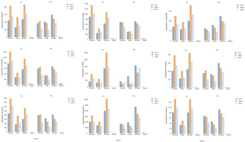
Mocci et al.                                                                                                                Transcriptomic Profile Stressed Comorbid Rat

TABLE 1 | Samples stratified by condition, tissue, and time elapsed since stress induction.

                                                   Week 1                                                                Week 7

Condition             Blood             Colon               DRG             Spinal               Blood        Colon               DRG             Spinal             Total

Naïve                    4                15                 5                 15                  4             7                 4                9                 63
SIH                      4                12                 4                 12                  2             6                 3                9                 52
CPH                      4                12                 4                 12                  4             8                 4               12                 60
Total                   12                39                13                 39                 10            21                11               30                175

TABLE 2 | Differentially expressed genes across conditions and time elapsed from stress induction across all tissues.

Tissue                                                            Week 1                                                               Week 7

                                     Total significant           Upregulated         Downregulated        Total significant         Upregulated         Downregulated
                                     DE genes (FDR                                                        DE genes (FDR
                                         ≤ 0.05)                                                              ≤ 0.05)

Blood          SIH_vs._Naive                   0                     0                      0                     23                     20                    3
               CPH_vs._Naive                   0                     0                      0                     44                     39                    5
               CPH_vs._SIH                   17                      0                     17                        3                    1                    2
Colon          SIH_vs._Naive                 23                      12                    11                   2,783                   1,342                1,441
               CPH_vs._Naive                433                     202                    231                   901                     348                 553
               CPH_vs._SIH                   94                      46                    48                    157                     62                   95
Spinal         SIH_vs._Naive               1352                     595                    757                   487                     226                 261
               CPH_vs._Naive               2,630                    1,329                 1,301                      3                                         3
               CPH_vs._SIH                   37                      7                     30                    451                     214                 237
DRG            SIH_vs._Naive                   0                     0                      0                        0                    0                    0
               CPH_vs._Naive                   0                     0                      0                        1                    1                    0
               CPH_vs._SIH                     0                     0                      0                        0                    0                    0

rhythm (adj P ≤ 5.6e-02) (Supplementary Figures 6A,B                                         We found 2,630 DEGs between CPH and naive rats
and Supplementary Table 3). Interestingly, Lyc2 alias Lyz2                                (Table 2      and     Supplementary Table 5B);      upregulated
was the top upregulated gene in CPH compared to SIH rats                                  genes     were      enriched    in    vascular     development
(FC = 14.01) and it has been recently found associated with                               (adj P = 7.4e-04) (Supplementary Figure 8A and
nerve injury-induced neuropathic pain (39). As to genes                                   Supplementary Table 3), while downregulated genes were
downregulated in CPH compared to SIH rats, they showed                                    significantly represented in RNAs metabolism (adj P = 2.6e-30)
downregulation of Fut9 (FC = −4.20) a gene involved in the                                (Supplementary Figure 8B, Supplementary Table 3). Capns1
synthesis of the Lewis motif; decreased levels of this protein                            was the most upregulated gene in the comparison of CPH and
have been associated with a reduction in neurite formation and                            naïve rats. Capns1 is a member of a calcium-activated protease
outgrowth (40).                                                                           family abundant in the CNS and calpain-1 loss leads to reduced
                                                                                          dendritic complexity and spine density deficits associated with
                                                                                          major deterioration in hippocampal long-term potentiation
Spinal                                                                                    and spatial memory (42). Fut9 (FC = −4.91) was the top
Week 1                                                                                    downregulated gene in CPH compared to naive rats at week 1.
After 1 week of stress induction, we observed 1,352                                          Thirty-seven genes were differentially expressed between
DEGs between SIH and naïve rats (Table 2 and                                              CPH and SIH rats (Table 2 and Supplementary Table 5C)
Supplementary Table 5A); the genes found up-regulated in                                  and they were significantly enriched in the translation GO
stressed rats were enriched in the GO response to insulin stimulus                        biological process (adj P = 5.37E-06) (Supplementary Figure 9
biological process (adj P = 7e-02) (Supplementary Figure 7A                               and Supplementary Table 3).
and Supplementary Table 3), while those downregulated were
enriched in protein modification GO terms (adj P = 1.8e-06)
(Supplementary Figure 7B). Two olfactory receptors, Olr35                                 Week 7
and Olr63, were 2-fold more expressed in SIH rats compared                                We observed 487 DEGs between SIH and naïve rats; the
to naïve. Olfactory perception is affected in stressed animals, as                        upregulated genes in stressed rats were significantly enriched
glucocorticoids might enhance odor detection, starting at the                             in the cell morphogenesis (adj P =1.7e-04) and histone
first step of detection (41).                                                             modification (adj P = 7.1e-04) (Supplementary Figure 10A

Frontiers in Pain Research | www.frontiersin.org                                      5                                            May 2022 | Volume 3 | Article 886042
Mocci et al. Transcriptomic Profile Stressed Comorbid Rat
and Supplementary Table3) and Atp1a4, a sodium/potassium- inflammatory and wound healing processes accompanied by a
transporting ATPase, was the most upregulated gene in decreased expression of genes involved in the metabolic pathway
SIH compared to naïve rats (FC = 3.25) (Table 2 and of oxidative phosphorylation (Figure 1C).
Supplementary Table 5D). Genes found downregulated in
stressed rats compared to naïve were significantly enriched in the Week 7
RNA metabolism (adj P =1.7e-10) (Supplementary Figure 10B SIH and CPH rats showed higher expression of genes enriched
and Supplementary Table 3). in oxidative phosphorylation when compared to naïve rats
Three genes (Ubd, Ciart, and Eral1) showed significantly (Figures 1D,E). Similarly, to what we observed at week 1, at week
lower expression in CPH compared to naïve rats at week 7 7 CPH rats compared to SIH showed activation of inflammatory
(Table 2 and Supplementary Table 5E) and ubiquitin D was the and stress response biological processes (Figure 1F).
most downregulated gene in CPH rats. Interestingly, activation
in the ubiquitin-proteasome system (UPS) targets and degrades Colon
proteins critical for the maintenance of chronic pain as those The entire gene expression profile extracted from the colon
involved in synaptic plasticity (43, 44). clustered into 40 modules (Supplementary Figure 13) and 26 of
We observed 451 DEGs between CPH and SIH conditions them were significantly associated with CPH or SIH conditions at
(Table 2 and Supplementary Table 5F). The top upregulated week 1 or week 7 from stress induction (Supplementary Table 7).
genes in CPH compared to SIH rats were Cyp2d3, a member
Week 1
of the p450 xenobiotic-inducible superfamily, and lysozyme 2
Similar to what we have observed in blood, stressed rats showed
(Lyc2), the enzyme that was also the top upregulated gene in CPH
downregulation of genes enriched in inflammation (cytokine-
compared to SIH rats at week 7 in the colon. Overall, upregulated
mediated), as opposed to an increased expression of genes
genes in CPH rats were enriched in the metabolism of RNA and
enriched in nervous system processes such as detection of
proteins terms (adj P = 1.3e-05)
external stimulus, axonogenesis, and dopaminergic synapsis
(Supplementary Figure 11A and Supplementary Table 3),
(Figure 2A and Supplementary Table 7). CPH rats compared to
while downregulated genes were represented
both naïve and SIH ones, showed higher expression of genes
in tube morphogenesis (adj P-value = 4e-03)
involved in neuronal functions, mostly related with synapses
(Supplementary Figure 11B and Supplementary Table 3).
(Figures 2B,C and Supplementary Table 7). In addition, we
DRG observed that genes involved in the response to an external
stimulus were downregulated in CPH compared to SIH rats
Week 1 and Week 7
(Figure 2C and Supplementary Table 7).
We did not observe any DEGs at week 1, whereas at week
7 only AABR07054370.1 alias Chmp4b was found significantly
Week 7
upregulated (FC = 1.72, P-value = 2.7e-07, FDR = 4.5e-03) in
Stressed rats compared to CPH and naïve, showed upregulation
CPH compared to naïve rats. An in vitro study reported that
of genes enriched in axon guidance, Notch, and Wnt signaling
Chmp4b may play a role in neuronal apoptosis and could be
pathways (Figures 2D,F and Supplementary Table 7). CPH rats
related to brain damage following intracerebral hemorrhage (45).
compared to naive showed upregulation of immune response
and response to stress genes, while genes enriched in oxidative
WGCNA-ROAST Analysis phosphorylation were mostly downregulated (Figure 2E and
Blood Supplementary Table 7).
By applying network analysis, the entire blood transcriptomic
profile was divided into 30 modules of genes grouped Spinal
by similarity of their eigengene expression profiles Network analysis applied to the whole spinal transcriptomic
(Supplementary Figure 12). Out of the 30 modules, 11 showed profile identified 37 modules (Supplementary Figure 14) and 27
at least 30% of their genes up or downregulated in CPH or SIH of them were found significantly up or downregulated in CPH
rats; for each one of these modules, we showed the top three most and/or SIH conditions at week 1 and/or week 7 from stress
significant GO biological processes and/or KEGG pathways, induction (Supplementary Table 7).
prioritizing those related with inflammatory and neurological
functions (Supplementary Table 6). As shown in Figure 1, we Week 1
schematize the results of this analysis and display the top 25 most Compared to naive, SIH and CPH rats showed decreased
significant DEGs for each contrast and time point. expression of genes linked to the organization of synapses
and circadian rhythm biological processes together with an
Week 1 increased expression of genes related to different biological
We observed a decreased expression of genes enriched in processes and pathways finalized to the production of new
inflammatory biological processes in stressed rats compared blood vessels, such as “angiogenesis” and “phosphatidylinositol
to naïve (Figure 1A), as opposed to stressed CPH rats that 3-kinase (PI3K) “(Figures 3A,B). Compared to SIH, CPH rats
compared to the naive ones, showed a higher expression of genes showed an increased expression of genes associated with the
with similar inflammatory functions (Figure 1B). Therefore, vascularization process along with a decreased expression of
CPH rats compared to stressed-only rats showed activation of genes linked to response to glucocorticoids (Figure 3C).
Frontiers in Pain Research | www.frontiersin.org 6 May 2022 | Volume 3 | Article 886042Mocci et al. Transcriptomic Profile Stressed Comorbid Rat
FIGURE 1 | Summarizes the results of both gene-set and gene differentially expressed (DE) in blood tissue across conditions and time. We focused on the top DE
gene-sets and top 25 most significant DEGs that were enriched mostly in inflammatory and neurological pathways. (A) SIH vs. Naïve at week 1, (B) CPH vs. Naïve at
week 1, (C) CPH vs. SIH at week 1, (D) SIH vs. Naïve at week 7, (E) CPH vs. Naïve at week 7, and (F) CPH vs. SIH at week 7.
Week 7 and time point (Supplementary Table 8). Figure 4 shows the
At week 7 after the induction of stress, we observed a reversal core enrichment of the top 25 DEGs in the related GO and
of the trend in the vascularization process; genes linked KEGG terms.
to the creation of new blood vessels as “angiogenesis”
“phosphatidylinositol 3-kinase and wound healing showed
higher expression in stressed compared to comorbid Week 1
rats (Figures 3D,E) We observed that a significant proportion of genes clustering in
different modules were downregulated in SIH compared to naïve
DRG rats. These genes were enriched in “chromatin modification,”
WGCNA network analysis applied to the whole DRG “Insulin signaling pathway,” “learning,” “locomotory behavior,”
transcriptomic profile divided the genes into 50 modules “Phosphatidylinositol signaling,” “plasma membrane transport,”
(Supplementary Figure 15); 20 of these modules showed ≥ 30% “protein methylation,” and “Wnt signaling pathway” (Figure 4A
of their genes significantly upregulated in at least one condition and Supplementary Table 8).
Frontiers in Pain Research | www.frontiersin.org 7 May 2022 | Volume 3 | Article 886042Mocci et al. Transcriptomic Profile Stressed Comorbid Rat FIGURE 2 | Summarizes the results of both gene-set and gene differentially expressed (DE) in colon tissue across conditions and time. We focused on the top DE gene-sets and top 25 most significant DEGs that were enriched mostly in inflammatory and neurological pathways. (A) SIH vs. Naïve at week 1, (B) CPH vs. Naïve at week 1, (C) CPH vs. SIH at week 1, (D) SIH vs. Naïve at week 7, (E) CPH vs. Naïve at week 7, and (F) CPH vs. SIH at week 7. Likewise, in the contrast between CPH and naïve rats Week 7 at week 1, we found that a significant proportion of genes We observed different enrichment patterns for all contrasts clustering within different modules were downregulated in CPH compared to those observed in week 1. SIH rats compared to rats (Supplementary Table 8). These genes were enriched in naïve showed upregulation of genes enriched in neurological inflammatory processes such as “adaptive immune response,” processes as “response to axon injury” and “synapse “cytokine–cytokine receptor interaction,” and “complement and organization. Conversely, genes downregulated in SIH coagulation cascades.” (Figure 4B). rats were overrepresented in neurodegenerative diseases When we compared directly CPH to SIH rats at week 1, we and the processing of different RNAs (Figure 4D and found that all genes were downregulated in CPH rats and the Supplementary Table 8). highest number of genes were enriched in “plasma membrane CPH rats compared to naive showed higher expression of transport,” followed by “embryonic forelimb morphogenesis” and genes enriched in oxidative phosphorylation and downregulation “amino acid transport” (Figure 4C). of genes involved in the nerve myelination (Figure 4E). Frontiers in Pain Research | www.frontiersin.org 8 May 2022 | Volume 3 | Article 886042
Mocci et al.                                                                                                           Transcriptomic Profile Stressed Comorbid Rat

 FIGURE 3 | Summarizes the results of both gene-set and gene differentially expressed (DE) in spinal tissue across conditions and time. We focused on the top DE
 gene-sets and top 25 most significant DEGs that were enriched mostly in inflammatory and neurological pathways. (A) SIH vs. Naïve at week 1, (B) CPH vs. Naïve at
 week 1, (C) CPH vs. SIH at week 1, (D) SIH vs. Naïve at week 7, (E) CPH vs. Naïve at week 7.

   When compared to SIH rats, CPH showed upregulation of                             of the mitochondrial membrane ATP synthase (Atp5me, Atp5mf,
genes involved in the oxidative phosphorylation GO biological                        and Atp5pd), representatives of the cytochrome c oxidase
process and Parkinson’s disease KEGG pathway (Figure 4F).                            (Cox6b1, Cox7c, and Cox8a), members of the mitochondrial
                                                                                     NADH: ubiquinone oxidoreductase (complex I) (Nd4l, Ndufa12,
Identification of CPH Biomarkers                                                     and Ndufb6) and components of the ubiquinol-cytochrome c
Finally, to detect specific CPH biomarkers from blood, we                            oxidoreductase complex (Uqcrb, Uqcrh, and Uqcrq). Despite the
focused our attention on the genes or gene-sets that are                             low abundance of these genes in blood compared to the other
distinctive of the comorbid condition and looked for any                             tissues, we noticed a common trend in the expression of these
correlation of their expression profiles across the solid tissues                    genes across all tissues, conditions, and time points (Figure 5).
(DRG, colon, and spinal) and blood. Both DE at the gene and                          At week 1, CPH rats showed the lowest expression of these genes
gene-set levels pointed to distinct mitochondrial genes: members                     compared to SIH and naïve, while at week 7 we observed a

Frontiers in Pain Research | www.frontiersin.org                                 9                                             May 2022 | Volume 3 | Article 886042
Mocci et al. Transcriptomic Profile Stressed Comorbid Rat FIGURE 4 | Summarizes the results of both gene-set and gene differentially expressed (DE) in DRG tissue across conditions and time. We focused on the top DE gene-sets and top 25 most significant DEGs that were enriched mostly in inflammatory and neurological pathways. (A) SIH vs. Naïve at week 1, (B) CPH vs. Naïve at week 1, (C) CPH vs. SIH at week 1, (D) SIH vs. Naïve at week 7, (E) CPH vs. Naïve at week 7, and (F) CPH vs. SIH at week 7. complete inversion of this trend, with the higher expression in DISCUSSION comorbid rats followed by naïve and SIH rats (Figure 5). The observed correlation in the expression of these Several studies have shown a correlation between stress and genes reflected a highly significant functional interaction pain and that chronic exposure to stress increases the intensity of their proteins; in fact, Search Tool for the Retrieval of the pain (1, 2). Stress is a major underlying factor in IBS of Interacting Genes/Proteins detected 66 protein-protein (1, 6, 9, 10) and TMD (46, 47), another chronic pain condition, interactions compared to the expected 5 (PPI, P ≤ 1.0e-16) and these conditions are significantly comorbid (11, 12). In this (Supplementary Figure 16) and reported high enrichment study, we explored if the genetic mechanisms that are triggered in the oxidative phosphorylation biological process (adj in the development of pain due to stress are the same as those P = 1.1e-09). evoked by the presence of multiple comorbid pain conditions. Frontiers in Pain Research | www.frontiersin.org 10 May 2022 | Volume 3 | Article 886042
Mocci et al.                                                                                                          Transcriptomic Profile Stressed Comorbid Rat

 FIGURE 5 | Potential CPH biomarkers in blood. The figure shows 9 genes, representative of mitochondrial membrane ATP synthase, cytochrome c oxidase, NADH:
 ubiquinone oxidoreductase (complex I) and ubiquinol-cytochrome c oxidoreductase complex. Since these genes showed high correlation of their expression profiles
 across the solid tissues (DRG, colon, and spinal) and blood, they represent potential diagnostic biomarkers for CPH.

To answer this question, we compared the entire transcriptomic                       Wnt and Notch pathways in SIH compared to CPH rats. The
profile of CPH rats to both SIH and control non-stressed rats and                    wnt/β-Catenin pathway has many critical regulatory functions,
focused on two major biological components involved in pain:                         including those related to the axis formation and synapse
inflammation and the nervous system.                                                 development and modulation (53). A recent report shows that the
    We carried out the analysis in four different tissues: at the                    Wnt/β-Catenin pathway boost ribosome biogenesis in response
systemic level in the blood, and in the colon, DRG, and spinal                       to stress, allowing cells to grow and survive (54). One week after
tissues. Blood was studied because, given the complexity of                          stress induction, the transcriptional profile from spinal tissue
overlapping pain conditions, easily available biomarkers may                         showed a similar picture in SIH and CPH rats; the two compared
provide the early identification and diagnosis of these disorders                    to naïve showed a decreased expression of genes linked to the
and eventually be applied to translational research on chronic                       synaptic organization, likely because of the damage in the nerve
visceral pain among human subjects. Colon tissue is commonly                         tissue due to stress exposure. Simultaneously, the exposure to
used in transcriptomic analyses applied to humans affected                           stress in these rats, particularly in CPH rats, determined an
with IBS and animal models, as it may pave the way for the                           increased expression of genes enriched in biological processes
gastrointestinal origin of stress-induced chronic visceral pain                      and pathways aimed at the production of new blood vessels,
(48, 49). The spinal cord and dorsal root ganglia are key                            angiogenesis, and phosphatidylinositol 3-kinase (PI3K), and
stations in sensory transduction and modulation, including pain                      neurotrophin pathways. The PI3K pathway plays an important
transmission (50).                                                                   role in regulating angiogenesis; hypoxia leads to the hypoxia-
    The analysis of the whole transcriptome profile in blood and                     inducible factor 1-alpha (HIF-1α) stabilization and is a major
colon tissues at week 1 showed activation of the inflammatory                        stimulus for increased vascular endothelial growth factor (VEGF)
response in CPH compared to SIH rats and in the colon, this                          production (55).
effect persisted after 7 weeks from stress induction. Previous                           The PI3K /Akt pathway has been reported as a regulator
studies described a long-term decrease in the number of T cells                      of spinal plasticity in the rat visceral pain model (56) and
in peripheral blood after repeated chronic mild stress induction                     associated with chronic pain (57). Previous studies described
(51, 52). Furthermore, in blood and colon tissues at week 1,                         an increased production of VEGF by intestinal mucosa cells of
CPH rats compared to SIH showed increased expression of                              patients with IBS and associated vascular endothelial growth
genes enriched in synapse organization, and neurotransmitter                         factors with visceral hyperalgesia, abdominal discomfort, and/or
transport biological processes. In the colon, at week 7 we                           pelvic pain (58, 59). Likewise, the neurotrophin pathway
observed significantly higher expression of genes enriched in                        promotes blood vessel growth (60); neurotrophins such as NT-3

Frontiers in Pain Research | www.frontiersin.org                                11                                            May 2022 | Volume 3 | Article 886042
Mocci et al. Transcriptomic Profile Stressed Comorbid Rat
control the sympathetic innervation of blood vessels (60, 61). cortisol and corticosterone), androgens (e.g., testosterone), and
Blood vessels play a crucial role in nerve regeneration; in estrogens (e.g., estriol) are synthesized in mitochondria(64).
physiological conditions, they transport ingredients (oxygen, Chronic pain has been associated with oxidative stress. The
nutrients, and hormones), remove metabolic waste, and facilitate hypothesized mechanism behind this link is the release of reactive
cell circulation, which provides a supportive microenvironment oxygen species, which affects mitochondrial function by reducing
for the nervous system (62). This mechanism may contribute the release of ATP in cells (65).
to the recovery from nervous damage by stress induction. In conclusion, our study highlights time and tissue-specific
Interestingly, our study highlights that the representative genes genetic signatures that help to differentiate the inflammatory and
to the angiogenesis-related terms differed between CPH and SIH neurological response to stress in SIH and comorbid TMD–IBS
rats. Furthermore, angiogenesis-related genes were more highly pain hypersensitivity (CPH) rats. Moreover, our data support the
expressed in CPH than SIH rats 1 week after stress, while the theory behind the association between pain and oxidative stress,
situation was reversed 7 weeks after stress exposure. as demonstrated by the decreased expression of mitochondrial
Genes related to “response to glucocorticoid” were genes in SIH and even more conspicuously in CPH rats compared
downregulated in the CPH compared to SIH rats, suggesting to naïve and reaffirm the value of antioxidants as a therapeutic
negative feedback in response to the stronger stress due to target for chronic pain. Finally, if we look at the switch in
persistent sensitization through 7 weeks (13). In addition, the expression levels of mitochondrial genes between 1 and 7
at 7 weeks from stress induction, “wound healing” was also weeks after stress exposure, and interpret our results in the
downregulated in CPH in comparison to SIH rats; it is well context of the energy required for cell growth and survival, this
known that stress and pain can deteriorate wound healing (63). study shows that the production of energy for repairing stress-
Our results suggest that chronic stressful condition in the induced damage in SIH rats starts and ends earlier than in CPH
CPH rats contributes to the delayed healing from tissue damage rats, where the need of energy to face stress consequences last
in the nervous system, resulting in the prolonged referred pain much longer.
observed in our previous study (13).
Finally, we analyzed the transcriptome profile obtained from DATA AVAILABILITY STATEMENT
DRG at week 1 and week 7 after stress induction. At week 1,
all the inflammatory and neurological response-related genes The data presented in the study are deposited in the
were downregulated in both SIH and CPH rats compared NCBI repository (https://www.ncbi.nlm.nih.gov/geo/), accession
to naive. However, at week 7, SIH rats compared to naïve number GSE199221. Expression data for genes analyzed across
showed upregulation of genes enriched in the synapse assembly, all tissues have been uploaded to the Neuroscience Multi-
Wnt and Notch pathways (48) as opposed to CPH rats that Omic (NeMO) (66) Analytics portal which enables web-
revealed a significant proportion of genes enriched in the based visualization available at the following link (https://
myelination process were downregulated in CPH compared to nemoanalytics.org/p?s=2f41a962) [accessed February 22, 2022],
naive. However, we did not observe genes enriched in any accessible previous registration.
neurological or inflammatory functions when comparing SIH
and CPH at 7 weeks, which is in line with the previous ETHICS STATEMENT
electrophysiological findings that peripheral sensitization was
present in both SIH and CPH rats at 7 weeks (13). The animal study was reviewed and approved by University of
Future studies could investigate the long-term effect of stress Maryland Institutional Animal Care and Use Committee.
on pain sensations among CPH rats to determine the impact
of the downregulated genes related to the myelination process AUTHOR CONTRIBUTIONS
among these rats. Our results may suggest greater damage in the
neurological components following stress in CPH compared to SD and RT contributed to the conception and design of the study.
SIH at 7 weeks, although these results are not consistent with SA contributed to the design of the study. EM performed the
behavioral changes. statistical analysis. EM, TG, and JC contributed in the manuscript
Our study highlights a common signature across tissues and preparation. All authors contributed to manuscript revision and
time elapsed from stress exposure; in all tissues, we noticed approved the submitted version.
that several mitochondrial genes, involved in the oxidative
phosphorylation pathway are downregulated in both CPH and FUNDING
SIH compared to naïve rats, with the lower expression in CPH
rats at week 1, while at week 7, the same or related genes This study was funded by the R01 grant: R01NR015472 to Dorsey
are upregulated in CPH in comparison to SIH and naïve rats. and Traub.
All biological processes activated by acute or chronic stress
require a substantial amount of energy. Mitochondria are the SUPPLEMENTARY MATERIAL
major source of cellular energy, via the transformation of
energetic substrates and oxygen into ATP (64). Additionally, all The Supplementary Material for this article can be found
steroid hormones, including progestogens (e.g., progesterone), online at: https://www.frontiersin.org/articles/10.3389/fpain.
mineralocorticoids (e.g., aldosterone), glucocorticoids (e.g., 2022.886042/full#supplementary-material
Frontiers in Pain Research | www.frontiersin.org 12 May 2022 | Volume 3 | Article 886042Mocci et al. Transcriptomic Profile Stressed Comorbid Rat
REFERENCES 20. Kim D, Langmead B, Salzberg SL. Hisat2. Nat Methods. (2015) 12:357–60.
doi: 10.1038/nmeth.3317
1. Crettaz B, Marziniak M, Willeke P, Young P, Hellhammer D, 21. Anders S, Pyl PT, Huber W. Genome analysis HTSeq-a Python framework to
Stumpf A, et al. Stress-induced allodynia-evidence of increased pain work with high-throughput sequencing data. Bioinformatics. (2015) 31:166–
sensitivity in healthy humans and patients with chronic pain after 9. doi: 10.1093/bioinformatics/btu638
experimentally induced psychosocial stress. PLoS One. (2013) 8:e69460. 22. Zhang Y, Parmigiani G, Johnson WE. ComBat-seq: batch effect
doi: 10.1371/journal.pone.0069460 adjustment for RNA-seq count data. NAR Genomics Bioinf. (2020) 2,
2. Larauche M, Mulak A, Taché Y. Stress and visceral pain: lqaa078. doi: 10.1093/nargab/lqaa078
from animal models to clinical therapies. Exp Neurol. (2012) 23. Johnson WE, Li C, Rabinovic A. Adjusting batch effects in microarray
233:49–67. doi: 10.1016/j.expneurol.2011.04.020 expression data using empirical Bayes methods. Biostatistics. (2007) 8:118–
3. Elsenbruch S. Abdominal pain in irritable bowel syndrome: a review of 27. doi: 10.1093/biostatistics/kxj037
putative psychological, neural and neuro-immune mechanisms. Brain Behav 24. Law CW, Chen Y, Shi W, Smyth GK. voom: precision weights unlock
Immun. (2011) 25:386–94. doi: 10.1016/j.bbi.2010.11.010 linear model analysis tools for RNA-seq read counts. Genome Biol. (2014)
4. Fukudo S. Stress and visceral pain: focusing on irritable bowel syndrome. Pain. 15:R29. doi: 10.1186/gb-2014-15-2-r29
(2013) 154:S63–S7. doi: 10.1016/j.pain.2013.09.008 25. Langfelder P, Horvath S. WGCNA: an R package for weighted
5. Moloney RD, Johnson AC, O’Mahony SM, Dinan TG, Greenwood-Van correlation network analysis. BMC Bioinformatics. (2008)
Meerveld B, Cryan JF. Stress and the microbiota–gut–brain axis in visceral 9:559. doi: 10.1186/1471-2105-9-559
pain: relevance to irritable bowel syndrome. CNS Neurosci Ther. (2016) 26. Wu D, Lim E, Vaillant F, Asselin-Labat ML, Visvader JE, Smyth GK. ROAST:
22:102–17. doi: 10.1111/cns.12490 rotation gene set tests for complex microarray experiments. Bioinformatics.
6. Creekmore AL, Hong S, Zhu S, Xue J, Wiley JW. Chronic stress-associated (2010) 26:2176–82. doi: 10.1093/bioinformatics/btq401
visceral hyperalgesia correlates with severity of intestinal barrier dysfunction. 27. Ritchie ME, Phipson B, Wu D, Hu Y, Law CW, Shi W, et al. Limma powers
Pain. (2018) 159:1777–89. doi: 10.1097/j.pain.0000000000001271 differential expression analyses for RNA-sequencing and microarray studies.
7. Ji Y, Hu B, Li J, Traub RJ. Opposing roles of estradiol and testosterone on Nucleic Acids Res. (2015) 43:e47. doi: 10.1093/nar/gkv007
stress-induced visceral hypersensitivity in rats HHS Public Access. J Pain. 28. Zhou Y, Zhou B, Pache L, Chang M, Khodabakhshi AH, Tanaseichuk
(2018) 19:764–76. doi: 10.1016/j.jpain.2018.02.007 O, et al. Metascape provides a biologist-oriented resource for the
8. Bradesi S, Svensson CI, Steinauer J, Pothoulakis C, Yaksh TL, Mayer analysis of systems-level datasets. Nat Commun. (2019) 10:1523.
EA. Role of spinal microglia in visceral hyperalgesia and NK1R up- doi: 10.1038/s41467-019-09234-6
regulation in a rat model of chronic stress. Gastroenterology. (2009) 136:1339– 29. Yu G, Wang LG, Han Y, He QY. ClusterProfiler: an R package for comparing
48.e2. doi: 10.1053/j.gastro.2008.12.044 biological themes among gene clusters. OMICS J Integrat Biol. (2012) 16:284–
9. Barbara G, Stanghellini V, de Giorgio R, Cremon C, Cottrell GS, Santini 7. doi: 10.1089/omi.2011.0118
D, et al. Activated mast cells in proximity to colonic nerves correlate 30. Wu T, Hu E, Xu S, Chen M, Guo P, Dai Z, et al. clusterProfiler 4.0: a
with abdominal pain in irritable bowel syndrome. Gastroenterology. (2004) universal enrichment tool for interpreting omics data. Innovation (N Y).
126:693–702. doi: 10.1053/j.gastro.2003.11.055 (2021) 2:100141. doi: 10.1016/j.xinn.2021.100141
10. Wiley JW, Zong Y, Zheng G, Zhu S, Hong S. Histone H3K9 31. Gene Ontology Consortium. The gene ontology resource: enriching a GOld
methylation regulates chronic stress and IL-6–induced colon epithelial mine. Nucleic Acids Res. (2021) 49:D325–34. doi: 10.1093/nar/gkaa1113
permeability and visceral pain. Neurogastroenterol Motility. (2020) 32. Kanehisa M, Furumichi M, Sato Y, Ishiguro-Watanabe M, Tanabe M. KEGG:
32:e13941. doi: 10.1111/nmo.13941 integrating viruses and cellular organisms. Nucleic Acids Res. (2021) 49:D545–
11. Bruno V, Catapano S, Mobilio N, Iovino P, Associato P, Gallotta S, et al. 51. doi: 10.1093/nar/gkaa970
High risk of temporomandibular disorder in irritable bowel syndrome: is 33. Jassal B, Matthews L, Viteri G, Gong C, Lorente P, Fabregat A, et al.
there a correlation with greater illness severity? Case Control Study. World The reactome pathway knowledgebase. Nucleic Acids Res. (2020) 48:D498–
J Gastroenterol. (2017) 23:103–9. doi: 10.3748/wjg.v23.i1.103 503. doi: 10.1093/nar/gkz1031
12. Aaron LA, Burke MM, Buchwald D. Overlapping conditions among patients 34. Grosshans HK, Fischer TT, Steinle JA, Brill AL, Ehrlich BE. Neuronal
with chronic fatigue syndrome, fibromyalgia, and temporomandibular Calcium Sensor 1 is up-regulated in response to stress to promote
disorder. Arch Intern Med. (2000) 160:221–7. doi: 10.1001/archinte.160.2.221 cell survival and motility in cancer cells. Mol Oncol. (2020) 14:1134–
13. Cao DY, Hu B, Xue Y, Hanson S, Dessem D, Dorsey SG, et al. Differential 51. doi: 10.1002/1878-0261.12678
activation of colonic afferents and dorsal horn neurons underlie stress- 35. Barbara B. Aquaporin biology and nervous system., Curr Neuropharmacol.
induced and comorbid visceral hypersensitivity in female rats. J Pain. (2021) (2010) 8:97–104. doi: 10.2174/157015910791233204
22:1283–93. doi: 10.1016/j.jpain.2021.04.004 36. Kamiya S, Russo MA, Lazar V, Bezirtzoglou empezirt E, Stavropoulou
14. Traub RJ, Cao D-Y, Karpowicz J, Pandya S, Ji Y, Dorsey SG, et al. A clinically E, Pircalabioru GG, et al. The role of cytochromes P450 in
relevant animal model of TMD and IBS co-morbidity. J Pain. (2014) 15:956– infection. Front Immunol. (2018) 9:89. doi: 10.3389/fimmu.2018.
66. doi: 10.1016/j.jpain.2014.06.008 00089
15. Ji Y, Hu B, Klontz C, Li J, Dessem D, Dorsey SG, et al. Peripheral mechanisms 37. Matsuoka Y, Yamashita A, Matsuda M, Kawai K, Sawa T, Amaya F. NLRP2
contribute to comorbid visceral hypersensitivity induced by preexisting inflammasome in dorsal root ganglion as a novel molecular platform
orofacial pain and stress in female rats. Neurogastroenterol Motil. (2020) that produces inflammatory pain hypersensitivity. Pain. (2019) 160:2149–
32:e13833 doi: 10.1111/nmo.13833 60. doi: 10.1097/j.pain.0000000000001611
16. Shen L, Yang X, Qian W, Hou X. The role of peripheral cannabinoid receptors 38. Shi R, Redman P, Ghose D, Hwang H, Liu Y, Ren X, et al. Shank
type 1 in rats with visceral hypersensitivity induced by chronic restraint stress. proteins differentially regulate synaptic transmission. eNeuro. (2017)
J Neurogastroenterol Motil. (2010) 16:281–90. doi: 10.5056/jnm.2010.16.3.281 4:ENEURO.0163-15.2017. doi: 10.1523/ENEURO.0163-15.2017
17. Machorru-Rojas N, Sainz-Espuñes T, Godínez-Victoria M, Castañeda- 39. Huang T, Wang J, Dai W, Liu X, Wang K, Yi D, et al. Identification of the
Sánchez JI, Campos-Rodríguez R, Pacheco-Yepez J, et al. Impact of chronic hub genes related to nerve injury-induced neuropathic pain. Front Neurosci.
immobilization stress on parameters of colonic homeostasis in BALB/c mice. (2020) 14:488. doi: 10.3389/fnins.2020.00488
Mol Med Rep. (2019) 20:2083–90. doi: 10.3892/mmr.2019.10437 40. Gouveia R, Schaffer L, Papp S, Grammel N, Kandzia S, Head SR,
18. Andrews S, Krueger F, Seconds-Pichon A, Biggins F, Wingett S. FastQC. et al. Expression of glycogenes in differentiating human NT2N neurons.
A Quality Control Tool for High Throughput Sequence Data. Babraham Downregulation of fucosyltransferase 9 leads to decreased Lewisx levels
Bioinformatics. Babraham Institute (2015). and impaired neurite outgrowth. Biochim Biophys Acta. (2012) 1820:2007–
19. Bolger AM, Lohse M, Usadel B. Genome analysis Trimmomatic: a flexible 19. doi: 10.1016/j.bbagen.2012.09.004
trimmer for Illumina sequence data. Bioinformatics. (2014) 30:2114– 41. Meunier N, Raynaud A, le Bourhis M, Grébert D, Dewaele A, Acquistapace
20. doi: 10.1093/bioinformatics/btu170 A, et al. The olfactory mucosa, first actor of olfactory detection, is
Frontiers in Pain Research | www.frontiersin.org 13 May 2022 | Volume 3 | Article 886042Mocci et al. Transcriptomic Profile Stressed Comorbid Rat
sensitive to glucocorticoid hormone. Eur J Neurosci. (2020) 51:1403– 57. Chen S-P, Zhou Y-Q, Liu D-Q, Zhang W, Manyande A, Guan X-H, et al.
18. doi: 10.1111/ejn.14564 PI3K/Akt Pathway: a potential therapeutic target for chronic pain. Curr Pharm
42. Duan H, Shen F, Li L, Tu Z, Chen P, Chen P, et al. Activation of Des. (2017) 23:1860–8. doi: 10.2174/1381612823666170210150147
the Notch signaling pathway in the anterior cingulate cortex is involved 58. Xie AX, Iguchi N, Clarkson TC, Malykhina AP. Pharmacogenetic inhibition
in the pathological process of neuropathic pain. Pain. (2021) 162:263– of lumbosacral sensory neurons alleviates visceral hypersensitivity
74. doi: 10.1097/j.pain.0000000000002014 in a mouse model of chronic pelvic pain. PLoS ONE. (2022)
43. Ossipov MH, Bazov I, Gardell LR, Kowal J, Yakovleva T, Usynin I, et al. 17:e0262769. doi: 10.1371/journal.pone.0262769
Control of chronic pain by the ubiquitin proteasome system in the spinal cord. 59. Malykhina AP, Lei Q, Erickson CS, Epstein ML, Saban MR, Davis
J Neurosci. (2007) 27:8226–37. doi: 10.1523/JNEUROSCI.5126-06.2007 CA, et al. VEGF induces sensory and motor peripheral plasticity, alters
44. Cheng J, Deng Y, Zhou J. Role of the ubiquitin system in chronic pain. Front bladder function, and promotes visceral sensitivity. BMC Physiol. (2012)
Mol Neurosci. (2021) 14:674914. doi: 10.3389/fnmol.2021.674914 12:15. doi: 10.1186/1472-6793-12-15
45. Shen J, Liu Y, Song Y, Li L, Duan C, Zhou Y, et al. CHMP4B, 60. Cristofaro B, Stone OA, Caporali A, Dawbarn D, Ieronimakis N, Reyes M,
ESCRT-III associating protein, associated with neuronal apoptosis et al. Neurotrophin-3 is a novel angiogenic factor capable of therapeutic
following intracerebral hemorrhage. Brain Res. (2015) 1597:1– neovascularization in a mouse model of limb ischemia. Arterioscler Thromb
13. doi: 10.1016/j.brainres.2014.11.043 Vasc Biol. (2010) 30:1143–50. doi: 10.1161/ATVBAHA.109.205468
46. Ohrbach R, Michelotti A. The role of stress in the etiology of oral parafunction 61. Randolph CL, Bierl MA, Isaacson LG. Regulation of NGF and NT-3 protein
and myofascial pain. Oral Maxillofac Surg Clin North Am. (2018) 30:369– expression in peripheral targets by sympathetic input. Brain Res. (2007)
79. doi: 10.1016/j.coms.2018.04.011 1144:59–69. doi: 10.1016/j.brainres.2007.01.099
47. Slade GD, Ohrbach R, Greenspan JD, Fillingim RB, Bair E, Sanders 62. Sheng Y, Zhu L. The crosstalk between autonomic nervous system and blood
AE, et al. Clinical review painful temporomandibular disorder: vessels. Int J Physiol Pathophysiol Pharmacol. (2018) 10:17–28.
decade of discovery from OPPERA studies. J Dent Res. (2016) 63. Christian LM, Graham JE, Padgett DA, Glaser R, Kiecolt-Glaser JK. Fax
95:1084–92. doi: 10.1177/0022034516653743 +41 61 306 12 34 E-Mail karger@karger.ch Stress and Wound Healing.
48. Kharbanda KK, Sabui S, Mirajul Hoque K, Zhang S, Chao G, Wang Z, et al. Neuroimmunomodulation. (2006) 13:337–46. doi: 10.1159/000104862
LncRNA H19 as a Competing Endogenous RNA to Regulate AQP Expression 64. Picard M, McEwen BS, Epel ES, Sandi C. An energetic view of
in the Intestinal Barrier of IBS-D Patients. Front Physiol. (2021) 11:602076. stress: focus on mitochondria. Front Neuroendocrinol. (2018) 49:72–
doi: 10.3389/fphys.2020.602076(2021) 85. doi: 10.1016/j.yfrne.2018.01.001
49. Tavakoli P, Vollmer-Conna U, Hadzi-Pavlovic D, Grimm MC. A review 65. Meeus M, Nijs J, Hermans L, Goubert D, Calders P. The role of mitochondrial
of inflammatory bowel disease: a model of microbial, immune and dysfunctions due to oxidative and nitrosative stress in the chronic pain or
neuropsychological integration. Public Health Rev. (2021) 42:1603990. chronic fatigue syndromes and fibromyalgia patients: peripheral and central
doi: 10.3389/phrs.2021.1603990 mechanisms as therapeutic targets? Expert Opin Ther Targets. (2013) 17:1081–
50. Garland EL. Pain Processing In The Human Nervous System: A Selective 9. doi: 10.1517/14728222.2013.818657
Review Of Nociceptive And Biobehavioral Pathways. Primary Care. (2012) 66. Orvis J, Gottfried B, Kancherla J, Adkins RS, Song Y, Dror
39:561–71. doi: 10.1016/j.pop.2012.06.013 AA, et al. gEAR: gene expression analysis resource portal for
51. Batuman OA, Sajewski D, Ottenweller JE, Pitman DL, Natelson BH. community-driven, multi-omic data exploration. Nat Methods. (2021)
Effects of repeated stress on T cell numbers and function in rats. 18:843–4. doi: 10.1038/s41592-021-01200-9
Brain Behav Immunity. (1990) 4:105–17. doi: 10.1016/0889-1591(90)
90013-G Conflict of Interest: The authors declare that the research was conducted in the
52. Iwamoto G, Sellami M, Lowder T, Castell M, Estruel-Amades S, Ruiz- absence of any commercial or financial relationships that could be construed as a
Iglesias P, et al. Changes in lymphocyte composition and functionality after potential conflict of interest.
intensive training and exhausting exercise in rats. Front Physiol. (2019)
10:1491. doi: 10.3389/fphys.2019.01491
Publisher’s Note: All claims expressed in this article are solely those of the authors
53. Mulligan KA, Cheyette BNR, Program NG. Wnt signaling in vertebrate neural
development and function. J Neuroimmune Pharmacol. (2012) 7:774–87. and do not necessarily represent those of their affiliated organizations, or those of
doi: 10.1007/s11481-012-9404-x the publisher, the editors and the reviewers. Any product that may be evaluated in
54. Dannheisig DP, Bächle J, Tasic J, Keil M, Pfister AS. The Wnt/β-catenin this article, or claim that may be made by its manufacturer, is not guaranteed or
pathway is activated as a novel nucleolar stress response. J Mol Biol. (2021) endorsed by the publisher.
433:166719. doi: 10.1016/j.jmb.2020.11.018
55. Karar J, Maity A, Fähling M, Sala C, Guha C, Einstein A. MOLECULAR Copyright © 2022 Mocci, Goto, Chen, Ament, Traub and Dorsey. This is an open-
NEUROSCIENCE PI3K/AKT/mTOR pathway in angiogenesis. Front Mol access article distributed under the terms of the Creative Commons Attribution
Neurosci. (2011) 4:51. doi: 10.3389/fnmol.2011.00051 License (CC BY). The use, distribution or reproduction in other forums is permitted,
56. Kay JC, Xia C-M, Liu M, Shen S, Yu SJ, Chung C, et al. Endogenous provided the original author(s) and the copyright owner(s) are credited and that the
PI3K/Akt and NMDAR act independently in the regulation of CREB original publication in this journal is cited, in accordance with accepted academic
activity in lumbosacral spinal cord in cystitis. Exp Neurol. (2013) 250:366– practice. No use, distribution or reproduction is permitted which does not comply
75. doi: 10.1016/j.expneurol.2013.10.015 with these terms.
Frontiers in Pain Research | www.frontiersin.org 14 May 2022 | Volume 3 | Article 886042You can also read