Extracellular Vesicles From Adipose Stem Cells Prevent Muscle Damage and Inflammation in a Mouse Model of Hind Limb Ischemia
←
→
Page content transcription
If your browser does not render page correctly, please read the page content below
Arteriosclerosis, Thrombosis, and Vascular Biology
Volume 40, Issue 1, January 2020; Pages 239-254
https://doi.org/10.1161/ATVBAHA.119.313506

TRANSLATIONAL SCIENCES

Extracellular Vesicles From Adipose Stem Cells Prevent Muscle Damage
and Inflammation in a Mouse Model of Hind Limb Ischemia
Role of Neuregulin-1

Federico Figliolini, Andrea Ranghino, Cristina Grange, Massimo Cedrino, Marta Tapparo, Claudia
Cavallari, Andrea Rossi, Gabriele Togliatto, Saveria Femminò, Maria Vittoria Gugliuzza, Giovanni
Camussi *, and Maria Felice Brizzi *

OBJECTIVES: Critical hindlimb ischemia is a severe consequence of peripheral artery disease.
Surgical treatment does not prevent skeletal muscle impairment or improve long-term patient
outcomes. The present study investigates the protective/regenerative potential and the
mechanism of action of adipose stem cell-derived extracellular vesicles (ASC-EVs) in a mouse
model of hindlimb ischemia.

APPROACH AND RESULTS: We demonstrated that ASC-EVs exert a protective effect on
muscle damage by acting both on tissue microvessels and muscle cells. The genes involved in
muscle regeneration were up-regulated in the ischemic muscles of ASC-EV-treated animals.
MyoD expression has also been confirmed in satellite cells. This was followed by a reduction in
muscle function impairment in vivo. ASC-EVs drive myoblast proliferation and differentiation in the
in vitro ischemia/reoxygenation model. Moreover, ASC-EVs have shown an anti-apoptotic effect
both in vitro and in vivo. Transcriptomic analyses have revealed that ASC-EVs carry a variety of
pro-angiogenic mRNAs, while proteomic analyses have demonstrated an enrichment of NRG1
(neuregulin 1). A NRG1 blocking antibody used in vivo demonstrated that NRG1 is relevant to
ASC-EV-induced muscle protection, vascular growth, and recruitment of inflammatory cells.
Finally, bioinformatic analyses on 18 molecules that were commonly detected in ASC-EVs,
including mRNAs and proteins, confirmed the enrichment of pathways involved in vascular growth
and muscle regeneration/protection.

CONCLUSIONS: This study demonstrates that ASC-EVs display pro-angiogenic and skeletal
muscle protective properties that are associated with their NRG1/mRNA cargo. We, therefore,
propose that ASC-EVs are a useful tool for therapeutic angiogenesis and muscle protection.

Key Words: endothelial cells ■ ischemia ■ myoblasts ■ neuregulin-1 ■ proteomics

Correspondence to: Maria Felice Brizzi, Department of Medical Sciences, University of Turin, Corso Dogliotti
14, 10126, Turin. Email mariafelice.brizzi@unito.it
Giovanni Camussi, Department of Medical Sciences, University of Turin, Corso Dogliotti 14, 10126, Turin.
Email giovanni.camussi@unito.it
* These authors contributed equally to this article.
For Sources of Funding and Disclosures, see page 253.
The       online-only     Data      Supplement     is     available      with     this     article       at
https://www.ahajournals.org/doi/suppl/10.1161/ATVBAHA.119.313506.
© 2019 American Heart Association, Inc.

                   Nonstandard Abbreviations and Acronyms
  ASC-EVs     adipose stem cell-derived extracellular vesicles
  BrdU        5-bromo-2-deoxyuridine
  EGR1        early growth response 1
  HMEC        human microvascular endothelial cells
  I/R         ischemia/reperfusion
  Myf5        myogenic factor 5
  MyoD        myoblast determination protein 1
  NRG1        neuregulin 1
  p38MAPK     P38 mitogen-activated protein kinase
  Pax7        paired box protein 7
  SCs         satellite cells

                                             Highlights
  ● Adipose stem cell-derived extracellular vesicles prevent muscle damage after acute hindlimb
    ischemia.
  ● Adipose stem cell-derived extracellular vesicles induce vascular growth and protect muscle against
    ischemia/reperfusion damage.
  ● Adipose stem cell-derived extracellular vesicles are enriched in NRG1 (neuregulin 1) and pro-
    angiogenic mRNAs.
  ● Adipose stem cell-derived extracellular vesicles via NRG1 impair inflammatory cell infiltration in
    muscles subjected to ischemia/reperfusion.

C
       ritical limb ischemia is a widespread disorder caused by the atherosclerosis of the peripheral
       arteries and is commonly found in patients with peripheral artery disease.1 Reduced blood
       supply to the ischemic limb leads to skeletal muscle damage. The standard therapies, which
are surgery and endovascular intervention, are not particularly effective, making patient
management a significant economic burden.2,3 The exponential growth of an aging population, the
onset of diabetes mellitus, and the high incidence of metabolic disease also pose a heavy social
encumbrance but are also driving forces for novel and effective therapeutic options. Regenerative
medicine and in particular cell-based therapy, has recently gained a great deal of attention.4
Mesenchymal stem cells, derived from a variety of sources, have been proposed to fulfill this role.4
Bone-marrow derived mesenchymal stem cells are currently the most widely studied stem cells in
regenerative medicine as they are known to home in and engraft injured tissues. However, special
clinical interest has recently been shown in the use of adipose-derived stem cells (ASCs) in the
ischemic setting as a range of useful properties, such as stimulation of angiogenesis, muscle
regeneration and inflammation suppression,5 have been attributed to them. Moreover, compared
with mesenchymal stem cells, ASCs could be easily obtained. However, the inefficient engraftment
of mesenchymal stem cell and ASC suggests that their positive effects depend on their secretome
(paracrine/autocrine hypothesis).6
   Stem cell-derived extracellular vesicles (EVs) have emerged over the past decade in their role
as important mediators of intercellular communication; they are involved in the transmission of
signals between cells to regulate a wide range of biological actions.7 Significant insight into the
functional role that they play in a number of clinical settings has been gained.7 In particular, it has
been found that stem cell-derived EVs can mimic the effect of the cell of origin via the horizontal
transfer of functional RNAs and proteins when systemically or locally administrated in regenerative
medicine. The use of EVs for genetic information transfer has, therefore, also been proposed.8
EVs that are released from stem cells can drive regenerative programs in cells that have survived
injury in various pathological settings, such as heart ischemia/reperfusion (I/R) injury,9 and
hepatectomy10 models. miRNAs, mRNAs, and proteins carried by EVs have been proven to favor
angiogenesis and tissue regeneration.
    Skeletal muscle damage is a hallmark of persistent (I/R) injury in patients with atherosclerosis
of the peripheral arteries. In response to skeletal muscle damage, resident stem cells, known as
satellite cells (SCs), are recruited and promptly participate in the regenerative processes by
undergoing cell division. Myogenic regulatory factors Myf5 (myogenic factor 5) and MyoD
(myoblast determination protein 1) are specific markers of the participation of differentiating
myoblasts in the complex network of events during myogenesis.11 However, tissue recovery after
damage also requires an efficient blood supply, which is impaired in acute I/R injury. The pro-
angiogenic properties of ASC-derived EVs have been reported to mainly depend on their miRNA
cargo.12 The present study investigates whether ASC-derived EVs may act on both vascular and
skeletal muscle cells to protect muscles upon acute I/R damage and by which mechanism this
may occur.

MATERIALS AND METHODS
The authors declare that all supporting data are available within the article and in the online-only
Data Supplement.

In Vivo Model of Hindlimb Ischemia
Animal studies were conducted in accordance with the Italian National Institute of Health Guide for
the Care and Use of Laboratory Animals. All procedures were approved by the Ethics Committee
of the University of Turin and the Italian Health Ministry (authorization number: 490/2015-PR).
Mice were housed according to the Federation of European Laboratory Animal Science
Association Guidelines. All experiments were performed in accordance with relevant guidelines
and regulations. C57BL/6J male mice (Charles River Laboratories), aged 7 to 8 weeks were used.
Since the female estrus cycle could introduce unexpected variables, only male mice have been
used in this study. Hindlimb ischemia was performed as previously described.13,14 Two different
ASC-EV administration routes were used in the preliminary study: route 1, ASC-EVs
(2×10¹⁰/mouse) were injected as detailed: 1×10¹⁰ administrated intravenously immediately after
intervention (T0), 0.5×10¹⁰ via intramuscular injection on day 1 (T1) and again on day 2 (T2);
route 2, ASC-EVs were injected intramuscularly at 1×10¹⁰ immediately after surgery (T0), and at
0.5×10¹⁰ at day 1 (T1) and day 2 (T2; n=8/each group/protocol). Animals were euthanized on
either day 3 (T3) or day 7 (T7) for molecular and histological analyses (n=8/group). In selected
experiments, ASC-EVs pretreated with a blocking antibody against NRG1 (neuregulin 1;
Raybiotech) were used (n=8). Semi-quantitative estimations of foot damage15 (by repeated
measures, analyzed using the ANOVA and Newman-Keuls Multiple Comparison tests) of foot
damage were performed serially using the following classification: 3=dragging of foot (foot
necrosis); 2=no dragging, but no plantar flexion (foot damage); 1=plantar flexion, but no toe flexion
(toe damage); and 0=flexing the toes to resist gentle traction on the tail (no damage).15 To monitor
blood flow, mice were placed on a heating plate at 37°C for 5 minutes, after anesthesia, to
minimize temperature variations. Hindlimb blood flow was measured using a Laser Doppler Blood
Perfusion analyzer (PeriScan PIM 3 System, Perimed, Stockholm, Sweden), immediately before
and after surgery, and at days 3 and 7 after surgery. Laser Doppler Blood Perfusion analysis was
performed on hind limbs and feet. Blood flow was reported as changes in the laser frequency,
using several color pixels. Images were analyzed to quantify blood flow using regions of interest
that were drawn freehand. Hindlimb blood flow was expressed as the ratio of left (ischemic) to
right (nonischemic) to avoid data variations that may be caused by ambient light and
temperature.13 To assess the area of muscle damage, at day 7 muscles were rapidly removed and
incubated for 20 minutes at 37°C in 0.1% solution of nitro-blue tetrazolium in phosphate buffer.
The necrotic mass was expressed as a percentage of total muscle mass (n=4 each/group
protocol).16 Details are reported in the online-only Data Supplement.

Histological and Immunofluorescence Analyses
Gastrocnemius muscle sections from ischemic and normo-perfused limbs were stained with
hematoxylin and eosin and Masson’s trichrome for histological analysis. Images of all the injured
areas of the ischemic limb sections were acquired for total muscle fiber counts. Random images
from total nonischemic limb sections were used as controls. Data were quantified as percentage of
damage area over total area. Capillary density was calculated as the number of capillaries per
muscle fiber in Masson’s trichrome sections. Ten randomly chosen microscopic fields, from 3
different sections in each tissue block, were examined and counted by 2 blind observers using
Image J software. Capillary density was expressed as the number of capillaries per muscle
fiber±SEM (magnification, ×400). Inflammatory cells in gastrocnemius muscles were quantified
using immunofluorescence analyses on optimal cutting temperature compound embedded
samples. Tissue slices (5 μm) were stained with a rat anti-mouse CD14 primary antibody
(PharMingen). Fifteen randomly chosen microscopic fields, from 3 different sections in each tissue
block, were examined for inflammatory cell count (2 blind observers). Inflammation was expressed
as the number of CD14 positive cells per high power field±SEM (magnification, ×400).
    Muscle cell proliferation was evaluated by immunohistochemistry. Gastrocnemius muscle
sections (paraffin-embedded samples) were stained with a monoclonal anti-proliferating cell-
nuclear antigen-antibody (Santa Cruz; at day 7). Apoptosis was evaluated using the TUNEL
(terminal deoxynucleotidyl transferase dUTP nick-end labeling) assay (ApopTagOncor,
Gaithersburg, MD). Number of proliferating cell-nuclear antigen-positive cells and TUNEL-positive
nuclei were evaluated by counting the number of positive nuclei per field in 10 randomly chosen
sections using ImageJ software. Details are reported in the online-only Data Supplement.

Isolation and Characterization of ASC-EVs
Male-derived ASCs were purchased from Lonza and cultured in ADSC growth medium (Lonza) in
T75 flasks. For EV isolation, ASCs (passage 2–7 and 60%–70% of confluence) were washed
several times with PBS (Lonza), to eliminate traces of serum and then cultured in alpha MEM
(Lonza) with penicillin/streptomycin and L-glutamine (Sigma-Aldrich, St Louis, MO) without FBS
overnight (16 hours) in 5% CO2 incubator at 37°C. Cell culture supernatants (8 mL each T75 flask)
were centrifuged twice at 4000 rcf, for 10 minutes, at 4°C and submitted to microfiltration (0.22 µm
PES from Meck Millipore, Tullagreen, Ireland) to eliminate cell debris and apoptotic bodies, and
then ultracentrifuged at 100 000 rcf, for 2 hours, at 4°C in a Beckman Coulter Optima L-90K
ultracentrifuge with rotor 70 Ti in polycarbonate tubes (355618; Beckman Coulter, Indianapolis,
IN). Pellets were resuspended in alpha MEM supplemented with 1% DMSO and stored at −80°C
for further experiments. In selected experiments, ASC-EVs were either treated with anti-NRG1
blocking antibody (Raybiotech) 5 µg/mL for 1 hour at RT, or with trypsin 0.1X (0.025% w/v) for 1
hour at 37°C, then washed with PBS, re-ultracentrifuged at 100 000 rcf for 2 hours, at 4°C and
resuspended, as previously described.13
To perform Nanosight Tracking Analyses, ASC-EVs were diluted (1:200) in sterile saline
solution (NaCl 0.9%) filtered with 0.1 µm pore filter and analyzed using a NanoSight NS300
equipped with Nanosight Tracking Analyses Analytical Software (Malvern Panalytical Ltd, Malvern,
United Kingdom). The number of ASC-EVs released per cell was calculated as reported in the
online Data Supplement.
Guava FACS Analysis
The characterization of EV surface molecules was performed by GUAVA FACS (fluorescence-
activated cell sorting) analysis (GUAVA EasyCyte 8, Millipore) using MACSPlex exosome kit
(Miltenyi biotech). Briefly, ASC-EVs were incubated with the antibody-coated capture Beads and
subsequently ASC-EVs, bound to the MACSPlex Exosome Capture Beads, were labeled with the
MACSPlex Exosome Detection Reagents containing different antibodies recognizing different
surface markers. Control antibodies were also used. Consequently, the sandwich complexes
formed among the MACSPlex Exosome Capture Bead, ASC-EVs, and the detection reagent were
analyzed based on the fluorescence generated by the MACSPlex Exosome Capture Bead and the
detection reagents. ASC-EVs fluorescence mean intensity was analyzed for each antibody and
compared with the controls following manufacturer’s instruction.
Transmission Electron Microscopy
Purified EV samples were analyzed as previously described placed on 200 mesh nickel formvar
carbon-coated grids (Electron Microscopy Science, Hatfield, Pennsylvania) and left to adhere for
20 minutes. Next, grids were incubated with 2.5% glutaraldehyde containing 2% sucrose for 10
minutes and extensively washed in distilled water. Samples were than negatively stained with
NanoVan (Nanoprobes, Yaphank, NY) and analyzed using a Jeol JEM 1010 electron microscope
(Jeol, Tokyo, Japan).17
NRG1 ELISA Assay
ELISA assay was performed on protein derived from different ASC-EV preparations. Briefly, 100
µL of ASC-EV proteins were loaded in a microtiter plate provided by NRG1 ELISA kit
(mybiosource) that has been coated with an anti-NRG1 specific antibody. Details are reported in
the online Data Supplement.
Molecular Characterization of ASC-EV Cargo
ASC-EV proteins from different preparations (n=8) were extracted using a lysis buffer provided by
the Human L1000 (Glass Slide) protein array kit (Ray biotech). This kit allows the detection of
1000 different proteins, including cytokines, chemokines, adipokines, growth factors, angiogenicfactors, proteases, soluble receptors, adhesion molecules, and other proteins to be simultaneously
investigated.
Approximately 25 µg of protein were loaded for each sample. The fluorescence signal was
subjected to background subtraction, and the cutoff threshold was set as >150 fluorescence
intensity AU.
A PCR (polymerase chain reaction) angiogenesis array was performed on total RNA that had
been extracted from ASC-EVs (n=7) using a miRVANA isolation kit and quantified using a
NanoDrop1000 spectrophotometer. The RT2 First Strand kit (SABiosciences) was employed for
cDNA synthesis, according to manufacturer’s instructions. Two hundred nanogram of cDNA was
run on an Angiogenesis RT2 Profiler PCR Array (PAHS 024, Qiagen, Frederick, MD) to profile 84
key genes involved in angiogenesis (list of genes available on website:
https://www.thermofisher.com).
Data from the protein and mRNA arrays were compared, and pathway enrichment analysis
was performed using Funrich 3.1.3.18 Data were considered significant at Papoptosis was evaluated by counting the fragmented nuclei stained with Hoechst staining. Briefly, differentiated C2C12, plated in 6 well plates, were cultured in hypoxic gas mixture (1% O2) for 8 hours and then cultured in normoxic conditions, both with and without ASC-EVs, for 16 hours. Normal and fragmented nuclei were counted in microphotographs that were obtained using a fluorescence microscope, and the percentage of apoptotic cells was calculate as the ratio between the number of fragmented nuclei and the number of total nuclei per microscopic field. In selected experiments, ASC-EVs pretreated with a blocking NRG1 antibody, trypsin (0.25% w/v) and rhNRG1 (45 pg equivalent to 1.5×10⁹ ASC-EVs/1.5×105 cells) were used. Details are reported in the online Data Supplement. RT-PCR Array Analysis The Skeletal Muscle Myogenesis and Myopathy RT2 Profiler PCR Array (PAMM-099Z, Qiagen) was used to characterize the gene expression profiles of the healthy and ischemic gastrocnemius muscles of C57BL/6J, both those treated with ASC-EVs and those untreated. Briefly, according to manufacturer’s instructions, RNA extracted from mouse muscles using the miRVANA kit was treated with gDNA elimination buffer to degrade DNA contamination, and then reverse-transcripted using RT2 First Strand kit. Data analysis was performed using the RT2 Profiler PCR Array Data Analysis tool provided by the manufacturer (thermofischer). The expression levels of each gene were normalized for housekeeping genes, according to manufacturer’s instructions. Details are reported in the online Data Supplement. Real-Time PCR Analysis The total RNA from the ischemic gastrocnemius muscles of C57BL/6J and hypoxic C2C12 (both those treated with ASC-EVs and those untreated) were analyzed. Different primers for mouse MyoD, Myogenin, Myf5, Pax7 (paired box protein 7), p21, and p27 (Table I in the online-only Data Supplement) were used. Data analysis was performed using ExpressionSuite software (Life Technologies). In selected experiments, the EGR1 (early growth response 1) gene was evaluated. The primer sequences are reported in Table I in the online-only Data Supplement. Details are reported in the online-only Data Supplement. Western Blot ASC-EVs, C2C12, and SCs were lysed in lysis buffer (RIPA buffer with proteinase inhibitors, Sigma Aldrich) for 1 hour at 4°C and centrifuged at 10 000 rcf. The supernatants were collected, quantified using the Bradford method, and analyzed by Western blot. The following primary antibodies were used: NRG1 (Raybiotech), CD63, MyoD, myogenin, CdK6, pho-p38MAPK, p38MAPK (P38 mitogen-activated protein kinase), Cyclin D1, p-Bcl-2, Bcl-XL, β-actin, and vinculin (Santa Cruz). Densitrometric analysis was performed by Image Lab Software (BioRad), and data were expressed as arbitrary unit±SD. Details are reported in the online-only Data Supplement. Statistical Analysis Between-group comparisons were performed by t test. Our data passed normality and equal variance tests. Comparisons between 3 or more groups were performed by one-way ANOVA, and significance was evaluated using the Newman-Keuls multi-comparison post hoc test. The cutoff for statistical significance was set at P
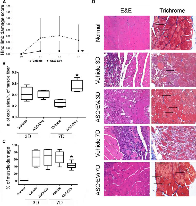
RESULTS

Characterization of ASC-EVs
ASC-EVs were analyzed for their surface markers using Guava FACS analysis (Figure I in the
online-only Data Supplement). ASC-EV modal particle size corresponds to 160 nm (Figure IA in
the online-only Data Supplement). No significant differences in ASC-EV number and size were
observed over the various batches by Nanosight analyses. ASCs produced 11 411±2316 EVs per
cell, after overnight starvation. Detailed information to estimate EV number is reported in the
online-only Data Supplement. Starting from 2.25×10¹¹ of total particles, the recovery of RNA and
protein was similar in all the samples analyzed (n=21); about 275±31 ng of RNA and about
30.52±2.27 µg of protein. CD63, CD81, and CD9 exosomal marker expression was also detected
using the MACSPlex kit for GUAVA FACS analysis (Figure IB in the online-only Data Supplement).
Moreover, ASC-EVs showed the expression of CD29, CD44, and CD105 (ASC markers) and HLA
I (Figure IB in the online-only Data Supplement). TEM was also performed and reported in Figure
IC in the online-only Data Supplement.

ASC-EVs Prevent Muscle Damage in a Mouse Model of Hindlimb Ischemia
Preliminary experiments to identify the best administration route were performed by comparing 2
different administration routes as indicated in Materials and Methods section. Route 1: 1×10¹⁰
administrated intravenously immediately after intervention (T0), 0.5×10¹⁰ via intramuscular
administration on day 1 (T1), and again on day 2 (T2); route 2: EVs were administered
intramuscularly with the same timing and doses (T0:1×10¹⁰; T1:0.5×10¹⁰; T2:0.5×10¹⁰; Figure II in
the online-only Data Supplement). Therefore, to evaluate ASC-EV protection, mice that were
subjected to the acute ischemic hindlimb process were treated with 2×10¹⁰ ASC-EVs using the
best administration route which corresponded to the route 1. The same volume of saline served as
control. Blood perfusion of the ischemic hindlimb analyzed immediately after surgery by Laser
Doppler, showed a strong reduction in blood flow (0.13±0.07 control group; 0.09±0.03 in ASC-EV
group; Figure III in the online-only Data Supplement). Although, no significant differences in large
vessel reperfusion were found in the treatment groups, the saline group showed significantly
higher functional damage scores than the ASC-EV-treated group (Figure 1A). To evaluate whether
functional differences were a result of improvement in tissue reperfusion, vessel number was
tallied in the ischemic muscles of treated animals at different time intervals (day 3 and 7); ASC-EV-
treated animals showed a higher number of vessels than the saline group on day 7 quantified as
number of small capillaries per number of muscle fiber (Figure 1B and 1D and Figure IV in the
online-only Data Supplement). No differences were obtained counting large vessels (not shown).
Furthermore, an analysis of gastrocnemius muscles revealed the presence of focal areas of
damage in the saline group. This effect was almost completely prevented by ASC-EV treatment
(Figure 1C and 1D). These results suggest that, in our model, ASC-EV treatment protects muscles
against ischemia-induced damage.
Figure 1. Effects of adipose stem cell-derived extracellular vesicles (ASC-EVs) on
ischemia/reperfusion damage. A, Foot damage score on different days (0, 1, 3, 7) after surgery in ASC-EV
treated and untreated mice. Data are expressed as mean±SD; *P value
Table 1. List of mRNAs Modulated in ASC-EV-Treated Muscles, Compared With Untreated Muscles (Table
view)

              Gene Symbol                                 Fold Regulation                        SD
 Myod1                                                         3.85                             ±0.30
 Myf5                                                          2.97                             ±0.18
 IL6                                                           2.89                             ±0.17
 Cav1                                                          2.33                             ±0.50
 Igf2                                                          2.03                             ±0.79
 Ppargc1a                                                      1.71                             ±0.59
 Ppargc1b                                                      1.71                             ±0.78
 Igf1                                                          1.69                             ±0.49
 Ctnnb1                                                        1.69                             ±0.67
 Capn2                                                         1.62                             ±0.31
 Capn3                                                         1.62                             ±0.51
 Prkaa1                                                        1.61                             ±0.58
 Lmna                                                          1.58                             ±0.24
 Cs                                                            1.57                             ±0.81
 IL1b                                                          1.55                             ±0.30
 Agrn                                                          1.55                             ±0.61
 Mapk14                                                        1.53                             ±0.75
 Prkag1                                                        1.52                             ±0.80
 Pax7                                                          1.52                             ±0.62
 Ikbkb                                                         0.65                             ±0.21
 Foxo1                                                         0.59                             ±0.15
 Myh1                                                          0.56                             ±0.18
 Tgfb1                                                         0.54                             ±0.25
 Lep                                                           0.24                             ±0.19

RQ (fold regulation) values are evaluated using: 2–ΔΔCt±SD (n=4). ASC-EV indicates adipose stem cell-derived
extracellular vesicles.
Figure 2. Adipose stem cell-derived extracellular vesicles (ASC-EVs) induce the expression of differentiation genes in muscles and in satellite cells exposed to ischemia/reperfusion (I/R) and induce proliferation on myoblast in vitro. A, Total RNA from I/R gastrocnemius muscles, both treated (ASC-EVs) and untreated (C) were analyzed for the expression of muscle differentiation genes (MyoD, Myogenin, Myf5, Pax7) by RT-PCR. Results are expressed as relative quantification (RQ) of data normalized for housekeeping genes. The results are representative of 3 different experiments (n=3; *P
These results were further validated by an analysis of the expression of cyclin kinase inhibitory
genes, p21 and p27 (Figure 2E).

ASC-EVs Induce Myoblast Differentiation and Prevent Apoptosis of Both
Myoblasts and Their Differentiated Counterparts
The effect of ASC-EVs on myoblast differentiation was also evaluated in vitro in murine myoblasts
subjected to I/R. Data reported in Figure 3A, demonstrate that ASC-EV-treated myoblasts
displayed a higher fusion index than untreated myoblasts. Moreover, the possibility that ASC-EVs
may prevent apoptosis was also investigated. A double-staining, using annexin V and 7AAD, was
performed. As shown in Figure 3B, ASC-EVs were able to increase the number of living cells and
to reduce the number of apoptotic myoblasts. The expression of Bcl-XL and the phosphorylation of
Bcl-2 were analyzed for validation purposes. As reported in Figure 3C, ASC-EV treatment was
associated with the increased expression of Bcl-XL and the phosphorylation of Bcl-2.19 The anti-
apoptotic effect of ASC-EVs was also evaluated in differentiated myoblasts by analyzing the
morphological alterations that occur in nuclei in the final stages of apoptosis. Data reported in
Figure 3D indicate that ASC-EV treatment decreased the percentage of fragmented nuclei in
differentiated myoblasts. Unlike proliferation (proliferating cell-nuclear antigen; Figure V in the
online-only Data Supplement), that occurs rapidly after I/R damage, we were able to demonstrate
the anti-apoptotic effects of ASC-EVs in vivo (Figure 3E). These data indicate that ASC-EVs can
protect muscles from I/R-induced damage by inducing myoblast proliferation, differentiation and by
preventing apoptosis.

Figure 3. Effects of adipose stem cell-derived extracellular vesicles (ASC-EVs) on myoblast
differentiation and apoptosis. A, After hypoxia, myoblasts were cultured in normoxic conditions in medium
supplemented with 10% FBS (C−) either with 2% horse serum (differentiation condition) with ASC-EVs (ASC-
EVs) or without ASC-EVs (C+; *P2 was considered
positive) and the total number of nuclei. B, Apoptosis assay on myoblasts cultured in hypoxic conditions in
medium with (C+) or without (C−) FBS either in the presence (ASC-EVs) or absence of ASC-EVs (*P
C+ and ASC-EVs vs C−). C, Representative images and relative densitometric analysis of p-Bcl-2 and Bcl-XL
protein expression in myoblasts treated as above and normalized for vinculin. The same blot was used to
evaluate p-Bcl-2 and Bcl-XL protein expression. Results are representative of all samples (n=5; *P
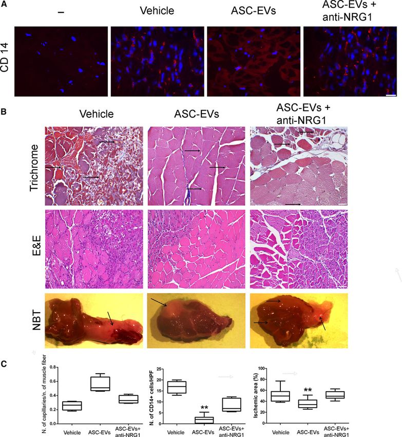
Bcl-XL were evaluated in myoblasts subjected to ASC-EV treatment. As shown in Figure 4B
through 4D, the NRG1 blockade prevented ASC-EV-mediated myoblast proliferation and the
transcription of EGR1, the NRG1 early response gene,23 while it was ineffective in preventing
protection against I/R induced apoptosis (Figure 4E). rhNRG1 served as positive internal control.
Moreover, the effect of the NRG1 blockade was also evaluated in endothelial cells subjected to
ischemia as the involvement of NRG1 in endothelial cell proliferation has already been reported.24
The results described in Figure 4F clearly demonstrate that ASC-EV-mediated endothelial cell
proliferation and EGR1 transcription (Figure VI in the online-only Data Supplement) were inhibited
by the NGR1 blockade. Finally, the pretreatment of ASC-EVs with trypsin demonstrates that ASC-
EV-mediated biological effects rely on having NRG1 bound to their membrane (Figure 4C and 4F).

Figure 4. Role of NRG1 (neuregulin 1) in adipose stem cell-derived extracellular vesicles (ASC-EV)-
mediated myoblast and endothelial cell proliferation. A, Expression of NRG1, CD63, and vinculin in ASC-
EVs, renal tubular (C−), and myotube cells (C+) were used as negative control and positive control
respectively.
B, BrdU (5-bromo-2-deoxyuridine) analysis of myoblasts cultured in hypoxic conditions, with (C+) and without
(C−) FBS, in the presence and in the absence of ASC-EVs (ASC-EVs), ASC-EVs treated with an anti-NRG1
blocking antibody (ASC-EVs+anti-NRG1) or with trypsin (ASC-EVs+trypsin; n=3; **P
with an anti-NRG1 blocking antibody (ASC-EVs+anti-NRG1) and with trypsin (ASC-EVs+trypsin; n=3 *P
negative control for primary antibodies is indicated as “−“.
B, Representative hematoxylin-eosin, and Masson’s trichrome, and NTB images of gastrocnemius muscles
of ischemic hindlimb in Vehicle, ASC-EVs and ASC-EVs+plus anti-NRG1 treated animals (original
magnification: ×200 for hematoxylin-eosin–scale bar: 50 µm and ×400–scale bar: 25 µm for Masson’s
trichrome). Arrows indicate the capillaries and the ischemic area in the NTB sections. C, Quantitative
analyses of capillaries (number of capillaries/number of muscle fiber), CD14+cells and the percentage of the
ischemic area in Vehicle, ASC-EV and ASC-EV plus anti-NRG1 ischemic hindlimb at day 7 after surgery are
reported. Data are expressed as mean±SEM; *P value
Gene Symbol         40–Ct (± SD)       Gene symbol        40–Ct, (± SD)       Gene Symbol         40–Ct, (± SD)
 EGF                    7.6±0.98           COL18A1            6.89±0.61             SPHK1             5.57±0.75
 FLT1                  7.57±0.64            PDGFA             6.85±0.59             PLAU              5.48±0.73
 IL8                   7.55±0.78             KDR              6.84±0.38             FGFR3             5.38±0.79
 IFNG                  7.51±0.88               F3             6.83±0.57               ID1             5.14±0.81
 IFNA1                 7.50±0.67           TGFBR1             6.81±1.19             S1PR1             5.08±0.83
 SERPINE1              7.46±0.75             BAI1             6.79±0.70             ERBB2             5.03±0.63
 FIGF                  7.45±0.50             NRP2             6.78±0.61            PECAM1             4.91±0.46
 TIMP3                 7.42±0.72           ANGPT2             6.75±0.48            NOTCH4             4.80±0.97
 JAG1                  7.39±0.92            MMP9              6.71±0.75             TYMP              4.51±0.61
 CXCL10                7.38±1.06            CXCL1             6.68±0.71              MDK              3.27±0.65

40–Ct values were calculated by subtracting the mean gene Ct from 40 total real-time polymerase chain reaction cycles
(±SD).

Figure 6. Adipose stem cell-derived extracellular vesicles (ASC-EV) cargo characterization and
pathway enrichment analysis. A, Venn diagram showing the comparison between mRNAs and proteins
carried by ASC-EVs.
B, List of the 18 molecules carried by ASC-EVs, such as mRNAs and proteins. C, Biological pathways in
which the 18 mRNA/proteins are mainly involved. Many enriched pathways are related to angiogenesis and
muscle regeneration. Bioinformatic analysis was performed using Funrich V3 software. The blue bar
represents the percentage of genes involved in the specific pathway; the red bar represents the significant P
value (P
Critical limb ischemia is a common feature of patients with peripheral artery disease.1,27 Since
current therapeutic options are still ineffective new therapeutic strategies with which to improve
patient outcomes are required. Adult stem cells are emerging as a cell-based therapy.4 Of the
stem cells available, ASCs are an ideal source.5 They are easy available, show low
immunogenicity and high proliferative capability5 and were found effective in preclinical models of
muscle atrophy28 and peripheral artery disease.29
  A number of evidence indicates that advantages may be provided by secretome derivatives of
stem cells, including EVs,5 in regenerative medicine and in therapeutic angiogenesis.30 Herein,
we investigated whether ASC-EVs could become a therapeutic tool with which to improve
angiogenesis in mice that recapitulate human critical limb ischemia. We have demonstrated that
ASC-EVs display pro-angiogenic effects in vivo, as shown by the number of capillary/muscle fiber
and by the analysis of capillary density. We have also found that mice treated with ASC-EVs
present a lower score for hindlimb functional damage than mice treated with the vehicle alone.
Accordingly, histological analyses have highlighted that mice subjected to ASC-EV treatment had
a reduced percentage of apoptotic cells and muscle damage. After damage, SCs enter the cell
cycle by activating p38MAPK and inducing the expression of MyoD in the daughter cells.11
Transcriptomic analysis, performed to detect early molecular events, has demonstrated that ASC-
EV treatment induced the expression of genes associated with myoblast proliferation and
differentiation, such as MyoD, Pax7, and Myf5, on day 3 after surgery. In accordance with these
results, SCs that were exposed to ASC-EVs in ischemic/reoxygenation conditions increased the
expression of MyoD. We further validated these data in vitro. Indeed, it has been demonstrated
that ASC-EV challenge leads to an increase in the number of BrdU positive myoblasts.
Accordingly, cyclin D1, CDK6, and p-p38/MAPK expression increased. Along with the effect on
myoblast proliferation, we have also been able to demonstrate that ASC-EVs induced myoblast
differentiation and prevented apoptotic signals in both myoblasts and their differentiated
counterparts. As a matter of fact, ASC-EVs were able to increase the expression of Bcl-XL and the
phosphorylation of Bcl219 in myoblasts and reduce the number of fragmented nuclei in
differentiated myoblasts. These results sustain the possibility that protection against apoptosis and
induction of vessel formation may well be the most relevant mechanisms via which a fast muscle
regeneration/protection occurs. In line with this hypothesis, we failed to detect an increased
number of regenerating myofibers and a decreased number of apoptotic cells at day 7 in ASC-EV-
treated animals.
    mRNAs, miRNAs, proteins, and bioactive lipids are all recognized as relevant mediators of EV
biological action in different regenerative processes.7 However, miRNAs are the most studied pro-
angiogenic drivers of stem-cell derived EV activities. This is also true for ASC-EVs.31–33 We have
demonstrated herein that ASC-EVs are also enriched in mRNAs that encode for a number of
angiogenic regulators. More importantly, proteomic analyses of ASC-EV cargo has indicated
relevant enrichment in NRG1, which is a member of the epidermal growth factor family that is also
produced by skeletal muscles.34,35 NRG1 regulates muscle cell homeostasis, SC survival and
many relevant biological processes in skeletal muscles.36 It exerts its biological effects by binding
to NRG1 receptors, including ErbB3 and ErbB4 and the co-receptors ErbB1 and ErbB2.35 We
herein report that ASC-EV-delivered NRG1 may be involved in muscle protection against I/R
damage, as is consistent with previous observations that demonstrate the involvement of the
NRG1/ErbB system in muscle regeneration.20 As a matter of fact, in vitro NRG1 blockade
prevented the activation of p-p38MAPK, the expression of cyclin D1 and the transcription of ERG1
in myoblasts treated with ASC-EVs and subjected to I/R. These biochemical events in vitro
translated into the inhibition of myoblast proliferation and in vivo in the prevention of ASC-EV-
mediated protection, as also supported by tetrazolium-staining. On the contrary, an NRG1-
independent mechanism seems to be associated with protection against I/R-induced apoptosis in
response to ASC-EV treatment. The IL-21R/IL-21 axis has been shown to prevent apoptosis and
favor cell survival in hypoxic conditions.37 Indeed, ASC-EVs were also enriched in IL-21,
suggesting that ASC-EV-treatment-associated apoptosis resistance may rely on the IL-21-
mediated signaling pathway. Further studies are required to validate this hypothesis.
It has been reported that endothelial cells express both NRG1 and its receptors,24 and Hedhli
et al22 have more recently demonstrated that NRG1 of endothelial origin is a crucial mediator of
both angiogenesis and arteriogenesis in a hindlimb ischemia model. In a finding that is consistent
with these results, we have demonstrated that, just like in myoblasts, ASC-EVs promote
endothelial cell proliferation and ERG1 transcription in hypoxic conditions and that these effects
rely on ASC-EV-membrane-bound NRG1.
The recruitment of inflammatory cells is necessary to promote vascular growth on damage.
However, it has been recently shown that ASC-EVs, obtained from patients undergoing abdominal
surgery, increased the recruitment of inflammatory cells without protecting muscles against I/R
damage.25 Of note, improvement of blood perfusion, protection of muscles, and a reduced
infiltration of inflammatory cells were observed in mice treated with ASC-EVs released in response
to PDGF (platelet-derived growth factor).25 The increase of IL-10 and TGF-β1 (transforming
growth factor beta 1) secretion by the inflammatory cells has been suggested to play a role in
PDGF-driven ASC-EV action. This suggests the possibility that immunomodulatory mechanisms
may be activated by ASC-EVs, including those mediated by NRG1.26 Nonetheless, possibly due
to the enrichment of pro-angiogenic mediators, ASC-EVs could induce a compensatory vascular
growth able to protect muscle from damage even in the presence of a reduced number of
inflammatory cells.
The existence of a dual ASC-EV biological effect, made up of angiogenesis and a hasty muscle
protection, is therefore sustained by our in vivo results. Moreover, pathway enrichment analysis
and, more importantly, the interaction graph further support the notion that cooperation between
the bioactive molecules carried by ASC-EVs sustain and boost biological effects in our preclinical
I/R model. This co-operative activity may provide a further explanation as to NRG1’s independent
protection against apoptosis in myoblasts and their differentiated counterparts, as well as to the
failure of 300 pg rhNRG1, corresponding to its ASC-EV content, to recapitulate ASC-EV
therapeutic effects.
EV therapeutic approaches have been proposed to solve a number of concerns about stem
cell-based therapy in a range of clinical settings.5 Our data demonstrate that the off-the-shelf ASC
secretome may be a useful therapeutic approach with which to improve vascular growth and to
protect muscles from I/R-mediated damage. Moreover, we have also identified NRG1 as one of
the most relevant mechanisms for ASC-EV biological activity. Effort should now be placed into
investigations of the therapeutic potential that ASC-EVs show for the treatment of a variety of
pathological conditions in which muscle protection is required.
Limitations
We are confident that the intrinsic curb of Laser Doppler Perfusion analysis, prevents the detection
of the profound vascular districts. However, histological analyses clearly demonstrated that, on
ASC-EV treatment, an increased number of capillary/number of muscle fiber are present in theischemic muscles. Moreover, concerns could be also raised by the protocol we adopted, owing to
the minimal residual blood flow (10%) detected after surgery (see Figure III in the online-only Data
Supplement). However, based on our results, it can be speculated that intravenous ASC-EVs
administration at T0, may have dual beneficial effects: locally, to boost of a hasty angiogenic
response, systemically, to activate signals in circulating inflammatory cells impairing their
recruitment into ischemic tissues, and prevent damage. Finally, since a low residual blood flow is a
common feature in critical limb ischemia patients undergoing revascularization, it could be
speculated that this protocol might be beneficial even in clinical settings.
ARTICLE INFORMATION
Received August 3, 2019; accepted October 17, 2019.
Affiliations
From the 2i3T Scarl University of Turin (F.F., M.C., C.C.), University of Turin, Italy. Department of Medical
Sciences (A. Ranghino, C.G., M.T., A. Rossi, G.T., S.F., M.V.G., G.C., M.F.B.), University of Turin, Italy.
Sources of Funding
This work was supported by grant number 071215 from Unicyte to 2i3T to G. Camussi and M.F. Brizzi.
Associazione Italiana per la Ricerca sul Cancro (AIRC) project IG 2015.17630 to M.F. Brizzi.
Disclosures
G. Camussi is a component of the Scientific Advisory Board of Unicyte AG. The other authors report no
conflicts.
REFERENCES
1. Olin, JW, White, CJ, Armstrong, EJ, Kadian-Dodov, D, Hiatt, WR. Peripheral artery disease. N Engl J
Med. 2016;67:558–587.
2. Vartanian, SM, Conte, MS. Surgical intervention for peripheral arterial disease. Circ Res.
2015;116:1614–1628. doi: 10.1161/CIRCRESAHA.116.303504 Crossref. PubMed.
3. Thukkani, AK, Kinlay, S. Endovascular intervention for peripheral artery disease. Circ Res.
2015;116:1599–1613. doi: 10.1161/CIRCRESAHA.116.303503 Crossref. PubMed.
4. Gupta, NK, Armstrong, EJ, Parikh, SA. The current state of stem cell therapy for peripheral artery
disease. Curr Cardiol Rep. 2014;16:447. doi: 10.1007/s11886-013-0447-2 Crossref. PubMed.
5. Vizoso, FJ, Eiro, N, Cid, S, Schneider, J, Perez-Fernandez, R. Mesenchymal stem cell secretome:
toward cell-free therapeutic strategies in regenerative medicine. Int J Mol Sci. 2017;18:E1852. Crossref.
PubMed.
6. Bronckaers, A, Hilkens, P, Martens, W, Gervois, P, Ratajczak, J, Struys, T, Lambrichts, I. Mesenchymal
stem/stromal cells as a pharmacological and therapeutic approach to accelerate angiogenesis.
Pharmacol Ther. 2014;143:181–196. doi: 10.1016/j.pharmthera.2014.02.013 Crossref. PubMed.
7. Camussi, G. Stem cell reviews and reports: microenvironment and extracellular microvesicles section.
Stem Cell Rev Rep. 2017;13:4. doi: 10.1007/s12015-017-9725-5 Crossref. PubMed.
8. Lee, Y, El Andaloussi, S, Wood, MJ. Exosomes and microvesicles: extracellular vesicles for genetic
information transfer and gene therapy. Hum Mol Genet. 2012;21(R1):R125–R134. doi:
10.1093/hmg/dds317 Crossref. PubMed.
9. Liu, L, Jin, X, Hu, CF, Li, R, Zhou, Z, Shen, CX. Exosomes derived from mesenchymal stem cells rescue
myocardial ischaemia/ reperfusion injury by inducing cardiomyocyte autophagy via AMPK and Aktpathways. Cell Physiol Biochem. 2017;43:52–68. doi: 10.1159/000480317 Crossref. PubMed.
10. Herrera, MB, Fonsato, V, Gatti, S, Deregibus, MC, Sordi, A, Cantarella, D, Calogero, R, Bussolati, B,
Tetta, C, Camussi, G. Human liver stem cell-derived microvesicles accelerate hepatic regeneration in
hepatectomized rats. J Cell Mol Med. 2010;14(6B):1605–1618. doi: 10.1111/j.1582-4934.2009.00860.x
Crossref. PubMed.
11. Jones, NC, Tyner, KJ, Nibarger, L, Stanley, HM, Cornelison, DD, Fedorov, YV, Olwin, BB. The
p38alpha/beta MAPK functions as a molecular switch to activate the quiescent satellite cell. J Cell Biol.
2005;169:105–116. doi: 10.1083/jcb.200408066 Crossref. PubMed.
12. Zhao, L, Johnson, T, Liu, D. Therapeutic angiogenesis of adipose-derived stem cells for ischemic
diseases. Stem Cell Res Ther. 2017;8:125. doi: 10.1186/s13287-017-0578-2 Crossref. PubMed.
13. Cavallari, C, Ranghino, A, Tapparo, M, Cedrino, M, Figliolini, F, Grange, C, Giannachi, V, Garneri, P,
Deregibus, MC, Collino, F, et al. Serum-derived extracellular vesicles (EVs) impact on vascular
remodeling and prevent muscle damage in acute hind limb ischemia. Sci Rep. 2017;7:8180. doi:
10.1038/s41598-017-08250-0 Crossref. PubMed.
14. Ranghino, A, Cantaluppi, V, Grange, C, Vitillo, L, Fop, F, Biancone, L, Deregibus, MC, Tetta, C, Segoloni,
GP, Camussi, G. Endothelial progenitor cell-derived microvesicles improve neovascularization in a
murine model of hindlimb ischemia. Int J Immunopathol Pharmacol. 2012;25:75–85. doi:
10.1177/039463201202500110 Crossref. PubMed.
15. Togliatto, G, Trombetta, A, Dentelli, P, Cotogni, P, Rosso, A, Tschöp, MH, Granata, R, Ghigo, E, Brizzi,
MF. Unacylated ghrelin promotes skeletal muscle regeneration following hindlimb ischemia via SOD-2-
mediated miR-221/222 expression. J Am Heart Assoc. 2013;2:e000376. doi: 10.1161/JAHA.113.000376
Crossref. PubMed.
16. Penna, C, Tullio, F, Femminò, S, Rocca, C, Angelone, T, Cerra, MC, Gallo, MP, Gesmundo, I, Fanciulli,
A, Brizzi, MF, et al. Obestatin regulates cardiovascular function and promotes cardioprotection through
the nitric oxide pathway. J Cell Mol Med. 2017;21:3670–3678. doi: 10.1111/jcmm.13277 Crossref.
PubMed.
17. Deregibus, MC, Figliolini, F, D’Antico, S, Manzini, PM, Pasquino, C, De Lena, M, Tetta, C, Brizzi, MF,
Camussi, G. Charge-based precipitation of extracellular vesicles. Int J Mol Med. 2016;38:1359–1366.
doi: 10.3892/ijmm.2016.2759 Crossref. PubMed.
18. Benito-Martin, A, Peinado, H. FunRich proteomics software analysis, let the fun begin! Proteomics.
2015;15:2555–2556. doi: 10.1002/pmic.201500260 Crossref. PubMed.
19. Campbell, TL, Mitchell, AS, McMillan, EM, Bloemberg, D, Pavlov, D, Messa, I, Mielke, JG, Quadrilatero,
J. High-fat feeding does not induce an autophagic or apoptotic phenotype in female rat skeletal muscle.
Exp Biol Med (Maywood). 2015;240:657–668. doi: 10.1177/1535370214557223 Crossref. PubMed.
20. Hirata, M, Sakuma, K, Okajima, S, Fujiwara, H, Inashima, S, Yasuhara, M, Kubo, T. Increased
expression of neuregulin-1 in differentiating muscle satellite cells and in motoneurons during muscle
regeneration. Acta Neuropathol. 2007;113:451–459. doi: 10.1007/s00401-007-0198-5 Crossref. PubMed.
21. Ho, AT Van, Hayashi, S, Bröhl, D, Auradé, F, Rattenbach, R, Relaix, F. Neural crest cell lineage restricts
skeletal muscle progenitor cell differentiation through neuregulin1-ErbB3 signaling. Dev Cell.
2011;21:273–287. Crossref. PubMed.
22. Hedhli, N, Dobrucki, LW, Kalinowski, A, Zhuang, ZW, Wu, X, Russell, RR 3rd, Sinusas, AJ, Russell, KS.
Endothelial-derived neuregulin is an important mediator of ischaemia-induced angiogenesis and
arteriogenesis. Cardiovasc Res. 2012;93:516–524. doi: 10.1093/cvr/cvr352 Crossref. PubMed.
23. Jacobson, C, Duggan, D, Fischbach, G. Neuregulin induces the expression of transcription factors and
myosin heavy chains typical of muscle spindles in cultured human muscle. Proc Natl Acad Sci U S A.
2004;101:12218–12223. doi: 10.1073/pnas.0404240101 Crossref. PubMed.
24. Russell, KS, Stern, DF, Polverini, PJ, Bender, JR. Neuregulin activation of ErbB receptors in vascular
endothelium leads to angiogenesis. Am J Physiol. 1999;277:H2205–H2211. doi:10.1152/ajpheart.1999.277.6.H2205 PubMed.
25. Lopatina, T, Favaro, E, Grange, C, Cedrino, M, Ranghino, A, Occhipinti, S, Fallo, S, Buffolo, F,
Gaykalova, DA, Zanone, MM, et al. PDGF enhances the protective effect of adipose stem cell-derived
extracellular vesicles in a model of acute hindlimb ischemia. Sci Rep. 2018;8:17458. doi:
10.1038/s41598-018-36143-3 Crossref. PubMed.
26. Ryzhov, S, Matafonov, A, Galindo, CL, Zhang, Q, Tran, TL, Lenihan, DJ, Lenneman, CG, Feoktistov, I,
Sawyer, DB. ERBB signaling attenuates proinflammatory activation of nonclassical monocytes. Am J
Physiol Heart Circ Physiol. 2017;312:H907–H918. doi: 10.1152/ajpheart.00486.2016 Crossref. PubMed.
27. Krishna, SM, Moxon, JV, Golledge, J. A review of the pathophysiology and potential biomarkers for
peripheral artery disease. Int J Mol Sci. 2015;16:11294–11322. doi: 10.3390/ijms160511294 Crossref.
PubMed.
28. Park, JU, Kwon, ST. Potential of autologous adipose-derived stem cells to regenerate atrophied muscle
in a rat model. Wound Repair Regen. 2017;25:944–955. doi: 10.1111/wrr.12598 Crossref. PubMed.
29. Rybalko, V, Hsieh, PL, Ricles, LM, Chung, E, Farrar, RP, Suggs, LJ. Therapeutic potential of adipose-
derived stem cells and macrophages for ischemic skeletal muscle repair. Regen Med. 2017;12:153–167.
doi: 10.2217/rme-2016-0094 Crossref. PubMed.
30. Camussi, G, Deregibus, MC, Quesenberry, PJ. Role of stem cell-derived extracellular RNA-carrying
vesicles cell reprogramming. Austin J Clin Pathol. 2014;1:1001.
31. Kang, T, Jones, TM, Naddell, C, Bacanamwo, M, Calvert, JW, Thompson, WE, Bond, VC, Chen, YE, Liu,
D. Adipose-derived stem cells induce angiogenesis via microvesicle transport of miRNA-31. Stem Cells
Transl Med. 2016;5:440–450. doi: 10.5966/sctm.2015-0177 Crossref. PubMed.
32. Lopatina, T, Bruno, S, Tetta, C, Kalinina, N, Porta, M, Camussi, G. Platelet-derived growth factor
regulates the secretion of extracellular vesicles by adipose mesenchymal stem cells and enhances their
angiogenic potential. Cell Commun Signal. 2014;12:26. doi: 10.1186/1478-811X-12-26 Crossref.
PubMed.
33. Togliatto, G, Dentelli, P, Gili, M, Gallo, S, Deregibus, C, Biglieri, E, Iavello, A, Santini, E, Rossi, C, Solini,
A, et al. Obesity reduces the pro-angiogenic potential of adipose tissue stem cell-derived extracellular
vesicles (EVs) by impairing miR-126 content: impact on clinical applications. Int J Obes (Lond).
2016;40:102–111. doi: 10.1038/ijo.2015.123 Crossref. PubMed.
34. Meyer, D, Yamaai, T, Garratt, A, Riethmacher-Sonnenberg, E, Kane, D, Theill, LE, Birchmeier, C.
Isoform-specific expression and function of neuregulin. Development. 1997;124:3575–3586. PubMed.
35. Britsch, S. The neuregulin-I/ErbB signaling system in development and disease. Adv Anat Embryol Cell
Biol. 2007;190:1–65. PubMed.
36. Gumà, A, Martínez-Redondo, V, López-Soldado, I, Cantó, C, Zorzano, A. Emerging role of neuregulin as
a modulator of muscle metabolism. Am J Physiol Endocrinol Metab. 2010;298:E742–E750. doi:
10.1152/ajpendo.00541.2009 Crossref. PubMed.
37. Wang, T, Cunningham, A, Dokun, AO, Hazarika, S, Houston, K, Chen, L, Lye, RJ, Spolski, R, Leonard,
WJ, Annex, BH. Loss of interleukin-21 receptor activation in hypoxic endothelial cells impairs perfusion
recovery after hindlimb ischemia. Arterioscler Thromb Vasc Biol. 2015;35:1218–1225. doi:
10.1161/ATVBAHA.115.305476 Crossref. PubMed.You can also read