Force-Induced Autophagy in Periodontal Ligament Stem Cells Modulates M1 Macrophage Polarization via AKT Signaling
←
→
Page content transcription
If your browser does not render page correctly, please read the page content below
ORIGINAL RESEARCH
                                                                                                                                              published: 26 May 2021
                                                                                                                                      doi: 10.3389/fcell.2021.666631

                                            Force-Induced Autophagy in
                                            Periodontal Ligament Stem Cells
                                            Modulates M1 Macrophage
                                            Polarization via AKT Signaling
                                            Nan Jiang 1† , Danqing He 2† , Yushi Ma 2 , Junxiang Su 3 , Xiaowen Wu 3 , Shengjie Cui 2 ,
                                            Zixin Li 2 , Yanheng Zhou 2 , Huajie Yu 4* and Yan Liu 2*
                                            1
                           Edited by:         Central Laboratory, Peking University School and Hospital of Stomatology & National Engineering Laboratory for Digital
            Naresh Kumar Rajendran,         and Material Technology of Stomatology & Beijing Key Laboratory of Digital Stomatology, Beijing, China, 2 Laboratory
University of Rochester, United States      of Biomimetic Nanomaterials, Department of Orthodontics, Peking University School and Hospital of Stomatology & National
                                            Engineering Laboratory for Digital and Material Technology of Stomatology & Beijing Key Laboratory of Digital Stomatology,
                     Reviewed by:
                                            Beijing, China, 3 Department of Endodontics, Shanxi Medical University School and Hospital of Stomatology, Shanxi, China,
                               Jie Li,      4
                                              The Fourth Division, Peking University School and Hospital of Stomatology, Beijing, China
 Chongqing Medical University, China
                     Tamara Kukolj,
      University of Belgrade, Serbia        Autophagy, a lysosomal degradation pathway, serves as a protective cellular mechanism
               Aleksandra Jaukovic,
      University of Belgrade, Serbia        in maintaining cell and tissue homeostasis under mechanical stimulation. As the
                   *Correspondence:         mechanosensitive cells, periodontal ligament stem cells (PDLSCs) play an important
                            Huajie Yu       role in the force-induced inflammatory bone remodeling and tooth movement process.
                yuhuajie666@163.com
                              Yan Liu
                                            However, whether and how autophagy in PDLSCs influences the inflammatory bone
                orthoyan@bjmu.edu.cn        remodeling process under mechanical force stimuli is still unknown. In this study,
     † These   authors have contributed     we found that mechanical force stimuli increased the expression of the autophagy
                    equally to this work
                                            protein LC3, the number of M1 macrophages and osteoclasts, as well as the ratio of
                    Specialty section:
                                            M1/M2 macrophages in the compression side of the periodontal ligament in vivo. These
         This article was submitted to      biological changes induced by mechanical force were repressed by the application
             Cell Growth and Division,
                                            of an autophagy inhibitor 3-methyladenine. Moreover, autophagy was activated in
                a section of the journal
  Frontiers in Cell and Developmental       the force-loaded PDLSCs, and force-stimulated PDLSC autophagy further induced
                                Biology     M1 macrophage polarization in vitro. The macrophage polarization could be partially
        Received: 10 February 2021          blocked by the administration of autophagy inhibitor 3-methyladenine or enhanced by
           Accepted: 22 April 2021
           Published: 26 May 2021
                                            the administration of autophagy activator rapamycin in PDLSCs. Mechanistically, force-
                               Citation:
                                            induced PDLSC autophagy promoted M1 macrophage polarization via the inhibition
     Jiang N, He D, Ma Y, Su J, Wu X,       of the AKT signaling pathway. These data suggest a novel mechanism that force-
    Cui S, Li Z, Zhou Y, Yu H and Liu Y
                                            stimulated PDLSC autophagy steers macrophages into the M1 phenotype via the AKT
     (2021) Force-Induced Autophagy
   in Periodontal Ligament Stem Cells       signaling pathway, which contributes to the inflammatory bone remodeling and tooth
           Modulates M1 Macrophage          movement process.
        Polarization via AKT Signaling.
       Front. Cell Dev. Biol. 9:666631.     Keywords: autophagy, macrophage polarization, bone remodeling, mechanical force, periodontal ligament stem
       doi: 10.3389/fcell.2021.666631       cells, inflammation, tooth movement, AKT signaling

Frontiers in Cell and Developmental Biology | www.frontiersin.org                 1                                              May 2021 | Volume 9 | Article 666631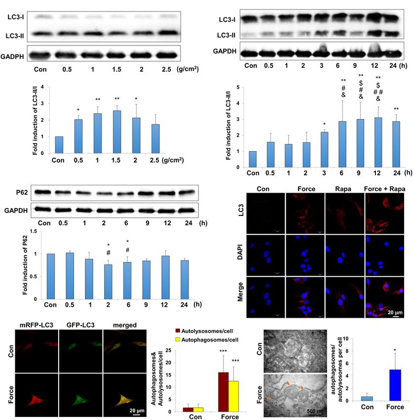
Jiang et al.                                                                                   PDLSC Autophagy Modulates Macrophage Polarization

INTRODUCTION                                                              mainly mediates the inflammation process. It could be
                                                                          activated by interferon (IFN)-γ or lipopolysaccharides, and
Mechanical force plays a vital role in maintaining tissue                 expressed inflammatory elements such as tumor necrosis
homeostasis and mediating pathological process under                      factor (TNF)-α and nitric oxide (NO); the M2 phenotype
physiological or pathological conditions (Smutny et al.,                  mainly participates in tissue remodeling, which could be
2017; Miroshnikova et al., 2018). In the alveolar bone                    activated by interleukin (IL)-4 or IL-13 and could produce
environment, mechanical force supports the homeostasis                    IL-10, arginase-1 (Arg-1) and DECTIN-1 (Murray, 2017;
during masticatory movements (Thompson et al., 2012),                     Shapouri-Moghaddam et al., 2018). Previously, we have
and also mediates the bone remodeling process during tooth                confirmed that M1 macrophage polarization is critical in
movement (Meikle, 2006). During tooth movement, aseptic                   the bone remodeling and root resorption process during
inflammatory microenvironment is developed in the periodontal             tooth movement (He et al., 2015a,b). Given the importance
tissues, which is characterized by elevated expressions of                of M1 macrophage polarization during tooth movement,
inflammatory cytokines, chemokines, and increased activations             we hypothesized that autophagy in PDLSCs influences
of inflammatory immune cells (Garlet et al., 2007; He et al.,             macrophage polarization under mechanical force stimuli
2015b; Yan et al., 2015). As the main mesenchymal stem                    and thus influences alveolar bone remodeling. This study
cells (MSCs) in the periodontal tissues, periodontal ligament             aims to verify whether and how force-induced autophagy in
stem cells (PDLSCs) constantly receive mechanical force                   PDLSCs modulates macrophage polarization and contributes
stimuli and contribute to the inflammatory responses and                  to periodontal inflammatory microenvironment and bone
bone remodeling process during tooth movement (Zhang                      remodeling during tooth movement.
et al., 2016; Huang et al., 2018). Increased expressions of
inflammatory cytokines, chemokines, and gas molecules
such as hydrogen sulfide have been found in the force-                    MATERIALS AND METHODS
stimulated PDLSCs (Lee et al., 2012; Liu et al., 2017; He
et al., 2020). However, how mechanical force modulates                    Animals and Treatments
PDLSC behaviors and therefore influences the inflammatory                 Adult Sprague-Dawley rats (180–200 g, 6–8 weeks, male) were
responses and the bone remodeling process under force stimuli             divided into three groups, including the group with force
is still obscure.                                                         loading (Force, n = 10), the group with force loading and 3-
    Autophagy has been gradually acknowledged as an important             methyladenine application (Force + 3-MA, n = 10) and the
protective cellular process to maintain cell and tissue homeostasis       control group without force loading (Con, n = 10). Orthodontic
under the external stimuli, such as stress, inflammation,                 force was applied for 7 days as previously described (He et al.,
hypoxia, and mechanical load (Hara et al., 2006; Jiang                    2015b). Briefly, a nickel–titanium coil spring (wire size 0.2 mm,
et al., 2010; Kroemer et al., 2010). Cells could degrade                  1 mm in diameter, and 1 mm in length, Smart Technology,
damaged organelles and misfolded proteins, and therefore                  China) was bonded between the upper first molar and incisors of
maintain themselves survival. The degraded components are                 rats to provide approximately 50–60 g force (Taddei et al., 2012).
encapsulated in autophagic vacuoles or autophagosomes, which              Volumetrically equivalent saline or 3-MA (30 mg/kg, Sigma, St.
fuse with lysosomes to form autophagolysosomes (Mizushima                 Louis, MO, United States) was injected intraperitoneally every
et al., 2008; Klionsky et al., 2012). Autophagy has also                  day during tooth movement (Jiang et al., 2010) in the Force or
been shown to participate in the pathological process of                  Force + 3-MA group, respectively.
inflammatory diseases, such as chronic intestinal inflammation,              The rats were sacrificed after 7 days, and the maxillae were
inflammatory bowel diseases and inflammatory periodontitis                harvested for further studies. The tooth movement distance
(Levine et al., 2011; An et al., 2016; Wang et al., 2019).                was measured between the midpoint of the distal-marginal
In the treatment of autoimmune encephalomyelitis, autophagy               ridge of the maxillary first molar and the midpoint of the
has been discovered to modulate the immunoregulatory                      mesial-marginal ridge of the maxillary second molar (He et al.,
properties of MSCs (Dang et al., 2014). In addition, autophagy            2015b). The measurement was repeated for three times and the
might also be adapted to mechanical load, which could                     average distance was calculated. All experimental protocols were
be activated in mechanosensitive cells such as osteoblasts,               approved by the Institutional Animal Use and Care Committee
endothelial cells, and PDLSCs (King et al., 2011; Ma et al.,              of Peking University (LA2013-92).
2013; Memmert et al., 2019). However, whether autophagy
in PDLSCs influenced inflammatory microenvironment of                     Human PDLSCs Culture and Mechanical
the periodontal tissues under mechanical force stimuli needs              Force Loading
further exploration.                                                      Human PDLSCs were isolated from the extracted healthy
    During the force-induced inflammatory bone remodeling                 premolars of volunteers receiving orthodontic treatment with
process, macrophages are regarded as one of the vital                     informed consent as previously described (Seo et al., 2004). The
immune cells (Feng and Teitelbaum, 2013; Horwood,                         ethic protocols were approved by the Peking University Ethical
2016). Depending on the different environmental signals,                  Committee (PKUSSIRB-201311103). Briefly, the periodontal
macrophages show a broad spectrum of activation phenotypes,               ligament was scraped from the root surface and minced into
described by M1 or M2 polarization. The M1 phenotype                      small pieces. Periodontal tissue explants were cultured in cell

Frontiers in Cell and Developmental Biology | www.frontiersin.org     2                                       May 2021 | Volume 9 | Article 666631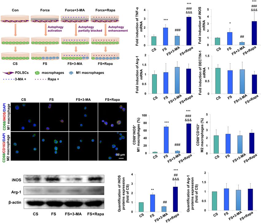
Jiang et al.                                                                                 PDLSC Autophagy Modulates Macrophage Polarization

culture flask upside down with DMEM medium (Invitrogen,                 were harvested from THP-1-derived macrophages for further
Carlsbad, CA, United States) with 20% fetal bovine serum (Gibco,        detection of macrophage-related markers.
MA, United States) and 1% Penicillin/Streptomycin. Cells were              To further explore the underlying mechanism, total
grown in a CO2 incubator at 37◦ C. The PDLSCs were identified           protein was harvested from THP-1-derived macrophages
following our previous protocols (Fu et al., 2016) and used at          after incubation in the conditioned medium for 2-4 h
passage four for further analysis.                                      to detect the changes of signaling pathway. Next, the
    To apply compressive force on PDLSCs, glass layers and 15 ml        activator of AKT signaling pathway IGF1 (100 ng/ml,
plastic tube caps containing weighed metal balls were used to           Abcam, United States) was applied to the macrophages of
apply compressive force (0.5–2.5 g/cm2 ) lasting for different          FS + Rapa group, and the inhibitor of AKT singnalling
time points, which was modified from a previously described             pathway GSK690693 (10 µM, Beyotime Biotechnology,
method (Kanzaki et al., 2002). After subjected to compressive           Jiangsu, China) was applied to the macrophages of
force for 12 h, PDLSCs were collected for further experiments           FS + 3-MA group. Total protein was extracted at 30 min
of transmission electronic microscopy (TEM), autophagy flux             to test the chemical drug efficiency. In addition, total
assay and immunofluorescence staining. Next, the culture                RNA and protein were harvested from THP-1-derived
medium from force-loaded PDLSCs (1.5 g/cm2 , 12 h) and force-           macrophages after 24 h treatment for further detection of
loaded PDLSCs with additional treatment of 3-MA (2.5 mM,                macrophage-related markers.
12 h) or rapamycin (0.25 µg/ml, 12 h, Sigma, St. Louis, MO,
United States) (Ma et al., 2018) were collected to treat THP-1-         Immunohistochemical Staining
derived macrophages.                                                    The trimmed maxillae were fixed in 10% neutral buffered
                                                                        formalin for 24 h, decalcified in 15% ethylenediaminetetraacetic
Transmission Electronic Microscopy                                      acid and embedded in paraffin. Consecutive 4-µm sagittal
Periodontal ligament stem cells with or without force loading           sections of the maxillary first molar were obtained and the middle
were fixed in 2.5% (v/v) glutaraldehyde, and treated with 3%            to apical third of the periodontal tissues was observed. A two-
potassium ferrocyanide and 1% (w/v) osmium tetroxide for 1 h            step detection kit (Zhongshan Golden Bridge Biotechnology,
at 4◦ C. Cells were then treated with 0.3% thiocarbohydrazide for       Beijing, China) was used following previous protocols (He
5 min at room temperature, and then incubated in 1% osmium              et al., 2015b). Primary antibodies including anti-microtubule
tetroxide for 20 min at 4◦ C. After dehydrated in the series of         associated protein 1 light chain 3 β (LC3B) (1:100, CST3868S,
ethanol, cells were embedded and the ultra-thin sections were           CST), anti-TNF-α (1:100, ab1793, Abcam), and anti-CD146
harvested and stained with solution containing 1% toluidine blue        (1:200, ab-75769, Abcam) were used. Five different slides from
and 2% borate. Images were observed with a JEM-1400-Plus                each sample (n = 10) were used for cell counting. Each slide was
transmission electron microscope (JEOL, Japan).                         measured for three times and the average positive cell numbers
                                                                        were calculated.
Autophagy Flux Assays
Periodontal ligament stem cells were transfected with the mRFP-         Tartrate-Resistant Acid Phosphatase
GFP-LC3 adenoviral (HanBio, Shanghai, China) according to the           Staining
manufacturer’s instructions. After infection, cells were cultured       Histology sections were stained with an acid phosphatase
for another 24 h and then subjected to compressive force                kit (387A-1KT; Sigma) for tartrate-resistant acid phosphatase
(1.5 g/cm2 ) for 12 h. After force loading, autophagosomes and          (TRAP) staining. TRAP-positive multinucleated cells (≥3 nuclei)
autolysosomes were observed and captured with a laser scanning          attached to the surface of the adjacent alveolar bone were
microscope (LSM 510, Zeiss, Jena, Germany) and the images               counted as osteoclasts.
were processed using LSM 5 Release 4.2 software. The intensity
of autophagy flux was determined by assessing the numbers of
                                                                        Immunofluorescence Staining
RFP/GFP-expressing cells.
                                                                        For immunofluorescence staining, tissue sections were
                                                                        double-stained with anti-CD68 (1:500; MCA341GA, Serotec,
Treatment of THP-1-Derived                                              United Kingdom) and anti-inducible nitric oxide synthase
Macrophages                                                             (iNOS) (1:100; ab-15323, Abcam) for M1 macrophages, or
THP-1 human monocytic cells (ATCCTIB-202) were                          anti-CD68 and anti-CD163 (1:100; sc-33560, Santa Cruz)
differentiated into macrophages with the treatment of 50                for M2 macrophages. PDLSCs were stained with anti-LC3B
ng/ml phorbol 12-myristate 13-acetate (PMA, Sigma) for                  (1:100, CST3868S, CST). Samples were incubated with primary
24 h. To determine whether the autophagy of force-stimulated            antibodies at 4◦ C overnight. On the following day, the samples
PDLSCs could influence macrophage polarization, THP-1-                  were incubated with fluorescein secondary antibody (1:200,
derived macrophages were incubated with supernatant from                Jackson Immuno Research Laboratories, West Grove, PA,
the force-loaded PDLSCs (FS) and force-loaded PDLSCs with               United States). Nuclei were counterstained with 4’,6-diamidino-
additional treatment of 3-MA (FS + 3-MA) or rapamycin                   2-phenylindole (DAPI). Samples were observed with a laser
(FS + Rapa). THP-1-derived macrophages incubated with                   scanning microscope (LSM 510, Zeiss), and the images were
supernatant from the PDLSCs without force-loaded served                 processed using LSM 5 Release 4.2 software. Three different
as control (CS). After 24 h, total RNA and total protein                slides from each sample (n = 6) were used for cell counting. Each

Frontiers in Cell and Developmental Biology | www.frontiersin.org   3                                       May 2021 | Volume 9 | Article 666631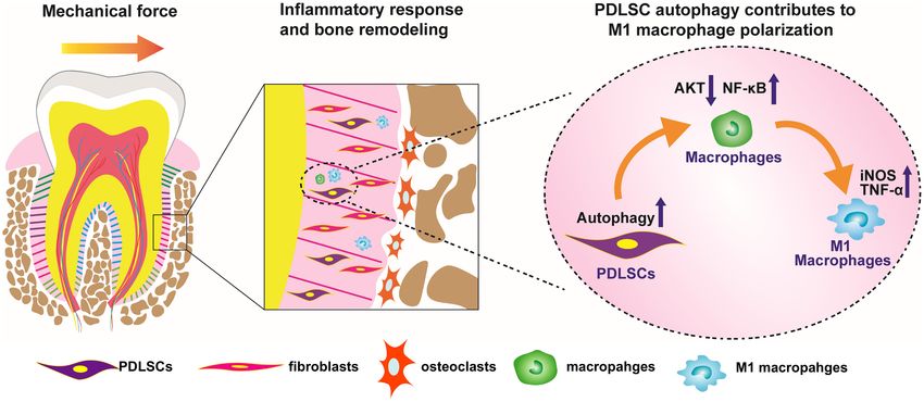
Jiang et al.                                                                                 PDLSC Autophagy Modulates Macrophage Polarization

slide or cell sample was measured for three times and the average        Statistical Analysis
positive cell numbers were calculated.                                   Data were presented as mean ± SD. Student’s t-tests or one-way
                                                                         analysis of variance (ANOVA) with post hoc tests were used to
Quantitative Real-Time Polymerase                                        compare the differences between groups using SPSS20.0. P < 0.05
Chain Reaction                                                           was considered statistically significant.
Total RNA was extracted using TRizol reagent (Invitrogen,
Carlsbad, CA, United States) in accordance with the
manufacturer’s instructions. RNA samples (2000 ng) were                  RESULTS
reverse transcribed into cDNA using RevertAid First Strand
cDNA Synthesis kit (Thermo Fisher Scientific). Then real-                Force-Induced Autophagy in PDLSCs
time Polymerase Chain Reaction (PCR) was performed using                 Contributes to M1 Macrophage
the FastStart Universal SYBR Green master kit (Roche) on                 Polarization in vivo
an Applied Biosystems 7500 Real-Time PCR System (Life                    To investigate whether mechanical force could activate
Technologies Corporation, United States) to determine the                autophagy, an experimental animal model of tooth movement
relative mRNA expression level of autophagy marker Beclin-               was set up and autophagy was systemically blocked with the
1. M1 macrophage markers including TNF-α, iNOS, and M2                   inhibitor 3-MA. After force loading for 7 days, the tooth
macrophage markers including arginase-1 (Arg-1) and DECTIN-              movement distance increased to 505.4 ± 72.4 µm, and was
1 were also detected. Fold changes of target genes were calculated       partially reversed to 327.8 ± 47.27 µm after 3-MA injection
with MMCT method using GAPDH as reference control. The                   (P < 0.001, Figure 1A). The expressions of the autophagy
sequences of human primers (designed by Primer Premier 5.0               protein LC3, the pro-inflammatory cytokine TNF-α and the
software) were listed as follows:                                        MSC surface marker CD146 increased in the compression
       GAPDH sence/antisence: 50 -ATGGGGAAGGTGAAGGT                      side of the periodontal tissues after force loading (P < 0.001).
       CG-30 /50 -GGGGTCATTGATGGCAACAATA-30 .                            Concomitantly, the number of TRAP+ osteoclasts also increased
       TNF-α sence/antisence: 50 -GAGGCCAAGCCCTGGTA                      after force loading (P < 0.001, Figures 1B,C). 3-MA injection
       TG-30 /50 -CGGGCCGATTGATCTCAGC-30 .                               significantly decreased the expressions of LC3, TNF-α and
       iNOS sence/antisence: 50 -TTCAGTATCACAACCTCAGC                    CD146, as well as the number of TRAP+ osteoclasts (P < 0.001).
       AAG-30 /50 -TGGACCTGCAAGTTAAAATCCC-30 .                           Nevertheless, the number of CD146+ MSCs and TRAP+
       DECTIN-1 sence/antisence: 50 -GGAAGCAACACATTGG                    osteoclasts was still upregulated in the Force + 3-MA group,
       AGAATGG-30 /50 -CTTTGGTAGGAGTCACACTGTC-30 .                       compared with the control group (P < 0.05, Figures 1B,C).
       arginase-1 sence/antisence: 50 -TGGACAGACTAGGAATT                    Mechanical force modulates M1 macrophage polarization,
       GGCA-30 /50 -CCAGTCCGTCAACATCAAAACT-30 .                          which contributes to bone remodeling and root resorption during
       Beclin-1 sence/antisence: 50 -ATTCGAGAGCAGCATCC                   tooth movement (He et al., 2015b). To assess the influence of
       AAC-30 /50 -AACAGGAAGCTGCTTCTCAC-30 .                             force-induced autophagy on macrophage polarization in vivo,
                                                                         immunofluorescence staining was performed to identify the
Western Blot Analysis                                                    changes of macrophage markers after force loading. The
Western blot analyses were performed following previous                  number of CD68+ iNOS+ M1 macrophages increased on the
protocols (He et al., 2015b). Total protein was extracted from           compression side of the periodontal ligament after force loading
cultured cells using RIPA lysis buffer (Sigma) with protein              (P < 0.001 versus Con) and decreased significantly after 3-
inhibitor cocktail (Roche) and PhosSTOP Phosphatase inhibitor            MA injection (P < 0.01 versus Force). Although the number of
cocktail (Roche, United States). Lysates were centrifuged at             CD68+ CD163+ M2 macrophages increased after force loading
12,000 × g for 15 min. Supernatants were collected for                   and 3-MA injection (P < 0.05 and 0.01, respectively), the
further use. Proteins were separated by 10% SDS-PAGE,                    ratio of M1/M2 macrophage polarization increased significantly
transferred onto PVDF membrane (Bio-Rad), blocked in 5% BSA              after force loading (P < 0.01 versus Con) and decreased
and then probed overnight with primary antibodies included               significantly after 3-MA injection (P < 0.01 versus Con and
anti-β-actin (1:10,000, a-5441, Sigma), anti-GAPDH (1:10,000,            Force, Figure 1D). These data suggest that autophagy modulates
60004, Proteintech), anti-LC3(1:500, CST3868S, CST), anti-               macrophage polarization after force loading and influences the
P62 (1:1,000, ab91526, Abcam), anti-iNOS (1:500, abs130136,              alveolar bone remodeling and tooth movement process.
Absin), anti-Arg-1 (1:5,000, sc-21050, Santa Cruz), anti-TNF-
α (1:200, sc-52746, Santa Cruz), anti-phospho-AKT1 (Ser473)              Mechanical Force Activates Autophagy
(1:500, 9018S, CST), anti-AKT (1:500, 9272, CST), and                    in PDLSCs
anti-NF-κB/P65 (1:1,000, 8242S, CST). The blots were then                Periodontal ligament stem cells, as the main MSCs in the
incubated with a horseradish peroxidase-conjugated secondary             periodontal ligament microenvironment, respond to mechanical
antibodies (1:5,000; Zhongshan Golden Bridge Biotechnology,              force and participate in the alveolar bone remodeling and tooth
Beijing, China). Finally, the protein bands were enhanced by             movement. To explore the autophagy activity under mechanical
chemiluminescence detection before photography. The relative             force, we performed western blot, immunofluorescence staining
density of at least three comparable results was measured by             autophagy flux assay and TEM on PDLSCs in vitro. LC3 is one
Image J software.                                                        of the most essentially monitored autophagy-related proteins.

Frontiers in Cell and Developmental Biology | www.frontiersin.org    4                                      May 2021 | Volume 9 | Article 666631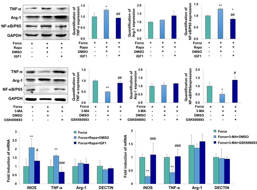
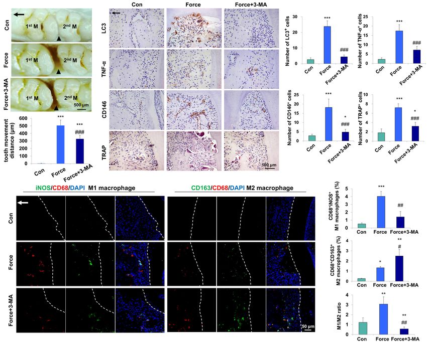
Jiang et al.                                                                                                   PDLSC Autophagy Modulates Macrophage Polarization

  FIGURE 1 | Mechanical force-induced autophagy contributes to M1 macrophage polarization and bone remodeling during tooth movement in vivo.
  (A) Representative images and semiquantification analysis of the tooth movement distance in rats. Force loading increased the tooth movement distance, which was
  partially reversed by 3-MA application. 1st M, the first molar; 2nd M, the second molar. Black triangles represent the tooth movement distance. (B) Representative
  immunohistochemical images of LC3, TNF-α and CD146 and TRAP staining. (C) Semiquantification analysis of the number of LC3+ , TNF-α+ and CD146+ and
  TRAP+ cells in panel (B). (D) Representative immunofluorescence images on the compression side of distobuccal roots and semiquantification analysis of
  double-labeled cells. M1 macrophage polarization was identified by CD68+ (red) and inducible nitric oxide synthase (iNOS)+ (green), whereas M2 macrophage
  polarization was identified as CD68+ (red) and CD163+ (green). Dashed lines mark the outline of distobuccal roots. Arrow represents the direction of force. Results
  are presented as mean ± SD. n = 10. *P < 0.05, **P < 0.01, ***P < 0.001 versus Con; # P < 0.05, ## P < 0.01, ### P < 0.001 versus Force.

Increased expression of LC3II/I was detected under compressive                        PDLSCs after compressive force stimulation for 12 h, which
force loading from 0.5 to 2.5 g/cm2 for 12 h. Nevertheless, 1.0 and                   was further enhanced after rapamycin application (Figure 2D).
1.5 g/cm2 compressive force triggered the strongest expressions                       Autophagosomes, also known as initial autophagic vacuoles,
of LC3II/I (Figure 2A), and 1.5 g/cm2 compressive force was                           are spherical structures with bilayers membranes containing
utilized in the following experiments. In addition, the expressions                   cytoplasmic components and/or organelles (Kliosnky et al.,
of LC3II/I started to increase increased after force stimulation for                  2016). mRFP-GFP-LC3 adenovirus were transfected into PDLSCs
3 h and persisted to 24 h (Figure 2B). Moreover, the expression                       to detect autophagic flux. Cells were applied with compressive
of another autophagy marker P62/SQSTM (P62) decreased after                           force for 12 h, and the flux rate of autophagy was detected with
compressive force loading for 2 and 6 h (P < 0.05, Figure 2C);                        microscopy. The numbers of autolysosomes (mRFP-positive, red
whereas the expression of Beclin1 increased after force stimuli                       dots) and autophagosomes (GFP/RFP double-positive, yellow
for 3 h (P < 0.05, Supplementary Figure 1A). Accordingly,                             dots) were significantly higher in the force group (P < 0.001,
real-time PCR showed that the mRNA expression of Beclin1                              Figure 2E). In addition, autophagosomes could be obviously
was significantly upregulated compared with the control group                         identified in the PDLSCs by TEM after compressive force
(P < 0.01, Supplementary Figure 1B). Immunofluorescence                               stimulation (Figure 2F). Taken together, these data reveal
staining showed that positive expression of LC3 aggregated in                         that under compressive force stimulation, autophagy can be

Frontiers in Cell and Developmental Biology | www.frontiersin.org                 5                                              May 2021 | Volume 9 | Article 666631
Jiang et al.                                                                                                   PDLSC Autophagy Modulates Macrophage Polarization

  FIGURE 2 | Autophagy is induced with the application of mechanical stimuli in PDLSCs. (A) Western blot of the expressions of LC3 in PDLSCs under different force
  values at protein level. The relative fold change of LC3-II/I was quantified. GAPDH served as an internal control for equal loading. Results are presented as
  mean ± SD. n = 3–4. ∗ P < 0.05, ∗∗ P < 0.01 versus Con. (B) Western blot of the expressions of LC3 in PDLSCs under force stimuli (1.5 g/cm2 ) at different time
  points. The relative fold change of LC3-II/I was quantified. GAPDH served as an internal control for equal loading. Results are presented as mean ± SD. n = 3–4.
  ∗ P < 0.05, ∗∗ P < 0.01 versus Con;$I P < 0.05 versus 0.5 h; # P < 0.05,## P < 0.01 versus 1 h; & P < 0.05, && P < 0.01 versus 2 h. (C) Western blot and

  quantification of P62 expression in PDLSCs under force stimuli (1.5 g/cm2 ) at different time points. Results are presented as mean ± SD. n = 3. ∗ P < 0.05 versus
  Con; # P < 0.05 versus 0.5 h. (D) Immunofluorescence staining of LC3 (red) in PDLSCs with the application of force stimuli (1.5 g/cm2 ) or rapamycin (Rapa).
  (E) PDLSCs were infected with adenovivus with mRFP-GFP-LC3. Cells were applied with static force for 6 h, and the autophagosomes and autolysosomes were
  detected in PDLSCs. Yellow-colored autophagosomes and red-colored autolysosomes were calculated. Results are presented as mean ± SD. n = 4. ∗∗∗ P < 0.001
  versus Con. (F) Ultrastructural features in PDLSC assessed by TEM without or with force. Arrows indicated the autophagosomes or autolysosomes in the
  cytoplasm. Numbers of autophagosomes/autolysosomes were quantified. Results are presented as mean ± SD. n = 3. ∗ P < 0.05 versus Con.

activated in a force-dependent and time-dependent tendency                            the force-loaded PDLSCs (FS), the force-loaded PDLSCs with
in PDLSCs.                                                                            additional treatment of an autophagy inhibitor 3-MA (FS + 3-
                                                                                      MA) or an autophagy activator rapamycin (FS + Rapa) was
                                                                                      utilized to treat THP-1-derived macrophages. THP-1-derived
Force-Stimulated PDLSC Autophagy                                                      macrophages incubated with supernatant from PDLSCs without
Induces M1 Macrophage Polarization                                                    force loading served as the control (CS) (Figure 3A).
in vitro                                                                                 The mRNA expression levels of M1 macrophage associated
To further detect whether autophagy in force-stimulated PDLSCs                        markers TNF-α and iNOS in THP-1-derived macrophages were
could affect macrophage polarization, the supernatant from                            upregulated in the FS group (P < 0.001 and P < 0.05 versus CS,

Frontiers in Cell and Developmental Biology | www.frontiersin.org                 6                                              May 2021 | Volume 9 | Article 666631
Jiang et al. PDLSC Autophagy Modulates Macrophage Polarization FIGURE 3 | Force-stimulated PDLSC autophagy induces M1 macrophage polarization in vitro. (A) Schematic illustration. (B) Relative mRNA expression of M1/M2-related genes of THP-1-derived macrophages with treatment of different conditioned medium. CS: control supernatant; FS: force-loaded supernatant. FS + 3-MA: force-loaded supernatant with 3-MA; FS + Rapa: force-loaded supernatant with rapamycin. Results are presented as mean ± SD. n = 6.∗ P < 0.05, ∗∗ P < 0.01, ∗∗∗ P < 0.001 versus CS; ## P < 0.01, ### P < 0.001 versus FS; &&& P < 0.001 versus FS + 3-MA. (C) Representative immunocytochemical images of THP-1-derived macrophages. M1 macrophage polarization [CD68+ (green) and iNOS+ (red)] increased in the FS group, which decreased significantly after 3-MA application or enhanced after rapamycin application. No change was observed in M2 macrophage polarization [CD68+ (green) and CD163+ (red)]. (D) Quantification of CD68+ /iNOS+ double positive cells and CD68+ /CD163+ double positive cells in panel (C). Results are presented as mean ± SD. n = 3–4. ∗∗∗ P < 0.001 versus CS; ### P < 0.001 versus FS; &&& P < 0.001 versus FS + 3-MA. (E) Western blot of the expressions of iNOS and arginase-1 in THP-1-induced macrophages. Beta-actin served as an internal control for equal loading. Protein expression level was quantified and presented as mean ± SD. n = 3. ∗∗ P < 0.01, ∗∗∗ P < 0.001 versus CS; ## P < 0.01 versus FS; &&& P < 0.001 versus FS + 3-MA. respectively), which were downregulated in the FS + 3-MA group (P < 0.001) group and enhanced in the FS + Rapa group (P < 0.001 and P < 0.01 versus FS, respectively) and further (P < 0.001). However, the proportion of CD68+ CD163+ enhanced in the FS + Rapa group (P < 0.001 versus CS, FS and M2 macrophages did not changed [Figures 3C,D and FS + 3-MA). However, no changes were observed of the mRNA Supplementary Figure 2 (Split images)]. Correspondingly, expressions of M2 macrophage associated markers arginase-1 western blot analysis showed that the expression of M1 and DECTIN-1 (Figure 3B). macrophage associated marker iNOS significantly increased in Immunocytochemical analyses showed that the proportion of the FS group (P < 0.01 versus CS), which was downregulated in CD68+ iNOS+ M1 macrophages increased significantly in the FS the FS + 3-MA group (P < 0.01 versus FS) and upregulated in group (P < 0.001), which was partially blocked in the FS + 3-MA the FS + Rapa group (P < 0.001 versus CS, P < 0.01 versus FS Frontiers in Cell and Developmental Biology | www.frontiersin.org 7 May 2021 | Volume 9 | Article 666631
Jiang et al. PDLSC Autophagy Modulates Macrophage Polarization
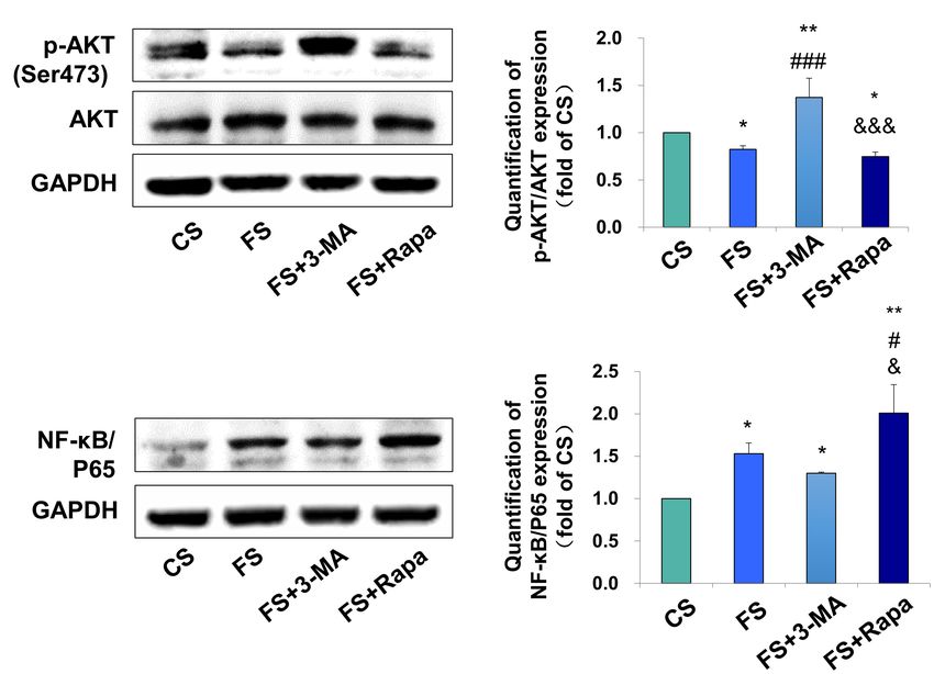
FIGURE 4 | Autophagy in PDLSCs under force stimuli suppressed AKT signaling in THP-1-derived macrophages. (A) Western blot of the expressions of
phosphor-AKT (Ser473) and AKT in THP-1-derived macrophages. GAPDH served as an internal control. The relative fold change of p-AKT/AKT was quantified.
Activation of autophagy could reduce active AKT activity in FS and FS + Rapa groups, while 3-MA (FS + 3-MA) reversed these effects. (B) Western blot of the
expressions of NF-κB/P65 in THP-1-derived macrophages. GAPDH served as an internal control. The expression of NF-κB/P65 increased after force loading, which
further enhanced after rapamycin application. The relative fold change was quantified. Results are presented as mean ± SD. n = 3. *P < 0.05, **P < 0.01 versus CS;
# P < 0.05, ### P < 0.001 versus FS; & P < 0.05, &&& P < 0.001 versus FS + 3-MA.
and P < 0.001 versus FS + 3-MA, Figure 3E). The same changes level of phospho-AKT (P < 0.05 versus CS, P < 0.001 versus
of TNF-α expression were detected (Supplementary Figure 3). FS + 3-MA, Figure 4A). In contrast, the expression of NF-κB/P65
However, the expression of M2 macrophage associated marker was upregulated after incubation in the supernatant from the
arginase-1 did not changed (Figure 3E). The application of force-loaded PDLSCs (P < 0.05 versus CS), which was further
3-MA or rapamycin exclusively did not change the expressions enhanced by the application of autophagy activator rapamycin
of TNF-α or arginase-1 in THP-1 macrophages (Supplementary (P < 0.01 versus CS, P < 0.05 versus FS and FS + 3-MA,
Figure 4). Taken together, these data suggest that autophagy Figure 4B).
in force-loaded PDLSCs could steer macrophage polarization To further confirm the regulatory mechanism, the AKT
toward the M1 phenotype in vitro. signaling activator IGF1 and inhibitor GSK690693 were applied
to the THP-1-derived macrophages. The activator efficiency of
Periodontal Ligament Stem Cell IGF1 was confirmed by the increased expression of phospho-
AKT/AKT (P < 0.01, Supplementary Figure 5A), whereas
Autophagy Modulates M1 Macrophage the inhibitor efficiency of GSK690693 was confirmed by
Polarization Through the Inhibition of the the decreased expression of phospho-AKT/AKT (P < 0.05,
AKT Signaling Pathway Supplementary Figure 5B). The expressions of TNF-α
After verifying the relationship between PDLSC autophagy and NF-κB/P65 increased in FS + Rapa + DMSO group
and macrophage polarization, we next explored the possible (P < 0.05 and P < 0.01 versus FS), which was decreased
underlying mechanism. AKT signaling has been acknowledged as after AKT activator IGF1 application (P < 0.01 versus
a crucial mediator of the macrophage survival and polarization FS + Rapa + DMSO, Figure 5A). No changes were observed
(Vergadi et al., 2017) and NF-κB is one of the key downstream of the expression the M2 marker arginase-1. Consistently,
signal of the AKT signaling. As a critical transcriptional factor the mRNA expression levels of M1 macrophage-related
in macrophages, increased NF-κB activity steers macrophage markers iNOS and TNF-α increased in FS + Rapa + DMSO
polarization toward the M1 phenotype (Kono et al., 2014; group (P < 0.01 versus FS) and decreased after IGF1
Hoover et al., 2020). We found the expression of phospho-AKT application (P < 0.05 and 0.001 versus FS + Rapa + DMSO),
in macrophages was significantly inhibited in the FS groups whereas no changes were observed in the expressions of
(P < 0.05 versus CS) when applying the conditioned medium into M2 macrophage-related markers arginase-1 and DECTIN-1
the THP-1-derived macrophages, whereas blocking autophagy (Figure 5C).
by 3-MA (FS + 3-MA) increased the expression of phospho- Furthermore, the expressions of TNF-α and NF-κB/P65
AKT (P < 0.01 versus CS, P < 0.001 versus FS) and enhancing decreased in FS + 3-MA + DMSO group (P < 0.01 and 0.05 versus
autophagy by rapamycin reversed the above-mentioned increase FS). After the application of AKT kinase inhibitor GSK690693,
Frontiers in Cell and Developmental Biology | www.frontiersin.org 8 May 2021 | Volume 9 | Article 666631Jiang et al. PDLSC Autophagy Modulates Macrophage Polarization
FIGURE 5 | Force induced autophagy in PDLSCs modulates M1 macrophage polarization through AKT signaling. (A) Western blot of the expressions of TNF-α,
Arg-1 and NF-κB/P65 by the application of force-loaded supernatant (FS) with rapamycin. AKT signaling activator IGF1 or equal volume of DMSO was added into
supernatant. The protein levels were quantified. Results are presented as mean ± SD. n = 3. *P < 0.05, **P < 0.01 versus the left column. ## P < 0.01 versus middle
column. (B) Western blot of the expressions of TNF-α, Arg-1 and NF-κB/P65 by the application of force-loaded supernatant (FS) with 3-MA. AKT kinase inhibitor
GSK690693 or equal volume of DMSO was added into conditioned medium. Results are presented as mean ± SD. n = 3. *P < 0.05, **P < 0.01 versus the left
column. # P < 0.05, ## P < 0.01 versus middle column. (C) Relative mRNA expression of M1/M2-related genes of THP-1-derived macrophages cultured with
different conditions, including FS, FS + Rapa + DMSO and FS + Rapa + IGF1. (D) Relative mRNA expression of M1/M2-related genes of THP-1-derived
macrophage cultured with different conditions, including FS, FS + 3 − MA + DMSO and FS + 3 − MA + GSK690693 groups. Results are presented as
mean ± SD. n = 3–4. **P < 0.01 versus left column; # P < 0.05, ### P < 0.001 versus middle column.
the expressions of TNF-α and NF-κB/P65 increased significantly load (Marino et al., 2014). However, it is still unclear that
at protein level (P < 0.01 and 0.05 versus FS + 3-MA + DMSO, whether and how autophagy influences bone remodeling
Figure 5B). Consistently, the mRNA expression levels of iNOS under mechanical force stimuli. In this study, we showed a
and TNF-α decreased in FS + 3-MA + DMSO group (P < 0.01 novel mechanism that force-induced autophagy in PDLSCs
versus FS) and increased after GSK690693 application (P < 0.001 contributed to M1 macrophage polarization, therefore
versus FS + 3-MA + DMSO), whereas no changes were observed promoting bone remodeling and tooth movement. First,
in the expressions of M2 macrophage-related markers arginase- force loading induced autophagy on the compression
1 and DECTIN-1 (Figure 5D). In sum, our data suggest that side of the periodontal tissues during tooth movement,
the autophagy activation in the force-loaded PDLSCs induces accompanied by the accumulation of M1 macrophages.
M1 macrophage polarization through the inhibition of the AKT Blockage of autophagy by 3-MA decreased the tooth movement
signaling pathway. distance and suppressed M1 macrophage polarization.
Second, compressive force in vitro stimulated autophagy
in PDLSCs, which further increased the expressions of M1
DISCUSSION macrophage-related inflammatory elements. These effects could
be suppressed or enhanced by the application of autophagy
Autophagy is an important protective cellular process to inhibitor 3-MA or autophagy activator rapamycin. Finally,
maintain cell and tissue homeostasis under mechanical the AKT signaling pathway participated in the regulation
Frontiers in Cell and Developmental Biology | www.frontiersin.org 9 May 2021 | Volume 9 | Article 666631Jiang et al. PDLSC Autophagy Modulates Macrophage Polarization FIGURE 6 | Scheme of the study. Force-stimulated autophagy in PDLSCs steers macrophages into M1 phenotype, which contributes to the bone remodeling and tooth movement process. of PDLSC autophagy on macrophage polarization under interpreted as follows. In the previous study, the trifurcation mechanical stimuli. area was detected for bone density. However, both compression Autophagy has been demonstrated playing a two-sided role in force and strain existed in the trifurcation area, which may inflammation and immune responses. On one hand, autophagy influence the assessment of bone density. Moreover, different in the epithelium decreases the chronic intestinal inflammation dosage of 3-MA was used in the present study, which may (Pott et al., 2018). Depletion of autophagy gene Atg16l1 in lead to different outcomes. Nevertheless, further studies on the T cells aggravates the spontaneous intestinal inflammation knockout mice should be developed to confirm the regulatory (Kabat et al., 2016). On the other hand, induction of intestinal role of autophagy in PDLSCs on macrophage polarization under autophagy may increase the severity of inflammatory bowel mechanical force stimuli. diseases (Zhang et al., 2018). Psychosocial stress may also enhance During inflammation and tissue remodeling process, MSCs intestinal autophagy and increase the M1/M2 macrophage could interact with immune cells such as macrophages by ratio in the remaining colon, thus aggravating the severity of expressing cytokines such as transforming growth factor-β and inflammatory bowel diseases (Wang et al., 2019). Moreover, secreting elements such as exosomes (Liu et al., 2019; Xu et al., higher level of autophagy has been found in the inflammatory 2019). In the periodontal tissues, PDLSCs are one type of periodontal tissues compared with normal periodontal tissues unique MSCs, which can interact with other cells including (An et al., 2016). During the force-induced tooth movement osteoclasts and immune cells under mechanical stimuli (Zhang process, aseptic inflammatory microenvironment is induced in et al., 2016; Liu et al., 2017). Previously, we have shown that the periodontal tissues (Garlet et al., 2007). Here, our data PDLSCs could secret inflammatory cytokines or gas molecule showed that autophagy induced by mechanical force participated such as hydrogen sulfide and influence macrophage polarization in the induction of inflammation in the periodontal tissues, under mechanical stimuli (He et al., 2015b, 2020). Moreover, which was characterized by the accumulation of M1 macrophages autophagy in PDLSCs under compressive force was found to be and the elevated expressions of M1 macrophage-related pro- regulated by lncRNA FER1L4 and involved in tissue remodeling inflammatory cytokines. Furthermore, blockage of autophagy (Huang et al., 2021). In this study, we show a novel mechanism level by 3-MA administration decreased the number of M1 that autophagy in PDLSCs could participate in regulating M1 macrophages, bone remodeling and tooth movement process. macrophage polarization under mechanical stimuli, and thus Consistent with the previous studies, the present study suggests contribute to the bone remodeling process. Given the important that force-induced autophagy in PDLSCs promotes inflammation roles of MSCs and macrophages during tissue regeneration in the periodontal tissues by activating M1 phenotypes, thus process, these findings indicate that modulating MSCs autophagy contributing to bone remodeling and tooth movement. may influence immune cells and therefore contribute to the tissue The role of force-induced autophagy on osteoclast activity regeneration process. is controversial. Previous study has been shown that blockage We also verify a novel signaling pathway of M1 phenotype of autophagy by 3-MA increased the expression of osteoclasts, activation under mechanical stimuli. Previous studies have shown decreased bone density and promoted tooth movement in that several signaling pathways, including β-catenin and STAT- the mouse model (Chen et al., 2019). The above findings 1, are involved in the inflammatory bone remodeling process varied different from our data. The underlying reasons can be under mechanical force stimuli (Mao et al., 2018; He et al., 2020). Frontiers in Cell and Developmental Biology | www.frontiersin.org 10 May 2021 | Volume 9 | Article 666631
Jiang et al.                                                                                                               PDLSC Autophagy Modulates Macrophage Polarization

In this study, we find that autophagy in PDLSCs influences M1                                   animal study was reviewed and approved by Institutional Animal
macrophage polarization through suppressing the AKT signaling                                   Use and Care Committee of the Peking University (LA2013-92).
pathway, which is a critical mediator in macrophage polarization
(Vergadi et al., 2017). Our data shed light on a novel mechanism
of how autophagy regulates macrophage polarization under                                        AUTHOR CONTRIBUTIONS
mechanical force in mechanical stimuli microenvironment.
   In conclusion, force-stimulated autophagy in PDLSCs steers                                   NJ and DH performed the experiments, analyzed the data, and
macrophages into the M1 phenotype via the inhibition of the                                     prepared the manuscript. YL and HY designed the experiments,
AKT signaling pathway, thereby contributing to bone remodeling                                  analyzed the data, and prepared and revised the manuscript.
and tooth movement (Figure 6). These results lead to a better                                   YM, JS, XW, SC, ZL, and YZ analyzed the data and revised the
understanding of how PDLSCs response to mechanical stimuli                                      manuscript. All authors reviewed the manuscript.
and interact with macrophage polarization, therefore modulate
alveolar bone remodeling. The findings also indicate a possibility
that modulating MSC autophagy may regulate inflammatory                                         FUNDING
bone remodeling and regeneration process.
                                                                                                The authors acknowledge the financial support from the
                                                                                                projects of the National Natural Science Foundations of
                                                                                                China No. 81871492 (YL), No. 81801031 (HY), and No.
DATA AVAILABILITY STATEMENT                                                                     81970901 (NJ), the Key R&D Plan of Ningxia Hui Autonomous
                                                                                                Region No. 2020BCG01001 (YL), Beijing Nova Program No.
The original contributions presented in the study are included                                  Z201100006820080 (NJ), the Peking University Medicine Seed
in the article/Supplementary Material, further inquiries can be                                 Fund for Interdisciplinary Research No. BMU2021MX001 (DH),
directed to the corresponding author/s.                                                         and the Peking University Clinical Medicine Plus X – Young
                                                                                                Scholars Project No. PKU2021LCXQ003 (NJ).

ETHICS STATEMENT
                                                                                                SUPPLEMENTARY MATERIAL
The studies involving human participants were reviewed
and approved by the Peking University Ethical Committee                                         The Supplementary Material for this article can be found
(PKUSSIRB-201311103). The patients/participants provided                                        online at: https://www.frontiersin.org/articles/10.3389/fcell.2021.
their written informed consent to participate in this study. The                                666631/full#supplementary-material

REFERENCES                                                                                      He, D., Kou, X., Yang, R., Liu, D., Wang, X., Luo, Q., et al. (2015b). M1-like
                                                                                                    Macrophage Polarization Promotes Orthodontic Tooth Movement. J. Dent. Res.
An, Y., Liu, W., Xue, P., Zhang, Y., Wang, Q., and Jin, Y. (2016). Increased                        94, 1286–1294.
   autophagy is required to protect periodontal ligament stem cells from apoptosis              He, D., Liu, F., Cui, S., Jiang, N., Yu, H., Zhou, Y., et al. (2020). Mechanical
   in inflammatory microenvironment. J. Clin. Periodontol. 43, 618–625. doi:                        load-induced H2S production by periodontal ligament stem cells activates M1
   10.1111/jcpe.12549                                                                               macrophages to promote bone remodeling and tooth movement via STAT1.
Chen, L., Mo, S., and Hua, Y. (2019). Compressive force-induced autophagy                           Stem Cell Res. Ther. 11:112. doi: 10.1186/s13287-020-01607-9
   in periodontal ligament cells downregulates osteoclastogenesis during tooth                  Hoover, A. A., Hufnagel, D. H., Harris, W., Bullock, K., Glass, E. B., Liu, E., et al.
   movement. J. Periodontal 90, 1170–1181.                                                          (2020). Increased canonical NF-kappaB signaling specifically in macrophages
Dang, S., Xu, H., Xu, C., Cai, W., Li, Q., Cheng, Y., et al. (2014). Autophagy                      is sufficient to limit tumor progression in syngeneic murine models of ovarian
   regulates the therapeutic potential of mesenchymal stem cells in experimental                    cancer. BMC Cancer 20:970. doi: 10.1186/s12885-020-07450-8
   autoimmune encephalomyelitis. Autophagy 10, 1301–1315. doi: 10.4161/auto.                    Horwood, N. J. (2016). Macrophage Polarization and Bone Formation: a review.
   28771                                                                                            Clin. Rev. Allergy Immunol. 51, 79–86.
Feng, X., and Teitelbaum, S. L. (2013). Osteoclasts: new Insights. Bone Res. 1,                 Huang, H., Yang, R., and Zhou, Y. H. (2018). Mechanobiology of Periodontal
   11–26.                                                                                           Ligament Stem Cells in Orthodontic Tooth Movement. Stem Cells Int.
Fu, Y., Liu, S., Cui, S. J., Kou, X. X., Wang, X. D., Liu, X. M., et al. (2016). Surface            2018:6531216.
   Chemistry of Nanoscale Mineralized Collagen Regulates Periodontal Ligament                   Huang, Y., Liu, H., Guo, R., Han, Y., Yang, Y., Zhao, Y., et al. (2021). Long Non-
   Stem Cell Fate. ACS Appl. Mat. Interfaces 8, 15958–15966. doi: 10.1021/acsami.                   coding RNA FER1L4 Mediates the Autophagy of Periodontal Ligament Stem
   6b04951                                                                                          Cells Under Orthodontic Compressive Force via AKT/FOXO3 Pathway. Front.
Garlet, T. P., Coelho, U., Silva, J. S., and Garlet, G. P. (2007). Cytokine                         Cell Dev. Biol. 9:631181. doi: 10.3389/fcell.2021.631181
   expression pattern in compression and tension sides of the periodontal                       Jiang, M., Liu, K., Luo, J., and Dong, Z. (2010). Autophagy is a renoprotective
   ligament during orthodontic tooth movement in humans. Eur. J. Oral Sci. 115,                     mechanism during in vitro hypoxia and in vivo ischemia-reperfusion injury.
   355–362.                                                                                         Am. J. Pathol. 176, 1181–1192.
Hara, T., Nakamura, K., Matsui, M., Yamamoto, A., Nakahara, Y., Suzuki-                         Kabat, A. M., Harrison, O. J., Riffelmacher, T., Moghaddam, A. E., Pearson, C. F.,
   Migishima, R., et al. (2006). Suppression of basal autophagy in neural cells                     Laing, A., et al. (2016). The autophagy gene Atg16l1 differentially regulates
   causes neurodegenerative disease in mice. Nature 441, 885–889.                                   Treg and TH2 cells to control intestinal inflammation. Elife 5:e12444. doi:
He, D., Kou, X., Luo, Q., Yang, R., Liu, D., Wang, X., et al. (2015a). Enhanced                     10.7554/eLife.12444
   M1/M2 macrophage ratio promotes orthodontic root resorption. J. Dent. Res.                   Kanzaki, H., Chiba, M., Shimizu, Y., and Mitani, H. (2002). Periodontal ligament
   94, 129–139. doi: 10.1177/0022034514553817                                                       cells under mechanical stress induce osteoclastogenesis by receptor activator

Frontiers in Cell and Developmental Biology | www.frontiersin.org                          11                                                 May 2021 | Volume 9 | Article 666631
Jiang et al. PDLSC Autophagy Modulates Macrophage Polarization of nuclear factor kappaB ligand up-regulation via prostaglandin E2 synthesis. Murray, P. J. (2017). Macrophage Polarization. Annu. Rev. Physiol. 79, 541–566. J. Bone Miner. Res. 17, 210–220. doi: 10.1359/jbmr.2002.17.2.210 Pott, J., Kabat, A. M., and Maloy, K. J. (2018). Intestinal Epithelial Cell Autophagy King, J. S., Veltman, D. M., and Insall, R. H. (2011). The induction of autophagy by Is Required to Protect against TNF-Induced Apoptosis during Chronic Colitis mechanical stress. Autophagy 7, 1490–1499. in Mice. Cell Host Microbe 23, 191–202.e4. doi: 10.1016/j.chom.2017.12.017 Klionsky, D. J., Abdalla, F. C., Abeliovich, H., Abraham, R. T., Acevedo-Arozena, Seo, B. M., Miura, M., Gronthos, S., Bartold, P. M., Batouli, S., Brahim, J., A., Adeli, K., et al. (2012). Guidelines for the use and interpretation of assays for et al. (2004). Investigation of multipotent postnatal stem cells from human monitoring autophagy. Autophagy 8, 445–544. periodontal ligament. Lancet 364, 149–155. Kliosnky, D., Wolvetang, E., and Walker, M. J. (2016). Guidelines for the Use and Shapouri-Moghaddam, A., Mohammadian, S., Vazini, H., Taghadosi, M., Interpretation of Assays for Monitoring Autophagy (3rd edition) (vol 12, pg 1, Esmaeili, S. A., Mardani, F., et al. (2018). Macrophage plasticity, 2015). Autophagy 12, 443–443. doi: 10.1080/15548627.2015.1100356 polarization, and function in health and disease. J. Cell Physiol. 233, Kono, Y., Kawakami, S., Higuchi, Y., Yamashita, F., and Hashida, M. (2014). In 6425–6440. Vitro Evaluation of Inhibitory Effect of Nuclear Factor-KappaB Activity by Smutny, M., Akos, Z., Grigolon, S., Shamipour, S., Ruprecht, V., Capek, D., et al. Small Interfering RNA on Pro-tumor Characteristics of M2-Like Macrophages. (2017). Friction forces position the neural anlage. Nat. Cell Biol. 19, 306–317. Biol. Pharm. Bull. 37, 137–144. doi: 10.1248/bpb.b13-00659 doi: 10.1038/ncb3492 Kroemer, G., Marino, G., and Levine, B. (2010). Autophagy and the integrated Taddei, S. R., Moura, A. P., Andrade, I. Jr., Garlet, G. P., Garlet, T. P., Teixeira, stress response. Mol. Cell 40, 280–293. M. M., et al. (2012). Experimental model of tooth movement in mice: a Lee, S. I., Park, K. H., Kim, S. J., Kang, Y. G., Lee, Y. M., and Kim, E. C. (2012). standardized protocol for studying bone remodeling under compression and Mechanical stress-activated immune response genes via Sirtuin 1 expression tensile strains. J. Biomech. 45, 2729–2735. doi: 10.1016/j.jbiomech.2012.09.006 in human periodontal ligament cells. Clin. Exp. Immunol. 168, 113–124. doi: Thompson, W. R., Rubin, C. T., and Rubin, J. (2012). Mechanical regulation of 10.1111/j.1365-2249.2011.04549.x signaling pathways in bone. Gene 503, 179–193. Levine, B., Mizushima, N., and Virgin, H. W. (2011). Autophagy in immunity and Vergadi, E., Ieronymaki, E., Lyroni, K., Vaporidi, K., and Tsatsanis, C. (2017). inflammation. Nature 469, 323–335. Akt Signaling Pathway in Macrophage Activation and M1/M2 Polarization. Liu, F., Qiu, H., Xue, M., Zhang, S., Zhang, X., Xu, J., et al. (2019). MSC- J. Immunol. 198, 1006–1014. secreted TGF-beta regulates lipopolysaccharide-stimulated macrophage M2- Wang, S. L., Shao, B. Z., Zhao, S. B., Chang, X., Wang, P., Miao, C. Y., et al. (2019). like polarization via the Akt/FoxO1 pathway. Stem Cell Res. Ther. 10:345. doi: Intestinal autophagy links psychosocial stress with gut microbiota to promote 10.1186/s13287-019-1447-y inflammatory bowel disease. Cell Death Dis. 10:391. doi: 10.1038/s41419-019- Liu, F., Wen, F., He, D., Liu, D., Yang, R., Wang, X., et al. (2017). Force-Induced 1634-x H2S by PDLSCs Modifies Osteoclastic Activity during Tooth Movement. Xu, R., Zhang, F., Chai, R., Zhou, W., Hu, M., Liu, B., et al. (2019). Exosomes J. Dent. Res. 96, 694–702. doi: 10.1177/0022034517690388 derived from pro-inflammatory bone marrow-derived mesenchymal stem Ma, K. G., Shao, Z. W., Yang, S. H., Wang, J., Wang, B. C., Xiong, L. M., et al. cells reduce inflammation and myocardial injury via mediating macrophage (2013). Autophagy is activated in compression-induced cell degeneration and polarization. J. Cell Mol. Med. 23, 7617–7631. doi: 10.1111/jcmm.14635 is mediated by reactive oxygen species in nucleus pulposus cells exposed to Yan, Y., Liu, F., Kou, X., Liu, D., Yang, R., Wang, X., et al. (2015). T compression. Osteoarthr. Cartil. 21, 2030–2038. Cells Are Required for Orthodontic Tooth Movement. J. Dent. Res. 94, Ma, Y., Qi, M., An, Y., Zhang, L., Yang, R., Doro, D. H., et al. (2018). Autophagy 1463–1470. controls mesenchymal stem cell properties and senescence during bone aging. Zhang, L., Liu, W., Zhao, J., Ma, X., Shen, L., Zhang, Y., et al. (2016). Aging Cell 17:e12709. Mechanical stress regulates osteogenic differentiation and RANKL/OPG ratio Mao, Y., Wang, L., Zhu, Y., Liu, Y., Dai, H., Zhou, J., et al. (2018). Tension in periodontal ligament stem cells by the Wnt/beta-catenin pathway. Biochim. force-induced bone formation in orthodontic tooth movement via modulation Biophys. Acta 1860, 2211–2219. doi: 10.1016/j.bbagen.2016.05.003 of the GSK-3beta/beta-catenin signaling pathway. J. Mol. Histol. 49, 75–84. Zhang, Y. S., Wang, F., Cui, S. X., and Qu, X. J. (2018). Natural dietary doi: 10.1007/s10735-017-9748-x compound naringin prevents azoxymethane/dextran sodium sulfate-induced Marino, G., Niso-Santano, M., Baehrecke, E. H., and Kroemer, G. (2014). Self- chronic colorectal inflammation and carcinogenesis in mice. Cancer Biol. Ther. consumption: the interplay of autophagy and apoptosis. Nat. Rev. Mol. Cell Biol. 19, 735–744. doi: 10.1080/15384047.2018.1453971 15, 81–94. doi: 10.1038/nrm3735 Meikle, M. C. (2006). The tissue, cellular, and molecular regulation of orthodontic Conflict of Interest: YM is now employed by the company Beijing Tason Biotech tooth movement: 100 years after Carl Sandstedt. Eur. J. Orthod. 28, 221–240. Co. Ltd. doi: 10.1093/ejo/cjl001 Memmert, S., Damanaki, A., Weykopf, B., Rath-Deschner, B., Nokhbehsaim, M., The remaining authors declare that the research was conducted in the absence of Gotz, W., et al. (2019). Autophagy in periodontal ligament fibroblasts under any commercial or financial relationships that could be construed as a potential biomechanical loading. Cell Tissue Res. 378, 499–511. doi: 10.1007/s00441-019- conflict of interest. 03063-1 Miroshnikova, Y. A., Le, H. Q., Schneider, D., Thalheim, T., Rubsam, M., Copyright © 2021 Jiang, He, Ma, Su, Wu, Cui, Li, Zhou, Yu and Liu. This is an Bremicker, N., et al. (2018). Adhesion forces and cortical tension couple cell open-access article distributed under the terms of the Creative Commons Attribution proliferation and differentiation to drive epidermal stratification. Nat. Cell Biol. License (CC BY). The use, distribution or reproduction in other forums is permitted, 20, 69–80. doi: 10.1038/s41556-017-0005-z provided the original author(s) and the copyright owner(s) are credited and that the Mizushima, N., Levine, B., Cuervo, A. M., and Klionsky, D. J. (2008). Autophagy original publication in this journal is cited, in accordance with accepted academic fights disease through cellular self-digestion. Nature 451, 1069–1075. doi: 10. practice. No use, distribution or reproduction is permitted which does not comply 1038/nature06639 with these terms. Frontiers in Cell and Developmental Biology | www.frontiersin.org 12 May 2021 | Volume 9 | Article 666631
You can also read


