P38 MAPK signaling in M1 macrophages results in selective elimination of M2 macrophages by MEK inhibition
←
→
Page content transcription
If your browser does not render page correctly, please read the page content below

Open access Short report
p38 MAPK signaling in M1
J Immunother Cancer: first published as 10.1136/jitc-2020-002319 on 20 July 2021. Downloaded from http://jitc.bmj.com/ on January 3, 2022 by guest. Protected by copyright.
macrophages results in selective
elimination of M2 macrophages by
MEK inhibition
Daniel Baumann,1,2 Jennifer Drebant,2 Tanja Hägele,2 Luisa Burger,2 Clara Serger,2
Claudia Lauenstein,2 Przemyslaw Dudys,3 Gerrit Erdmann,3 Rienk Offringa 1,2
To cite: Baumann D, Drebant J, ABSTRACT indications that do not respond to either of
Hägele T, et al. p38 MAPK M2 macrophages promote tumor progression and the single-agent treatments. We and others
signaling in M1 macrophages therapy resistance, whereas proimmunogenic M1
results in selective elimination
recently reported that pharmacological inhi-
macrophages can contribute to the efficacy of cytostatic bition of mitogen- activated protein kinase
of M2 macrophages by
and immunotherapeutic strategies. The abundance of M2 (MAPK) kinase (MEK) constitutes a highly
MEK inhibition. Journal for
ImmunoTherapy of Cancer macrophages in the immune infiltrate of many cancer
promising cytostatic strategy in this respect,
2021;9:e002319. doi:10.1136/ types has prompted the search for strategies to target
and eliminate this subset. From our prior experiments in that combination with immunostimula-
jitc-2020-002319
in syngeneic mouse tumor models, we learned that tory antibodies results in increased antitumor
►► Additional supplemental pharmacological inhibition of mitogen-activated protein efficacy compared with single- agent treat-
material is published online only. kinase kinase (MEK) did not merely result in tumor cell ment in syngeneic mouse tumor models.
To view, please visit the journal death, but also in the modulation of the tumor immune This was shown for ICB using PD-L1-blocking
online (http://dx.d oi.org/10. infiltrate. This included a prominent decrease in the antibodies as well as for agonist antibodies
1136/j itc-2020-0 02319). numbers of macrophages as well as an increase in the addressing the immunostimulatory receptors
M1/M2 macrophage ratio. Investigation of the mechanism OX40, CD137, and CD40.3–5
Accepted 23 May 2021 underlying this finding in primary murine macrophage
An important aspect of the mechanism
cultures revealed that M2 macrophages are significantly
more sensitive to MEK inhibition-induced cell death
of action of MEK inhibitors (MEKi) in this
than their M1 counterparts. Further analyses showed context is the induction of tumor cell death
that the p38 MAPK pathway, which is activated in M1 without hampering the function of dendritic
macrophages only, renders these cells resistant to cells and T cells. Furthermore, we found that
death by MEK inhibition. In conclusion, the dependency the antitumor efficacy of MEK inhibition is
of M2 macrophages on the MEK/extracellular-signal not only limited to the induction of tumor
regulated kinase (ERK) pathway empowers MEK inhibitors cell death, but also involves modulation of
to selectively eliminate this subset from the tumor the tumor immune cell infiltrate at several
microenvironment.
levels.5 This includes the elimination of the
vast majority of macrophages, which are
© Author(s) (or their
employer(s)) 2021. Re-use INTRODUCTION known to accumulate in many experimental
permitted under CC BY-NC. No Immune checkpoint blockade (ICB) shows and human cancer types, thereby contrib-
commercial re-use. See rights significant clinical efficacy against tumors uting to the subversion of the antitumor
and permissions. Published by
harboring high numbers of somatic muta- T-cell response.6 7 As a consequence of MEKi
BMJ.
1 tions, corresponding to elevated tumor treatment, the balance between macrophage
Department of Surgery,
immunogenicity resulting from the expres- subsets in the tumor microenvironment was
University Hospital Heidelberg,
Heidelberg, Baden-Württemberg, sion of mutanome- encoded neoantigens. found to shift from the tumor- nurturing
Germany The therapeutic impact of ICB on less immu- M2 type toward the proimmune M1 type,
2
Division of Molecular Oncology nogenic tumors is, however, still limited.1 suggesting that M2 macrophages are more
of Gastrointestinal Tumors, sensitive to this drug than their M1 counter-
German Cancer Research
Tumor immunogenicity can be enhanced
Center, Heidelberg, Baden- through treatment with cytostatic drugs. parts. In this study, we investigated the mech-
Württemberg, Germany This results in cell-death-related proinflam- anism underlying this differential impact
3
NMI TT Pharmaservices, Berlin, matory ‘danger’ signals and increases the of MEKi on macrophage subsets. We found
Germany availability of tumor antigen for dendritic that activation of the p38 MAPK pathway in
Correspondence to cell-mediated cross-presentation to T cells.2 M1 macrophages makes these cells indepen-
Professor Rienk Offringa; Therefore, regimens combining cytostatic dent of the MEK/ERK pathway and thereby
r.offringa@dkfz.de drugs with ICB may be used to treat cancer rescues them from death by MEK inhibition,
Baumann D, et al. J Immunother Cancer 2021;9:e002319. doi:10.1136/jitc-2020-002319 1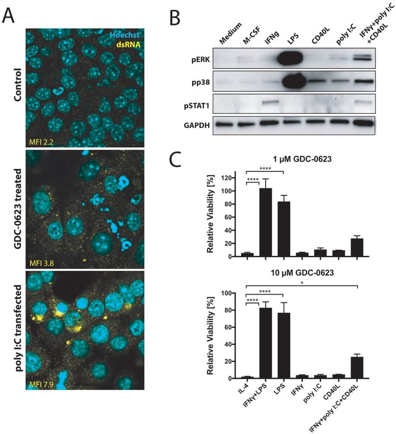
Open access
while the survival of M2 macrophages critically depends Southampton, UK) and produced as described in online
J Immunother Cancer: first published as 10.1136/jitc-2020-002319 on 20 July 2021. Downloaded from http://jitc.bmj.com/ on January 3, 2022 by guest. Protected by copyright.
on the MEK/ERK pathway. supplemental methods.
Macrophage OT-I assay
Murine bone marrow-derived macrophages were gener-
METHODS ated, cultured, and MEKi-treated as described before.5
Mice and in vivo experiments After 72 hours of MEKi treatment (100 nM or 1 µM),
C57BL/6-Ly5.1 mice were bred in animal facilities of the macrophages were pulsed with 1.25 ng/mL of SIIN-
German Cancer Research Center. Detailed information FEKL peptide for 3 hours. Remaining MEKi and SIIN-
on in vivo experiments, including the handling and dosing FEKL peptide were washed away, and peptide- loaded
of small-molecule drugs and antibodies, can be found in macrophages were co-cultured with carboxyfluorescein
online supplemental methods. All animal experiments succinimidyl ester (CFSE)-labeled (5 µM) naïve OT-I cells
were performed on the basis of prior approval by the freshly isolated from spleens and lymph nodes of OT-I
ethical authorities. Mice were sacrificed if signs of distress mice and magnetic activated cell sorting (MACS) puri-
were noticed, when termination criteria were reached, or fied using the mouse CD8+ T-cell isolation kit (Miltenyi
analyses were performed at specific time points. Biotec) according to the manufacturer’s instructions.
After 72 hours of co-culture, OT-I cells were harvested
Patient samples and analyzed by flow cytometry. Staining and data analysis
Informed written consent was obtained from all partic- were performed as described in Baumann et al.5 Count-
ipants before sample collection. Collection of human Bright absolute counting beads (Thermo Fisher Scien-
material and clinical data was approved by the local tific) were added to determine absolute T-cell numbers.
ethics committees and conducted in accordance with the
regulations of the tissue biobanks and the Declaration of Flow cytometry
Helsinki. Extraction of cells from tumor tissue and flow cytometry
staining of cells from tumors or in vitro-generated macro-
Cell lines, culture, and cytotoxicity assays phages were performed as described in online supple-
Murine PDA30364 cells and bone marrow-derived macro- mental methods and in Baumann et al.5
phages were generated, cultured, and used in cytotoxicity Western blot analysis
assays as described in Baumann et al5 and online supple- Cells were incubated in the presence of 500 nM MEKi
mental methods. Dual cytotoxicity assays were performed GDC-0623 or dimethyl sulfoxide for 1 hour before addi-
in the presence of 2 µM BIRB 796 and varying concentra- tion of stimuli. Stimulation periods and exact exper-
tions of MEKi GDC-0623. imental setups are described in detail in the figure
Human monocytes were isolated from peripheral blood legends. Staining with primary antibodies was performed
mononuclear cells using the pan monocyte isolation kit overnight at 4°C and with secondary antibodies for
(Miltenyi Biotec) and differentiated ex vivo with cyto- 2 hours at room temperature. The following antibodies
kines into M1-like or M2-like macrophages as described in and dilutions were used: anti- mouse pERK1/2 (clone
online supplemental methods. Cytokine concentrations 20G11, 4376, 1:1000; Cell Signaling), anti-mouse pp38
are indicated in the figure legends. These cells were used (D3F9, 4511, 1:1000; Cell Signaling), anti-mouse pSTAT1
in cytotoxicity assays as described before5 and in online (D4A7, 7649, 1:1000; Cell Signaling), anti-mouse GAPDH
supplemental methods. (GTX100118, 1:1000; GeneTex), and goat anti- rabbit
IgG-HRP (7074, 1:3000; Cell Signaling). Clarity Western
Drugs and reagents ECL substrate (170-5060; Bio-Rad) was used for chemi-
MEKi GDC-0623 (A-1181; Chemgood), p38 MAPKi BIRB luminescence reaction according to the manufacturer’s
796 (S1574; DP, Selleckchem), murine multimeric CD40L instructions. Images were processed using the Image Lab
(AG-40B-0020; Adipogen), poly I:C (tlrl-pic; InvivoGen), Software (V.6.0.1; Bio-Rad).
lipopolysaccharide (LPS) (L4391; Sigma), recombinant
murine interferon-γ (IFNγ) (12343536; ImmunoTools), DigiWest pathway activity analysis
murine macrophage-colony stimulating factor (M-CSF) DigiWest (NMI TT) was conducted as published in Treindl
(12343115; ImmunoTools), murine interleukin (IL)-4 et al.8 In brief, proteins were blotted onto polyvinylidene
(12340042; ImmunoTools), murine IL-10 (12340105; fluoride membranes and biotinylated. Each sample lane
ImmunoTools), anti- mouse CSF1R (BP0213; BioX- was cut into 96 molecular weight fractions and proteins
Cell), recombinant human M-CSF (11343115; Immuno- were eluted. Each molecular weight fraction was bound
Tools), human IFNγ (11343536; ImmunoTools), human onto color- coded Luminex beads and incubated with
IL-4 (11340045; ImmunoTools), human transforming primary, followed by secondary antibodies. Readout was
growth factor-β1 (TGFβ1) (11343160; ImmunoTools), performed on a Luminex FlexMAP 3D. Antibody lists
and human IL-10 (11340103; ImmunoTools). Chimeric can be provided upon request. For quantification of the
agonist anti-mouse CD40 antibody (mIgG1, clone 3-23) antibody-specific signals, the analysis tool is described in
was obtained through Martin Glennie (University of detail in Treindl et al.8 Protein expression values were
2 Baumann D, et al. J Immunother Cancer 2021;9:e002319. doi:10.1136/jitc-2020-002319
Open access
normalized on the total protein amount loaded onto one cell line PDA30364 was derived from a pancreatic tumor
J Immunother Cancer: first published as 10.1136/jitc-2020-002319 on 20 July 2021. Downloaded from http://jitc.bmj.com/ on January 3, 2022 by guest. Protected by copyright.
lane, and non-parametric testing was conducted using that arose in a genetically engineered mouse model
the MultiExperiment Viewer (MeV) V.4.9.0.9 Data were featuring pancreas- specific expression of the KRAS-
mean-centered and log2- transformed prior to utiliza- G12D oncogene as well as mutant P53-R172H5 (online
tion of the MeV software, and hierarchical clustering was supplemental methods). As a result, this tumor line also
performed on significant genes using Euclidean distance provides a benchmark for the cytotoxic impact of MEK
and complete linkage. inhibition. In line with our previously published data,5
single-agent MEKi treatment results in tumor stasis in
dsRNA immunofluorescence staining this model (figure 1D). Ablation of macrophages with an
PDA30364 tumor cells were treated for 48 hours with 1 µM anti-CSFR1 antibody also significantly suppresses tumor
GDC-0623. After treatment, cells were fixed in 4% para- growth in this experiment, in line with findings reported
formaldehyde and blocked for 1 hour in 5% bovine serum by others,7 11 pointing at the protumorigenic role of
albumin (BSA)/phosphate-buffered saline. Staining for tumor-associated macrophages in these tumors. Analysis
double-stranded RNA (dsRNA) was performed using the of the immune infiltrate confirmed that both MEKi and
anti-dsRNA antibody K1 (Scicons) at a concentration of anti-CSF1R antibody profoundly reduced the numbers
1:200. As a secondary antibody, Alexa Flour 594 goat anti- of CD206+ intratumoral macrophages while having only
mouse IgG was used at a concentration of 1:500. Nuclei a limited effect on the iNOS+ counterparts (figure 1E).
were counterstained with Hoechst. Images were acquired As shown in our previous report, complementation of
with Leica TCS SP5 confocal microscope. MEKi with agonist anti-CD40 antibody resulted in supe-
rior antitumor efficacy compared with either single-agent
Immunohistochemistry treatment (figure 1D). Notably, CD40 antibody treatment
Murine tumors were fixed in 5% formaldehyde for 24 equally increased the frequency of iNOS+ macrophages
hours at 4°C, dehydrated in ethanol, and embedded in in the absence or presence of MEKi (figure 1E), further
paraffin. Tissue blocks were sectioned into 4 µm slice indicating that the viability of this macrophage subset is
using a rotatory microtome. Slides were deparaffinized not significantly affected by MEK inhibition.
in Roticlear (Carl Roth) and rehydrated in ethanol. We zoomed in on the differential MEKi sensitivity of
Antigen retrieval was carried out by microwaving sections macrophage subsets by performing reductionist in vitro
for 30 min in 10 mM citrate buffer (pH 6.0). Endoge- experiments with iNOS+ M1- like and CD206+ M2- like
nous peroxidases were blocked by 3% H2O2. Blocking macrophage cultures. For this, primary mouse macro-
of sections was performed in rat serum (Vector Labora- phage cultures were generated from bone marrow
tories) and washed with Tris-buffered saline+0.1% BSA. precursors in the presence of, respectively, IFNγ/LPS
Immunohistochemical (IHC) staining was performed and IL-4 (online supplemental methods; online supple-
with rat Vectastain ABC (PK-6104) according to the manu- mental figure S1B). Monitoring of the viability of these
facturer’s instructions. F4/80-specific antibody (BM8; cultures in the presence of MEKi demonstrated that the
Abcam) was used at a dilution of 1:100. IHC stainings of M1 macrophages are relatively resistant; even very high
human tumor specimens were performed and analyzed concentrations of MEKi induce only partial death. In
as described in Poschke et al10 using antibodies reactive contrast, M2 macrophages are highly sensitive, compa-
against human CD68 (KP1; Abcam), CD163 (EDHu-1; rable to the mutant Kras-transformed PDA30364 tumor
AbD Serotec), and CD3 (PS1; Leica). Nuclei were coun- cells (figure 1F,G). As alternative M2 activation in the
terstained with hematoxylin (Novocastra). After staining, tumor microenvironment can occur in the presence of
slides were rehydrated using increasing ethanol concen- TGFβ and IL-10,7 we included this M2-like subtype in our
trations, followed by Roticlear. Slides were mounted in experiments, showing that also these macrophages were
Permount mounting medium (Thermo Fisher Scientific). highly sensitive to MEK inhibition (figure 2A,B). The
survival of M1 macrophages under MEK inhibition raised
the question of whether these cells had maintained their
RESULTS proinflammatory state. As shown in figure 2C, MEKi-
M2 macrophages are highly sensitive to MEK inhibition treated M1 macrophages maintained high levels of CD86
We examined the mechanism of action of MEKi toward and CD40 and even showed somewhat increased levels
the macrophage infiltrate in our transplantable pancre- of iNOS. Interestingly, the remaining M2 macrophages
atic cancer model PDA30364, one of the three models after MEKi treatment showed reduced expression of
used in our prior study,5 because these tumors—like the M2 marker CD206 and increased CD86 expression,
human pancreatic ductal adenocarcinoma6 7 (see also suggesting partial reprogramming under the influence
below)— are highly infiltrated with M2-like macrophages of MEK inhibition (figure 2D). When loaded with the
(figure 1A,B), which are characterized by the marker peptide SIINFEKL derived from chicken ovalbumin,
CD206+ and lower surface levels of major histocompati- MEKi-treated M1 macrophages maintained their capacity
bility complex (MHC)-II and CD86 compared with induc- to present antigen to SIINFEKL- specific OT-
I T cells,
ible nitric oxide synthase (iNOS)+ M1-like macrophages as well as the expression of proinflammatory markers
(figure 1C; online supplemental figure S1A). The tumor (figure 2C–F). Under the same experimental conditions,
Baumann D, et al. J Immunother Cancer 2021;9:e002319. doi:10.1136/jitc-2020-002319 3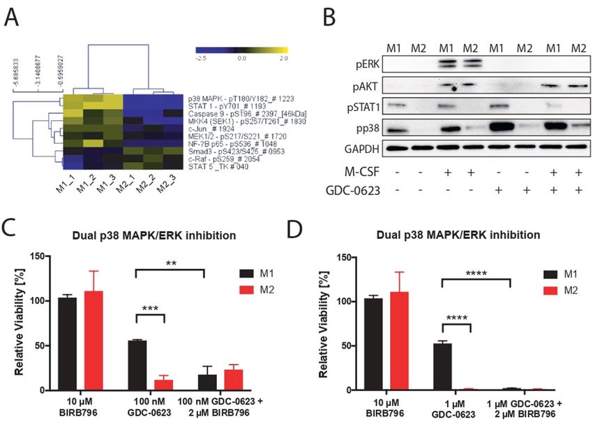
Open access
J Immunother Cancer: first published as 10.1136/jitc-2020-002319 on 20 July 2021. Downloaded from http://jitc.bmj.com/ on January 3, 2022 by guest. Protected by copyright.
Figure 1 M2 macrophages are highly sensitive to MEK inhibition in vivo and ex vivo. (A) Immunohistochemistry of a PDA30364
tumor using an F4/80-specific antibody. Scale bar: 100 µm. (B) Flow cytometric identification of iNOS+ M1 and CD206+ M2
macrophages in a freshly dissociated tumor sample upon gating on the live CD45+, CD11b+, F4/80+ cell subset. (C) MHC-
II (IA/IE) and CD86 surface levels of macrophages gated in (B). Mean±SEM, n=4. (D) Mice bearing PDA30364 tumors were
treated with MEKi 30 mg kg−1 GDC-0623 (daily), anti-CSF1R antibody (1 mg once, followed by 0.5 mg every other day), or anti-
CD40 antibody (200 µg on days 10, 12, 14, and 17 after tumor inoculation). Mean±SEM, n=5. Two-way ANOVA with post hoc
Tukey test. (E) Flow cytometry-based quantification of M2 (CD206+) and M1 (iNOS+) macrophages in PDA30364 tumors from
mice treated as described in (C). Mean±SEM, n=4. One-way ANOVA with post hoc Dunnett test. (F) Dose–response curves of
PDA30364 and M1 (20 ng mL−1 M-CSF+1 ng mL−1 IFNγ/LPS) and M2 (20 ng mL−1 M-CSF+2.5 ng mL−1 IL-4) polarized murine
macrophages treated with GDC-0623. Mean±SEM, n=3. Numbers indicate best-fit inhibitory concentration (IC)50 values. (G)
Viability of PDA30364 and murine M1 and M2 macrophages treated with GDC-0623 at 1 µM. Mean±SEM, n>4. One-way ANOVA
with post hoc Tukey test. Significance levels are indicated by asterisks (*p≤0.05; **p≤0.01; ***p≤0.001: ****p≤0.0001). ANOVA,
analysis of variance; IFNγ, interferon-γ; IL, interleukin; LPS, lipopolysaccharide; M-CSF, macrophage-colony stimulating factor;
MEK, mitogen-activated protein kinase kinase.
the numbers of M2 macrophages were decimated, online supplemental figure S2A). To assess whether
resulting in the loss of antigen presentation in the OT-1 human M1 and M2 macrophages display differential
T-cell assay (figure 2E,F). sensitivity to MEK inhibition, we generated monocyte-
As mentioned above, human pancreatic tumors are derived human macrophage cultures and polarized these
rich in M2-like macrophages, which in the human setting with, respectively, IFNγ+LPS, IL-4 or TGFβ+IL-10. As
are characterized by expression of the markers CD163 previously reported, CD206 can be induced by IL-4 in in
and CD68,12 as well as the marker CD206 (figure 2G,H; vitro macrophages cultures13 (online supplemental figure
4 Baumann D, et al. J Immunother Cancer 2021;9:e002319. doi:10.1136/jitc-2020-002319Open access
J Immunother Cancer: first published as 10.1136/jitc-2020-002319 on 20 July 2021. Downloaded from http://jitc.bmj.com/ on January 3, 2022 by guest. Protected by copyright.
Figure 2 M1 macrophages are more resistant to MEK/ERK inhibition and maintain their proinflammatory state. (A) Dose–
response curves of M1 (20 ng mL–1 M-CSF+1 ng mL–1 IFNγ/LPS) and M2 (20 ng mL–1 M-CSF+2.5 ng mL–1 IL-4 or 20 ng mL–1
TGFβ+10 ng mL–1 IL-10) polarized murine macrophages treated with GDC-0623. Mean±SEM, n=3. (B) Viability of murine M1
and M2 macrophages treated with 1 µM GDC-0623. Mean±SEM, n=6 (two independent experiments). One-way ANOVA with
post hoc Tukey test. (C, D) Flow cytometric analysis of surface markers and costimulatory molecules on macrophage cultures
as described in (A). Mean±SEM, n=3. Two-way ANOVA with post hoc Sidak test. (E) Co-culture OT-I T cells with SIINFEKL
peptide (1.25 ng/mL) pulsed and washed macrophages pretreated with cell culture medium and 1 µM GDC-0623, respectively.
Representative CFSE dilution blots. (F) Quantification of total OT-I numbers per well. Mean±SEM, n=3. Unpaired two-tailed t-
test. (G) Representative IHC stainings of human PDAC tissue specimen sections for the indicated markers and quantification
of samples from mutiple PDAC patients. n = 90-100 . Scale bar: 100 µm. (H) Flow cytometric analysis of macrophages in
freshly dissociated human PDAC sample upon gating on the live CD45+, CD11b+ subset. (I) Dose–response curves of
monocyte-derived human macrophages. M1 (50 ng mL −1 M-CSF+50 ng mL−1 IFNγ+20 ng mL−1 LPS) and M2 (50 ng mL−1 M-
CSF+20 ng mL−1 IL-4 or 20 ng mL−1 TGFβ+10 ng mL−1 IL-10) macrophages were treated with GDC-0623. Mean±SEM, n=3.
Numbers indicate best-fit IC50 values. (J) Flow cytometric analysis of surface markers and costimulatory molecules on human
macrophage cultures as described in (I). Mean±SEM, n=3. Two-way ANOVA with post hoc Sidak test. Significance levels are
indicated by asterisks (*p≤0.05; **p≤0.01; ***p≤0.001: ****p≤0.0001). ANOVA, analysis of variance; FACS, fluorescence-activated
cell sorting; IFNγ, interferon-γ; IHC, immunohistochemistry; IL, interleukin; LPS, lipopolysaccharide; M-CSF, macrophage-
colony stimulating factor; MEK, mitogen-activated protein kinase kinase; PDAC, pancreatic ductal adenocarcinoma; TGFβ,
transforming growth factor-β.
Baumann D, et al. J Immunother Cancer 2021;9:e002319. doi:10.1136/jitc-2020-002319 5Open access
J Immunother Cancer: first published as 10.1136/jitc-2020-002319 on 20 July 2021. Downloaded from http://jitc.bmj.com/ on January 3, 2022 by guest. Protected by copyright.
Figure 3 p38 MAPK signaling acts as a compensatory survival pathway in M1 macrophages. (A) DigiWest phospho-protein
pathway activity analysis, log2 Fc; non-parametric Wilcoxon rank-sum test (pOpen access
J Immunother Cancer: first published as 10.1136/jitc-2020-002319 on 20 July 2021. Downloaded from http://jitc.bmj.com/ on January 3, 2022 by guest. Protected by copyright.
Figure 4 CD40 ligation and dsRNA treatment stimulate p38 signaling in macrophages. (A) Immunofluorescence analysis of
dsRNA untreated or treated with 1 µM GDC-0623 for 48 hours. Poly I:C transfection served as a positive straining control. (B)
Western blot of mouse BMDM stimulated with culture medium containing 20 ng mL−1 M-CSF, 10 ng mL−1 IFNγ, 1 ng mL−1 LPS,
1 µg mL−1 CD40L, 100 ng mL−1 poly I:C, or the combination of 10 ng mL−1 IFNγ+100 ng mL−1 poly I:C+1 µg mL−1 CD40L . (C)
Viability of differently stimulated macrophages (20 ng mL−1 M-CSF+2.5 ng mL−1 IL-4, 10 ng mL−1 IFNγ+1 ng mL−1 LPS, 1 ng mL−1
LPS, 10 ng mL−1 IFNγ, 100 ng mL−1 poly I:C, 1 µg mL−1 CD40L, or the combination of 10 ng mL−1 IFNγ+100 ng mL−1 poly I:C+1 µg
mL−1 CD40L) treated with GDC-0623 for 3 days. Mean±SEM, n=3. One-way ANOVA with post hoc Dunnett test. Significance
levels are indicated by asterisks (*p≤0.05; ****p≤0.0001). ANOVA, analysis of variance; BMDM, bone marrow-derived
macrophages; dsRNA, double-stranded RNA; IFNγ, interferon-γ; IL, interleukin; LPS, lipopolysaccharide; M-CSF, macrophage-
colony stimulating factor; MEK, mitogen-activated protein kinase kinase.
cycle inhibitors.20 dsRNA is known to induce p38 MAPK shift in M1/M2 ratio when combining MEK inhibition
through the cytosolic innate receptor RIG-I.21 Indeed, with agonist anti-CD40 antibodies, which deliver a direct
culturing of PDA30364 tumor cells in the presence of trigger to macrophages through their activatory CD40
MEKi induced dsRNA release, as detected by anti-dsRNA receptor as well as an indirect IFNγ signal derived from
antibody in immunofluorescence stainings (figure 4A). activated T cells.5 We therefore also evaluated p38 MAPK
Furthermore, incubation of bone marrow- derived induction by CD40 ligand (CD40L), IFNγ, and their
macrophages with poly I:C induced phosphorylation of combination with poly I:C, showing that also CD40L and,
p38 MAPK, although to a much lesser extent than LPS to a lesser extent, IFNγ can trigger a modest activation of
(figure 4B). Notably, we observed the most prominent p38 MAPK signaling (figure 4B). Accordingly, treatment
Baumann D, et al. J Immunother Cancer 2021;9:e002319. doi:10.1136/jitc-2020-002319 7Open access
of macrophages with the combination of all three factors of the latter pathway were found to interfere with the
J Immunother Cancer: first published as 10.1136/jitc-2020-002319 on 20 July 2021. Downloaded from http://jitc.bmj.com/ on January 3, 2022 by guest. Protected by copyright.
results in stronger p38 MAPK activation. Although the immunosuppressive function of tumor-associated macro-
activation levels are still much less as those induced by phages.26 27 We therefore conclude that both CSF1R
LPS, this triple stimulation of macrophages does mark- downstream pathways are important for M2 macrophage
edly improve the resistance of these cells to MEK inhi- survival.
bition (figure 4C). Taken together, these data support Another potential advantage of MEK inhibition over
the notion that the p38 MAPK pathway contributes to CSF1R targeting is the direct cytostatic action against
the survival of M1 macrophages in MEKi-treated tumors, tumors, at least in cancer types in which oncogenic growth
especially when applied in conjunction with agonist anti- is driven by an activated MEK/ERK pathway. Due to the
CD40 antibodies. lack of this dual function, the single-agent therapeutic
efficacy of CSF1R inhibitors is limited. Consequently,
many ongoing clinical studies involve combination with
DISCUSSION chemotherapy or targeted cytostatic drugs. In the majority
In addition to mediating direct antiproliferative and cyto- of further clinical studies, CSF1R inhibitors are applied
toxic effects against tumor cells, MEKi have the capacity in conjunction with immune checkpoint inhibitory anti-
to increase the immunogenicity of tumor cells and to bodies.7 11 17 Taken together, this implies that CSF1R
make the tumor microenvironment more permissive inhibitors are ideally combined with both a cytostatic and
for immunotherapy through depletion of immunosup- a T-cell stimulatory drug. Due to the ‘two-in-one’ effect
pressive macrophages.5 Our study demonstrates that of MEK inhibition, one would expect that combination
the dependency of M2 macrophages on the MEK/ERK with an immunostimulatory antibody suffices for thera-
pathway renders these cells highly sensitive to death by peutic efficacy, as is suggested by experimental data from
MEK inhibition. In contrast, the activation of the p38 preclinical tumor models reported by us and others.3–5
MAPK pathway in M1 macrophages rescues these cells. As we have shown, one option in this respect is the use
Consequently, MEKi treatment of tumors results in the of an agonist anti-CD40 antibody. This drug also has a
selective elimination of M2 macrophages and thereby in
dual mechanism of action, in that it indirectly enhances
an increase in the M1/M2 ratio.
T-cell immunity while at the same time modulating the
To date, the most extensively explored strategy for
macrophage infiltrate, thereby synergizing with the
eliminating tumor-associated macrophages involves the
impact of MEKi on this immune cell subset.5–7 Our data
inhibition of CSF1R by means of blocking antibodies or
suggest that part of this synergy may be related to the
small-molecule receptor tyrosine kinase inhibitors. CSF1R
capacity of CD40 agonists to activate p38 MAPK signaling
stimulation is essential for the differentiation and survival
in macrophages, both directly through CD40 ligation on
of M2 macrophages. Pharmacological blockade of this
the macrophages and indirectly through stimulation of
pathway was shown to decrease macrophage numbers in
Th1/CTL immunity, thereby rescuing M1 macrophages
preclinical models and in human cancers.7 11 Depending
from MEKi- induced cell death. Notably, other studies
on the preclinical model, this depletion was found to
equally affect M2 and M1 intratumoral macrophages,22 have shown that p38 MAPK signaling is associated with
or preferentially the M2 macrophages23 as also observed repolarization of M2 macrophages toward M1 type.28 29
in our study. Irrespective of this difference, MEK inhi- Furthermore, p38 MAPK signaling does not only rescue
bition has the advantage over CSF1R blockade that—at M1 macrophages from MEKi- induced cell death, but
least in tumors dependent on the MEK/ERK pathway—it also activated T cells that receive costimulatory signals
suppresses tumor cell growth and induces immunogenic through their receptors OX40 and 4-1BB.3
tumor cell death.3–5 In summary, our study further underpins the notion
The presence of M1 macrophages in human cancers that MEKi do not merely exert a direct cytostatic effect
was found to be associated with favorable prognosis and on MEK/ERK pathway-driven tumors, but also reshape
a type I immune infiltrate,24 while selective recruitment the tumor microenvironment, thereby increasing the
of M1 macrophages induced by low-dose radiation was therapeutic efficacy of immunostimulatory antibodies.
shown to facilitate infiltration of tumors by effector T Graphical abstract of the paper has been provided online
cells.25 Therefore, strategies that selectively target M2 supplemental file 4.
macrophages would be preferable. Exactly this is achieved
Contributors RO and DB designed the study and wrote the manuscript. DB, JD,
by MEK inhibition. The MEK/ERK pathway is one of and TH designed and performed experiments, and analyzed data. DB and GE
the two main signaling cascades triggered downstream performed statistical analyses. JD, LB, CS, and TH. performed ex vivo macrophage
of CFS1R engagement, the other one being the PI3K/ and OT-I experiments. DB, JD, and TH performed in vivo therapy experiments
AKT pathway.16 17 The currently prevailing view is that and subsequent flow cytometric analyses. DB and JD performed dsRNA staining.
JD generated protein lysates and performed western Blot analysis. DigiWest
MEK signaling primarily drives proliferative responses analysis were performed and analyzed by PD and GE. CL and JD performed the
in macrophages, while PI3K signaling is important for immunohistochemistry stainings.
macrophage survival. Interestingly, our data show that Funding This study was funded by the K.H. Bauerstiftung, the Helmholtz
MEKi efficiently induce M2 macrophage death, though Foundation (Immunology &Inflammation Future Theme) and German Cancer
not affecting PI3K signaling. Notably, also inhibitors Research Center PhD programme.
8 Baumann D, et al. J Immunother Cancer 2021;9:e002319. doi:10.1136/jitc-2020-002319Open access
Competing interests None declared. 10 Poschke I, Faryna M, Bergmann F, et al. Identification of a tumor-
J Immunother Cancer: first published as 10.1136/jitc-2020-002319 on 20 July 2021. Downloaded from http://jitc.bmj.com/ on January 3, 2022 by guest. Protected by copyright.
reactive T-cell repertoire in the immune infiltrate of patients with
Patient consent for publication Not required. resectable pancreatic ductal adenocarcinoma. Oncoimmunology
Provenance and peer review Not commissioned; externally peer reviewed. 2016;5:e1240859.
11 Cannarile MA, Weisser M, Jacob W, et al. Colony-stimulating factor 1
Data availability statement Data are available upon reasonable request. Antibody receptor (CSF1R) inhibitors in cancer therapy. J Immunother Cancer
lists used in DigiWest analyses can be provided upon request. 2017;5:53.
12 Cheng S, Li Z, Gao R, et al. A pan-cancer single-cell transcriptional
Supplemental material This content has been supplied by the author(s). It has
atlas of tumor infiltrating myeloid cells. Cell 2021;184:792–809.
not been vetted by BMJ Publishing Group Limited (BMJ) and may not have been 13 Semnani RT, Mahapatra L, Moore V, et al. Functional and phenotypic
peer-reviewed. Any opinions or recommendations discussed are solely those characteristics of alternative activation induced in human monocytes
of the author(s) and are not endorsed by BMJ. BMJ disclaims all liability and by interleukin-4 or the parasitic nematode Brugia malayi. Infect
responsibility arising from any reliance placed on the content. Where the content Immun 2011;79:3957–65.
includes any translated material, BMJ does not warrant the accuracy and reliability 14 Ono K, Han J. The p38 signal transduction pathway: activation and
of the translations (including but not limited to local regulations, clinical guidelines, function. Cell Signal 2000;12:1–13.
terminology, drug names and drug dosages), and is not responsible for any error 15 Barnholt KE, Kota RS, Aung HH, et al. Adenosine blocks IFN-
and/or omissions arising from translation and adaptation or otherwise. gamma-induced phosphorylation of STAT1 on serine 727 to reduce
macrophage activation. J Immunol 2009;183:6767–77.
Open access This is an open access article distributed in accordance with the 16 Stanley ER, Chitu V. CSF-1 receptor signaling in myeloid cells.
Creative Commons Attribution Non Commercial (CC BY-NC 4.0) license, which Cold Spring Harb Perspect Biol 2014;6. doi:10.1101/cshperspect.
permits others to distribute, remix, adapt, build upon this work non-commercially, a021857. [Epub ahead of print: 02 Jun 2014].
and license their derivative works on different terms, provided the original work is 17 Osipov A, Saung MT, Zheng L, et al. Small molecule
properly cited, appropriate credit is given, any changes made indicated, and the use immunomodulation: the tumor microenvironment and overcoming
is non-commercial. See http://c reativecommons.org/licenses/by-nc/4.0 /. immune escape. J Immunother Cancer 2019;7:224.
18 Kuma Y, Sabio G, Bain J, et al. BIRB796 inhibits all p38 MAPK
Author note Daniel Baumann and Jennifer Drebant are now working at Boehringer isoforms in vitro and in vivo. J Biol Chem 2005;280:19472–9.
Ingelheim Pharma GmbH & Co. KG, Cancer Immunology and Immune Modulation, 19 Gruenbaum LM, Schwartz R, Woska JR, et al. Inhibition of pro-
Biberach an der Riß, Germany. inflammatory cytokine production by the dual p38/JNK2 inhibitor
BIRB796 correlates with the inhibition of p38 signaling. Biochem
ORCID iD Pharmacol 2009;77:422–32.
Rienk Offringa http://orcid.org/0000-0001-6310-1026 20 Goel S, DeCristo MJ, Watt AC, et al. CDK4/6 inhibition triggers anti-
tumour immunity. Nature 2017;548:471–5.
21 Mikkelsen SS, Jensen SB, Chiliveru S, et al. Rig-I-mediated
activation of p38 MAPK is essential for viral induction of interferon
and activation of dendritic cells: dependence on TRAF2 and TAK1. J
REFERENCES Biol Chem 2009;284:10774–82.
1 Chan TA, Yarchoan M, Jaffee E, et al. Development of tumor 22 Ruffell B, Chang-Strachan D, Chan V, et al. Macrophage IL-10 blocks
mutation burden as an immunotherapy biomarker: utility for the CD8+ T cell-dependent responses to chemotherapy by suppressing
oncology clinic. Ann Oncol 2019;30:44–56. IL-12 expression in intratumoral dendritic cells. Cancer Cell
2 Kroemer G, Galluzzi L, Kepp O, et al. Immunogenic cell death in
2014;26:623–37.
cancer therapy. Annu Rev Immunol 2013;31:51–72.
23 Zhu Y, Knolhoff BL, Meyer MA, et al. CSF1/CSF1R blockade
3 Dushyanthen S, Teo ZL, Caramia F, et al. Agonist immunotherapy
reprograms tumor-infiltrating macrophages and improves response
restores T cell function following MEK inhibition improving efficacy in
to T-cell checkpoint immunotherapy in pancreatic cancer models.
breast cancer. Nat Commun 2017;8:606.
Cancer Res 2014;74:5057–69.
4 Ebert PJR, Cheung J, Yang Y, et al. Map kinase inhibition promotes
T cell and anti-tumor activity in combination with PD-L1 checkpoint 24 Thorsson V, Gibbs DL, Brown SD, et al. The immune landscape of
blockade. Immunity 2016;44:609–21. cancer. Immunity 2018;48:812–30.
5 Baumann D, Hägele T, Mochayedi J, et al. Proimmunogenic 25 Klug F, Prakash H, Huber PE, et al. Low-dose irradiation
impact of MEK inhibition synergizes with agonist anti-CD40 programs macrophage differentiation to an iNOS⁺/M1 phenotype
immunostimulatory antibodies in tumor therapy. Nat Commun that orchestrates effective T cell immunotherapy. Cancer Cell
2020;11:2176. 2013;24:589–602.
6 Ugel S, De Sanctis F, Mandruzzato S, et al. Tumor-induced myeloid 26 De Henau O, Rausch M, Winkler D, et al. Overcoming resistance to
deviation: when myeloid-derived suppressor cells meet tumor- checkpoint blockade therapy by targeting PI3Kγ in myeloid cells.
associated macrophages. J Clin Invest 2015;125:3365–76. Nature 2016;539:443–7.
7 Mantovani A, Marchesi F, Malesci A, et al. Tumour-associated 27 Kaneda MM, Messer KS, Ralainirina N, et al. PI3Kgamma is a
macrophages as treatment targets in oncology. Nat Rev Clin Oncol molecular switch that controls immune suppression. Nature
2017;14:399–416. 2016;539:437–42.
8 Treindl F, Ruprecht B, Beiter Y, et al. A bead-based western for 28 Cheng Y, Zhu Y, Xu W, et al. PKCalpha in colon cancer cells
high-throughput cellular signal transduction analyses. Nat Commun promotes M1 macrophage polarization via MKK3/6-p38 MAPK
2016;7:12852. pathway. Mol Carcinog 2018;57:1017–29.
9 Saeed AI, Sharov V, White J, et al. Tm4: a free, open-source system 29 Lu HT, Yang DD, Wysk M, et al. Defective IL-12 production in
for microarray data management and analysis. Biotechniques mitogen-activated protein (MAP) kinase kinase 3 (Mkk3)-deficient
2003;34:374–8. mice. Embo J 1999;18:1845–57.
Baumann D, et al. J Immunother Cancer 2021;9:e002319. doi:10.1136/jitc-2020-002319 9You can also read