Glucose Metabolism: The Metabolic Signature of Tumor Associated Macrophage
←
→
Page content transcription
If your browser does not render page correctly, please read the page content below
MINI REVIEW
                                                                                                                                               published: 29 June 2021
                                                                                                                                      doi: 10.3389/fimmu.2021.702580

                                          Glucose Metabolism: The Metabolic
                                          Signature of Tumor Associated
                                          Macrophage
                                          Qi Zhang 1,2,3,4,5, Junli Wang 1,2, Dipesh Kumar Yadav 1,2,3,4,5, Xueli Bai 1,2,3,4,5
                                          and Tingbo Liang 1,2,3,4,5*
                                          1 Department of Hepatobiliary and Pancreatic Surgery, The First Affiliated Hospital, Zhejiang University School of Medicine,
                                          Hangzhou, China, 2 Zhejiang Provincial Key Laboratory of Pancreatic Disease, The First Affiliated Hospital, Zhejiang University
                                          School of Medicine, Hangzhou, China, 3 Zhejiang Clinical Research Center of Hepatobiliary and Pancreatic Diseases,
                                          Hangzhou, China, 4 The Innovation Center for the Study of Pancreatic Diseases of Zhejiang Province, Hangzhou, China,
                                          5 Zhejiang University Cancer Center, Hangzhou, China

                                          Macrophages exist in most tissues of the body, where they perform various functions at
                                          the same time equilibrating with other cells to maintain immune responses in numerous
                                          diseases including cancer. Recently, emerging investigations revealed that metabolism
                                          profiles control macrophage phenotypes and functions, and in turn, polarization can
                                          trigger metabolic shifts in macrophages. Those findings implicate a special role of
                                          metabolism in tumor-associated macrophages (TAMs) because of the sophisticated
                            Edited by:    microenvironment in cancer. Glucose is the major energy source of cells, especially for
                      Dunfang Zhang,      TAMs. However, the complicated association between TAMs and their glucose
      National Institute of Dental and
      Craniofacial Research (NIDCR),      metabolism is still unclearly illustrated. Here, we review the recent advances in
                         United States    macrophage and glucose metabolism within the tumor microenvironment, and the
                    Reviewed by:          significant transformations that occur in TAMs during the tumor progression.
                      Xingrong Du,
             Fudan University, China
                                          Additionally, we have also outlined the potential implications for macrophage-based
Xue Jiao, Shandong University, China      therapies in cancer targeting TAMs.
                  *Correspondence:
                                          Keywords: macrophage, glucose metabolism, polarization, cancer, therapy
                        Tingbo Liang
              liangtingbo@zju.edu.cn

                  Specialty section:      INTRODUCTION
        This article was submitted to
                         Inflammation,     Cancer is a major public health burden worldwide, with a significantly high incidence of mortality.
              a section of the journal    The environment around the tumor is called as tumor microenvironment (TME), which assists
             Frontiers in Immunology
                                          cancer cells in growth and progression (1). Over the last few years, TME has extensively been
            Received: 29 April 2021       studied for the effective treatment of cancer. Though TME has diverse tumor-infiltrating immune
           Accepted: 10 June 2021         cells like the T-cells, regulatory T-cells (Treg), myeloid-derived suppressor cells (MDSC), tumor-
           Published: 29 June 2021
                                          associated neutrophils, dendritic cells, and tumor-associated macrophages (TAMs), macrophages
                           Citation:      are the most abundant (2). A large number of studies suggest that TAMs serve as a key promoter of
 Zhang Q, Wang J, Yadav DK, Bai X
                                          metastasis in cancer, by releasing extracellular signals, growth factors, proteolytic enzymes, and
         and Liang T (2021) Glucose
Metabolism: The Metabolic Signature
                                          inhibitory proteins for T cells (3). Thus, targeting TAMs to prevent tumor progression and
  of Tumor Associated Macrophage.         metastasis has been a hot spot in current cancer research.
         Front. Immunol. 12:702580.           Traditionally, macrophages are the large phagocytes that pose various forms in tissues
   doi: 10.3389/fimmu.2021.702580         throughout the body (e.g., Kupffer cells in the liver, alveolar macrophages in the lungs, microglia

Frontiers in Immunology | www.frontiersin.org                                     1                                           June 2021 | Volume 12 | Article 702580
Zhang et al. Macrophage Glucose Metabolism
in the cerebrum) and typically play an important role glucose metabolism pathways, glucose can further be
in homeostatic and immune responses during the disease metabolized via the hexosamine biosynthesis pathway (HBP)
process (4, 5). Moreover, macrophages are highly plastic and (2–5%) and eventually leading to the generation of a donor
can modify their properties subsequently according to the molecule uridine diphosphate N-acetylglucosamine (UDP-
microenvironment (6). Inactive macrophages (M0) typically GlcNAc) (24–26).
represent undifferentiated cells and can reprogram themselves Macrophages preferentially attach the surface of glucose
into polarized cells when exposed to certain stimuli. Depending transporter 1 (GLUT1) to meet their energy requirements (27).
on the cell surface markers, cytokines release, and metabolic Under normal conditions, naïve M0 macrophages get energy by
signatures, macrophages are conventionally classified into two efficiently employing OXPHOS (28). Whereas, polarized
subtypes, i.e. classically activated pro-inflammatory M1 macrophages (M1 and M2) rely more on their characteristic
macrophages, and alternatively activated anti-inflammatory M2 metabolic signatures for energy prerequisite within the tissue
macrophages (5, 7–9). microenvironment (23).
In recent years, increasing evidence has put forward that
TAMs can unanimously adopt distinct metabolic signatures to
execute proper effector functions required for the TME (10–13).
It has been traditionally assumed that cancer cells primarily
GLUCOSE METABOLISM AND THE M1
metabolize glucose via glycolysis to produce sufficient energy and MACROPHAGES
other key metabolites necessary for survival (Warburg effect)
Traditional pro-inflammatory cytokine such as interferon g
(14–17), which essentially perplexes the metabolic profiles of
(IFN-g), tumor necrosis factor a (TNF-a), and
immune cells especially TAMs (18). However, a fresh study
lipopolysaccharide (LPS) stimulates M0 macrophages to
astonishingly revealed that TAMs are the main consumer of
differentiate into classical M1 phenotype (29–31). M1
glucose in cancers rather than cancer cells themselves (19). Yet,
macrophages exhibit profound inflammatory cytokines
how glucose metabolism influences TAMs functions in cancer
secretion (including IL-1b, IL-6, IL-23, TNF-a) and precise
and vice versa are still obscure. Consequently, the complex
antigen presentation (Table 1). To uphold dramatic pro-
correlation between glucose metabolism and TAMs in TME is
inflammatory functions, M1 macrophages trigger energy
worthy to investigate adequately. In this review, we have focused
expenditure by the magnified aerobic glycolysis and PPP in
on the modifications that consistently occur in glucose
conjunction with decreased OXPHOS and fatty acid oxidation
metabolism and TAMs in TME, and the potential implications
(FAO). Glycolysis and PPP are fundamental for macrophage
for macrophage-based therapies in cancer.
functional adjustments and preventing the body from harmful
events within an exigent time.
In parallel, glycolytic enzymes are found to have remarkable
GLUCOSE METABOLISM PATHWAYS alternations within the LPS microenvironment (32).
Traditionally, glycolysis is mainly regulated by three major
Glucose traditionally serves as the primary source of energy for enzymes: hexokinase (HK), phosphofructokinase 1 (PFK1),
supporting the normal functions of the cells including and pyruvate kinase (PK), which catalyze irreversible steps in
macrophages. After being transported across the plasma this process (21). Under LPS stimulation, HK acts as the glucose
membrane, glucose is principally metabolized through three sensor and mediates the phosphorylation of glucose for
pathways, i.e. glycolysis, pentose phosphate pathway (PPP), subsequent utilization, crucially contributing to the pro-
and Krebs or Tricarboxylic Acid (TCA) cycle (20, 21). inflammatory cytokine secretion in M1 macrophages (33).
Glycolysis is a metabolic pathway typically takes place in the Recently, an inducible form of PFK1, 6-phosphofructo-2-
cytosol, which breaks down glucose into pyruvate in aerobic kinase/fructose-2,6-bisphosphatase 3 (PFKFB3) stepped into
environment and lactate in anaerobic settings and produces research (34, 35). Once PFKFB3 is stimulated with IFN-g/LPS,
adenosine triphosphate (ATP). Pyruvate produced from it further induces progressive production of fructose 2,6-
aerobic glycolysis further enters the Krebs cycle and is oxidized bisphosphate, and thus, promotes overall glycolysis flux in M1
through a series of reactions called oxidative phosphorylation macrophages to meet its energy demand (36). On the other hand,
(OXPHOS) to produce more ATPs. On the other hand, M1 macrophages significantly upregulate the key metabolic
glycolysis also supplies glucose-6-phosphate to the PPP, regulator, an isoform 2 of the pyruvate kinase (PKM2) under
provoking the production of nicotinamide adenine LPS activation to bind IL-1b promotor region concerning
dinucleotide phosphate (NADPH) and ribose-5-phosphate. increased inflammatory response (37, 38).
Though glycolysis possesses a lower capacity for ATP Besides, overexpression of GLUT1 in M1 macrophages
generation than OXPHOS, (only two ATP per molecule of promotes glucose metabolism and metabolites production in
glucose), it is a more rapid source of energy for macrophages the PPP, striking a complex pro-inflammatory signature (39). It
and other cells and contributes metabolic intermediates for has been found that long-term glucose exposure reduces the
biosynthetic pathways to support the synthesis of ribose, phagocytic ability of M1 macrophages, probably because of
amino acids, and fatty acids that are crucial for metabolic impaired glycolytic capacity (40). Interestingly, the constitutive
adaptation (22, 23). Apart from the above-mentioned three expression of sedoheptulose kinase (CARKL), a carbohydrate
Frontiers in Immunology | www.frontiersin.org 2 June 2021 | Volume 12 | Article 702580Zhang et al. Macrophage Glucose Metabolism
TABLE 1 | The complexity between macrophage phenotypes and glucose metabolism.
M1 macrophage M2 macrophage Tumor-associated Macrophage
Activation stimuli IFN-g, LPS, TNF-a IL-4, IL-13 Tumor microenvironment, such as hypoxia, adenosine
Inflammatory IL-1b, IL-6, IL-12, IL-23, TNF-a IL-1, IL-6, IL-10, TGF-b Both, mainly anti-inflammatory cytokines
cytokines secretion
Marker expression CD68, CD86, CD80, MHC-II, CD163, CD206, MHC-II, CXCR1, Both M1 & M2 markers, mainly immunosuppressive molecules
INOS, TLR-4 CXCR2, TLR1, TLR8
Chemokine CXCL3, CXCL5, CCL2, CCL3, CCL17, CCL18, CCL22, CCL24 CCL1, CCL5, CCL10
secretion CCL4, CCL5, CCL8-11
Antigen Yes No Yes
presentation
Glucose Glycolysis, PPP, HBP OXPHOS, FAO, HBP OXPHOS & FAO, with increased glycolysis, PPP, HBP
metabolism pattern
Glucose HK, PFKFB3, PKM2, PDK1 PDK1, CARKL, PFKFB1 both
metabolism
enzymes
Signaling pathways HIF-1a, STAT1, STAT5, IRF3, mTORC2, IRF4, STAT3, STAT6 AKT/mTOR, HIF-1a, NF-kb
IRF5, NF-kb
Functions Pro-inflammatory, tissue damage Anti-inflammatory, phagocytosis; tumor M2a, M2b, M2c, M2d and others subtypes; promoting tumor
formation and progression progression; immune suppression; immune scape
kinase-like protein that is involved in the conversion of b) (8, 29–31). In contrast to M1 macrophages, M2 macrophages
sedoheptulose into sedoheptulose-7-phosphate, decreases the preferentially utilize FAO and OXPHOS to execute cellular
glycolytic flux of glucose and results in defective M1 behaviors and activities (Table 1) (48–50). Although some
polarization (41) These findings portray an interlaced network evidence demonstrated that FAO is typical for M2
that pro-inflammatory molecules stimulate glucose metabolism polarization, researchers believe that M2 macrophages retain
in macrophages. Conversely, glucose uptake in macrophages the same dependence on glycolysis and exhibit modest glucose
supervises pro-inflammatory phenotype. The pro-inflammatory consumption (51, 52). Glucose can fuel fatty acid synthesis to
environment and increased glucose levels might guide each other support increased FAO in M2 macrophages, linking glycolysis,
in a self-perpetuating cycle, among which hypoxia-induced fatty acid synthesis, and FAO.
factor 1 alpha (HIF-1a) (37, 42–44) plays an essential role. An integrative analysis demonstrated that glucose oxidation,
Previously, HBP was identified to promote inflammation in but not that of fatty acids, is necessary for the early differentiation
macrophages that associated with O-linked b-N- of M2 macrophages and PDK-1 plays an ineffable role in this
acetylglucosamine (O-GlcNAc) signaling (45, 46). Nevertheless, conversion (53). Glucose uptake was increased over time in
a study surprisingly observed a decreased HBP activity and macrophages when stimulated by IL-4. This observation
protein O-GlcNAcylation in LPS-stimulated macrophages. pioneeringly spiked interest of glycolysis in M2 macrophages
Subsequently, they proved that the O-GlcNAcylation of the (54). Another point as recognized, CARKL is upregulated in M2
receptor-interacting serine/threonine-protein kinase 3 macrophages, which can lead to the production of ribose-5P,
contributed to an unexpected inhibitory effect (47). Indeed, enhancing the nonoxidative steps of PPP (41). Moreover, a
Yang et al. observed a similar immunosuppressive role of O- selective expression of the glycolytic enzyme 6-phosphofructo
GlcNAc signaling in macrophage activation. Macrophages 2-kinase B1 (PFKFB1), was consistently found in M2
presented suppressed O-GlcNAc signaling during M1 macrophage, it can catabolize fructose-2,6-bisphosphate more
polarization even though the increased glucose uptake. efficiently than PFKFB3.
Therefore, macrophage O-GlcNAc signaling is an important Alluringly, it was found that blocking glycolysis with 2-
regulator of integrating glucose metabolism and inflammatory deoxyglucose (2-DG) diminished the IL-4-induced expression
response. Taken together, those results indicated that metabolic of the M2 phenotype, and the mTORC2 signaling upstream of
changes are not just the result of the inflammatory response, but IRF4 expression played a critical role (54, 55). Interestingly,
rather a critical modulator of the entire process. similar results were acquired from macrophages cultured in a
glucose-free medium (55). Depletion of glucose or substitution of
glucose with galactose remarkably suppresses glycolysis but does
not affect OXPHOS and M2 macrophages activation (51). This
GLUCOSE METABOLISM AND THE M2 phenomenon indicates that glycolysis is not mandatory for M2
MACROPHAGES activation if OXPHOS is intact, but becomes necessary if
OXPHOS is compromised (56). At the same time, HBP was
Alternatively activated M2 macrophages are primarily induced also found dispensable for anti-inflammatory M2-like
by IL-4 and IL-13 that are secreted from innate and adaptive polarization (57). Thus, glucose looks like energy support for
immune cells, and are characterized by an anti-inflammatory OXPHOS in M2 macrophages, probably triggering a spurt
profile mainly IL-10 and transforming growth factor-beta (TGF- mitochondrial respiratory activity.
Frontiers in Immunology | www.frontiersin.org 3 June 2021 | Volume 12 | Article 702580Zhang et al. Macrophage Glucose Metabolism
GLUCOSE METABOLISM SIGNATURE OF glycolysis in TAMs and have recognized more subtypes of TAMs
TUMOR-ASSOCIATED MACROPHAGES in cancer like CD68+ TAM in non-small cell lung cancer
(NSCLC) (74), CD169+ macrophages in PADC (75, 76),
As stated earlier, TAMs constitute the largest population of CD163+ macrophages in epithelial ovarian cancer (77), and
immune cells within the tumor, and are immunosuppressive in PD-1+ macrophages in primary mouse and human cancer
nature during tumor progression. Upregulation of the expression (78). Hence, all various phenotypes of TAMs can contribute to
of ectonucleoside triphosphate diphosphohydrolase 1 (ENTPD the tumor progression, depending on the metabolism balance
also known as CD39), 5’-nucleotidase Ecto (NT5E also known as in TME.
CD73) (58, 59), or programmed cell death ligand 1 (PDL-1) (60) By integrating data from the ImmGen project, Schultze et al.
were comprehensively detected in TAMs. As cancer cells proposed a core signature for human and murine macrophages
themselves are typically dependent on glucose, they consume expanding our understanding (79). Correspondingly, Sarukhan
most glucose from the surrounding microenvironment and et al. discussed the potential underlying mechanisms regulating
administrate glycolysis to supply rapidly growing energy TAMs specialization (80). These studies allowed us better
requirements. Consequently, TAMs domestically shift toward understand the heterogeneity of TAMs in tumors. Nevertheless,
OXPHOS and FAO metabolism and exhibit functions primarily the question that how glucose metabolism influence
similar to M2 macrophages in a poor glucose TME to maintain macrophages’ switch in the tumor microenvironment, involving
their immunosuppressive roles (61, 62). Wenes et al. recently the recruitment of circulating precursors or the re-education of
revealed that in hypoxic conditions of solid tumors, TAMs cells in situ still existed. Our group recently identified a novel
promoted neoangiogenesis and tumor metastasis by shift subtype of CD19+ TAMs in HCC, results showed that glycolysis
towards oxidative metabolism with decreased glycolysis may be an innate feature that prefers the tumor progression
through activation of mTOR signaling pathways (63). In the (unpublished data). A recent study also supported that glucose
meanwhile, results showed that enhanced glucose flux through use was modulated by cell-intrinsic programs of cells through
the HBP propelled cancer progression by boosting O- mTORC1 signaling in tumor (19). In fact, tumor cells rely more
GlcNAcylation in TAMs (64). on glucose to support their growth than TAMs, such nutrient
However, slightly distinction of environment stimulus can competition between tumor cells and immune cells apparently are
elicit substantially different macrophage phenotypes and adverse for the ready proliferation of tumor cells. Additionally,
metabolism profiles (65, 66). Even though given the same how macrophages glucose metabolism affects other immune cells
stimuli, macrophages can display differential responsiveness. in tumor is incompletely explored. Hence, more profound work is
Considering the complexity of the TME, the plasticity and required to develop the underlying process.
adaptability of macrophages, it should be noted that such a Delightfully, advances in technology for single-cell RNA
defined 2D spectrum of M1–M2 polarization adopted from in- sequencing (81, 82) and high-dimensional cytometry by
vitro experiments may not properly map the metabolism fluorescence or mass cytometry (cytometry by time of flight
signatures of macrophage in-vivo, it has to be considered as an (CyTOF)) (83) significantly promoted the high-dimensional
extremely dynamic and mixed 3D spectrum. More recently single-cell analyses. In the past few years, numerous profound
researches revealed that TAMs actually have higher glucose and novel views of metabolic flux and TAMs have been stated
uptake (67) and a high level of glycolytic metabolism similar to (84, 85). In further study, the complete and elaborate description
M1 macrophages to support their cytokine profiles and of TAMs subpopulations landscape remains to establish to
functions. Proteomic analyses revealed that glycolytic enzymes explain the macrophages evolution and glucose metabolism.
including hexokinase 2 are upregulated in macrophages
stimulated by tumor extract solution from breast cancer
patients (68), consistent with the findings in pancreatic ductal
adenocarcinoma (PDAC) (69) and non-medullary thyroid TARGET GLUCOSE METABOLISM IN
carcinoma (70). Simultaneously, lactic acid released by MACROPHAGE FOR CANCER THERAPY
glycolytic cancer cells into the TME also upregulates HIF-1a
expression in TAMs responsible for increased glycolysis and M2- Given the important role of TAMs in promoting tumor
like state (71, 72). Additionally, in-vivo, macrophages are capable development and the complex landscape of the macrophages
of repolarization from M2 to dichotomous M1 phenotype, they which are heterogeneously evolved under the selective pressure
can co-express both M1 and M2 polarization hallmarks of TME, manipulating macrophages tentatively may serve as a
following tumor progression (56). We recently identified a promising approach for controlling tumor progression
subtype of pro-inflammatory M2-type (CD206+IL-1b+) TAMs (Figure 1A). Previously, TAMs-targeted antitumor strategies
characterized as stable mitochondrial respiration, enhanced were mainly based on the inhibition of macrophages
glycolysis, and elevated O-GlcNAcylation protein levels in recruitment (86, 87) or depletion of M2-like TAMs. However,
hepatocellular carcinoma. This novel subtype of macrophages a recent study discovered that interruption of C–C motif
shares similar cell markers and cellular metabolism with classic chemokine ligand 2 (CCL2) inhibition was associated with
M2-like phenotype while playing a pro-inflammatory M1-like increased cancer cell mobility and neovascularization, leading
function (73). Other researchers too have found such enhanced to accelerated metastasis and cancer death (88). Furthermore,
Frontiers in Immunology | www.frontiersin.org 4 June 2021 | Volume 12 | Article 702580Zhang et al. Macrophage Glucose Metabolism
A
B
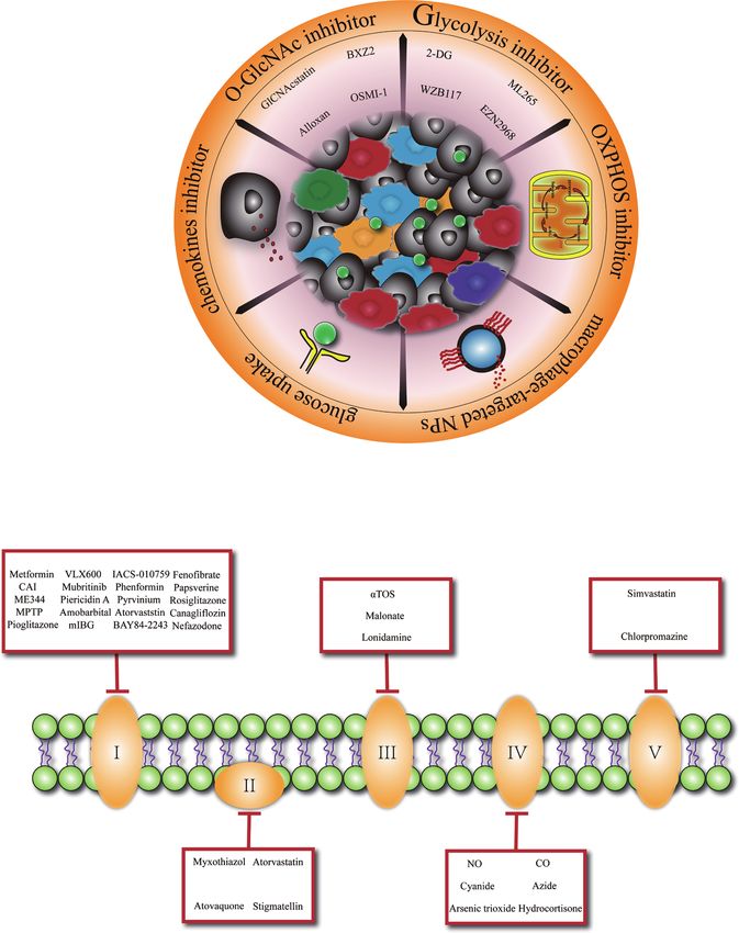
FIGURE 1 | Glucose metabolism basis macrophage-targeted therapy for cancer. (A) Overview of promising cancer therapy based on glucose metabolism
characteristic in tumor-associated macrophage. 2-DG, 2-deoxyglucose. (B) Specific presentation of OXPHOS (oxidative phosphorylation) inhibitors involved
mitochondrial complex I, II, III, IV, V. CAI, carboxyamidotriazole; MPTP, 1-methyl 4-phenyl 1,2,3,6 tetrahydropyridine; mIBG2, meta-iodobenzylguanidine; aTOS, a-
tocopheryl succinate; NO, nitric oxide; CO, carbon monoxide.
far-ranging macrophage depletion could bring outside effects, activities of macrophages with dimethyl malonate treatment
such as other immunosuppressive cells’ compensation (89–91). exhibited markedly delayed tumor growth (93). Similarly, FAO
Since macrophages glucose metabolism is inextricably inhibitors are developed to achieve the phenotypic transition of
connected to its functionality, metabolic reprogramming of M2- macrophages and inhibit tumor development. Furthermore,
like TAMs toward an anti-tumoral phenotype at the same time researchers have revealed that acriflavine (ACF), a heteroaromatic
rupture cancer cell metabolism might be an elegant way. In the dye with an antibacterial and antiviral effect, shifted macrophage
context of a profound relationship between OXPHOS and the polarization to an M1-like anti-tumoral phenotype by blocking the
differentiation of M2 macrophages especially in TAMs, inhibiting HIF-1a pathway and enhancing glucose uptake in PDAC (94). This
OXPHOS pathway (Figure 1B) has been explored as a promising phenomenon shows that increasing the glucose utilization of TAMs
approach to promote TAMs transition to M1 macrophages (92). may be a promising direction.
Blocking the expression of succinate dehydrogenase complex As above mentioned, glycolysis is important in the early
flavoprotein subunit A (SDHA) and oxidative phosphorylation differentiation of TAMs, the maintenance of an M2-like profiles
Frontiers in Immunology | www.frontiersin.org 5 June 2021 | Volume 12 | Article 702580Zhang et al. Macrophage Glucose Metabolism
also dependent on a high glycolytic flow. Consequently, glycolysis CONCLUSION
inhibition (with decreased lactate derived from the tumor) of TAMs
is certainly hopeful for cancer therapy. Chitin administration As previously described, macrophages might respond diversely
significantly decreased anti-inflammatory M2 macrophage depending on the heterogeneity in miscellaneous tissue
polarization and prevented disease progression in a series of microenvironment and cell subpopulations ongoing changed.
mouse models (95). Also, dichloroacetic acid profoundly prevented Hence, macrophages should be considered as dynamic
macrophage migration in a lung tumor xenograft model by alternations in the different phases of cancer where they adapt
inhibiting macrophages glycolysis (42). Several O-GlcNAcylation various phenotypes and also metabolic signatures; the enhanced or
inhibitors had been proved to inhibit cancer cell growth (96, 97). decreased glucose metabolism of macrophages should also not be
Nevertheless, specific targeting of one of the metabolic pathways for taken as favorable or harmful effects for TME. On the other hand,
macrophages is potentially deflective. Proper adjustment of glucose advanced tools such as spatial transcriptomics and multiplex
metabolism in macrophages, instead of a simple one-way increase or immunohistochemistry need to be developed to dig the
decrease, presents a potential therapeutic strategy. association of glucose metabolism and macrophages. Whatever,
In addition, owing to distinct cell populations of the TME share currently, the most effective strategies to target cancer will have to
common metabolic profiles and all metabolic pathways are precisely combine TAM-targeted prodrugs delivery systems with
important for normal cells, sustained modifications of core complex cell glucose metabolism pathways and real-time imaging
metabolic pathways may have marginally immunological effects systems in cancer. In summary, depth work is required to probe
that are difficult to predict. Alternatively, the use of prodrugs that the macrophage-response specificity, tissue-type sensitivity, and
are specifically activated macrophages according to the metabolism-pattern availability, especially constricting the gap
embellishment of glucose metabolism in TME could be between research and clinic with the help of precision medicine.
considered for future therapy. For instance, esterase-sensitive
motif (ESM) inhibitors were prosperously tested as clinical
agents targeting macrophages (98). More than that, with the AUTHOR CONTRIBUTIONS
development of nanotechnology, drug delivery systems based on
QZ and JW wrote the manuscript. DY, XB, and TL revised the
nanoparticles (NPs) have been in the generation of therapeutic
manuscript. QZ and JW contributed equally to this work. All authors
agents for several features, they are avirulent and can easily
contributed to the article and approved the submitted version.
penetrate physiological barriers with a stable consistency.
Glucose-based NPs have been used as biocompatible polymers
to re-educate TAMs (99). Meanwhile, 18F-FDG PET (100–102) FUNDING
has been proposed as a non-invasive strategy to detect glucose
uptake and orbit underlying macrophage polarization This work was financially supported by the National Key
mechanisms. The application of biological or chemical materials Research and Development Program (2020YFA0804300),
in targeted therapy makes it possible for the natural modulation of National Natural Science Foundation of China (Nos. 81871320,
macrophage glucose metabolism in-vivo, favoring an optimal 82071865) and the Distinguished Young Scholar Foundation of
metabolic balance of macrophages to display functions in TME. Zhejiang Province, China (LR20H160002).
9. Yao Y, Xu XH, Jin L. Macrophage Polarization in Physiological and
REFERENCES Pathological Pregnancy. Front Immunol (2019) 10:792. doi: 10.3389/
1. Joyce JA, Fearon DT. T Cell Exclusion, Immune Privilege, and the Tumor fimmu.2019.00792
Microenvironment. Sci (New York N.Y.) (2015) 348:74–80. doi: 10.1126/ 10. Langston PK, Shibata M, Horng T. Metabolism Supports Macrophage
science.aaa6204 Activation. Front Immunol (2017) 8:61. doi: 10.3389/fimmu.2017.00061
2. Netea-Maier RT, Smit JWA, Netea MG. Metabolic Changes in Tumor Cells 11. DeNardo DG, Ruffell B. Macrophages as Regulators of Tumour Immunity
and Tumor-Associated Macrophages: A Mutual Relationship. Cancer Lett and Immunotherapy. Nat Rev Immunol (2019) 19:369–82. doi: 10.1038/
(2018) 413:102–9. doi: 10.1016/j.canlet.2017.10.037 s41577-019-0127-6
3. Lin Y, Xu J, Lan H. Tumor-Associated Macrophages in Tumor Metastasis: 12. Vitale I, Manic G, Coussens LM, Kroemer G, Galluzzi L. Macrophages And
Biological Roles and Clinical Therapeutic Applications. J Hematol Oncol Metabolism in the Tumor Microenvironment. Cell Metab (2019) 30:36–50.
(2019) 12:76. doi: 10.1186/s13045-019-0760-3 doi: 10.1016/j.cmet.2019.06.001
4. Ovchinnikov DA. Macrophages in the Embryo and Beyond: Much More Than 13. Su P, Su P, Su P, Ma X, Liu L, Yang M, et al. Enhanced Lipid Accumulation
Just Giant Phagocytes. Genesis (2008) 46:447–62. doi: 10.1002/dvg.20417 and Metabolism are Required for the Differentiation and Activation of
5. Italiani P, Boraschi D. New Insights Into Tissue Macrophages: From Their Tumor-Associated Macrophages. Cancer Res (2020) 80:1438–50.
Origin to the Development of Memory. Immune Netw (2015) 15:167–76. doi: 10.1158/0008-5472.CAN-19-2994
doi: 10.4110/in.2015.15.4.167 14. Warburg O, Wind F, Negelein E. The Metabolism OF Tumors IN the Body.
6. Viola A, Munari F, Sá nchez-Rodrı́guez R, Scolaro T, Castegna A. The J Gen Physiol (1927) 8:519–30. doi: 10.1085/jgp.8.6.519
Metabolic Signature of Macrophage Responses. Front Immunol (2019) 15. Warburg O. On Respiratory Impairment in Cancer Cells. Sci (New York
10:1462. doi: 10.3389/fimmu.2019.01462 N.Y.) (1956) 124:269–70.
7. Mills CD, Kincaid K, Alt JM, Heilman MJ, Hill AM. M-1/M-2 Macrophages 16. Vander Heiden MG, Cantley LC, Thompson CB. Understanding the
and the Th1/Th2 Paradigm. J Immunol (2000) 164:6166–73. doi: 10.4049/ Warburg Effect: The Metabolic Requirements of Cell Proliferation. Sci
jimmunol.1701141 (New York N.Y.) (2009) 324:1029–33. doi: 10.1126/science.1160809
8. Gordon S. Alternative Activation of Macrophages. Nat Rev Immunol (2003) 17. Jang M, Kim SS, Lee J. Cancer Cell Metabolism: Implications for Therapeutic
3:23–35. doi: 10.1038/nri978 Targets. Exp Mol Med (2013) 45:e45. doi: 10.1038/emm.2013.85
Frontiers in Immunology | www.frontiersin.org 6 June 2021 | Volume 12 | Article 702580Zhang et al. Macrophage Glucose Metabolism
18. Hsu PP, Sabatini DM. Cancer Cell Metabolism: Warburg and Beyond. Cell 38. Xie M, Yu Y, Kang R, Zhu S, Yang L, Zeng L, et al. PKM2-Dependent
(2008) 134:703–7. doi: 10.1016/j.cell.2008.08.021 Glycolysis Promotes NLRP3 and AIM2 Inflammasome Activation. Nat
19. Reinfeld BI, Madden MZ, Wolf MM, Chytil A, Bader JE, Patterson AR, et al. Commun (2016) 7:13280. doi: 10.1038/ncomms13280
Cell-Programmed Nutrient Partitioning in the Tumour Microenvironment. 39. Freemerman AJ, Johnson AR, Sacks GN, Milner JJ, Kirk EL, Troester MA,
Nature (2021) 593:282–8. doi: 10.1038/s41586-021-03442-1 et al. Metabolic Reprogramming of Macrophages: Glucose TRANSPORTER
20. O’Neill LA, Kishton RJ, Rathmell J. A Guide to Immunometabolism for 1 (Glut1)-Mediated GLUCOSE Metabolism Drives A Proinflammatory
Immunologists. Nat Rev Immunol (2016) 16:553–65. doi: 10.1038/ Phenotype*. J Biol Chem (2014) 289:7884–96. doi: 10.1074/jbc.M113.522037
nri.2016.70 40. Pavlou S, Lindsay J, Ingram R, Xu H, Chen M. Sustained High Glucose
21. Werner C, Doenst T, Schwarzer M. Metabolic Pathways and Cycles. Scientist’s Exposure Sensitizes Macrophage Responses to Cytokine Stimuli But Reduces
Guide to Cardiac Metab (2016) 39–55. doi: 10.1016/B978-0-12-802394-5.00004-2 Their Phagocytic Activity. BMC Immunol (2018) 19:24. doi: 10.1186/s12865-
22. Xie N, Zhang L, Gao W, Huang C, Huber PE, Zhou X, et al. NAD(+) 018-0261-0
Metabolism: Pathophysiologic Mechanisms and Therapeutic Potential. 41. Haschemi A, Kosma P, Gille L, Evans CR, Burant CF, Starkl P, et al. The
Signal Transduction Targeted Ther (2020) 5:227. doi: 10.1038/s41392-020- Sedoheptulose Kinase CARKL Directs Macrophage Polarization Through
00311-7 Control of Glucose Metabolism. Cell Metab (2012) 15:813–26. doi: 10.1016/
23. Liu Y, Xu R, Gu H, Zhang E, Qu J, Cao W, et al. Metabolic Reprogramming j.cmet.2012.04.023
in Macrophage Responses. Biomarker Res (2021) 9:1. doi: 10.1038/s41392- 42. Semba H, Takeda N, Isagawa T, Sugiura Y, Honda K, Wake M, et al. Hif-1a-
020-00311-7 PDK1 Axis-Induced Active Glycolysis Plays an Essential Role in
24. Kreppel LK, Blomberg MA, Hart GW. Dynamic Glycosylation of Nuclear Macrophage Migratory Capacity. Nat Commun (2016) 7:11635.
and Cytosolic Proteins: CLONING and CHARACTERIZATION of A doi: 10.1038/ncomms11635
Unique O-GlcNAc Transferase WITH Multiple TETRATRICOPEPTIDE 43. van Uden P, Kenneth NS, Rocha S. Regulation of Hypoxia-Inducible factor-
Repeats*. J Biol Chem (1997) 272:9308–15. doi: 10.1074/jbc.272.14.9308 1alpha by NF-Kappab. Biochem J (2008) 412:477–84. doi: 10.1042/BJ20080476
25. Love DC, Hanover JA. The Hexosamine Signaling Pathway: Deciphering the 44. Rius J, Guma M, Schachtrup C, Akassoglou K, Zinkernagel AS, Nizet V, et al.
“O-GlcNAc Code”. Science’s STKE Signal Transduction Knowl Environ NF-Kappab Links Innate Immunity to the Hypoxic Response Through
(2005) 2005):re13. doi: 10.1126/stke.3122005re13 Transcriptional Regulation of HIF-1alpha. Nature (2008) 453:807–11.
26. Yang X, Ongusaha PP, Miles PD, Havstad JC, Zhang F, So WV, et al. doi: 10.1038/nature06905
Phosphoinositide Signalling Links O-GlcNAc Transferase to Insulin 45. Chang YH, Weng CL, Lin KI, Liu C, Li L, Herring LE. O-GlcNAcylation and
Resistance. Nature (2008) 451:964–9. doi: 10.1038/nature06668 its Role in the Immune System. J BioMed Sci (2020) 27:57. doi: 10.1186/
27. Fukuzumi M, Shinomiya H, Shimizu Y, Ohishi K, Utsumi S. Endotoxin- s12929-020-00648-9
induced Enhancement of Glucose Influx Into Murine Peritoneal 46. Li T, Li X, Attri KS, Liu C, Li L, Herring LE, et al. O-Glcnac Transferase
Macrophages Via GLUT1. Infect Immun (1996) 64:108–12. doi: 10.1128/ Links Glucose Metabolism to MAVS-Mediated Antiviral Innate Immunity.
iai.64.1.108-112.1996 Cell Host Microbe (2018) 24:791–803 e796. doi: 10.1016/j.chom.2018.11.001
28. Van den Bossche J, Baardman J, de Winther MP. Metabolic Characterization 47. Li X, Gong W, Wang H, Li T, Attri KS, Lewis RE, et al. O-Glcnac Transferase
of Polarized M1 and M2 Bone Marrow-Derived Macrophages Using Real- Suppresses Inflammation and Necroptosis by Targeting Receptor-
Time Extracellular Flux Analysis. J Visualized Exp JoVE (2015) 105:53424. Interacting Serine/Threonine-Protein Kinase 3. Immunity (2019) 50:576–
doi: 10.3791/53424 590 e576. doi: 10.1016/j.immuni.2019.01.007
29. Murray PJ, Allen JE, Biswas SK, Fisher JA, Gilroy DW, Goerdt S, et al. 48. Vats D, Mukundan L, Odegaard JI, Zhang L, Smith KL, Morel CR, et al.
Macrophage Activation and Polarization: Nomenclature and Experimental Oxidative Metabolism and PGC-1b Attenuate Macrophage-Mediated
Guidelines. Immunity (2014) 41:14–20. doi: 10.1016/j.immuni.2014.06.008 Inflammation. Cell Metab (2006) 4:13–24. doi: 10.1016/j.cmet.2006.05.011
30. Murray PJ. Macrophage Polarization. Annu Rev Physiol (2017) 79:541–66. 49. Namgaladze D, Brune B. Fatty Acid Oxidation is Dispensable for Human
doi: 10.1146/annurev-physiol-022516-034339 Macrophage IL-4-induced Polarization. Biochim Biophys Acta (2014)
31. Ramond E, Jamet A, Coureuil M, Charbit A. Pivotal Role of Mitochondria in 1841:1329–35. doi: 10.1016/j.bbalip.2014.06.007
Macrophage Response to Bacterial Pathogens. Front Immunol (2019) 50. Wu L, Zhang X, Zheng L, Zhao H, Yan G, Zhang Q, et al. Ripk3 Orchestrates
10:2461. doi: 10.3389/fimmu.2019.02461 Fatty Acid Metabolism in Tumor-Associated Macrophages and
32. Freemerman AJ, Johnson AR, Sacks GN, Milner JJ, Kirk EL, Troester MA, Hepatocarcinogenesis. Cancer Immunol Res (2020) 8:710–21. doi: 10.1158/
et al. Metabolic Reprogramming of Macrophages: Glucose Transporter 1 2326-6066.CIR-19-0261
(GLUT1)-Mediated Glucose Metabolism Drives a Proinflammatory 51. Wang F, Zhang S, Vuckovic I, Jeon R, Lerman A, Folmes CD, et al. Glycolytic
Phenotype. J Biol Chem (2014) 289:7884–96. doi: 10.1074/jbc.M113.522037 Stimulation is Not a Requirement for M2 Macrophage Differentiation. Cell
33. Moon JS, Hisata S, Park MA, DeNicola GM, Ryter SW, Nakahira K, et al. Metab (2018) 28:463–475.e464. doi: 10.1016/j.cmet.2018.08.012
Mtorc1-Induced HK1-Dependent Glycolysis Regulates Nlrp3 52. de–Brito –N, Duncan–Moretti J, da–Costa H, Saldanha–Gama R, Paula–
Inflammasome Activation. Cell Rep (2015) 12:102–15. doi: 10.1016/ Neto HA, Dorighello G, et al. Aerobic Glycolysis is a Metabolic Requirement
j.celrep.2015.05.046 to Maintain the M2-like Polarization of Tumor-Associated Macrophages.
34. Rodrı́guez-Prados JC, Travé s PG, Cuenca J, Rico D, Aragoné s J, Martı́n– Biochim Biophys Acta Mol Cell Res (2020) 1867:118604. doi: 10.1016/
Sanz P, et al. Substrate Fate in Activated Macrophages: A Comparison j.bbamcr.2019.118604
Between Innate, Classic, and Alternative Activation. J Immunol (2010) 53. Tan Z, Xie N, Cui H, Moellering DR, Abraham E, Thannickal VJ, et al.
185:605–14. doi: 10.4049/jimmunol.0901698 Pyruvate Dehydrogenase Kinase 1 Participates in Macrophage Polarization
35. Boscá L, Gonzá́ lez-Ramos S, Prieto P, Ferná́ ndez-Velasco M, Mojena M, Via Regulating Glucose Metabolism. J Immunol (2015) 194:6082–9.
Martı́n -Sanz P, et al. Metabolic Signatures Linked to Macrophage doi: 10.4049/jimmunol.1402469
Polarization: From Glucose Metabolism to Oxidative Phosphorylation. 54. Covarrubias AJ, Aksoylar HI, Yu J, Snyder NW, Worth AJ, Iyer SS, et al. Akt-
Biochem Soc Trans (2015) 43:740–4. doi: 10.1042/BST20150107 mTORC1 Signaling Regulates Acly to Integrate Metabolic Input to Control
36. Jiang H, Shi H, Sun M, Wang Y, Meng Q, Guo P, et al. Pfkfb3-Driven of Macrophage Activation. eLife (2016) 5:e11612. doi: 10.7554/eLife.11612
Macrophage Glycolytic Metabolism is a Crucial Component of Innate 55. Huang SC, Smith AM, Everts B, Colonna M, Pearce EL, Schilling JD, et al.
Antiviral Defense. J Immunol (2016) 197:2880–90. doi: 10.4049/ Metabolic Reprogramming Mediated by the Mtorc2-IRF4 Signaling Axis Is
jimmunol.1600474 Essential for Macrophage Alternative Activation. Immunity (2016) 45:817–
37. Palsson-McDermott EM, Curtis AM, Goel G, Lauterbach MA, Sheedy FJ, 30. doi: 10.1016/j.immuni.2016.09.016
Gleeson LE, et al. Pyruvate Kinase M2 Regulates Hif-1a Activity and IL-1b 56. Van den Bossche J, Baardman J, Otto NA, van der Velden S, Neele AE, van den
Induction and Is a Critical Determinant of the Warburg Effect in LPS- Berg SM, et al. Mitochondrial Dysfunction Prevents Repolarization of
Activated Macrophages. Cell Metab (2015) 21:65–80. doi: 10.1016/ Inflammatory Macrophages. Cell Rep (2016) 17:684–96. doi: 10.1016/
j.cmet.2014.12.005 j.celrep.2016.09.008
Frontiers in Immunology | www.frontiersin.org 7 June 2021 | Volume 12 | Article 702580Zhang et al. Macrophage Glucose Metabolism
57. Yang Y, Li X, Luan HH, Zhang B, Zhang K, Nam JH, et al. OGT Suppresses 76. Pan Y, Lu F, Fei Q, Yu X, Xiong P, Yu X, et al. Single-Cell RNA Sequencing
S6K1-mediated Macrophage Inflammation and Metabolic Disturbance. Proc Reveals Compartmental Remodeling of Tumor-Infiltrating Immune Cells
Natl Acad Sci U States Am (2020) 117:16616–25. doi: 10.1073/pnas.1916121117 Induced by anti-CD47 Targeting in Pancreatic Cancer. J Hematol Oncol
58. Zanin RF, Braganhol E, Bergamin LS, Campesato LF, Filho AZ, Moreira JC, (2019) 12:124. doi: 10.1186/s13045-019-0822-6
et al. Differential Macrophage Activation Alters the Expression Profile of 77. Cheng H, Wang Z, Fu L, Xu T. Macrophage Polarization in the Development
NTPDase and Ecto-5’-Nucleotidase. PloS One (2012) 7:e31205. doi: 10.1371/ and Progression of Ovarian Cancers: An Overview. Front Oncol (2019)
journal.pone.0031205 9:421. doi: 10.3389/fonc.2019.00421
59. Murphy PS, Wang J, Bhagwat SP, Munger JC, Janssen WJ, Wright TW, et al. 78. Arlauckas SP, Garris CS, Kohler RH, Kitaoka M, Cuccarese MF, Yang KS,
CD73 Regulates Anti-Inflammatory Signaling Between Apoptotic Cells and et al. In Vivo Imaging Reveals a Tumor-Associated Macrophage-Mediated
Endotoxin-Conditioned Tissue Macrophages. Cell Death Differentiation Resistance Pathway in anti-PD-1 Therapy. Sci Trans Med (2017) 9:eaal3604.
(2017) 24:559–70. doi: 10.1038/cdd.2016.159 doi: 10.1126/scitranslmed.aal3604
60. Hartley GP, Chow L, Ammons DT, Wheat WH, Dow SW. Programmed Cell 79. Xue J, Schmidt SV, Sander J, Draffehn A, Krebs W, Quester I, et al.
Death Ligand 1 (Pd-L1) Signaling Regulates Macrophage Proliferation and Transcriptome-Based Network Analysis Reveals a Spectrum Model of
Activation. Cancer Immunol Res (2018) 6:1260–73. doi: 10.1158/2326- Human Macrophage Activation. Immunity (2014) 40:274–88.
6066.CIR-17-0537 doi: 10.1016/j.immuni.2014.01.006
61. de Goede KE, Driessen AJM, Van den Bossche J. Metabolic Cancer- 80. Van Overmeire E, Laoui D, Keirsse J, Van Ginderachter JA, Sarukhan A.
Macrophage Crosstalk in the Tumor Microenvironment. Biol (Basel) Mechanisms Driving Macrophage Diversity and Specialization in Distinct
(2020) 9:380. doi: 10.3390/biology9110380 Tumor Microenvironments and Parallelisms With Other Tissues. Front
62. Puthenveetil A, Dubey S. Metabolic Reprograming of Tumor-Associated Immunol (2014) 5:127. doi: 10.3389/fimmu.2014.00127
Macrophages. Ann Transl Med (2020) 8:1030. doi: 10.21037/atm-20-2037 81. Chen H, Ye F, Guo G. Revolutionizing Immunology With Single-Cell RNA
63. Wenes M, Shang M, Di Matteo M, Goveia J, Martı́n-Pé rez R, Serneels J, et al. Sequencing. Cell Mol Immunol (2019) 16:242–9. doi: 10.1080/
Macrophage Metabolism Controls Tumor Blood Vessel Morphogenesis and 09546634.2019.1630701
Metastasis. Cell Metab (2016) 24:701–15. doi: 10.1016/j.cmet.2016.09.008 82. Hartmann FJ, Mrdjen D, McCaffrey E, Glass DR, Greenwald NF, Bharadwaj
64. Rodrigues Mantuano N, Stanczak MA, Oliveira IA, Kirchhammer N, Filardy A, et al. Single-Cell Metabolic Profiling of Human Cytotoxic T Cells. Nat
AA, Monaco G, et al. Hyperglycemia Enhances Cancer Immune Evasion by Biotechnol (2021) 39:186–97. doi: 10.1038/s41587-020-0651-8
Inducing Alternative Macrophage Polarization Through Increased O- 83. Hartmann FJ, Bendall SC. Immune Monitoring Using Mass Cytometry and
Glcnacylation. Cancer Immunol Res (2020) 8:1262–72. doi: 10.1158/2326- Related High-Dimensional Imaging Approaches. Nat Rev Rheumatol (2020)
6066.CIR-19-0904 16:87–99. doi: 10.1038/s41584-019-0338-z
65. Shapouri-Moghaddam A, Mohammadian S, Vazini H, Taghadosi M, 84. Zhou B, Magana L, Hong Z, Huang LS, Chakraborty S, Tsukasaki Y, et al.
Esmaeili SA, Mardani F, et al. Macrophage Plasticity, Polarization, and The Angiocrine Rspondin3 Instructs Interstitial Macrophage Transition Via
Function in Health and Disease. J Cell Physiol (2018) 233:6425–40. Metabolic-Epigenetic Reprogramming and Resolves Inflammatory Injury.
doi: 10.1002/jcp.26429 Nat Immunol (2020) 21:1430–43. doi: 10.1038/s41590-020-0764-8
66. Locati M, Curtale G, Mantovani. Diversity A. Mechanisms, and Significance 85. Artyomov MN, Van den Bossche J. Immunometabolism in the Single-Cell
of Macrophage Plasticity. Annu Rev Pathol (2020) 15:123–47. doi: 10.1146/ Era. Cell Metab (2020) 32:710–25. doi: 10.1016/j.cmet.2020.09.013
annurev-pathmechdis-012418-012718 86. Ruffell B, Coussens LM. Macrophages and Therapeutic Resistance in Cancer.
67. Madden MZ, Reinfeld BI, Wolf MM, Cohen AS, Manning HC, Rathmell Cancer Cell (2015) 27:462–72. doi: 10.1016/j.ccell.2015.02.015
WK, et al. Nutrient Partitioning in the Tumor Microenvironment and FDG- 87. Mantovani A, Marchesi F, Malesci A, Laghi L, Allavena P. Tumour-
PET Imaging. J Immunol (2020) 204:240.244. Associated Macrophages as Treatment Targets in Oncology. Nat Rev Clin
68. Liu D, Chang C, Lu N, Wang X, Lu Q, Ren X, et al. Comprehensive Oncol (2017) 14:399–416. doi: 10.1038/nrclinonc.2016.217
Proteomics Analysis Reveals Metabolic Reprogramming of Tumor- 88. Bonapace L, Coissieux MM, Wyckoff J, Mertz KD, Varga Z, Junt T, et al.
Associated Macrophages Stimulated by the Tumor Microenvironment. Cessation of CCL2 Inhibition Accelerates Breast Cancer Metastasis by
J Proteome Res (2017) 16:288–97. doi: 10.1021/acs.jproteome.6b00604 Promoting Angiogenesis. Nature (2014) 515:130–3. doi: 10.1038/nature13862
69. Penny HL, Sieow JL, Adriani G, Yeap WH, See Chi P, San Luis B, et al. 89. Lee B, Qiao L, Kinney B, Feng GS, Shao J. Macrophage Depletion Disrupts
Warburg Metabolism in Tumor-Conditioned Macrophages Promotes Immune Balance and Energy Homeostasis. PloS One (2014) 9:e99575.
Metastasis in Human Pancreatic Ductal Adenocarcinoma. Oncoimmunology doi: 10.1371/journal.pone.0099575
(2016) 5:e1191731. doi: 10.1080/2162402X.2016.1191731 90. Wu CL, McNeill J, Goon K, Little D, Kimmerling K, Huebner J, et al.
70. Arts RJ, Plantinga TS, Tuit S, Ulas T, Heinhuis B, Tesselaar M, et al. Conditional Macrophage Depletion Increases Inflammation and Does Not
Transcriptional and Metabolic Reprogramming Induce an Inflammatory Inhibit the Development of Osteoarthritis in Obese Macrophage Fas-
Phenotype in non-Medullary Thyroid Carcinoma-Induced Macrophages. Induced Apoptosis-Transgenic Mice. Arthritis Rheumatol (Hoboken N.J.)
Oncoimmunology (2016) 5:e1229725. doi: 10.1080/2162402X.2016.1191731 (2017) 69:1772–83. doi: 10.1002/art.40161
71. Goetze K, Walenta S, Ksiazkiewicz M, Kunz-Schughart LA, Mueller-Klieser 91. Cassetta L, Pollard JW. Targeting Macrophages: Therapeutic Approaches in
W. Lactate Enhances Motility of Tumor Cells and Inhibits Monocyte Cancer. Nat Rev Drug Discovery (2018) 17:887–904. doi: 10.1038/nrd.2018.169
Migration and Cytokine Release. Int J Oncol (2011) 39:453–63. 92. Yu Q, Wang Y, Dong L, He Y, Liu R, Yang Q, et al. Regulations of Glycolytic
doi: 10.3892/ijo.2011.1055 Activities on Macrophages Functions in Tumor and Infectious Inflammation.
72. Colegio OR, Chu NQ, Szabo AL, Chu T, Rhebergen AM, Jairam V, et al. Front Cell Infect Microbiol (2020) 10:287. doi: 10.3389/fcimb.2020.00287
Functional Polarization of Tumour-Associated Macrophages by Tumour- 93. Ashton TM, McKenna WG, Kunz-Schughart LA, Higgins GS. Oxidative
Derived Lactic Acid. Nature (2014) 513:559–63. doi: 10.1038/nature13490 Phosphorylation as an Emerging Target in Cancer Therapy. Clin Cancer Res
73. Zhang J, Zhang Q, Lou Y, Fu J, Chen J, Wei J, et al. Hypoxia-Inducible (2018) 24:2482–90. doi: 10.1158/1078-0432.CCR-17-3070
Factor-1a/Interleukin-1b Signaling Enhances Hepatoma Epithelial- 94. Bulle A, Dekervel J, Deschuttere L, Nittner D, Van Cutsem E, Verslype C,
Mesenchymal Transition Through Macrophages in a Hypoxic- et al. Anti-Cancer Activity of Acriflavine as Metabolic Inhibitor of OXPHOS
Inflammatory Microenvironment. Hepatol (Baltimore Md.) (2018) in Pancreas Cancer Xenografts. Onco Targets Ther (2020) 13:6907–16.
67:1872–89. doi: 10.1002/hep.29681 doi: 10.2147/OTT.S245134
74. Jeong H, Kim S, Hong BJ, Lee CJ, Kim YE, Bok S, et al. Tumor-Associated 95. Zhao Q, Chu Z, Zhu L, Yang T, Wang P, Liu F, et al. 2-Deoxy-D-Glucose
Macrophages Enhance Tumor Hypoxia and Aerobic Glycolysis. Cancer Res Treatment Decreases Anti-inflammatory M2 Macrophage Polarization in
(2019) 79:795–806. doi: 10.1158/0008-5472.CAN-18-2545 Mice With Tumor and Allergic Airway Inflammation. Front Immunol
75. Kurahara H, Shinchi H, Mataki Y, Maemura K, Noma H, Kubo F, et al. (2017) 8:637. doi: 10.3389/fimmu.2017.00637
Significance of M2-polarized Tumor-Associated Macrophage in Pancreatic 96. Chaiyawat P, Chokchaichamnankit D, Lirdprapamongkol K, Srisomsap C,
Cancer. J Surg Res (2011) 167:e211–219. doi: 10.1016/j.jss.2009.05.026 Svasti J, Champattanachai V, et al. Alteration of O-GlcNAcylation Affects
Frontiers in Immunology | www.frontiersin.org 8 June 2021 | Volume 12 | Article 702580Zhang et al. Macrophage Glucose Metabolism
Serine Phosphorylation and Regulates Gene Expression and Activity of Hypoxia-Inducible Factor-1a Activation Through Nox2-dependent
Pyruvate Kinase M2 in Colorectal Cancer Cells. Oncol Rep (2015) 34:1933– Reactive Oxygen Species Generation. J Nucl Med (2014) 55:1699–705.
42. doi: 10.3892/or.2015.4178 doi: 10.2967/jnumed.114.139428
97. Wang Z, Qin J, Zhao J, Li J, Li D, Popp M, et al. Inflammatory IFIT3 Renders 102. Tavakoli S, Short JD, Downs K, Nguyen HN, Lai Y, Zhang W, et al.
Chemotherapy Resistance by Regulating Post-Translational Modification of Differential Regulation of Macrophage Glucose Metabolism by
VDAC2 in Pancreatic Cancer. Theranostics (2020) 10:7178–92. doi: 10.7150/ Macrophage Colony-Stimulating Factor and Granulocyte-Macrophage
thno.43093 Colony-Stimulating Factor: Implications for (18)F FDG Pet Imaging of
98. Needham LA, Davidson AH, Bawden LJ, Belfield A, Bone EA, Brotherton Vessel Wall Inflammation. Radiology (2017) 283:87–97. doi: 10.1148/
DH, et al. Drug Targeting to Monocytes and Macrophages Using Esterase- radiol.2016160839
Sensitive Chemical Motifs. J Pharmacol Exp Ther (2011) 339:132–42.
doi: 10.1124/jpet.111.183640 Conflict of Interest: The authors declare that the research was conducted in the
99. Reichel D, Tripathi M, Perez JM. Biological Effects of Nanoparticles on absence of any commercial or financial relationships that could be construed as a
Macrophage Polarization in the Tumor Microenvironment. Nanotheranostics potential conflict of interest.
(2019) 3:66–88. doi: 10.7150/ntno.30052
100. Tavakoli S, Zamora D, Ullevig S, Asmis R. Bioenergetic Profiles Diverge Copyright © 2021 Zhang, Wang, Yadav, Bai and Liang. This is an open-access article
During Macrophage Polarization: Implications for the Interpretation of 18F- distributed under the terms of the Creative Commons Attribution License (CC BY).
FDG PET Imaging of Atherosclerosis. J Nucl Med (2013) 54:1661–7. The use, distribution or reproduction in other forums is permitted, provided the
doi: 10.2967/jnumed.112.119099 original author(s) and the copyright owner(s) are credited and that the original
101. Lee SJ, Thien Quach CH, Jung KH, Paik JY, Lee JH, Park JW, et al. Oxidized publication in this journal is cited, in accordance with accepted academic practice. No
Low-Density Lipoprotein Stimulates Macrophage 18F-FDG Uptake Via use, distribution or reproduction is permitted which does not comply with these terms.
Frontiers in Immunology | www.frontiersin.org 9 June 2021 | Volume 12 | Article 702580You can also read