HIGH LEVELS OF ANTI-LEISHMANIA IGG3 AND LOW CD4+ T CELLS COUNT WERE ASSOCIATED WITH RELAPSES IN VISCERAL LEISHMANIASIS - BMC ...
←
→
Page content transcription
If your browser does not render page correctly, please read the page content below
Kuschnir et al. BMC Infectious Diseases  (2021) 21:369
https://doi.org/10.1186/s12879-021-06051-5

 RESEARCH                                                                                                                                            Open Access

High levels of anti-Leishmania IgG3 and low
CD4+ T cells count were associated with
relapses in visceral leishmaniasis
Renata Caetano Kuschnir1, Leonardo Soares Pereira2, Maria Rita Teixeira Dutra2, Ludmila de Paula2,
Maria Luciana Silva-Freitas1, Gabriela Corrêa-Castro1,3, Simone da Costa Cruz Silva4, Glaucia Cota5,
Joanna Reis Santos-Oliveira1,3 and Alda Maria Da-Cruz1,6,7*

  Abstract
  Background: Visceral leishmaniasis (VL) is severe and potentially fatal. Brazil is one of the countries with the
  greatest endemicity for the disease in the world. The reduction of CD4+ T lymphocytes, B cells activation and high
  levels of inflammatory cytokines (IL-6/IL-8/TNF/IL-1β), plasma LPS, soluble CD14, anti-Leishmania IgG3 and low leptin
  levels are involved in the immunopathogenesis of VL, most associated with severe VL. Despite relapses occurring in
  about 4–5% of patients with VL not associated with HIV infection, the factors underlying relapses are little known.
  Our aim was to identify clinical, laboratory and immunological parameters that may be associated with recurrences
  in VL.
  Methods: Fifteen VL patients recruited from Hospital Eduardo de Menezes (BH-MG) were grouped into relapsing
  (R-VL, n = 5) and non-relapsing (NR-VL, n = 10) and evaluated during active disease, immediately after treatment
  (post-treatment) and 6 months post-treatment (6mpt). Clinical and laboratory data obtained from medical records
  were correlated with CD4+ and CD8+ T cell counts and anti-Leishmania Igs and IL-6 plasma levels and compared to
  those parameters of ten healthy controls.
  Results: During the active phase of VL, despite similarity in the clinical symptoms, the rates of thrombocytopenia,
  elevated transaminases (AST and ALT) and hyperbilirubinemia were higher in the NR-VL group compared to R-VL
  (p < 0.05), a profile reversed during the post-treatment phase. All patients had low CD4+ T counts in active phase,
  however, NR-VL patients had a higher gain of this cell type than R-VL in the post-treatment (p < 0.05). There was a
  significant reduction in IgG3 levels during the follow-up in the NR-VL group compared to the R-VL, especially at
  6mpt (p < 0.05). In addition, IgG3 levels were negatively correlated with CD4+ T counts in the R-VL group (r = −
  0.52). Elevated levels of IL-6 were observed in active VL and correlated with clinical markers of severity.
  (Continued on next page)

* Correspondence: alda@ioc.fiocruz.br
1
 Laboratório Interdisciplinar de Pesquisas Médicas, Instituto Oswaldo Cruz,
FIOCRUZ, Rio de Janeiro, Rio de Janeiro, Brazil
6
 Disciplina de Parasitologia, DMIP, Faculdade de Ciências Médicas, UERJ, Rio
de Janeiro, Brazil
Full list of author information is available at the end of the article

                                         © The Author(s). 2021 Open Access This article is licensed under a Creative Commons Attribution 4.0 International License,
                                         which permits use, sharing, adaptation, distribution and reproduction in any medium or format, as long as you give
                                         appropriate credit to the original author(s) and the source, provide a link to the Creative Commons licence, and indicate if
                                         changes were made. The images or other third party material in this article are included in the article's Creative Commons
                                         licence, unless indicated otherwise in a credit line to the material. If material is not included in the article's Creative Commons
                                         licence and your intended use is not permitted by statutory regulation or exceeds the permitted use, you will need to obtain
                                         permission directly from the copyright holder. To view a copy of this licence, visit http://creativecommons.org/licenses/by/4.0/.
                                         The Creative Commons Public Domain Dedication waiver (http://creativecommons.org/publicdomain/zero/1.0/) applies to the
                                         data made available in this article, unless otherwise stated in a credit line to the data.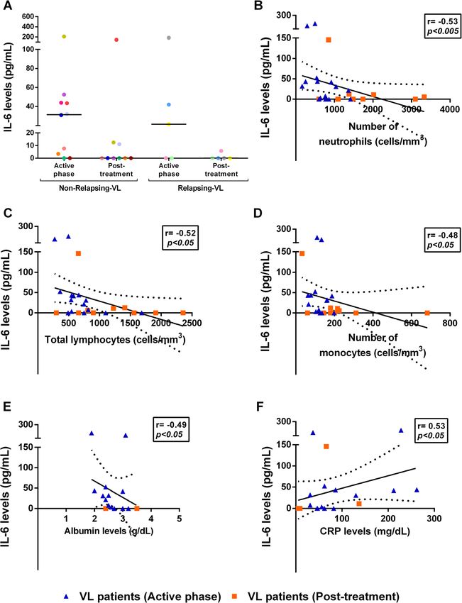
Kuschnir et al. BMC Infectious Diseases   (2021) 21:369                                                         Page 2 of 14

  (Continued from previous page)
  Conclusions: During active phase of VL, the NR-VL patients presented more severe laboratorial abnormalities
  compared to R-VL, probably because the latter had already received previous treatment. On the other hand, R-VL
  exhibited greater impairment of immune reconstitution and a high degree of B lymphocyte activation, which must
  be a factor that favored relapses.
  Keywords: Visceral leishmaniasis, Relapses, Clinical follow-up, Immune response

Background                                                       Relapses are characterized by the resurgence of signs
Visceral leishmaniasis (VL) is caused by Leishmania (L.)       and symptoms after an initial improvement of a disease
infantum in Brazil, being transmitted to mammals by Lut-       manifestation [21]. It is considered a risk factor for death
zomyia longipalpis sand fly [1]. VL is endemic in more         in VL, especially in HIV-co-infected patients [22, 23].
than 90 countries or territories, however, in 2017, few        Recently, our group demonstrated that the maintenance
countries as Brazil, Ethiopia, India, Kenya, South Sudan       of high levels of cell activation, microbial translocation
and Sudan have concentrated more than 90% of the cases         products, anti-Leishmania IgG3 and a low CD4+ T cell
[2]. In the Americas, 3562 new cases of VL were diag-          reconstitution could be associated with VL relapse in
nosed in 2018 and Brazil was responsible for 97% of them       VL/HIV coinfected patients [23]. Moreover, this low im-
[3]. Belo Horizonte, located in Minas Gerais state, is one     mune reconstitution has been related to a greater im-
of the Brazilian cities with the highest number of VL pa-      pairment of the thymic output among VL/HIV-relapsing
tients, with 2378 reported cases from 2007 to 2019 [4].        patients [24]. The inability to reconstitute the effector
   Pathogen and host’s immune system interaction leads         response seems to exert a key role in the VL relapses.
to different clinical presentation, predisposing a high          In Brazil, the official frequency of relapses after VL
variety of outcomes, since asymptomatic disease to high        treatment among non HIV-infected patients is underes-
severity and risk of death. Commonly, most patients af-        timated, although the compulsory notification form has
fected by VL respond well to anti-Leishmania treatment         a specific field to inform this clinical presentation [25].
and evolve to the remission. However, there are those          In the literature, the vast majority of studies are related
who evolve to the severe form of VL, with a high lethal-       to therapeutic failure reports [26]. There are few VL re-
ity rate [5]. In addition, relapse is also observed in med-    lapses reports and those which have it, present a great
ical practice [6, 7].                                          variability in the incidence of relapses according to the
   Clinical and laboratory markers such as age, bleeding,      population and regions studied [27–30]. In a Spanish co-
edema, jaundice, dyspnea, bacterial infection, HIV/AIDS        hort, VL relapse rate was 12% [27] while in Sudan, the
co-infection, leukocyte count below 1500 cells/mm3,            clinical follow-up during an epidemic in Babar showed
thrombocytopenia below 50,000 cells/mm3 and renal              5.7% of relapses, most of patients concentrated in few
failure have also been linked to VL severity [8, 9]. Also,     families [31]. A previous Brazilian study showed relapse
several soluble molecules, such as elevated levels of IL-      in 2.3% of children diagnosed with VL between 2006
2R, IL-1β, IL-6, IL-8, IL-27, TNF, soluble CD14 (sCD14)        and 2011 [30]. Based on secondary data, an analysis of
[10–13], soluble CD163 (sCD163) [14] and specific anti-        VL cases also in Brazil from 2001 to 2010 showed that
Leishmania immunoglobulins [15, 16], as well as low            relapses occurred in 3.1% of patients not co-infected
leptin levels [17] have already been associated with se-       with HIV [29].
verity of VL. Interestingly, IL-6 levels were also associ-       Factors underlying clinical relapses in VL alone have
ated with risk of death in VL [12].                            not been deeply addressed. Clinical characteristics as
   Indeed, besides parasite specific immunosuppression,        male gender, extreme of age and discreet reduction of
the exuberant inflammatory condition constitutes the           splenomegaly were risk factors associated with relapses
key mechanism in the physiopathology of L. infantum            in Indian VL patients [32, 33]. In Georgia, a pediatric co-
infection. Similar to what is seen in sepsis, severe dengue    hort showed that children younger than one-year-old
and severe malaria, high degree of cell activation and         had a higher chance of relapse as the ones who were late
high levels of cytokines are seen in the active VL [12,        diagnosed [34]. More recently, a Brazilian study showed
18–20]. These mechanisms contribute to the immune              that in addition to HIV infection, thrombocytopenia,
response impairment, which in turn have a negative im-         lower limb edema and secondary pneumonia were fac-
pact on the effector capacity to control the parasite.         tors independently associated with relapse [28].
Therefore, these features can influence the clinical evo-        VL relapsing patients should not achieve an effector
lution of VL patients, in terms of clinical cure status, se-   immune response able to maintain the remission of the
verity/death and disease recurrences/relapses.                 disease. Herein, we actively monitored patients with VL,
Kuschnir et al. BMC Infectious Diseases   (2021) 21:369                                                        Page 3 of 14

followed from the active phase of the disease up to 12        treatment, and 6 months after treatment (6mpt). Some
months after treatment and confirmed the presence of          participants were followed up to 12 months after treat-
two distinct groups: those with primary VL without re-        ment (NR-VL = 5 and R-VL = 3). The post-treatment
lapse along 6 months (assumed as single/life episode)         time point varied from patient to patient, due to differ-
and those with two or more VL episodes/life, either with      ences in therapeutic regimens, but for all of them, this
relapse identified at the cohort enrollment or during the     moment refers to the last day of medication (Table 1).
follow-up period. Our aim was to identify possible differ-      Patient’s data were obtained from the medical records:
ences in clinical and immunological parameters that           1) clinical signs and symptoms - fever, asthenia, hypor-
could infer mechanisms involved in recurrence of VL.          exia, weight loss, vomiting, diarrhea, bleeding, pallor,
We evidenced that after therapy, both groups improved         edema, splenomegaly, hepatomegaly, jaundice and
hematological and biochemical parameters and reduced          underlying bacterial infection; 2) laboratory data - blood
IL-6 levels. In contrast, VL relapsing patients maintained    cell counts, biochemical tests related to liver and kidney
increased IgG3 levels besides lower CD4+ T cell counts        function.
in comparison to non-relapsing VL patients. These re-           In each study visit, 45 mL of peripheral blood were
sults suggest that VL relapsing patients keep a B cellular    collected from each patient, divided into 3 tubes with
activation status along with a deficient T cell compart-      heparin (10 mL each) and 3 tubes with EDTA (5 mL
ment reconstitution.                                          each). The whole blood conserved in EDTA was used
                                                              for the CD4+/CD8+ T lymphocytes count, while the
Methods                                                       plasma in heparin tube was used to evaluate the anti-
Casuistic and study design                                    Leishmania IgG immunoglobulins levels and the IgG1
Fifteen VL patients were recruited for a prospective co-      and IgG3 subclasses, as well as the levels of IL-6. The
hort study carried out in an infectious diseases referral     VL therapy was defined on individual basis according to
hospital in Belo Horizonte, MG, Brazil (Hospital              the service routine and Brazilian Minister of Health rec-
Eduardo de Menezes – Fundação Hospitalar do Estado            ommendations [1].
de Minas Gerais/ HEM-FHEMIG) from May 2018 to
September 2019 (Flow diagram). The inclusion criteria         Absolute T-cell counts quantification
in this cohort was presence of fever, cytopenia or spleno-    Absolute T lymphocyte counts were determined using
megaly, age over 18 years, independently of gender.           the BD Multitest monoclonal antibodies anti-CD45-
Pregnant women and patients with HIV-infection were           PerCP, anti-CD3-FITC, anti-CD4-APC, and anti-CD8-
excluded. The leishmaniasis diagnosis was confirmed by        PE (BD® Biosciences, Franklin Lakes, NJ, USA) according
direct visualization of amastigotes in bone marrow aspir-     to the manufacturer’s instructions and as described pre-
ate or presence of anti-Leishmania antibodies in serum.       viously by Silva-Freitas et al. [23]. The counts were ac-
The patients were divided in two groups: those who had        quired using a FACSCalibur and later the FACSVia. The
experienced only one VL episode throughout life (non-         data were analyzed with Multiset software (BD®, USA).
relapsing – NR) and those experiencing more than one          The results are expressed as the number of cells per
VL episode, either previously or during the prospective       cubic millimeter (cells/mm3) and was performed at the
follow-up (relapsing – R – group). Also, ten healthy sub-     reference service of the Instituto Nacional de Infectolo-
jects were included (HS, n = 10) as controls.                 gia Evandro Chagas, FIOCRUZ.
   Cure was defined clinically by the disappearance of
fever associated to recovery of cytopenia and splenomeg-      Anti-Leishmania immunoglobulin assessment
aly involution, if present. Relapses were parasitologicaly    An ELISA was performed as previously described in
confirmed by direct examination or culture in NNN             Silva-Freitas et al. [23], with some modifications. Briefly,
medium in patients presenting resurgence of fever, wors-      L. (L.) infantum (MHOM/BR/1974/PP75) soluble pro-
ening cytopenia or increased splenomegaly, compared to        mastigote (40 μg/mL) was used to coat a polystyrene
the previous condition, as defined by Cota et al. [21]. Pa-   flat-bottom microtiter plate (Nunc-Immuno, Roskilde,
tients with clinical symptoms and parasitological con-        Denmark). In this assay, plasma samples from VL pa-
firmation of active VL were included only after               tients were diluted as follows: 1:10,000 to IgG, 1:2000 to
appropriate written informed consent was obtained. This       IgG1 and 1:200 to IgG3. Then, diluted peroxidase-
study was approved by the Ethical Committees of Insti-        conjugated mouse monoclonal anti-human immuno-
tuto Oswaldo Cruz - FIOCRUZ, HEM-FHEMIG and                   globulin G (IgG) (1:1000) (Invitrogen, San Francisco,
Instituto René Rachou – FIOCRUZ.                              CA, USA) and diluted monoclonal anti-human IgG1 (1:
   Patients were followed prospectively for 6 months and      500) and IgG3 (1:400) (Zymed Laboratories Inc., San
evaluated in three visits: active phase of the disease (be-   Francisco, CA, USA) were used. The absorbance was
fore anti-Leishmania treatment), after anti-Leishmania        measured with a Benchmark microplate reader (Bio-Rad
Kuschnir et al. BMC Infectious Diseases (2021) 21:369 Page 4 of 14
Table 1 Clinical characteristics of non-relapsing and relapsing VL patients
Patient Comorbidities Active phase Cumulative Previous Time Relapse Time Total Total
treatment Amph B episodes between during between number follow-
dose of VL first VL follow- current of VL up
in VL active episode up active episodes
phase and current phase
active and
phase relapse
Non- VL01 Smoker Amph. B deoxychol 28.5 mg/kg No NA No NA 1 12
Relapsing Previous 50 mg/day per 12 months
marijuana use days followed by
Occasional use Liposomal Amph. B
of alcohol 20 mg/kg
VL02 No information Liposomal Amph. B 20 mg/kg No NA No NA 1 12
20 mg/kg months
VL03 Occasional use Amph. B deoxychol 21.7 mg/kg No NA No NA 1 12
of alcohol 50 mg/day per 3 days months
Positive followed by
serology for Liposomal Amph. B
Chagas Disease 20 mg/kg
VL04 Virchowian Amph. B deoxychol 22 mg/kg No NA No NA 1 12
hanseniasis 50 mg/day per 4 days months
ADHD followed by
Development Liposomal Amph. B
delay – 20 mg/kg
infantilized
behavior
VL05 Dyslipidemia Amph. B deoxychol - No NA No NA 1 Post-
Past of drug 1000 mga treatment
addiction
VL08 Arterial Amph. B Lipid 20 mg/kg No NA No NA 1 12
hypertension Complex 20 mg/kg months
Smoker
Alcoholic
VL11 Alcoholic Amph. B Lipid 20 mg/kg No NA No NA 1 6 months
Ex-smoker Complex 200 mg/day
Past of for 4 days followed
schistosomiasis by Liposomal Amph.
and gonorrhea B 20 mg/kg (in 7
weeks)
VL12 Drug addicted Amph. B deoxychol 23 mg/kg No NA No NA 1 6 months
150 mg followed by
Liposomal Amph. B
20 mg/kg
VL13 Alcoholic Meglumine 20 mg/kg No NA No NA 1 Post-
Smoker antimoniate 20 treatment
mgSb+ 5/Kg/day for
3 days followed by
Liposomal Amph. B
20 mg/kg
VL14 Arterial Liposomal Amph. B 20 mg/kg No NA No NA 1 Post-
hypertension 20 mg/kg in7 days treatment
Ex-smoker
Relapsing VL06 Arterial Amph. B Lipid 20 mg/kg Yes (1) 4 months No NA 2 12
hypertension Complex 20 mg/kg months
Diabetes
mellitus type 2
Metabolic
syndrome
VL07 Occasional use Amph. B Lipid 40 mg/kg Yes (3) 15 months Yes 4 months 5 12
of alcohol Complex 40 mg/kg Jan, months
2019
VL09 Chronic renal Amph. B Lipid 20 mg/kg Yes (2) 15 months Yes 12 months 4 12
disease Complex 20 mg/kg Oct, monthsKuschnir et al. BMC Infectious Diseases (2021) 21:369 Page 5 of 14
Table 1 Clinical characteristics of non-relapsing and relapsing VL patients (Continued)
Patient Comorbidities Active phase Cumulative Previous Time Relapse Time Total Total
treatment Amph B episodes between during between number follow-
dose of VL first VL follow- current of VL up
in VL active episode up active episodes
phase and current phase
active and
phase relapse
Hypersplenim 2019
Past of
schistosomiasis
VL10 Chagasic Amph. B Lipid 25 mg/kg No NA Yes 4 months 2 6 months
cardiac Complex 25 mg/kg Feb,
insufficiency 2019
VL15 Ex-smoker Liposomal Amph. B 40 mg/kg Yes (2) 5 months No NA 3 Post-
Pulmonary 40 mg/kg treatment
hypertension
Subclinic
hypothyroidism
VL visceral leishmaniasis, yo years old, ADHD attention deficit hyperactivity disorder, Amph. B deoxychol Amphotericin B deoxycholate, Liposomal Amph. B
Liposomal amphotericin B, Amph. B Lipid Complex Amphotericin B lipid complex
a
no exact dose information available NA not applicable
Laboratories, Hercules, CA, USA) at 492 nm. The results GraphPad Prism software (version 6.0, San Diego, CA,
were expressed as an ELISA index (EI), which is based USA). Differences were considered statistically signifi-
on the division of the average optical density (OD) of cant when the p value was < 0.05.
the duplicates of the patient samples, by the average OD
obtained from the negative controls. Results
Clinical and laboratory characteristics of non-relapsing
Quantitation of IL-6 levels in plasma and relapsing visceral leishmaniasis patients in active
IL-6 levels were quantified in plasma samples stored at phase and after anti-Leishmania treatment
− 70 °C using a commercial kit (IL-6 Quantikine ELISA, Fifteen VL patients were enrolled in the study (Table 1).
R&D Systems, Minneapolis, Maryland, USA), according During active VL phase, fever, asthenia and weight loss
to the manufacturer’s recommendations. A standard were reported by 14 (93%), 11 (73%) and 12 (80%) pa-
seven-point curve diluted in calibration reagent, as well tients, respectively. Splenomegaly was identified in all
as plasma samples were quantified in duplicate. The op- patients based on physical examination and/or imaging
tical density was determined by the Microplate reader tests, and 12 patients had a concomitant liver enlarge-
Benchmark equipment (Bio-Rad Laboratories, Hercules, ment. Considering the severity markers defined by the
CA, USA) at 450 nm. The results were expressed in pi- Brazilian Minister of Health, three patients complained
cograms per milliliter (pg/mL), and the minimum detec- of abdominal pain, two patients reported bleeding prior
tion limit was 3.13 pg/mL. to hospitalization and one presented hematemesis dur-
ing the clinical evolution. Ten patients received anti-
Statistical analysis biotic therapy for suspected or confirmed concomitant
Laboratorial parameters were expressed in medians with bacterial infection.
interquartile ranges shown in square brackets. The gain Only one patient completed treatment using ampho-
of CD4+ and CD8+ T lymphocytes was performed based tericin B deoxycholate, whereas most patients required
on the ratio between the number of cells present at a the use of amphotericin B lipid formulations (Table 1).
given time and the number of cells present in the active R-VL patients showed higher accumulated dose during
phase of VL. Comparisons between the two groups of the treatment in active phase, although without signifi-
VL patients (NR and R) and between each VL group and cant difference between groups (Table 1 and 2).
the control group were performed using the unpaired Regarding liver function, transaminases levels (AST
and non-parametric, Mann Whitney t-test. Wilcoxon and/or ALT) were augmented in the majority of NR-VL
tests for paired variables with skewed distributions were (7 out 10), but in only one out 5 of the R-VL group (p <
also used for comparisons involving the same individual 0.05, Table 2). The total bilirubin was at normal levels in
at different times. Spearman’s test was used for correl- both groups; however, the direct bilirubin (DB) levels
ation analysis. The statistical analyses were performed were more elevated in NR-VL (7 out 10) than in R-VL,
using SPSS® version 16 (multivariate analysis) and in which only one patient presented a slight increase ofKuschnir et al. BMC Infectious Diseases (2021) 21:369 Page 6 of 14
Table 2 Clinical and laboratory differences between relapsing and non-relapsing visceral leishmaniasis patients
Analyzed parameters Visceral leishmaniasis patients
[median (IQR)]
NR (n = 10) R (n = 5) p-value
Sex (M/F) 8/2 4/1 NA
Age (years) 38 (30.8–46.5) 44 (30–61) 0.49
Total number of VL episodes (n) 1 3 (2–5) NA
Accumulated dose of Amph. B in the current active VL phase (mg/kg) 20 (20–22.5) 25 (20–40) 0.24
C-reactive protein (mg/dL) 72 (42.3–154.5) 54.5 (39.5–174.5) 0.76
3 3
Platelet (x10 cel/mm ) 68 (42.8–80.8) 107 (88–216.5) 0.01
Aspartate aminotransferase (U/L) 69.5 (56.8–195.8) 38 (24–79) 0.03
Total bilirubin (mg/dL) 0.9 (0.7–1.0) 0.5 (0.5–0.8) 0.03
Fig. 1 Laboratorial indices of non-relapsing (NR-) and relapsing (R-) visceral leishmaniasis (VL) patients. Comparison between the median values of
active phase and early post-treatment of NR (a and c) and R (b and d) VL patients. Asterisks denote statistically significant differences between
the phases of clinical follow-up: *p < 0.05Kuschnir et al. BMC Infectious Diseases (2021) 21:369 Page 7 of 14
DB (Table 2 and Supplementary Table 1). A high degree 12mpt, the CD8+ T-cells reached counts very similar to
of inflammatory activity can be inferred from the eleva- those found in HS (Fig. 2c) in both groups.
tion of C-reactive protein (NR-VL = 72 mg/dL [42.3– Interestingly, the CD8+ T-cell counts were positively
154.5 mg/dL] and R = 54.5 mg/dL [39.5–174.5 mg/dL]), correlated with the transaminase levels in the active
compared to normal parameters (< 10 mg/dL), with no phase of the disease: CD8+ T-cells and AST (r = 0.64;
statistical difference between groups of patients (Table 2 p < 0.05) and with ALT (r = 0.63; p < 0.05 - Fig. 2d and
and Supplementary Table 1). e).
Considering hematological parameters, all patients had
anemia, leukopenia and thrombocytopenia. NR-VL pa- Relapsing visceral leishmaniasis patients maintain
tients presented the lowest cell types counts compared elevated IgG3 anti-Leishmania levels
to R-group, but only differences on platelets counts were The IgG anti-Leishmania levels were measured by Elisa
statistically significative (p < 0.05) (Table 2 and Supple- Index (EI). Despite no significant difference, NR-VL pa-
mentary Table 1). After treatment, NR-VL patients tients presented lower levels of anti-Leishmania IgG1
showed a significant increase in total leukocytes from than R-VL group up to 6mpt (Fig. 3a). It is interesting to
1300 cells/mm3 [975–1775 cells/mm3] to 2950 cells/ note that NR-VL group has already presented a reduc-
mm3 [1825–3675 cells/mm3]. From these, lymphocytes, tion in these levels soon after the anti-Leishmania treat-
monocytes, and neutrophils significantly augmented ment, whereas higher antibodies levels persisted up to
after therapy (p < 0.05) (Fig. 1). This increase of leuco- 6mpt in R patients (NR-VL = 20.2 [5.8–126.5]; R-VL =
cytes also occurred in R patients, but the differences 75.4 [42.8–105.3]) (Fig. 3a). After 12 months, IgG1 levels
were not significant (Fig. 1). Finally, the NR-VL group showed a tendency to decrease in the NR-VL group
also showed a significant increase in platelet counts im- (12.5 [3.8–43.8], p = 0.06) in relation to the active phase
mediately after treatment in relation to the active phase (Fig. 3a).
of VL. In terms of IgG3 levels, NR-VL patients showed a
gradual reduction in these levels right after anti-Leish-
mania treatment in relation to the active phase of VL
Visceral leishmaniasis relapses were associated with (active phase: 24.3 [11.2–36.6]; post-treatment: 17.8
maintenance of low CD4+ T-cells [9.6–35.1], 6mpt: 6.4 [3.9–14.6], 12mpt: 3.6 [2.7–12.2],
All VL patients, regardless of being from R- or NR-VL p = 0.06) (Fig. 3b). However, it is notable that such de-
group, had lower CD4+ T cell counts (NR-VL - 312.5 crease was higher in NR-VL group when compared to
cells/mm3 [205.8–509 cells/mm3] and R - 232 cell/mm3 R-VL (p < 0.05), whose IgG3 levels remained elevated up
[76.5–368.5 cell/mm3]) during active VL compared to to 6mpt (R-VL: active phase: 33.1 [29.9–61.8]; post-
HS (1115 cell/mm3 [630.5–1258 cell/mm3]) (Fig. 2a). treatment: 31.9 [22.2–60.9]; 6mpt: 27.3 [16.3–54.6]).
However, immediately after treatment a significant in- (Fig. 3b).
crease of CD4+ T-cell counts was observed in the NR- Finally, IgG3 anti-Leishmania levels correlated nega-
VL group, but not in R group. Likewise, the gain of tively with CD4+ T cell counts (r = − 0.52, p < 0.05, Fig.
CD4+ T lymphocytes after anti-Leishmania treatment 3c) in R-VL patients in all phases of the clinical follow-
was 1.69 times [1.41–2.05 times] in relation to the active up, which reinforces that relapsing patients whose CD4+
phase of the disease (p < 0.05); while the gain in patients T counts are lower are also those who have higher IgG3
R was 0.99 [0.95–1.48] (Fig. 2b). levels.
After 6 months of treatment (6mpt), NR-VL patients
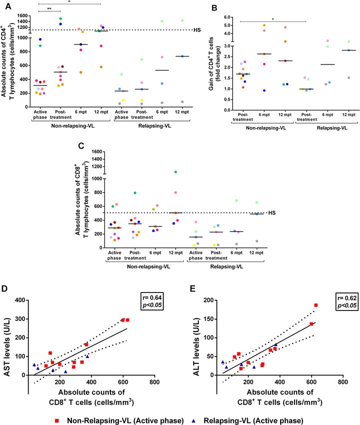
still maintained higher CD4+ T lymphocyte counts than IL-6 levels correlated with laboratorial parameters of
R patients (NR-VL - 906 cells/mm3 [664–1097 cells/ severity in visceral leishmaniasis patients
mm3]; R-VL - 532 cells/mm3 [131–532 cells/mm3]) (Fig. In the active phase, IL-6 levels were above the minimum
2a and b). Some patients were followed up to 12mpt and detection limit in 8 out 10 NR-VL and in 3 out 5 R-VL
among NR-VL, four patients presented CD4+ T counts in relation to the HS, whose median was 0.1 pg/mL
above than 800 cells/mm3 (1074 cell/mm3 [731.5–1226 [0.1–165 pg/ml]. However, a reduction in these levels
cell/mm3] very similar to those found in HS (1115 cell/ were observed immediately after the treatment in most
mm3 [630.5–1258 cell/mm3]) (Fig. 2a). On the other patients of both groups (Fig. 4a). For both VL groups,
hand, even at a long term post-therapy, R-VL patients IL-6 levels were associated with several VL severity
still presented the lowest CD4+ T cell counts (734 cell/ markers, during the active and post-treatment phases. A
mm3 [76–1438 cell/mm3]) (Fig. 2a and b). negative correlation was verified between IL-6 levels and
Regarding CD8+ T lymphocytes, both NR-VL and R- neutrophils (r = − 0.53, p < 0.05, Fig. 4b), lymphocytes
VL patients showed lower numbers of this subpopula- (r = − 0.52; p < 0.05, Fig. 4c), monocytes (r = − 0.48, p <
tion than those found in HS (Fig. 2c) up to 6mpt. At 0.05, Fig. 4d) and albumin (r = − 0.49; p < 0.05, Fig. 4e).Kuschnir et al. BMC Infectious Diseases (2021) 21:369 Page 8 of 14 Fig. 2 Evaluation of immune impairment in visceral leishmaniasis (VL) patients throughout clinical follow-up. Absolute counts of CD4+ (a) and CD8+ (c) T lymphocytes of non-relapsing (NR) and relapsing (R) VL patients. Gain of CD4+ T lymphocytes during clinical follow-up in relation to the active phase of VL (b). Positive correlation between the absolute counts of CD8+ T lymphocytes and the aspartate aminotransferase - AST (d) and alanine aminotransferase - ALT (e) levels during the active phase of VL (Spearman correlation, r = 0.64 and r = 0.62, respectively, p < 0.05). Each point represents a VL patient and each color represents the same patient in the different stages of clinical follow-up. The black dashed line represents the median value of healthy subjects (HS). The horizontal bars represent the median values of each group. Post-treatment: Early post-treatment. Mpt: months post-treatment. Asterisks denote significant differences between the phases of clinical follow-up within the R or NR group itself or even between the R and NR group: *p < 0.05. ** p < 0.01
Kuschnir et al. BMC Infectious Diseases     (2021) 21:369                                                                                 Page 9 of 14

 Fig. 3 Anti-Leishmania infantum Igs levels in non-relapsing (NR) and relapsing (R) VL patients. IgG1 (a) and IgG3 (b) levels in NR-VL and R-VL
 patients throughout clinical follow up. Negative correlation between the absolute counts of CD4+ T lymphocytes and levels of anti-L. infantum
 IgG3 during all the follow-up (c) (Spearman correlation, p < 0.05, r = − 0.52). Post-treatment: Early post-treatment. Mpt: months post-treatment.
 Each point represents a VL patient and each color represents the same patient in the different stages of clinical follow-up. The horizontal bars
 represent the median values of each group. Asterisks denote statistically significant differences between the phases of clinical follow-up within
 the R or NR group itself or even between the R and NR group: *p < 0.05

On the other hand, IL-6 levels correlated positively with                    Discussion
C-reactive protein levels (r = 0.53; p < 0.05) (Fig. 4f).                    There are many studies carried out in India [33], Sudan
                                                                             [32], Georgia [34] and Brazil [28] addressing
Kuschnir et al. BMC Infectious Diseases (2021) 21:369 Page 10 of 14 Fig. 4 Assessment of IL-6 levels and correlation with laboratory parameters in visceral leishmaniasis (VL) patients. Plasma IL-6 levels in non- relapsing (NR) and relapsing (R) VL patients throughout clinical follow-up (a). Negative correlation between the IL-6 levels during VL active phase and early post-treatment and the neutrophil counts (b, Spearman correlation, r = − 0.53; p < 0.005), lymphocyte counts (c, r = − 0.52, p < 0.05), monocyte counts (d, r = − 0.48, p < 0.05 ) and albumin levels (e, r = − 0.49, p < 0.05). Positive correlation between the IL-6 levels at active phase and early post-treatment and C-reactive protein (CRP) levels (f, r = 0.53, p < 0.05). Post-treatment: Early post-treatment. Each point represents a VL patient and each color represents the same patient in the different stages of clinical follow-up. The horizontal bars represent the median values of each group retrospectively non-HIV infected patients presenting a VL group. As R-VL patients have previously undergone VL relapsing course, but divergent findings in terms of anti-Leishmania treatment, we believe that it may have clinical and laboratorial aspects associated with relapses contributed to a partial clinical recovery, with a reduc- were found. In the present study, the patients’ symptoms tion in the damage caused by the infection. However, were quite similar in the active phase, and it was not this was not enough to restore the R-VL organic possible to identify significant differences between R- functions. and NR-VL groups. However, more intense laboratorial Splenomegaly at study enrollment was observed in abnormalities were observed in NR-VL compared to R- both groups, being a usual manifestation of VL [9].
Kuschnir et al. BMC Infectious Diseases   (2021) 21:369                                                             Page 11 of 14

Splenomegaly and thrombocytopenia have already been                  Also, endogenous IL-10 secretion has already been as-
identified as markers of VL relapses [28, 32, 33]. NR-VL          cribed to the CD8+ T cells in VL which may contribute
patients showed significant platelet elevation after treat-       to the immunosuppression. Simultaneously, the chronic
ment, different from observed for R-VL group. It is               immune activation may lead CD8+ T lymphocytes to ex-
known that spleen histological disorganization affects its        press molecules with inhibitory function on their sur-
functionality [35] which implies in higher consumption            face, such as PD-1, CTLA-4 [54, 55], TIM-3 and LAG3
of platelets, as a confounding factor, resulting in elevated      [16]. In these situations of exhaustion of the cellular re-
parasitemia [9], and immunodepression related to its im-          sponse, CD8+ T lymphocytes function may be severely
munity function [32].                                             impaired in active phase of VL [56].
  The responsible medical team decided each patient                  In parallel, the levels of specific anti-Leishmania IgG
treatment in an individualized way, based on clinic charac-       and their subclasses after active VL may have an import-
teristics and side effects of the avaiable drugs. It is import-   ant role as marker of cure and/or predisposition to re-
ant to highlight that although Amph. B Lipid Complex is           lapse [17, 57]. Here, IgG3 levels were persistently
not considered a drug of first choice in VL treatment in          increased in R-VL group in relation to NR-VL, which
international and Brazilian official guidelines, there are        may indicate the presence of continuous parasite stimu-
studies that were able to show the effectiveness of this          lation, without a complete control by the immune sys-
drug in VL cases, including in HIV coinfected patients            tem. L. donovani Indian relapsing patients [58, 59] have
[36–39]. Therefore, we do not believe that the treatment          shown similar behavior for IgG1, whose levels remained
with this specific Amph. B formulation is related with the        high after the active episode of VL. In other studies [60–
occurrence of new VL episodes in these patients.                  62], a reduction in IgG1 and IgG3 levels was found in
  After VL treatment, a recovery of bone marrow cellu-            patients infected either by L. infantum or by L. donovani,
larity was observed in NR-VL patients, especially with an         who were considered clinically cured few months after
increase in leukocytes levels compared to the active              the treatment. In addition, we have found that a negative
phase of VL. A significant and faster gain of CD4+ T              association between IgG3 and CD4+ T-cell count in R-
cells was observed in NR-VL patients immediately after            VL patients, corroborating the link between persistence
anti-Leishmania treatment, but not in R-VL patients.              of high levels of IgG3 and relapse.
Similar results were observed in VL/HIV coinfection                  Finally, we observed that IL-6, a cytokine known to be in-
[23]. This deficit in the recovery of CD4+ T lymphocytes          flammatory and already associated with a more severe
can be related to an impaired input of cells originated           prognosis in the evolution of VL [12, 17], was correlated to
from bone marrow, deficient replication rate or thymic            laboratory severity markers, such as hypoalbuminemia,
dysfunction [24, 40, 41]. Low CD4+ T cell counts in VL/           leukopenia and thrombocytopenia. As expected, CRP levels
HIV coinfected patients during the active phase of VL             also correlated positively with IL-6 since its binding with its
was a predictor of a poor prognosis: death or recurrence          receptor activates immunocompetent and hematological
[42, 43]. Thus, patients who are unable to restore the            cells leading to the production of acute phase responses, as
CD4+ T counts may be prone to relapse. Interestingly, in          CRP [63]. However, there were patients that despite pre-
the current series, all VL patients maintained low CD8+           senting clinical signs and laboratorial data associated with
T lymphocyte counts up to 6 months of follow-up, in re-           severity did not present elevated IL-6 levels, showing that
lapsing and not relapsing groups. Whether CD8+ T cells            other mechanisms in VL can trigger immunopathological
contribute to protection, immunopathogenesis or even              mechanisms related to severity. This study design and the
to the immunosuppression in VL is still unclear. Like-            analysis performed until now are not sufficient to state the
wise, the function of these cells has already been de-            role of IL-6 may in the VL relapsing course. This study has
scribed for human cutaneous leishmaniasis during the              limitations, including the small sample, the short follow-up
active and healing phases [44–47]. Some studies have              time and mainly the comparison between patients enrolled
showed that CD8+ T lymphocyte cytotoxic activity, se-             at the primary but also at a relapsing VL episode, which
cretion of cytokines and chemokines, granzyme B levels            can add differences in the total time of infection and in pre-
and lymphoproliferation may contribute to the parasite            vious exposure to anti-Leishmania treatments. However,
control in experimental and human VL [48–51]. At the              our results are hypothesis generator and rise important
same time, there was a positive correlation between the           questions to be evaluated in a prospective study, as the po-
CD8+ T lymphocytes and transaminases levels, which                tential of CD4+ T-cell count or IgG3 as biomarkers for VL
raises the hypothesis that liver damage could be associ-          relapses.
ated with cytotoxic activity. Herein, this occurred re-
gardless of the VL clinical outcome. Such an association          Conclusion
has already been demonstrated in hepatitis B [52] and in          Therefore, the VL relapsing course among patients not
infectious mononucleosis by Epstein-Barr virus [53].              infected    with    HIV      and     without    other
Kuschnir et al. BMC Infectious Diseases         (2021) 21:369                                                                                       Page 12 of 14

recognized associated immunosuppression is a challen-                            Rachou (number 68118117.3.3001.5091) and Instituto Oswaldo Cruz -
ging reality considering the lack of understanding of the                        Fundação Oswaldo Cruz (number 68118117.3.1001.5248). The patients/
                                                                                 participants provided their written informed consent to participate in this
pathophysiological mechanisms involved, of reliable                              study. We also confirm that all methods were performed in accordance with
prognostic markers and clinical protocols for addressing                         the relevant guidelines and regulations.
the condition. Our findings suggest a deficit in T-cell re-
                                                                                 Consent for publication
constitution and maintenance of B-cell compartment ac-
                                                                                 Not applicable.
tivation as possible immunomechanisms underlying the
VL relapse.                                                                      Competing interests
                                                                                 The authors declare that they have no competing interests.
Abbreviations
6mpt: Six months post-treatment; 12mpt: Twelve months after treatment;           Author details
                                                                                 1
ADHD: Attention deficit hyperactivity disorder; ALT: Alanine aminotransferase;    Laboratório Interdisciplinar de Pesquisas Médicas, Instituto Oswaldo Cruz,
Amph. B deoxychol: Amphotericin B deoxycholate; Amph. B Lipid                    FIOCRUZ, Rio de Janeiro, Rio de Janeiro, Brazil. 2Hospital Eduardo de
Complex: Amphotericin B lipid complex; AST: Aspartate aminotransferase;          Menezes, Fundação Hospitalar do Estado de Minas Gerais, Belo Horizonte,
CRP: C-reactive protein; DB: Direct bilirubin; EI: Elisa Index; F: Female;       Minas Gerais, Brazil. 3Núcleo de Ciências Biomédicas Aplicadas, Instituto
HS: Healthy subjects; IQR: Interquartile range; Liposomal Amph. B: Liposomal     Federal de Educação, Ciência e Tecnologia – IFRJ, Rio de Janeiro, Rio de
amphotericin B; LPS: Lipopolysaccharide; M: Male; NA: Not applicable; NR-        Janeiro, Brazil. 4Instituto Nacional de Infectologia Evandro Chagas, FIOCRUZ,
VL: Non-relapsing patients; OD: Optical density; Post-treatment: Right after     Rio de Janeiro, Rio de Janeiro, Brazil. 5Instituto René Rachou, FIOCRUZ, Belo
treatment; R-VL: Relapsing patients; sCD14: Soluble CD14; sCD163: Soluble        Horizonte, Minas Gerais, Brazil. 6Disciplina de Parasitologia, DMIP, Faculdade
CD163; Scr: Serum creatinine; VL: Visceral leishmaniasis; yo: Years old          de Ciências Médicas, UERJ, Rio de Janeiro, Brazil. 7Rede de Pesquisas em
                                                                                 Saúde do Estado do Rio de Janeiro/ FAPERJ, Rio de Janeiro, Brazil.
Supplementary Information                                                        Received: 11 December 2020 Accepted: 8 March 2021
The online version contains supplementary material available at https://doi.
org/10.1186/s12879-021-06051-5.
                                                                                 References
 Additional file 1: Supplementary Table 1. Laboratorial characteristics          1. Ministério da Saúde (BR), Secretaria de Vigilância em Saúde. Guia de
 of non-relapsing and relapsing VL patients during the active phase.                 vigilância em saúde. 2nd. Brasil: Ministério da Saude; 2017. Available at:
 Additional file 2. Flow diagram of the study.                                       https://portalarquivos2.saude.gov.br/images/PDF/2017/outubro/16/Volume-
                                                                                     Unico-2017.pdf. Accessed Dec 2019.
                                                                                 2. World Health Organization. Leishmaniasis. 2018. Available at: https://www.
Acknowledgements                                                                     who.int/leishmaniasis. Accessed Dec 2019.
We would like to thank all the Eduardo de Menezes hospital staff who             3. Pan American Health Organization, World Health Organization.
helped with the logistics of collecting and sending of the samples. We also          Leishmaniasis. Epidemiological report of the Americas: 2019. Available at:
acknowledge the participation of all the patients and healthy individuals            www.paho.org/leishmaniasis. Accessed Dec 2019.
enrolled in this study. We would also like to thank Dr. Adriano Gomes-Silva      4. Brasil, Ministério da Saúde. Sistema de Informação de Agravos de
who helped with the standardization of anti-Leishmania immunoglobulin de-            Notificação - Banco de dados do Sistema Único de Saúde - DATASUS.
tection assays.                                                                      Available at: http://tabnet.datasus.gov.br/cgi/deftohtm.exe?sinannet/cnv/
                                                                                     leishvmg.def. Accessed Sept 2020.
Authors’ contributions                                                           5. Saporito L, Giammanco GM, De Grazia S, Colomba C. Visceral leishmaniasis:
RCK, MLS-F, GC-C, JRS-O and AMD-C: formal analysis, investigation, method-           host–parasite interactions and clinical presentation in the
ology, organized the database, and wrote the draft of the manuscript. JRS-O          immunocompetent and in the immunocompromised host. Int J Infect Dis.
and AMD-C: conceptualization, funding acquisition, project administration,           2013;17(8):e572–6. https://doi.org/10.1016/j.ijid.2012.12.024.
review and editing of the manuscript. GC: formal analysis and critically re-     6. Rodrigues da Silva J. Leishmaniose visceral (calazar). Rio de Janeiro: Tese
vised the manuscript for intellectual content. LP, LSP, MRTD: recruitment and        [Concurso para provimento efetivo do cargo de professor catedrático da
clinical follow-up of the patients. SCCS: methodology and critically revised         cadeira de Clinica das Doenças Tropicais e Infectuosas da Faculdade
the manuscript for intellectual content. All authors read and approved the           Nacional de Medicina da Universidade do Brasil]. Sociedade Editora e
final manuscript.                                                                    Gráfica Ltda; 1957.
                                                                                 7. Rossi M, Fasel N. How to master the host immune system? Leishmania
Funding                                                                              parasites have the solutions! Int Immunol. 2017. https://doi.org/10.1093/
This work was supported by the Instituto Oswaldo Cruz (internal funds: PAEF          intimm/dxx075.
II-IOC-23-FIO-18–2-53), CNPq (Universal - 433637/2018–8), FAPERJ (E-26/          8. Sampaio MJAQ, Cavalcanti NV, Alves JGB, Fernandes Filho MJC, Correia JB.
202.944/2016) and IFRJ (Pro-Ciência/2019). MLS-F received a fellowship from          Risk factors for death in children with visceral Leishmaniasis. PLoS Negl Trop
CAPES. GC-C received a fellowship from FAPERJ. AMD-C receive research fel-           Dis. 2010;4(11):e877. https://doi.org/10.1371/journal.pntd.0000877.
lowships from CNPq and FAPERJ. GC is currently receiving a grant from            9. Costa DL, Rocha RL, Chaves EBF, Batista VGV, Costa HL, Costa CHN.
CNPq [301384/2019].                                                                  Predicting death from kala-azar: construction, development, and validation
                                                                                     of a score set and accompanying software. Rev Soc Bras Med Trop. 2016;
Availability of data and materials                                                   49(6):728–40. https://doi.org/10.1590/0037-8682-0258-2016.
All data generated and/or analysed during this study are included in this        10. Barral-Netto M, Badaró R, Barral A, Almeida RP, Santos SB, Badaró F, et al.
published article [and its supplementary information files].                         Tumor necrosis factor (Cachectin) in human visceral leishmaniasis. J Infect
The datasets generated and/or analysed during the current study are not              Dis. 1991;163(4):853–7. https://doi.org/10.1093/infdis/163.4.853.
publicly available due individual privacy of patients could be compromised       11. Santos-Oliveira JR, Regis EG, Leal CRB, Cunha RV, Bozza PT, Da-Cruz AM.
but are available from the corresponding author on reasonable request.               Evidence that lipopolysaccharide may contribute to the cytokine storm and
                                                                                     cellular activation in patients with visceral leishmaniasis. PLoS Negl Trop Dis.
Declarations                                                                         2011;5(7):e1198. https://doi.org/10.1371/journal.pntd.0001198.
                                                                                 12. Costa DL, Rocha RL, Carvalho RMA, Lima-Neto AS, Harhay MO, Costa CHN,
Ethics approval and consent to participate                                           et al. Serum cytokines associated with severity and complications of kala-
This study has been reviewed and approved by the Ethics Committee of                 azar. Pathog Glob Health. 2013;107(2):78–87. https://doi.org/10.1179/204
Hospital Eduardo de Menezes (number 68118117.3.3002.5124), Instituto Rene            7773213Y.0000000078.
Kuschnir et al. BMC Infectious Diseases (2021) 21:369 Page 13 of 14
13. Santos PL, de Oliveira FA, Santos MLB, Cunha LCS, Lino MTB, de Oliveira Monooxygenase gene as a cause of relapses in visceral Leishmaniasis in
MFS, et al. The Severity of Visceral Leishmaniasis Correlates with Elevated children, in Sudan. J Infect Dis. 2017;216(1):22–8. https://doi.org/10.1093/
Levels of Serum IL-6, IL-27 and sCD14. PLoS Negl Trop Dis. 2016;10(1): infdis/jix277.
e0004375. https://doi.org/10.1371/journal.pntd.0004375. 32. Gorski S, Collin SM, Ritmeijer K, Keus K, Gatluak F, Mueller M, et al. Visceral
14. Silva RLL, Santos MB, Almeida PLS, Barros TS, Magalhães L, Cazzaniga RA, leishmaniasis relapse in southern Sudan (1999-2007): a retrospective study
et al. sCD163 levels as a biomarker of disease severity in leprosy and visceral of risk factors and trends. PLoS Negl Trop Dis. 2010;4(6):e705. https://doi.
leishmaniasis. PLoS Negl Trop Dis. 2017;11(3):e0005486. https://doi.org/10.13 org/10.1371/journal.pntd.0000705.
71/journal.pntd.0005486. 33. Burza S, Sinha PK, Mahajan R, Lima MA, Mitra G, Verma N, et al. Risk factors
15. Ansari NA, Kumar R, Raj A, Salotra P. Elevated levels of IgG3 and IgG4 for visceral Leishmaniasis relapse in Immunocompetent patients following
subclass in paediatric cases of kala azar. Parasite Immunol. 2008;30(8):403–9. treatment with 20 mg/kg liposomal amphotericin B (Ambisome) in Bihar,
https://doi.org/10.1111/j.1365-3024.2008.01036.x. India. PLoS Negl Trop Dis. 2014;8(1):e2536. https://doi.org/10.1371/journal.
16. Singh B, Bhushan Chauhan S, Kumar R, Singh SS, Ng S, Amante F, et al. A pntd.0002536.
molecular signature for CD8+ T cells from visceral leishmaniasis patients. 34. Kajaia M, Morse DL, Kamkamidze G, Butsashvili M, Chubabria G, Zenaishvili O,
Parasite Immunol. 2019;41(11):e12669. https://doi.org/10.1111/pim.12669. et al. Risk factors for relapse of visceral leishmaniasis in Georgia. Tropical Med
17. Fievez AMdC, Silva-Freitas ML, Sousa AdQ, Santos-Oliveira JR, Da-Cruz AM. Int Health. 2011;16(2):186–92. https://doi.org/10.1111/j.1365-3156.2010.02694.x.
Lower levels of leptin are associated with severity parameters in visceral 35. Sundar S, Mehta H, Suresh AV, Singh SP, Rai M, Murray HW. Amphotericin B
leishmaniasis patients. PLoS One. 2019;14(3):e0214413. https://doi.org/10.13 treatment for Indian visceral leishmaniasis: conventional versus lipid
71/journal.pone.0214413. formulations. Clin Infect Dis. 2004;3(3):377–83. https://doi.org/10.1086/3
18. Bozza FA, Cruz OG, Zagne SMO, Azeredo EL, Nogueira RMR, Assis EF, et al. 80971.
Multiplex cytokine profile from dengue patients: MIP-1beta and IFN-gamma 36. Hermida MD, de Melo CVB, Lima IDS, Oliveira GGS, dos Santos WLC.
as predictive factors for severity. BMC Infect Dis. 2008;8(1):86. https://doi. Histological disorganization of spleen compartments and severe visceral
org/10.1186/1471-2334-8-86. Leishmaniasis. Front Cell Infect Microbiol. 2018;8:394. https://doi.org/10.33
19. Andrade BB, Reis-Filho A, Souza-Neto SM, Clarêncio J, Camargo LMA, Barral 89/fcimb.2018.00394.
A, et al. Severe Plasmodium vivax malaria exhibits marked inflammatory 37. Sundar S, Agrawal NK, Sinha PR, Horwith GS, Murray HW. Short-course, low-
imbalance. Malar J. 2010;9(1):13. https://doi.org/10.1186/1475-2875-9-13. dose amphotericin B lipid complex therapy for visceral leishmaniasis
20. Costa CHN, Werneck GL, Costa DL, Holanda TA, Aguiar GB, Carvalho AS, unresponsive to antimony. Ann Intern Med. 1997;127(2):133–7. https://doi.
et al. Is severe visceral leishmaniasis a systemic inflammatory response org/10.7326/0003-4819-127-2-199707150-00007.
syndrome? A case control study. Rev Soc Bras Med Trop. 2010;43(4):386–92. 38. Laguna F, Vilela S, Jiménez-Mejías ME, Sirera G, Torre-Cisneros J, Ribera E,
https://doi.org/10.1590/s0037-86822010000400010. et al. Amphotericin B lipid complex versus meglumine antimoniate in the
21. Cota GF, Sousa MR, Assis TSM, Pinto BF, Rabello A. Exploring prognosis in treatment of visceral leishmaniasis in patients infected with HIV: a
chronic relapsing visceral leishmaniasis among HIV infected patients: randomized pilot study. J Antimicrob Chemoter. 2003;52(3):464–8. https://
circulating Leishmania DNA. Acta Trop. 2017;172:186–91. https://doi.org/10.1 doi.org/10.1093/jac/dkg356.
016/j.actatropica.2017.05.011. 39. Goldsmith DR, Perry CM. Amphotericin B lipid complex in visceral
22. Cota GF, Sousa MR, Mendonça ALP, Patrocinio A, Assuncao LS, de Faria SR, Leishmaniasis. Drugs. 2004;64(17):1905–11. https://doi.org/10.2165/000034
et al. Leishmania-HIV co-infection: clinical presentation and outcomes in an 95-200464170-00004.
urban area in Brazil. PLoS Negl Trop Dis. 2014;8(4):e2816. https://doi.org/1 40. Carvalho EM, Bacellar O, Brownell C, Regis T, Coffman RL, Reed SG.
0.1371/journal.pntd.0002816. Restoration of IFN-gamma production and lymphocyte proliferation in
23. Silva-Freitas ML, Cota GF, Machado-de-Assis TS, Giacoia-Gripp C, Rabello A, visceral leishmaniasis. J Immunol. 1994;152(12):5949–56.
Da-Cruz AM, et al. Immune activation and bacterial translocation: a link 41. da Silva AVA, Souza TL, Figueiredo FB, Mendes-Jr AAV, Ferreira LC, Filgueira
between impaired immune recovery and frequent visceral Leishmaniasis CPB, et al. Detection of amastigotes and histopathological alterations in the
relapses in HIV-infected patients. PLoS One. 2016;11(12):e0167512. https:// thymus of Leishmania infantum-infected dogs. Immun Inflamm Dis. 2020;
doi.org/10.1371/journal.pone.0167512. 8(2):127–39. https://doi.org/10.1002/iid3.285.
24. Silva-Freitas ML, Corrêa-Castro G, Cota GF, Giacoia-Gripp C, Rabello A, Dutra 42. Cota GF, Sousa MR, Rabello A. Predictors of visceral Leishmaniasis relapse in
JT, et al. Impaired Thymic output can be related to the low immune HIV-infected patients: a systematic review. PLoS Negl Trop Dis. 2011;5(6):
reconstitution and T cell repertoire disturbances in relapsing visceral e1153. https://doi.org/10.1371/journal.pntd.0001153.
Leishmaniasis associated HIV/AIDS patients. Front Immunol. 2020;11:953. 43. Távora LGF, Nogueira MB, Gomes ST. Visceral Leishmaniasis/HIV co-infection
https://doi.org/10.3389/fimmu.2020.00953. in Northeast Brazil: evaluation of outcome. Braz J Infect Dis. 2015;19(6):651–
25. Brasil, Ministério da Saúde. Sistema de Informação de Agravos de 6. https://doi.org/10.1016/j.bjid.2015.07.004.
Notificação. Available at: http://portalsinan.saude.gov.br/images/ 44. Da-Cruz AM, Conceição-Silva F, Bertho AL, Coutinho SG. Leishmania-reactive
documentos/Agravos/Leishmaniose%20Visceral/LV_v5.pdf. Accessed Sept CD4+ and CD8+ T cells associated with cure of human cutaneous
2019. leishmaniasis. Infect Immun. 1994;62(6):2614–8. https://doi.org/10.1128/IAI.
26. Gebreyohannes EA, Bhagvathula AS, Abegaz TM, Seid MA. Treatment 62.6.2614-2618.1994.
outcomes of visceral leishmaniasis in Ethiopia from 2001 to 2017: a 45. Brodskyn CI, Barral A, Boaventura V, Carvalho E, Barral-Netto M. Parasite-
systematic review and meta-analysis. Infect Dis Poverty. 2018;7(1):108. driven in vitro human lymphocyte cytotoxicity against autologous infected
https://doi.org/10.1186/s40249-018-0491-7. macrophages from mucosal leishmaniasis. J Immunol. 1997;159(9):4467–73.
27. Horrillo L, Castro A, Matía B, Molina L, García-Martínez J, Jaqueti J, et al. 46. Novais FO, Carvalho LP, Graff JW, Beiting DP, Ruthel G, Roos DS, et al.
Clinical aspects of visceral leishmaniasis caused by L. infantum in adults. Ten Cytotoxic T cells mediate pathology and metastasis in cutaneous
years of experience of the largest outbreak in Europe: what have we leishmaniasis. PLoS Pathog. 2013;9(7):e1003504. https://doi.org/10.1371/
learned? Parasit Vectors. 2019;12(1):359. https://doi.org/10.1186/s13071-019-3 journal.ppat.1003504.
628-z. 47. Novais FO, Carvalho AM, Clark ML, Carvalho LP, Beiting DP, Brodsky IE, et al.
28. Simão JC, Victória C, Fortaleza CMCB. Predictors of relapse of visceral CD8+ T cell cytotoxicity mediates pathology in the skin by inflammasome
leishmaniasis in inner São Paulo state, Brazil. Int J Infect Dis. 2020;95:44–9. activation and IL-1β production. PLoS Pathog. 2017;13(2):e1006196. https://
https://doi.org/10.1016/j.ijid.2020.02.028. doi.org/10.1371/journal.ppat.1006196.
29. Gomes MLS. Coinfecção leishmaniose visceral e Aids no Brasil, 2001 a 2010. 48. Mary C, Auriault V, Faugére B, Dessein AJ. Control of Leishmania infantum
Rio de Janeiro: Dissertação [Mestrado em Epidemiologia e Saúde Pública] - infection is associated with CD81 and gamma interferon- and Interleukin-5-
Escola Nacional de Saúde Pública Sergio Arouca; 2012. producing CD41 antigen-specific T cells. Infect Immun. 1999;67(11):5559–66.
30. Caldas AJM, Lisbôa LLC, Silva PF, Coutinho NPS. SilvaTC. Perfil das crianças com https://doi.org/10.1128/IAI.67.11.5559-5566.1999.
leishmaniose visceral que evoluíram para óbito, falha terapêutica e recidiva em 49. Tsagozis P, Karagouni E, Dotsika E. CD8(+) T cells with parasite-specific
hospital de São Luís, Maranhão. Rev Pesq Saúde. 2013;14(2):91–5. cytotoxic activity and a Tc1 profile of cytokine and chemokine secretion
31. Marquet S, Bucheton B, Reymond C, Argiro L, EL-Safi SH, Kheir MM, et al. develop in experimental visceral leishmaniasis. Parasite Immunol. 2003;
Exome sequencing identifies two variants of the Alkylglycerol 25(11–12):569–79. https://doi.org/10.1111/j.0141-9838.2004.00672.x.Kuschnir et al. BMC Infectious Diseases (2021) 21:369 Page 14 of 14
50. Tsagozis P, Karagouni E, Dotsika E. Function of CD8+ T lymphocytes in a
selfcuring mouse model of visceral leishmaniasis. Parasitol Int. 2005;54(2):
139–46. https://doi.org/10.1016/j.parint.2005.02.005.
51. Kaushal H, Bras-Gonçalves R, Negi NS, Lemesre JL, Papierok G, Salotra P.
Role of CD8+ T cells in protection against Leishmania donovani infection in
healed visceral Leishmaniasis individuals. BMC Infect Dis. 2014;14(1):653.
https://doi.org/10.1186/s12879-014-0653-6.
52. Lopes TGSL, Schinoni MI. Aspectos gerais da hepatite B. R Ci Med Biol. 2011;
10(3):337–44.
53. Carvalho LH. Mononucleose infecciosa [infectious mononucleosis]. J Pediatr.
1999;75(Suppl 1):S115–25.
54. Day CL, Kaufmann DE, Kiepiela P, Brown JA, Moodley ES, Reddy S, et al. PD-
1 expression on HIV-specific T cells is associated with T-cell exhaustion and
disease progression. Nature. 2006;443(7109):350–4. https://doi.org/10.1038/
nature05115.
55. Kaufmann DE, Kavanagh DG, Pereyra F, Zaunders JJ, Mackey EW, Miura T,
et al. Upregulation of CTLA-4 by HIV-specific CD4+ T cells correlates with
disease progression and defines a reversible immune dysfunction. Nat
Immunol. 2007;8(11):1246–54. https://doi.org/10.1038/ni1515.
56. Gautam S, Kumar R, Singh N, Singh AK, Rai M, Sacks D, et al. CD8 T cell
exhaustion in human visceral leishmaniasis. J Infect Dis. 2014;209(2):290–9.
https://doi.org/10.1093/infdis/jit401.
57. Marlais T, Bhattacharyya T, Singh OP, Mertens P, Gilleman Q, Thunissen C,
et al. Visceral Leishmaniasis IgG1 Rapid Monitoring of Cure vs. Relapse, and
Potential for Diagnosis of Post Kala-Azar Dermal Leishmaniasis. Front Cell
Infect Microbiol. 2018;8:427. https://doi.org/10.3389/fcimb.2018.00427.
58. Bhattacharyya T, Ayandeh A, Falconar AK, Sundar S, El-Safi S, Gripenberg
MA, et al. IgG1 as a potential biomarker of post-chemotherapeutic relapse
in visceral Leishmaniasis and adaptation to a rapid diagnostic test. PLoS
Negl Trop Dis. 2014;8(10):e3273. https://doi.org/10.1371/journal.pntd.00032
73.
59. Mollett G, Hinckel BCB, Bhattacharyya T, Marlais T, Singh OP, Mertens P,
et al. Detection of immunoglobulin G1 against rK39 improves monitoring of
treatment outcomes in visceral Leishmaniasis. Clin Infect Dis. 2019;69(7):
1130–5. https://doi.org/10.1093/cid/ciy1062.
60. Ravindran R, Anam K, Bairagi BC, Saha B, Pramanik N, Guha SK, et al.
Characterization of immunoglobulin G and its subclass response to Indian
Kala-Azar infection before and after chemotherapy. Infect Immun. 2004;
72(2):863–70. https://doi.org/10.1128/iai.72.2.863-870.2004.
61. Ganguly S, Das NK, Panja M, Pal S, Modak D, Rahaman M, et al. Increased
levels of interleukin-10 and IgG3 are hallmarks of Indian post-kala-azar
dermal leishmaniasis. J Infect Dis. 2008;197(12):1762–71. https://doi.org/10.1
086/588387.
62. Gomes IT, Carvalho SFG, Rocha RDR, Peruhype-Magalhaes V, Dietze R,
Martins-Filho OA, et al. Anti-Leishmania chagasi immunoglobulin G3
detected by flow cytometry for early cure assessment in American visceral
leishmaniasis. J Immunol Methods. 2010;360(1–2):76–83. https://doi.org/10.1
016/j.jim.2010.06.011.
63. Tanaka T, Narazaki M, Masuda K, Kishimoto T. Regulation of IL-6 in immunity
and diseases. Adv Exp Med Biol. 2016;941:79–88. https://doi.org/10.1007/
978-94-024-0921-5_4.
Publisher’s Note
Springer Nature remains neutral with regard to jurisdictional claims in
published maps and institutional affiliations.You can also read