Hyperphosphatemic Tumoral Calcinosis: Case presentation and Clinical-radio-pathological Correlation
←
→
Page content transcription
If your browser does not render page correctly, please read the page content below
case report

                                 Hyperphosphatemic Tumoral
                                 Calcinosis: Case presentation
                                 and Clinical-radio-pathological
                                 Correlation
                                 Calcinosis tumoral hiperfosfatémica: Presentación de un
                                 caso y correlación clínico-radiopatológica

                                                                                                      Ana María Henao González1
                                                                                                                  Julián Garzón2
                                                                                                                   Oscar Messa3
                                                                                                                    Karina Ruiz4
    Key words (MeSH)
                                 		                                                                      Ennya L. Leonel Triana5
    Calcinosis
    Soft tissue neoplasms
    Hyperphosphatemia
                                 Summary
                                 Introduction: Hyperphosphatemic tumoral calcinosis is a rare, benign condition characterized
                                 by calcified masses in the periarticular tissues which is associated with high blood phosphate
                                 levels as a result of increase in renal tubular reabsorption. Case presentation: In this report
                                 we describe an unusual case of hyperphosphatemic tumoral calcinosis in an adolescent who
                                 had history of trauma in the gluteal region and was surgically treated. Discussion: Tumoral
    Palabras clave (DeCS)        calcinosis, also known as Teutschlaender disease, belongs to a broad spectrum of entities
    Calcinosis                   characterized by calcified soft tissue masses, which according to their location, morphology
    Neoplasias de los tejidos    of the calcifications, signal intensity characteristics and sparing of the bony structures,
      blandos                    should be considered as a diagnostic possibility. Conclusion: Due to the large number of
    Hiperfosfatemia              differential diagnoses, such as surface osteosarcoma, chondrosarcoma, myositis ossificans,
                                 among others, radiological and histological findings are crucial in the diagnosis and proper
                                 management of this entity. This has to go along with the proper classification of the patient
                                 as hyperphosphatemic, normophosphatemic or with secondary hyperphosphatemia in order
1
 Doctor. Third year resident
                                 to guarantee an optimal treatment.
of the postgraduate course
in radiology, Pontificia Uni-
versidad Javeriana. Bogotá,      Resumen
Colombia.
                                 Introducción: La calcinosis tumoral hiperfosfatémica es una condición rara, benigna,
2
 Doctor. Radiologist, De-
partment of Radiology and
                                 caracterizada por masas calcificadas en los tejidos periarticulares asociada a niveles altos
Diagnostic Imaging. National     de fosfato en sangre por aumento en la reabsorción tubular renal. Presentación de caso:
Cancer Institute. Bogotá,        Se presenta un caso inusual de calcinosis tumoral hiperfosfatémica en un adolescente con
Colombia
                                 antecedente de trauma en la región glútea quien fue manejado quirúrgicamente. Discusión:
3
 Doctor. Pathologist, National
Cancer Institute. Bogotá,
                                 La calcinosis tumoral, también conocida como enfermedad de Teutschlaender, hace parte
Colombia                         del amplio espectro de entidades que cursan con masas calcificadas de tejidos blandos y
4
 Doctor. Fourth year resident    que por sus características de localización, morfología de las calcificaciones, intensidad
of the postgraduate course       de señal y respeto óseo debe ser considerada como posibilidad diagnóstica. Conclusión:
in pathology, Universidad de
Cartagena. Bogotá, Colombia      Debido a la gran cantidad de diagnósticos diferenciales, como el osteosarcoma de
                                 superficie, condrosarcoma y miositis osificante, entre otras, los hallazgos radiológicos e
5
  Odontologist. Second year
resident of the postgraduate     histológicos son determinantes en el diagnóstico y manejo apropiado de dicha entidad.
course in pathology and          Esto debe ir de la mano con la clasificación adecuada del paciente como hiperfosfatémico,
oral surgery, Pontificia Uni-
versidad Javeriana. Bogotá,      normofosfatémico o con hiperfosfatemia secundaria, con el fin de garantizar un tratamiento
Colombia                         óptimo.

4692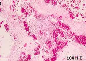
case report
Introduction
Hyperphosphatemic tumor calcinosis is a rare condition, generated
by the deposition of calcium phosphate crystals, of which there is little a
b
published information. This entity appears as a benign calcified tumor
mass, located in the soft tissues of the periarticular regions, associated
with high blood phosphate levels, secondary to a mutation in the genes Figure 1. (a) Hypominera-
coding for proteins that perform tubular reabsorption. Multiple secondary lization of the enamel in
the cervical third of the
etiologies have also been described, including chronic renal failure and upper anterior teeth. b)
hyperparathyroidism. The most frequent locations are shoulders, hip and Areas of gingivitis.
elbows (1-5).
This paper describes the case of a young patient with this condition
and discusses the importance of radiological and histological findings to
be considered within the spectrum of differential diagnoses of calcified
soft tissue masses.
Case presentation
A 14-year-old male patient, who consults for the appearance of a
non-painful, right thigh mass that relates to mild trauma seven months
ago. There are no other antecedents or symptoms of importance.
The physical examination identifies a large mass in the lateral
region of the proximal third of the right thigh, slightly painful and
Figure 2. Radiograph: Lar-
without inflammatory signs. In addition, the oral cavity showed signs ge mass of soft tissues on
of gingivitis and changes in the enamel structure, such as areas of the lateral aspect of the
hypoplasia and hypomineralization (figure 1). proximal third of the femur,
with lobulated, confluent
An x-ray is performed in which calcifications are observed, well calcifications that resemble
defined, located in the right periarticular coxofemoral region, without “clouds”.
erosion of the cortical bone or signs of fracture (figure 2). For the
adequate characterization of this lesion, a magnetic resonance (MR)
with contrast medium, gadolinium, is used, in which a mass with very
heterogeneous signal intensity is observed in T1 and T2-weighted se-
quences, areas with marked low signal due to the presence of calcium,
Figure 3. Coronal plane
as well as cystic component, liquid-calcium levels and, as in the radio- MRI enhanced in T1: Mass
graph, without compromise of the cortex of the adjacent bone (figures located in the gluteal
3 and 4). This lesion enhances the septa following the administration muscular plane, with
heterogeneous signal
of contrast medium (figure 5). There is no evidence of restriction in intensity and some low
the diffusion sequence (figure 6). signal foci.
In the blood tests of this patient, high phosphorus was found in
7.5 mg / dl (normal range 2.5-4.5 mg / dl), calcium levels with normal
values (9.66 mg / dl) and without alterations of renal function.
The patient underwent orthopedic surgery, which consisted of
complete excision of a mass of approximately 24 × 14 × 6.5 cm, ap-
proximately 875 grs, light brown color, soft consistency and multino-
dular surface.
In the anatomopathological findings, a yellowish-white, lobed
mass with a milky material outlet was observed at the time of the cut
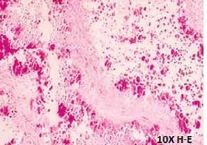
(figure 7). Microscopically, calcifications were found surrounded by
a chronic inflammatory reaction with multinuclear giant cells of the
‘foreign body’ type. No osteocartilaginous component was found, fu-
socellular proliferation with “zonal” change as in ossifying myositis
or areas of atypia, mitosis or necrosis to be considered as of malignant
origin (figure 8). With the radiological, clinical and histopathological Figure 4. Axial plane, T2-weighted sequence: High signal mass,
findings, the definitive diagnosis of hyperphosphatemic tumor calci- heterogeneous, with cystic component showing liquid-calcium
levels inside (arrow).
nosis is reached.
Rev. Colomb. Radiol. 2017; 28(2): 4692-6 4693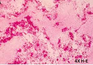
case report

                                                                                a

Figure 5. Heterogeneous enhancement after administration of
contrast medium; Preservation of cortical integrity is observed.                b

       a

       b                                                                        c

Figure 6. a) Diffusion sequence and b) ADC map: Absence of
mass restriction.

                                                                                d

                                       Figure 7. Macroscopic
                                       view of the tumor lesion.
                                       Surface is observed with
                                       areas of liquefaction and      Figure 8. a) Panoramic view in 4x. b and c) Increase in 10x: His-
                                       calcifications.                tiocytes and cystic cavities with calcifications. d) Increase of 40x.

4694                                          Hyperphosphatemic Tumoral Calcinosis: Case Presentation and Clinical-radio-pathological Correlation.
                                                                                                 Henao A., Garzón J., Messa O., Ruiz K., Leonel E.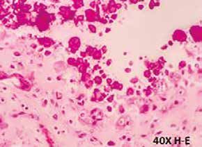
case report

Discussion                                                                 In the diffusion sequences these lesions do not restrict (figure 8)
     Tumor calcinosis is a rare, generalized and progressive di-           (1,10-13).
sease in which calcium salts are not deposited at the usual sites,              In macroscopic pathology, a mass of cystic composition is
which is why it is classified into heterotopic calcifications. It was      described, with yellow lump material corresponding to the crys-
first described by Duret in 1899 as a large calcified and multilo-         tals of calcium hydroxyapatite (1) and in histology is characteri-
bulated mass of soft periarticular tissues. It is commonly found in        zed by the aggregation of foamy histiocytes; These are transfor-
adolescents as a non-painful, firm and mobile mass characteristica-        med, with the participation of collagenolysis, into cystic cavities
lly located in the soft tissues of the periarticular regions of the hip,   coated by osteoclasts, such as giant cells and histiocytes. Mo-
shoulders and elbows, and foot involvement is uncommon (1-5).              vement and friction are key forces in the generation of periar-
     Under normal conditions, serum phosphate levels depend                ticular lesions. In this process there are two calcifying events,
on three factors: intestinal absorption, skeletal storage and re-          possibly driven by concurrent hyperphosphatemia or endogenous
nal resorption. Primary tumoral calcinosis may be of the hy-               hypervitaminosis D. The first occurs in membranous antiprotease
perphosphatemic or normo-phosphatemic variety. The primary                 fragments, which contain large cytoplasmic vesicles in osteoclast
hyperphosphatemic variety is an autosomal recessive disorder,              giant cells and mononuclear cells, which cover the cavities of
by mutation of the genes FGF23, GALNT3 and KL (1,4-6).                     tumor calcinosis; The second, in tumor calcinosis loci, membra-
Phosphate reabsorption in the kidneys is carried out in the pro-           nous debris and lining cells of the cavity and erythrocytes. Fina-
ximal tubules by means of sodium-phosphate type II and type                lly, the cavities are filled with calcified material, lose their syno-
III cotransporters. The sodium-phosphate cotransporter type II             vial lining, are encapsulated by fibrous tissue and ossify (1,14).
is responsible for 70-80% of phosphate reabsorption (7,8). This                 The difficulty in this patient is due to the possible differential
process is regulated by the fibroblast growth factor 23.0 encoded          diagnoses for his age and to the clinical manifestation as well as
by the FGF23 gene, which is mutated in this pathology (6-8).               the radiological findings (1,11,12,15). In the evaluation of this case
     Secondary tumoral calcinosis is associated with conditions            we consider in the differential diagnosis the following entities:
such as hyperparathyroidism, chronic kidney disease, vitamin D                  Surface osteosarcoma, or juxtacortical: one of the histological
toxicity, milk-alkali syndrome and osteolysis (1,6).                       variants of osteosarcoma, which in turn is divided into paraostal and
     It appears more frequently between the first and second de-           periostal, and may appear in patients of greater age range than classic
cade of life, in the afro-descendant community. Clinically it ma-          osteosarcoma. This tumor has a broad cortical base, cortical thicke-
nifests with a non-painful mass, which decreases the range of              ning and is composed of mature bone matrix that is characteristically
motion of the adjacent joint. The relationship between trauma              of central location. Although both are masses with calcified soft tissue
and hyperphosphatemic tumor calcinosis is rare, but may appear             component, in osteosarcoma there is contact and compromise of the
in the normophosphatemic variety (1).                                      cortical, without evidence of a radiolucent line separating them; in
     In the oral cavity it can manifest with root malformations that       addition, the morphology of “cloud” calcifications and their random
include tartar and obliteration of the pulp, with local thickening         location do not coincide with the dense central calcifications (osteoid
of the roots of the permanent teeth, specific findings for this di-        matrix) observed in the osteosarcoma. In MRI, although both are he-
sorder that can serve as a phenotypic marker; These malforma-              terogeneous lesions in T1 and T2 and enhance after administration
tions seem to fit somewhere between the classic descriptions of            of contrast medium, osteosarcoma presents diffusion restriction and
dental dysplasia type I and II (DD-I and DDII), a hallmark of              liquid-calcium levels are not characteristic.
hyperphosphatemic diseases, such as tumor calcinosis (9).                       Ossifying myositis corresponds to the formation of soft tissue
     Laboratory tests in these patients demonstrate hyperphospha-          bone due to different etiologies but, mainly, secondary to trauma; It
temia (less common normophosphatemia), normocalcemia, nor-                 is considered that there are progenitor cells producing osteoid in the
mal or elevated vitamin D levels, normal parathyroid hormone               affected soft tissues, which with a stimulus such as trauma become
and normal renal function (1).                                             osteoblasts and form osteoid. In pictures different patterns are found
     Conventional radiography is the first diagnostic study in             depending on the stages of evolution: initially, subcutaneous plane
which there are typically amorphous and multilobulated calci-              edema and distortion of the muscular plane. Subsequently, between
fications described in the literature to appear as “in clouds”, lo-        the third and fourth week, development of periosteal reaction; Six to
cated on the extensor surfaces of the periarticular regions. The radio-    eight weeks, peripheral bone formation and central immature bone;
lucent line interposed between the conglomerate of calcifications and      and from one and a half to six months, development of mature pe-
the cortical bone shows respect for the bone (figure 2). Computed          ripheral bone. In radiography the peripheral arrangement of calcifi-
tomography (CT) helps to characterize the cystic component with            cations and the evolution in time of the images do not coincide with
liquid-calcium levels, which some authors call “a sign of sedi-            the pattern described in tumor calcinosis. In MRI, myositis ossificans
mentation” (1). MRI shows a mass with a very heterogeneous                 may show liquid-liquid, but not liquid-calcium levels.
cystic component, both in T1 and T2, by the calcified zones that
reflect high intensity signals (figures 2 and 5), with characte-
ristic levels of liquid-calcium (the latter is very low signal in          Conclusions
all sequences) (figure 5), as well as heterogeneous and septa en-              Hyperphosphatemic tumor calcinosis is a rare entity that should
hancement after administration of contrast medium (figure 7).              be considered within the spectrum of differential diagnoses of patients

Rev. Colomb. Radiol. 2017; 28(2): 4692-6                                                                                                  4695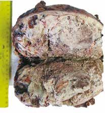
case report

with calcified soft tissue masses. Complete physical examination, in-
cluding the oral cavity, and characteristic imaging findings, such as
lobular calcifications that respect cortical and fluid-calcium levels, are
essential for a proper diagnostic approach and treatment.

References
1.    Olsen KM. Tumoral calcinosis: Pearls, polemics, and alternative possibilities.
      Radiographics. 2006;26:871-5.
2.    Bittmann S, Günther MW, Ulus H. Tumoral calcinosis of the gluteal region
      in a child: Case report with overview of different soft-tissue calcifications. J
      Pediatric Surg. 2003;38:E4-7.
3.    Mallick S, Ahmad Z, Gupta AK, Mathur SR. Hyperphosphatemic tumoral cal-
      cinosis. BMJ Case Reports. 2013;2013:bcr2013008728.
4.    Farrow EG, Imel EA, White KE. Hiperphosphatemic familial tumoral cal-
      cinosis (FGF23, GALNT3 and Klotho). Best Pract Res Clin Rheumatol.
      2011;25:735-47.
5.    Chefetz I, Sprecher E. Familial tumoral calcinosis and the role of O-glycos-
      ylation in the maintenance of phosphate homeostasis. Biochimica Biophysica.
      2009;1972:847-52.
6.    Bergwitz C, Jüppner H. Disorders of phosphate homeostasis and tissue minera-
      lization. Endocr Dev. 2009;16:133-56.
7.    DiMeglio LA, et al. Disorders of phosphate metabolism. Endocrinol Metab
      Clin N Am. 2000;29(3).
8.    Vieira AR, Moses L, Vairo F, et al. Root anomalies and dentin dysplasia in
      autosomal recessive hyperphosphatemic familial tumoral calcinosis (HFTC).
      Oral Surg Oral Med Oral Pathol Oral Radiol. 2015;120:e235-9.
9.    Espinoza A, et al. Calcinosis tumoral en pediatría: reporte de un caso clínico y
      revisión de la literatura. Rev Chil Radiol. 2008;14(2).
10.   Balach T, et al. The clinical evaluation of soft tissue tumors. Radiol Clin N AM.
      2011;49:1185-96.
11.   Stock H. Tumoral calcinosis and its mimics. Radiol Corner. 2014;78(8).
12.   Galindo Gómez A. Calcinosis tumoral hiperfosfatémica en pediatría: Reporte
      de un caso. Bol Med Hosp Infant Mex. 2014;71:167-73.
13.   Fathi I, Sakr M. Review of tumoral calcinosis: a rare clinic-pathological entity.
      World J Clin Cases. 2014;2:409-14.
14.   Leung YY, Lai R. Tumoral calcinosis: a case report. J Orthopaedic Surg.
      2011;19:108-12.
15.   McCarthy EF. Pseudotumors and reactive lesions. Surg Pathol. 2012;5:257-86.

Correspondence

      Ana María Henao González
      Calle 138 # 10A-10
      Bogotá, Colombia
      anitahego@hotmail.com

Received for evaluation: May 13, 2016
Accepted for publication: March 19, 2017

4696                                                            Hyperphosphatemic Tumoral Calcinosis: Case Presentation and Clinical-radio-pathological Correlation.
                                                                                                                   Henao A., Garzón J., Messa O., Ruiz K., Leonel E.
You can also read