It's About Time: Time-Dependent Tissue Damage in the Adult Porcine Retina After Enucleation

Page created by Bernard Mitchell
 
CONTINUE READING
It's About Time: Time-Dependent Tissue Damage in the Adult Porcine Retina After Enucleation
Tissue Engineering – Regenerative Medicine /
                                                             Research Article

                                                             Cells Tissues Organs 2021;210:58–65                                                  Received: November 13, 2020
                                                                                                                                                  Accepted: January 22, 2021
                                                             DOI: 10.1159/000514795                                                               Published online: May 26, 2021

It’s About Time: Time-Dependent Tissue
Damage in the Adult Porcine Retina After
Enucleation
Frida Svare a Bo Åkerström b Fredrik Ghosh a
aDepartment
              of Ophthalmology, Lund University, Lund, Sweden; bSection for Infection Medicine, Department of
Clinical Sciences, Lund University, Lund, Sweden

Keywords                                                                                         compared with 90-min counterparts. In the culture experi-
Porcine retina · Large animal model · Enucleation ·                                              ment, no significant difference in overall tissue damage was
Apoptosis · Culture                                                                              found between the 2 groups, however, apoptosis was sig-
                                                                                                 nificantly increased, and ganglion cell survival decreased in
                                                                                                 the cultured 240-min group. In addition, a significantly in-
Abstract                                                                                         creased LDH medium activity was found in the 240-min
The ex vivo large animal retina is extensively used in research                                  group compared with the 90-min counterpart at all time
ranging from discovery of disease mechanisms to future                                           points. The adult porcine retina is relatively resistant to tis-
treatment paradigms. Due to limited standardization when                                         sue damage 90 min after enucleation but displays distinct
harvesting the tissue, the time after enucleation is often ex-                                   signs of injury after 240 min. The importance of these time
tended for several hours, a factor that so far has not yet been                                  points is further highlighted when retinal explants are cul-
fully characterized. The purpose of this study was to investi-                                   tured. Our results strongly suggest that time after enucle-
gate the relationship between time after enucleation and                                         ation is a crucial factor that should be considered in experi-
retinal tissue damage. Adult, porcine retinal explants were                                      ments involving the ex vivo adult porcine retina.
dissected and fixed 90 or 240 min after enucleation. In a sep-                                                                                   © 2021 The Author(s)
arate experiment, explants were cultured for 48 h, following                                                                                     Published by S. Karger AG, Basel

dissection either 90 or 240 min after enucleation. Retinas
were analyzed morphologically using hematoxylin and eo-                                              Introduction
sin for overall tissue damage, TUNEL staining for detection
of apoptosis, and RBPMS immunohistochemistry for evalua-                                            The isolated adult retina is extensively used in contem-
tion of ganglion cell survival. In addition, medium from the                                     porary research for explorations of pathological disease
cultured explants was sampled after 2, 24, and 48 h of culture                                   mechanisms and tissue engineering, as well as in treat-
and assessed for the cell damage marker lactate dehydroge-                                       ment paradigms ranging from pharmacological interven-
nase (LDH). Retinas examined 240 min after enucleation dis-                                      tion to experimental retinal transplantation [for a review,
played a significant increase in overall tissue damage, in-                                      see Rettinger and Wang, 2018]. Most experiments have
creased apoptosis, and decreased ganglion cell survival                                          explored retinal tissue from small animals, but research

karger@karger.com      © 2021 The Author(s)                                                      Correspondence to:
www.karger.com/cto     Published by S. Karger AG, Basel                                          Frida Svare, frida.svare_johanson @ med.lu.se
                       This is an Open Access article licensed under the Creative Commons
                       Attribution-NonCommercial-4.0 International License (CC BY-NC)
                       (http://www.karger.com/Services/OpenAccessLicense), applicable to
                       the online version of the article only. Usage and distribution for com-
                       mercial purposes requires written permission.
It's About Time: Time-Dependent Tissue Damage in the Adult Porcine Retina After Enucleation
involving large animal models, including porcine and bo-             were dissected from the centrally located area on the temporal and
vine eyes, has gained interest in recent years due to an             nasal side of the optic nerve head between the 3 major vascular
                                                                     arcades as previously described [Taylor et al., 2013]. After dissec-
enhanced similarity to the human retina.                             tion, retinal pieces were either immediately fixed in 4% parafor-
    When working with large animal models, the tissue                maldehyde in 0.1 M phosphate buffer, pH 7.2 for 2 h at room tem-
used for retinal experiments is frequently harvested from            perature, or cultured as described below.
local abattoirs, which limits the possibility of a standard-              For exploration of time-dependent tissue damage after enucle-
ized handling process, and the time after enucleation is             ation, retinal pieces were immediately fixed in 4% paraformalde-
                                                                     hyde in 0.1 M phosphate buffer, 90 or 240 min (±10 min) after
often extended for several hours [Schnichels et al., 2019].          enucleation (n = 12 in each group).
The retina degenerates postmortem after enucleation due                   For the culture experiment, additional pieces from each time
to global ischemia as well as axotomy of ganglion cell ax-           point (n = 12 in each group) were explanted onto Millicell–PCF
ons. To limit tissue damage, enucleated eyes can be kept             0.4 mm culture plate inserts (Millipore, Billerica, MA, USA), with
under hypothermic conditions, which in a few studies has             the inner retina facing the membrane. This orientation of explants
                                                                     was chosen to optimize tissue survival in vitro [Taylor et al., 2014].
been shown to, at least partly, attenuate ganglion cell              The 24 retinal explants were cultured in Dulbecco’s modified Ea-
death [Reinhard et al., 2016; Schultheiss et al., 2016].             gle’s medium F12 (DMEM/F12; Invitrogen). The medium was
However, the influence of time from enucleation on over-             supplemented with 10% fetal calf serum (Sigma-Aldrich, St Louis,
all retinal tissue damage, as well as any impact on subse-           MA, USA) and 1% antibiotics (2 mML–glutamine, 100 U/mL pen-
quent investigations in the large animal model eye, has              icillin, and 100 ng/mL streptomycin; Sigma Aldrich). Explants
                                                                     were maintained at 37°C (95% humidity and 5% CO2 + 95% air)
not yet been thoroughly explored.                                    for 48 h after which they were fixed as described above. The culture
    For the present study, we wanted to expand the knowl-            time points were chosen based on previous experiments in which
edge on time after enucleation as a relevant factor for ex-          the tissue was relatively well preserved [Åkerström et al., 2017].
perimental research involving the adult retina in a large
animal model, with the aim of systematic characteriza-                   Histology
                                                                         After several rinses in phosphate buffered saline (PBS), the
tion of time-dependent tissue damage per se and explora-             fixed tissue explants were infiltrated with 0.1 M Sörensens solution
tion of any extended effects in vitro. Previous in vivo and          with increasing concentrations of sucrose up to 25%. They were
clinical investigations have indicated that the retinal tol-         then embedded in egg albumin/gelatine medium, cryosectioned at
erance time to ischemia is approximately 90 min and that             12 µm, and transferred to glass slides. For light microscopy, slide
extensive tissue destruction is evident after 240 min                1, 10, 20, and 25 were stained with hematoxylin and eosin (H&E).
                                                                     For immunohistochemical labeling of ganglion cells, slides were
[Hayreh et al., 2004]. These time points were therefore              rinsed 3 times with PBS and then incubated with PBS buffer con-
chosen for the present experiment in which we investi-               taining 0.25% Triton-X and 1% bovine serum albumin (BSA), for
gated retinal tissue damage in porcine eyes 90 or 240 min            20 min at room temperature. The specimens were then incubated
after enucleation, with the addition of experiments in               overnight at 4°C with an antibody raised against RNA-binding
which we cultured retinal explants after these time points           protein with multiple splicing (RBPMS; PhosphoSolutions, Au-
                                                                     rora, CO, USA), a specific ganglion cell marker [Rodriguez et al.,
to explore whether an increased time after enucleation               2014]. The specimens were then rinsed in PBS and incubated for
has an extended impact in vitro.                                     45 min with a rhodamine red-conjugated secondary antibody
                                                                     (Jackson ImmunoResearch, West Grove, PA, USA), and finally
                                                                     mounted in Vectashield mounting medium with DAPI (Vector
   Materials and Methods                                             Laboratories Inc., Burlingame, CA, USA). Negative control exper-
                                                                     iments were performed as above, replacing the primary antibody
    Animals                                                          with PBS containing 0.25% Triton-X and 1% BSA.
    Adult pigs aged between 4 and 6 months were transported from         Terminal deoxynucleotidyl transferase dUTP nick end labeling
a local breeder to the Biomedical Center at Lund University where    (TUNEL) staining was performed using the TMR red In Situ Cell
all experiments were performed. Animals were sacrificed after se-    Death Detection kit supplied by Roche Diagnostics GmbH,
dation by an intravenous overdose of sodium pentobarbital (Apo-      Mannheim, Germany.
teket, Umeå, Sweden), after which both eyes were enucleated and
placed in a vial containing CO2-independent culture medium (In-          Lactate Dehydrogenase Assay of Medium from Cultured
vitrogen, Paisley, UK) at 4°C on ice. Eyes were immediately trans-       Explants
ported to the laboratory and dissected at room temperature 90 or         For assessment of overall cell viability, 240 µL of medium de-
240 min after enucleation as follows. The anterior segment was       rived from retinal explants cultured 90 and 240 min after enucle-
removed by sharp incision in the pars plana 360°, and the vitreous   ation, was sampled after 2, 24, and 48 h and stored at −80°C. After
carefully removed using sterilized tissue paper. The neuroretinas    thawing, the samples were diluted (1:3) with PBS and analyzed for
were gently dissected free from the pigment epithelium with mi-      lactate dehydrogenase (LDH) activity using the CytoTox 96®
cro-forceps, and the optic nerve was cut using micro-scissors. Six   Non-Radioactive Cytotoxicity Assay (Promega, Madison, WI,
full-thickness retinal pieces measuring approximately 5 × 5 mm       USA), according to the instructions from the manufacturers.

Time-Dependent Tissue Damage in the                                  Cells Tissues Organs 2021;210:58–65                                59
Adult Retina                                                         DOI: 10.1159/000514795
Table 1. Tissue damage score for quantification of the morphology in the inner and outer retina in H&E-stained retinal explants

                    Score        Inner retina                                                           Outer retina

Lamination          0            Normal organization of cell bodies in the INL                          Normal organization of cell bodies in the ONL

                    1            Mild displacement and/or disorganization of cell bodies in the         Mild displacement and/or disorganization of
                                 INL                                                                    photoreceptor cell bodies in the ONL
                    2            Moderate disorganization and displacement of cell bodies in the        Moderate disorganization of photoreceptor cell
                                 INL                                                                    bodies in the ONL with displacement into the IS/OS
                                                                                                        region
                    3            Severe disorganization of the INL                                      Severe disorganization of the ONL

Pyknosis            0            No pyknotic cells in the INL or GCL                                    No pyknotic cells in the ONL

                    1            Singular pyknotic cell bodies (15
                                 majority of cells non-pyknotic; singular pyknotic cell bodies in the   in total), the majority of cells non-pyknotic
                                 GCL
                    3            The majority of cell bodies in the INL and GCL pyknotic                The majority of cell bodies in the ONL pyknotic

Vacuolization       0            Normal vacuoles found in the outer part of the INL, and in the         No vacuoles in the ONL
                                 GCL; no vacuoles in the IPL
                    1            Vacuoles found within the INL; no increase in size of normal           Singular vacuoles found within the ONL
                                 vacuoles
                    2            Moderately increased number of vacuoles within the INL and             Moderately increased number of vacuoles found
                                 GCL; vacuoles found in the IPL; no increased size of the normal        within the ONL
                                 vacuoles
                    3            Severe increase in number of vacuoles within the INL, IPL, and         Severe increase in number of vacuoles found within
                                 GCL; increased size of the normal vacuoles                             the ONL disrupting the normal cellular architecture

     OS, outer segments; IS, inner segments; ONL, outer nuclear layer; INL, inner nuclear layer; IPL, inner plexiform layer; GCL, ganglion cell layer.

    Quantification and Statistical Analysis                                      ble 1): 0 representing normal morphology, 1 mild, 2 moderate, and
    All morphological data from retinas examined 90 and 240 min                  3 severe damage. The following parameters were evaluated: pyk-
after enucleation were compared with in vivo controls and ana-                   nosis, lamination, and vacuolization with the inner and outer ret-
lyzed using one-way ANOVA. In the culture experiment, morpho-                    ina graded separately. The average score for the inner and outer
logical and LDH data from the 2 groups (cultured 48 h, 90 or 240                 retina was calculated from the 3 parameters above and analyzed
min after enucleation) was analyzed using Student’s t-test. Graph-               statistically. The tissue damage score was graded individually in a
Pad Prism version 8.4.2 for Mac (GraphPad Software, La Jolla, CA,                masked manner by 2 observers.
USA) was used for all statistical analyses, and p < 0.05 was consid-                 Since examination of the ganglion cell layer is precarious in
ered significant.                                                                H&E staining, the specific ganglion cell marker RBPMS men-
    For morphological analysis (H&E, TUNEL, and RBPMS), 4 tis-                   tioned above was used to ascertain the number of remaining gan-
sue sections from each retinal specimen were photographed in a                   glion cells. Labeled cells on each image were counted in a masked
masked manner under the microscope using the ×20 objective                       manner. Only completely labeled cells, positioned in the ganglion
(Olympus BX53; Olympus, Münster, Germany). For each section,                     cell layer were counted. Debris and fragmented cellular structures
3 photographs (1920 × 1200 pixels) were taken using a digital cam-               were excluded.
era system (Olympus DP74), one from the very center of the sec-                      TUNEL labeling was quantified in a masked manner using Im-
tion and one on each side, leaving approximately one image frame                 ageJ (US National Institutes of Health, Bethesda, MD, USA;
of space between photographs. Thus, for each staining/labeling,                  https://imagej.nih.gov/ij/). On each image, the inner (from the in-
each retinal specimen generated 12 images, and each group 144                    ner nuclear layer to the inner limiting membrane) and outer retina
images to be analyzed. Furthermore, 24 images per staining/label-                (from the outer plexiform layer to the outer segments) were delin-
ing derived from 2 normal porcine eyes were used as in vivo con-                 eated using the polygon selection tool, and TUNEL-positive cells
trols.                                                                           automatically counted using the ImageJ-plugin, Macro cell coun-
    For quantification of overall retinal tissue damage (H&E stain-              ter [Maidana et al., 2015].
ing), images were graded according to a tissue damage score (Ta-

60                          Cells Tissues Organs 2021;210:58–65                                             Svare/Åkerström/Ghosh
                            DOI: 10.1159/000514795
Results                                                       after enucleation compared with 90-min counterparts (p
                                                                 < 0.0001).
   Morphological Analysis of Tissue Damage after
   Enucleation                                                      Morphological and LDH Analysis of Tissue Damage
   The in vivo controls, representing the normal retina,            after Enucleation and Tissue Culture
displayed the expected laminated appearance with no                 Retinal tissue explanted after 90 or 240 min and then
pyknotic cells in the cellular layers (Fig. 1A). Small vacu-     cultured for 48 h, displayed a varying degree of disorga-
oles were present in the ganglion cell layer (GCL), as well      nized lamination, pyknotic cells, as well as vacuoles in all
as the in the outer part of the inner nuclear layer (INL).       3 cellular layers in H&E-stained sections (Fig. 2A, B). A
No vacuoles were detected in the outer nuclear layer             statistical analysis of the tissue damage score revealed no
(ONL).                                                           difference between the 2 groups (Fig. 2C; 90 min + cul-
   Retinas examined 90 min after enucleation, in general,        ture: inner retina 0.77 ± 0.42, outer retina 0.96 ± 0.62; 240
displayed well-defined lamination, but revealed tissue           min + culture: inner retina 0.80 ± 0.31, outer retina 0.91
damage in the inner and outer retina in the form of loss         ± 0.45).
of cell organization in the INL and ONL, an increase in             TUNEL staining indicated apoptotic cells in all 3 nu-
vacuolization in the INL, and an increased amount of             clear layers in both culture groups, especially in the inner
pyknotic cells when compared with the in vivo controls           retina (Fig. 2D, E). Retinas cultured 240 min after enucle-
(Fig. 1B, D; tissue damage score inner retina 0.12 ± 0.25        ation displayed a significant increase of TUNEL-positive
mean ± SD, p < 0.05; outer retina 0.18 ± 0.22, p < 0.001).       cells in the inner and outer retina compared with 90-min
   Retinas examined 240 min after enucleation displayed          counterparts (Fig. 2F; 90 min + culture: inner retina 86.83
a highly significant increase in tissue damage compared          ± 67.74, outer retina 56.99 ± 47.06; 240 min + culture: in-
with 90-min counterparts in the form of increased vacu-          ner retina 171.78 ± 60.86, outer retina 67.47 ± 38.54, p <
olization, especially in the GCL and INL, increased pyk-         0.0001 (inner), p < 0.05 (outer)).
nosis, and loss of cellular organization (Fig. 1C, D; inner         A disparity between the 2 time points was also evident
retina 0.67 ± 0.32, p < 0.0001; outer retina 0.34 ± 0.23, p      in RBPMS-labeled sections where explants cultured 240
< 0.0001).                                                       min after enucleation displayed a strong significant de-
   TUNEL staining of in vivo retinas displayed back-             crease of ganglion cells when compared with 90-min
ground fluorescence only with no cellular labeling               counterparts (Fig. 2G–I; 90 min + culture: 9.51 ± 4.73; 240
(Fig. 1E). Occasional TUNEL-labeled cells could be seen          min + culture: 4.62 ± 5.12, p < 0.0001).
in retinas examined 90 and 240 min after enucleation,               The level of the intracellular enzyme LDH was mea-
however, only background fluorescence was present in             sured at 2, 24, and 48 h in the culture medium to assess
the majority of images (Fig. 1F, G). Statistical analysis of     cellular viability in the explants. LDH analysis revealed
the number of TUNEL-labeled cells revealed no signifi-           significantly higher activity (absorbance level) in culture
cant difference when comparing the in vivo retinas and           medium derived from explants cultured 240 min after
retinas examined 90 min after enucleation (Fig. 1H; inner        enucleation compared with 90-min counterparts at all
retina 0.35 ± 0.67; outer retina 0.67 ± 3.41). Retinas exam-     time points (Fig. 3; 90 min + culture 2 h: 0.33 ± 0.12; 240
ined 240 min after enucleation displayed a significant in-       min + culture 2 h: 0.86 ± 0.52, p < 0.001; 90 min + culture
crease in labeled cells in the inner retina compared with        24 h: 1.12 ± 0.49; 240 min + culture 24 h: 2.04 ± 0.52, p <
the in vivo, as well as 90-min counterparts (Fig. 1H; inner      0.0001; 90 min + culture 48 h: 2.20 ± 0.86; 240 min + cul-
retina 0.68 ± 1.52, p < 0.01 and p < 0.05). No significant       ture 48 h: 2.53 ± 0.76, p < 0.05).
difference in labeled cells was seen in the outer retina
(0.23 ± 0.51).
   RBPMS immunohistochemistry revealed well-labeled,                Discussion
large cell bodies in the GCL in the in vivo retinas (Fig. 1I).
A highly significant decline in labeled retinal ganglion            Retinal Tissue Damage after Enucleation Is Highly
cells was detected in retinas examined 90 and 240 min af-           Time-Dependent
ter enucleation compared to in vivo controls (Fig. 1I–L;            In this paper, we have explored the impact of time after
in vivo 35.42 ± 16.70; 90 min 17.86 ± 12.09; 240 min 11.90       enucleation in relation to tissue damage in the adult por-
± 7.31, p < 0.0001). In addition, the number of labeled          cine retina. We focused our exploration on overall retinal
cells was significantly lower in retinas examined 240 min        morphological damage and apoptosis in the inner and

Time-Dependent Tissue Damage in the                              Cells Tissues Organs 2021;210:58–65                       61
Adult Retina                                                     DOI: 10.1159/000514795
Inner retina               Outer retina
        A                           B                           C                           D
                        HTX/EOSIN

                                                                                                                            1.5

                                                                                                  HTX tissue damage score
            NFL
            GCL
                                                                                                                            1.0
            IPL

            INL                                                                                                             0.5
            OPL
            ONL
            IS                                                                                                              0.0
            OS

                                                                                                                                          vo

                                                                                                                                   24 ns

                                                                                                                                           s

                                                                                                                                                                 vo

                                                                                                                                                          24 ns

                                                                                                                                                                  s
                                                                                                                                         in

                                                                                                                                                               in
                                                                                                                                  vi

                                                                                                                                        i

                                                                                                                                                              vi

                                                                                                                                                               i
                                                                                                                                       m

                                                                                                                                       m

                                                                                                                                                              m

                                                                                                                                                              m
                                                                                                                             in

                                                                                                                                                         in
                                                                                                                                    90

                                                                                                                                     0

                                                                                                                                                              90

                                                                                                                                                            0
                  in vivo               90 mins                          240 mins
                                                                                                                                   Inner retina               Outer retina
        E                   TUNEL   F                           G                           H
                                                                                                                                                                    ns        ns

                                                                                                                             5

                                                                                             Number of TUNEL-positive
                                                                                                                                                                         ns

            GCL
                                                                                                                             4
                                                                                                                                             ns

                                                                                                   cells/image
                                                                                                                             3

            INL                                                                                                              2

                                                                                                                             1
            ONL
                                                                                                                             0

                                                                                                                                                          vo

                                                                                                                                                                   s

                                                                                                                                                                          s
                                                                                                                                   vo

                                                                                                                                           s

                                                                                                                                                   s
                  in vivo               90 mins

                                                                                                                                                                 in

                                                                                                                                                                         in
                                                                                                                                         in

                                                                                                                                                  in
                                                                         240 mins

                                                                                                                                                          vi
                                                                                                                                  vi

                                                                                                                                                                m

                                                                                                                                                                      m
                                                                                                                                         m

                                                                                                                                                m

                                                                                                                                                         in
                                                                                                                             in

                                                                                                                                                              90

                                                                                                                                                                    0
                                                                                                                                    90

                                                                                                                                             0

                                                                                                                                                                   24
                                                                                                                                           24
        I                           J                           K                           L
                        RBPMS

                                                                                                                            60
            GCL
                                                                                            ganglion cells/image
                                                                                             Number of labeled

            IPL                                                                                                             40

                                                                                                                            20

                                                                                                                             0
                                                                                                                                    vo

                                                                                                                                            s

                                                                                                                                                     s
                                                                                                                                         in

                                                                                                                                                  in

                  In vivo               90 mins
                                                                                                                                  vi

                                                                                                                                       m

                                                                                                                                              m

                                                                         240 mins
                                                                                                                             in

                                                                                                                                    90

                                                                                                                                             0
                                                                                                                                          24

Fig. 1. Adult porcine retinal explants examined 90 or 240 min after       er retina. I–K Ganglion cells; RBPMS immunohistochemistry. I
enucleation. A–C Overall morphology; H&E staining. A Adult in             Adult in vivo control, showing well labeled ganglion cells in the
vivo control, showing normal lamination of the retinal layers. B          ganglion cell layer. J, K Retinal explants examined 90 and 240 min
Retinal explant examined 90 min after enucleation, revealing signs        after enucleation, revealing a decline of labeled ganglion cells com-
of minor tissue damage in the inner and outer retina. C Retina ex-        pared to the in vivo control. The retina examined 240 min after
amined 240 min after enucleation, with increased vacuolization,           enucleation, shows a decrease of labeled ganglion cells compared
loss of cell organization and pyknotic cells compared to the 90-min       to the 90 min-counterparts. L Statistical analysis (one-way ANO-
counterparts. D Statistical analysis (one-way ANOVA) of the tis-          VA) of the number of labeled cells. D, H, L Bars represent mean
sue damage score in the inner and outer retina. E–G Apoptosis;            values, error bars SD. * p < 0.05; ** p < 0.01; *** p < 0.001; **** p <
TUNEL staining. E Adult in vivo control, with no apoptotic cells.         0.0001. OS, outer segments; IS, inner segments; ONL, outer nucle-
F, G Retinal explants examined 90 and 240 min after enucleation,          ar layer; OPL, outer plexiform layer; INL, inner nuclear layer; IPL,
revealing 1 apoptotic cell each (arrows). H Statistical analysis (one-    inner plexiform layer; GCL, ganglion cell layer; NFL, nerve fiber
way ANOVA) of the number of labeled cells in the inner and out-           layer. Scale bars, 100 μm.

62                      Cells Tissues Organs 2021;210:58–65                                                                 Svare/Åkerström/Ghosh
                        DOI: 10.1159/000514795
A                             B                               C                                     Inner retina                   Outer retina
                                             HTX/EOSIN
                                                                                                                                 2.0                                       ns

                                                                                                       HTX tissue damage score
                                                                                                                                 1.5             ns

                              NFL
                              GCL                                                                                                1.0
                              IPL
                                                                                                                                 0.5
                              INL
                              OPL
                                                                                                                                 0.0
                              ONL

                                                                                                                                                                            re

                                                                                                                                                                           re
                                                                                                                                         e

                                                                                                                                                      e
                                                                                                                                          r

                                                                                                                                                    r

                                                                                                                                                                        ltu

                                                                                                                                                                        ltu
                                                                                                                                       tu

                                                                                                                                                 tu
                              IS

                                                                                                                                        l

                                                                                                                                                   l

                                                                                                                                                                      cu

                                                                                                                                                                     cu
                                                                                                                                     cu

                                                                                                                                                cu
                              OS

                                                                                                                                                                   s+

                                                                                                                                                                   s+
                                                                                                                                  s+

                                                                                                                                             s+
                                     90 mins+culture 240 mins+culture

                                                                                                                                                                 in

                                                                                                                                                                in
                                                                                                                          in

                                                                                                                                         in
                                                                                                                         m

                                                                                                                                        m

                                                                                                                                                               m

                                                                                                                                                               m
                                                                                                           90

                                                                                                                                         0

                                                                                                                                                        90

                                                                                                                                                             0
                                                                                                                                       24

                                                                                                                                                           24
                              D                             E                                                                     Inner retina                   Outer retina
                                               TUNEL                                          F
                                                                                                                                 250

                                                                                          Number of TUNEL-positive
                                                                                                                                 200
                              GCL

                                                                                                cells/image
                                                                                                                                 150

                                                                                                                                 100
                              INL
                                                                                                                                  50

                              ONL                                                                                                  0

                                                                                                                                        re

                                                                                                                                                      re

                                                                                                                                                                  re

                                                                                                                                                                                re
                                                                                                                                       ltu

                                                                                                                                                 ltu

                                                                                                                                                                 ltu

                                                                                                                                                                           ltu
                                                                                                                                   cu

                                                                                                                                              cu

                                                                                                                                                               cu

                                                                                                                                                                       cu
                                  90 mins+culture           240 mins+culture

                                                                                                                                  s+

                                                                                                                                             s+

                                                                                                                                                             s+

                                                                                                                                                                       s+
                                                                                                                                 in

                                                                                                                                            in

                                                                                                                                                            in

                                                                                                                                                                      in
                                                                                                                        m

                                                                                                                                        m

                                                                                                                                                           m

                                                                                                                                                                  m
                                                                                                         90

                                                                                                                                        0

                                                                                                                                                        90

                                                                                                                                                                  0
                                                                                                                                       24

                                                                                                                                                                 24
                              G                             H                                     I
                                             RBPMS
                                                                                                                                  60
                                                                                             ganglion cells/image
                                                                                              Number of labeled

                               GCL                                                                                                40

                               IPL
                                                                                                                                  20

                                                                                                                                   0
                                                                                                                                        re

                                                                                                                                                      re
                                                                                                                                       ltu

                                                                                                                                                 ltu
                                                                                                                                    cu

                                                                                                                                               cu
                                                                                                                                  s+

                                                                                                                                             s+

                                  90 mins+culture           240 mins+culture
                                                                                                                                 in

                                                                                                                                            in
                                                                                                                         m

                                                                                                                                        m
                                                                                                          90

                                                                                                                                         0
                                                                                                                                       24

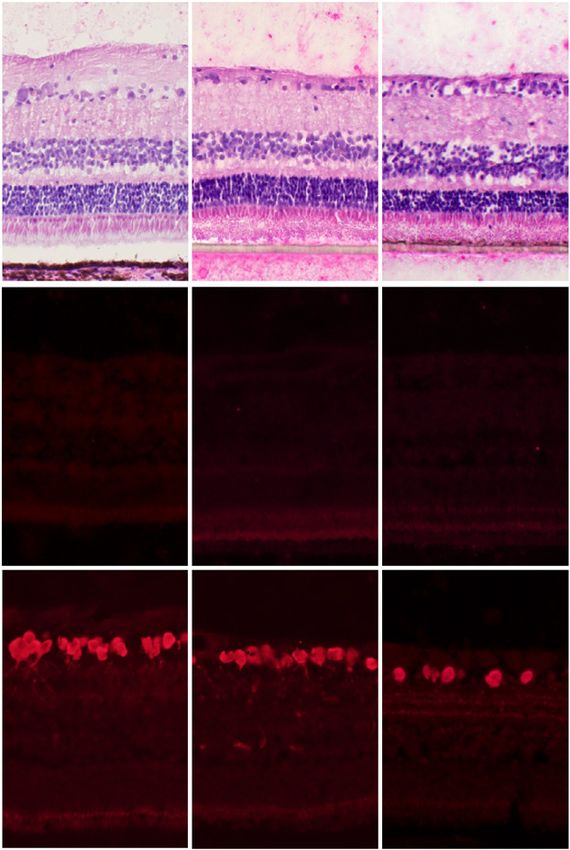
Fig. 2. Adult porcine retinal explants cultured for 48 h 90 or 240      nal explant cultured 240 min after enucleation for 48 h displays loss
min after enucleation. A, B Overall morphology; H&E staining.           of ganglion cells compared with 90-min counterpart. I Statistical
Both retinal explants show signs of a disorganization of cell bodies,   analysis (Student’s t-test) of the number of labeled cells. C, F, I Bars
scattered pyknotic cell bodies, and vacuoles. C Statistical analysis    represent mean values, error bars SD. * p < 0.05; **** p < 0.0001.
(Student’s t-test) of the tissue damage score in the inner and outer    OS, outer segments; IS, inner segments; ONL, outer nuclear layer;
retina. D, E Apoptosis; TUNEL staining. Retinal explants show           OPL, outer plexiform layer; INL, inner nuclear layer; IPL, inner
apoptotic cells in all 3 nuclear layers. F Statistical analysis (Stu-   plexiform layer; GCL, ganglion cell layer; NFL, nerve fiber layer.
dent’s t-test) of the number of labeled cells in the inner and outer    Scale bars, 100 μm.
retina. G, H Ganglion cells; RBPMS immunohistochemistry. Reti-

Time-Dependent Tissue Damage in the                                     Cells Tissues Organs 2021;210:58–65                                                                          63
Adult Retina                                                            DOI: 10.1159/000514795
[2016] reported that ganglion cell activity in the mini-pig
                                                                                 retina was maintained for at least 50 h after enucleation
                              4
                                                              90 mins+culture    when the tissue was kept under hypothermic conditions
     (absorbance at 490 nm)

                              3
                                                              240 mins+culture   at 4°C. Similarly, Schultheiss et al. [2016] found that hy-
                                                                                 pothermia prolonged ganglion cell function and pre-
          LDH release

                              2                                                  served the retinal structure for up to 340 min in bovine
                                                                                 eyes after enucleation. To minimize tissue damage, we
                              1                                                  ensured that our eyes were kept cold at 4°C after enucle-
                                                                                 ation until the time of dissection. In accordance with the
                              0                                                  previous studies, our retinas examined 240 min after enu-
                                  2h   24h     48h
                                                                                 cleation displayed only a minimal increase in TUNEL de-
                                                                                 tectable apoptosis compared with 90-minute counter-
                                                                                 parts, however, overall morphology and ganglion cell sur-
Fig. 3. Statistical analysis (Student’s t-test) of the cell damage
marker lactate dehydrogenase (LDH) in culture medium derived                     vival were profoundly affected, as evident in H&E-stained
from retinal explants cultured 90 or 240 min for 2, 12, and 24 h.                sections and RBPMS immunohistochemistry, suggesting
* p < 0.05; *** p < 0,001; **** p < 0.0001. Bars represent mean val-             that at least in our experimental setup, hypothermia failed
ues, error bars SD. Time-dependent retinal tissue damage after                   to protect the retina 240 min after enucleation.
enucleation.
                                                                                    Time after Enucleation Has a Significant Impact on
                                                                                    Retinal Tissue Damage in vitro
outer retina and used immunohistochemistry to explore                               To further elucidate any possible extended time-de-
ganglion cell loss specifically. The results show that with                      pendent effects in vitro, we examined retinas harvested 90
the exception of ganglion cells, the retina is relatively well                   or 240 min after enucleation with subsequent culturing
preserved 90 min after enucleation, whereas profound                             for 48 h. The in vitro retinal explant setup is increasingly
changes in the inner and outer retina are seen after 240                         used for a wide array of research activities, including dis-
min. Enucleation of the eye induces global ischemia, and                         ease mechanism exploration, pharmacological interven-
the increase in tissue damage seen 240 min after enucle-                         tion as well as transplantation [Engelsberg and Ghosh,
ation corresponds well to that reported in previous in                           2007; Rettinger and Wang, 2018]. Immature retinal por-
vivo experiments where 90 min of ischemia have been                              cine tissue can be cultured for extended time periods and
reported to be well tolerated whereas 240 min are detri-                         even show much of the normal development, however,
mental [Hayreh et al., 1980, 2004].                                              the adult retina degenerates rapidly within the in vitro
   Even though retinas examined 90 min after enucle-                             environment, which has been attributed to axotomy and
ation displayed only minimal morphological deteriora-                            the loss of retinal pigment epithelium-photoreceptor
tion and apoptosis, RBPMS labeling of ganglion cells was                         contact [Winkler et al., 2002; Engelsberg et al., 2005]. We
significantly decreased when compared to in vivo con-                            have previously shown that cell survival in adult retinal
trols. In addition to global ischemia, the enucleation par-                      porcine explants, including ganglion cells and photore-
adigm also includes transection of the optic nerve, which                        ceptors, can be significantly enhanced by placing the ex-
is well-known to produce ganglion cell death. However,                           plant with the inner limiting membrane facing the culture
this phenomenon is usually not detectable until after lon-                       membrane [Taylor et al., 2014]. To limit the culture-in-
ger time periods [Watanabe and Fukuda, 2002]. When                               duced effect, we thus used this method for the culture
isolating the tissue prior to fixation, additional axonal                        experiment.
trauma is induced within the retina, more central to the                            The cultured retinas of both groups displayed a similar
ganglion cell body. Therefore, we cannot exclude the pos-                        degree of overall tissue damage in H&E staining. How-
sibility that the loss of RBPMS labeling in the ganglion                         ever, a strong significant increase in TUNEL-labeled cells
cells 90 min after enucleation is primarily the result of                        in the inner retina combined with profound loss of gan-
axonal trauma and not ischemia.                                                  glion cells in the 240-min group indicates that the initial
   Our results are at least in part in contrast to earlier                       discrepancy in tissue damage discussed above is indeed
findings concerning time-dependency after enucleation                            extended in vitro. The increased tissue damage state of
and may, therefore, be relevant to experimental research                         retinas 240 min after enucleation was further confirmed
involving the large animal adult retina. Reinhard et al.                         in the culture medium analysis in which the 240-min ex-

64                                     Cells Tissues Organs 2021;210:58–65                            Svare/Åkerström/Ghosh
                                       DOI: 10.1159/000514795
plants already after 2 h of culture displayed a highly sig-                        Statement of Ethics
nificant increase in LDH activity. The implication of
                                                                                   All procedures and handling of animals were in accordance
these findings is relevant to a range of research involving                    with the guidelines and requirements of the Government Commit-
large animal adult retinal tissue experiments and may be                       tee on Animal Experimentation at Lund University and also com-
of particular interest for explorations in vitro.                              plied with the ARVO guidelines for animal experimentation.
   To summarize, we here show that time after enucle-
ation is a crucial factor for retinal tissue survival in a large
animal model. The adult porcine retina displays pro-                               Conflict of Interest Statement
found tissue damage 240 min after enucleation, whereas
retinas examined after 90 min remain relatively intact,                            The authors have no conflicts of interest to declare.
and this discrepancy is extended when the tissue is placed
under culture conditions. These time points are in accor-
dance with previous in vivo experiments and should be                              Funding Sources
considered in experiments involving the ex vivo adult                              The study was supported by the Swedish Research Council
porcine retina.                                                                (grant number 2015-02,772), The King Gustaf V and Queen Vic-
                                                                               toria Freemason Foundation.
   Acknowledgements

   The authors extend their gratitude to Hodan Abdshill for excel-                 Author Contributions
lent technical assistance and knowledge regarding all aspects of the
study, Linnéa Taylor for advice on technical aspects of the culture                F.S.: Conception, experimentation, data collection and analy-
experiment, and Jesper Bergwik and Amanda Kristiansson at the                  sis, manuscript preparation, correspondence. B.Å.: Conception,
Section for Infection Medicine, Dept. of Clinical Sciences, Lund               data analysis (LDH), proof reading. F.G.: Conception, data collec-
University, for guidance regarding the LDH analysis.                           tion and analysis, manuscript preparation.

   References
Åkerström B, Cederlund M, Bergwik J, Ma-             Reinhard K, Mutter M, Gustafsson E, Gustafsson      Schultheiss M, Schnichels S, Hermann T, Hurst J,
   nouchehrian O, Arnér K, Taylor IH, et al. The         L, Vaegler M, Schultheiss M, et al. Hypother-      Feldkaemper M, Arango-Gonzalez B, et al.
   Role of Mitochondria, Oxidative Stress, and           mia promotes survival of ischemic retinal          Hypothermia Protects and Prolongs the Tol-
   the Radical-binding Protein A1M in Cultured           ganglion cells. Invest Ophthalmol Vis Sci.         erance Time of Retinal Ganglion Cells against
   Porcine Retina. Curr Eye Res. 2017 Jun;42(6):         2016 Feb;57(2):658–63.                             Ischemia. PLoS One. 2016 Feb; 11(2):
   948–61.                                           Rettinger CL, Wang HC. Current Advancements            e0148616.
Engelsberg K, Ghosh F. Transplantation of cul-           in the development and characterization of      Taylor L, Arnér K, Engelsberg K, Ghosh F. Effects
   tured adult porcine full-thickness retina. Cell       full–thickness adult neuroretina organotypic       of glial cell line-derived neurotrophic factor
   Transplant. 2007 Jan;16(1):31–9.                      culture systems. Cells Tissues Organs. 2018;       on the cultured adult full-thickness porcine
Engelsberg K, Johansson K, Ghosh F. Develop-             206(3):119–32.                                     retina. Curr Eye Res. 2013 Apr;38(4):503–15.
   ment of the embryonic porcine neuroretina in      Rodriguez AR, de Sevilla Müller LP, Brecha NC.      Taylor L, Arnér K, Taylor IH, Ghosh F. Feet on
   vitro. Ophthalmic Res. 2005 Mar–Apr; 37(2):           The RNA binding protein RBPMS is a selec-          the ground: physical support of the inner ret-
   104–11.                                               tive marker of ganglion cells in the mamma-        ina is a strong determinant for cell survival
Hayreh SS, Kolder HE, Weingeist TA. Central ret-         lian retina. J Comp Neurol. 2014 Apr;522(6):       and structural preservation in vitro. Invest
   inal artery occlusion and retinal tolerance           1411–43.                                           Ophthalmol Vis Sci. 2014 Apr;55(4):2200–13.
   time. Ophthalmology. 1980 Jan;87(1):75–8.         Schnichels S, Kiebler T, Hurst J, Maliha AM,        Watanabe M, Fukuda Y. Survival and axonal re-
Hayreh SS, Zimmerman MB, Kimura A, Sanon A.              Löscher M, Dick HB, et al. Retinal Organ Cul-      generation of retinal ganglion cells in adult
   Central retinal artery occlusion. Retinal sur-        tures as Alternative Research Models. Altern       cats. Prog Retin Eye Res. 2002 Nov;21(6):529–
   vival time. Exp Eye Res. 2004 Mar;78(3):723–          Lab Anim. 2019 Mar;47(1):19–29.                    53.
   36.                                                                                                   Winkler J, Hagelstein S, Rohde M, Laqua H. Cel-
Maidana DE, Tsoka P, Tian B, Dib B, Matsumoto                                                               lular and cytoskeletal dynamics within organ
   H, Kataoka K, et al. A novel ImageJ macro for                                                            cultures of porcine neuroretina. Exp Eye Res.
   automated cell death quantitation in the reti-                                                           2002 Jun;74(6):777–88.
   na. Invest Ophthalmol Vis Sci. 2015 Oct;
   56(11):6701–8.

Time-Dependent Tissue Damage in the                                            Cells Tissues Organs 2021;210:58–65                                     65
Adult Retina                                                                   DOI: 10.1159/000514795
You can also read