Lysimachiae Herba Inhibits Inflammatory Reactions and Improves Lipopolysaccharide/D-Galactosamine-Induced Hepatic Injury - MDPI
←
→
Page content transcription
If your browser does not render page correctly, please read the page content below
antioxidants

Article
Lysimachiae Herba Inhibits Inflammatory Reactions and
Improves Lipopolysaccharide/D-Galactosamine-Induced
Hepatic Injury
Yun Hee Jeong, Tae In Kim                 , You-Chang Oh *          and Jin Yeul Ma *

                                          Korean Medicine (KM)-Application Center, Korea Institute of Oriental Medicine, 70 Cheomdanro, Dong-gu,
                                          Daegu 41062, Korea; runxi0333@kiom.re.kr (Y.H.J.); tikim@kiom.re.kr (T.I.K.)
                                          * Correspondence: ulivuli@kiom.re.kr (Y.-C.O.); jyma@kiom.re.kr (J.Y.M.); Tel.: +82-53-940-3882 (Y.-C.O.);
                                            +82-53-940-3812 (J.Y.M.)

                                          Abstract: This study aimed to determine the anti-inflammatory and hepatoprotective effects of
                                          Lysimachiae Herba ethanolic extract (LHE) in lipopolysaccharide (LPS)-stimulated macrophages and
                                          in a LPS/D-galactosamine (GalN)-induced acute hepatitis mouse model. Then, the production of in-
                                          flammatory mediators and the activation of related pathways in macrophages were explored. Finally,
                                          we assessed the serum aminotransferase levels and the expression of inflammatory/antioxidant
                                          molecules in liver tissues in mice. Results revealed that LHE treatment significantly inhibited the
                                          production of inflammatory mediators in LPS-stimulated RAW 264.7 macrophages. Molecular data
                                          showed that LHE remarkably increased the activities of the antioxidant pathway and inhibited

                                   the phosphorylation of mitogen-activated protein kinase as well as the transcriptional activity of
                                          nuclear factor-κB induced by LPS. Furthermore, it prevented acute liver damage caused by LPS/D-
Citation: Jeong, Y.H.; Kim, T.I.; Oh,
                                          GalN-induced hepatitis by inhibiting aminotransferase levels and histopathological changes in mice.
Y.-C.; Ma, J.Y. Lysimachiae Herba
                                          Moreover, treatment with LHE significantly inhibited the activation of inflammatory pathways
Inhibits Inflammatory Reactions and
Improves Lipopolysaccharide/
                                          and increased the expression of antioxidant molecules including heme oxygenase-1/Nuclear factor
D-Galactosamine-Induced Hepatic           erythroid 2-related factor 2. In conclusion, LHE has potent anti-inflammatory and hepatoprotective
Injury. Antioxidants 2021, 10, 1387.      effects in LPS-stimulated macrophages and the LPS/D-GalN-induced acute hepatitis mouse model.
https://doi.org/10.3390/antiox            Thus, it can be a treatment option for inflammation, hepatitis, and liver injury.
10091387
                                          Keywords: Lysimachiae Herba; anti-inflammatory; antioxidant; hepatoprotective; lipopolysaccharide;
Academic Editor: Guillermo                D-galactosamine
R. Schinella

Received: 6 August 2021
Accepted: 26 August 2021
                                          1. Introduction
Published: 30 August 2021
                                               Lysimachiae Herba (LH) is a dried whole part of Lysimachia christinae, known in Korea
                                          as “Geumjeoncho”, and is commonly found in East Asia. This herb has been commonly
Publisher’s Note: MDPI stays neutral
                                          used as a traditional herbal medicine for the treatment of inflammatory diseases, viral
with regard to jurisdictional claims in
published maps and institutional affil-
                                          hepatitis, cholecystitis, urinary stones, and jaundice [1–3]. LH contains flavonoids and
iations.
                                          phenols as major components, and these compounds have been studied for their anti-
                                          cholecystitis efficacy and cholagogic activity [4]. Many previous studies have investigated
                                          the efficacy of LH on liver-related diseases, but scientific studies of its anti-inflammatory
                                          properties and precise cellular molecular mechanisms are lacking, and the effect of LH on
                                          lipopolysaccharide (LPS)/D-galactosamine (GalN)-induced acute hepatitis has not been
Copyright: © 2021 by the authors.
                                          studied.
Licensee MDPI, Basel, Switzerland.
                                               Fulminant hepatitis is a severe liver disease pathologically by extensive hepatocellular
This article is an open access article
distributed under the terms and
                                          apoptosis and hemorrhagic necrosis leading to multiple organ failures [5,6]. Fulminant
conditions of the Creative Commons
                                          hepatic injury is caused by a variety of factors that can cause liver failure, including
Attribution (CC BY) license (https://     alcohol, chemicals, oxidizing agents, and hepatitis virus [7]. LPS and D-GalN are the
creativecommons.org/licenses/by/          most common agents that cause fulminant hepatic injury [8,9]. LPS is a component of
4.0/).                                    the gut Gram-negative bacteria [10], and plays an important role in the initiation stage

Antioxidants 2021, 10, 1387. https://doi.org/10.3390/antiox10091387                                     https://www.mdpi.com/journal/antioxidants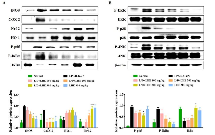
Antioxidants 2021, 10, 1387                                                                                              2 of 18

                              of endotoxic damage and activates inflammatory cytokines, causing liver tissue damage.
                              D-GalN causes hepatocyte necrosis upon acute exposure, and cirrhosis and cell tumor
                              during chronic exposure [11]. Additionally, D-GalN is able to sensitize toxic effects of
                              the liver toward endotoxins such as LPS and results in fulminant hepatic failure within
                              a few hours [12]. LPS/D-GalN exposure results in liver damage due to overgrowth of
                              intestinal bacteria, disruption of intestinal barrier function, and an increase in permeability
                              to endotoxin and bacteria [13]. Therefore, hepatic injury induced by LPS in combination
                              with D-GalN is well-accepted animal model which is significantly similar to acute hepatic
                              failure in the clinical setting [7] and is widely used to study the underlying mechanism of
                              fulminant hepatitis and its potential therapeutic drugs [14,15]. Previous studies reported
                              that LPS/D-GalN induced secretion of inflammatory cytokines, such as tumor necrosis
                              factor (TNF)-α, interleukin (IL)-6, and IL-1β in mice model of hepatic liver injury [16,17],
                              which promote liver cell necrosis, hepatic failure [18,19] and reduce antioxidant enzyme
                              activity [20]. Therefore, reducing the inflammatory cytokines and activating the antioxidant
                              enzymes may be key to the prevention and treatment of fulminant hepatitis.
                                    The production of cytokines is closely involved in the activation of mitogen-activated
                              protein kinase (MAPK) and nuclear factor (NF)-κB signaling pathway [21]. When acute
                              liver injury is induced with LPS/D-GalN, LPS binds to the Toll-like receptor of Kupffer
                              cells and promotes the transcription and production of inflammatory cytokines such as
                              TNF-α, IL-6, and IL-1β [22,23]. Thus, NF-κB and MAPK signaling pathway are critical
                              contributors to the mechanism of the LPS/D-GalN-induced fulminant hepatitis. Nuclear
                              factor E2-related factor 2 (Nrf-2) is a major transcription factor that regulates cellular defense
                              mechanism against oxidative stress and inflammation by regulating the level of several
                              endogenous cytoprotective enzyme, including heme oxygenase (HO)-1. Previous studies
                              reported that the protective effect of the Nrf-2 pathway in acute liver inflammation and
                              liver fibrosis [24–26]. Another study has shown that HO-1 has anti-inflammatory activity
                              against acute and chronic inflammatory diseases, including experimental colitis, bronchitis,
                              and hepatitis [27,28]. Therefore, Nrf-2/HO-1 signaling pathway plays a positive role in the
                              pathogenesis of inflammatory diseases including fulminant hepatitis induced by LPS/D-
                              GalN. Based on these previous findings, we also hypothesized that the hepatoprotective
                              effect of LHE may be closely associated with strong anti-inflammatory activity. Therefore,
                              in this study, we not only investigated the hepatoprotective effect of LHE on LPS/D-GalN-
                              induced acute hepatitis mice models in vivo, but also investigated whether LHE has an
                              inhibitory effect on inflammatory responses in RAW 264.7 macrophages stimulated with
                              LPS in vitro levels. We also explored chemical components of LHE by high-performance
                              liquid chromatography (HPLC) analysis.

                              2. Materials and Methods
                              2.1. Materials and Reagents
                                   Roswell park memorial institute (RPMI) 1640 medium, fetal bovine serum (FBS), and
                              antibiotics were obtained from Hyclone (Logan, UT, USA). LPS, dexamethasone (Dex),
                              bovine serum albumin (BSA), and D-GalN were purchased from Sigma-Aldrich (St. Louis,
                              MO, USA). Enzyme-linked immunosorbent assay (ELISA) antibody sets were obtained
                              from eBioscience (San Diego, CA, USA). Cell culture dish and well plates were purchased
                              from SPL Life Sciences (Pocheon, Korea). The cell counting kit (CCK) was obtained from
                              Dojindo Molecular Technologies, Inc. (Kumamoto, Japan). Bradford reagent was pur-
                              chased from Bio-Rad (Hercules, CA, USA). Nitrocellulose (NC) membrane was obtained
                              from Millipore (Bedford, MA, USA). Various primary and horseradish peroxidase (HRP)-
                              conjugated secondary antibodies for protein analysis were purchased from Cell Signaling
                              Technology, Inc. (Boston, MA, USA). RNA extraction reagent and DNA synthesizing kit
                              were obtained from iNtRON Biotech (Daejeon, Korea). Oligonucleotide primers for tumor
                              necrosis factor (TNF)-α, interleukin (IL)-6, IL-1β, inducible nitric oxide synthase (iNOS), cy-
                              clooxygenase (COX)-2, HO-1, and β-actin were synthesized from Bioneer (Daejeon, Korea).
                              The quantitative polymerase chain reaction (qPCR) reaction reagent kit was purchased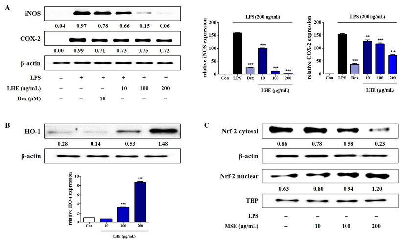
Antioxidants 2021, 10, 1387                                                                                          3 of 18

                              from Bioneer. Protocatechuic acid, catechin, quercitrin, quercetin, and kaempferol were
                              purchased from Sigma-Aldrich. HPLC-grade acetonitrile was provided by Merck KFaA
                              (Darmstadt, Germany). ACS reagent-grade formic acid was obtained from Sigma-Aldrich.
                              Water analyzed via HPLC was prepared using the Puris-Evo RO water system (Mirae ST
                              Co., Ltd., Anyang-si, Korea).

                              2.2. Preparation of LHE
                                   LH was purchased as a dried herb from Yeongcheonhyundai Herbal Market (Yeongcheon,
                              Korea) and was identified by Prof. KiHwan Bae (College of Pharmacy, Chungnam Na-
                              tional University, Korea). All voucher specimens were deposited in an herbal bank at the
                              KM-Application Center, Korea Institute of Oriental Medicine (voucher number: E207). The
                              dried herb (30.0 g) was extracted with 390 mL of 70% ethanol in a 40 ◦ C shaking incubator
                              (100 rpm) for 24 h. In some previous studies using plant extracts, 70% ethanol was used as
                              the extraction solvent, and we set the extraction conditions for LH with reference to the
                              studies [29,30]. The extract solution was filtered using a 150 mm filter paper (Whatman,
                              Piscataway, NJ, USA) and was concentrated to 100 mL using a rotary vacuum evaporator
                              (Buchi, Tokyo, Japan). Samples were then freeze-dried and stored in desiccators at −20 ◦ C
                              before use. The sample yield was 12.2627%, and 3.6788 g of extract was obtained.

                              2.3. Cell Culture and Drug Treatment
                                   Murine macrophage RAW 264.7 cells were obtained from Korea Cell Line Bank (Seoul,
                              Korea) and were cultured in complete RPMI 1640 medium (10% FBS and 1% antibiotics
                              (v/v)). The cells were then incubated in humidified 5% CO2 atmosphere at 37 ◦ C. To
                              stimulate the cells, LPS (200 ng/mL) was added in the presence or absence of LHE (10, 100,
                              or 200 µg/mL) at the indicated periods.

                              2.4. Cell Viability Assay
                                   LHE-induced cytotoxicity was analyzed using CCK reagent. The macrophages were
                              seeded in 96-well culture plates at a density of 5 × 10⁴ cells/well in 100 µL medium. After
                              18 h of incubation, LHE was added to the cells and was incubated for 24 h at 37 ◦ C with 5%
                              CO2 . Then, 10 µL of CCK solutions was applied to each well, and the cells were incubated
                              for another 1 h. Then, the optical density was read at 450 nm using ELISA reader (infinite
                              M200, TECAN, Mannedorf, Switzerland).

                              2.5. Analysis of NO Production
                                   NO production was analyzed by measuring nitrite levels in the supernatants of
                              cultured macrophage cells. Macrophage cells (5 × 10⁴ cells/well in 100 µL medium) were
                              plated, incubated with LHE, and stimulated with LPS for 24 h. The supernatant was mixed
                              with the same volume of Griess reagent (1% sulfanilamide, 0.1% naphthylethylenediamine
                              dihydrochloride, and 2.5% phosphoric acid) and was incubated at room temperature (RT)
                              for 5 min. The absorbance at 570 nm was read. The concentration of nitrite was calculated
                              with sodium nitrite as the standard.

                              2.6. Inflammatory Cytokine Production
                                   To determine the effects of LHE, the production of pro-inflammatory cytokines, such
                              as TNF-α, IL-6, and IL-1β, was assessed using ELISA. That is, 2.5 × 10⁵ cells/well in 500 µL
                              medium were seeded on 24 well plates and were incubated overnight. The cells were
                              pretreated with three concentrations of LHE for 1 h and further challenged with LPS for
                              an additional 24 h at 37 ◦ C with 5% CO2 . The cytokine levels in the supernatants were
                              measured using ELISA antibody sets according to the manufacturer’s instructions.

                              2.7. Preparation of Whole Cell, Cytosolic, Nuclear, and Mice Liver Tissue Extracts
                                  To obtain whole cell and liver tissue lysates, cell pellets or mouse liver tissue samples
                              were resuspended in radioimmunoprecipitation assay lysis buffer (Millipore) containing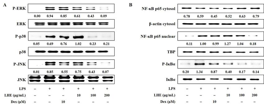
Antioxidants 2021, 10, 1387                                                                                             4 of 18

                              protease and phosphatase inhibitors. Cytosol and nuclear fractions were isolated using
                              NE-PER nuclear and cytoplasmic extraction reagents (Thermo Scientific, Rockford, IL,
                              USA), according to the manufacturer’s instruction. The fractions were stored at −80 ◦ C
                              before use.

                              2.8. Western Blotting for Protein Analysis
                                    Western blotting was performed to evaluate the effects of LHE on the expression of
                              various inflammatory response-related proteins or inflammatory pathway proteins in the
                              whole cell, cytosol, nucleus, or mice liver tissues. For cell protein analysis, the cells were
                              pretreated with LHE and stimulated with LPS at indicated periods. After incubation, the
                              cells were collected via scrapping and were washed twice with ice-cold phosphate buffered
                              saline (PBS). For liver protein analysis, mouse liver tissue samples were collected and gently
                              rinsed twice with PBS. The total proteins were determined using the Bradford’s method.
                              Equal amounts of proteins were subjected to sodium dodecyl sulfate–polyacrylamide gel
                              electrophoresis after transferring them into an NC membrane with a glycine transfer buffer
                              (192 mM glycine, 25 mM Tris-HCl [pH 8.8], and 20% MeOH [v/v]). Then, after blocking
                              the nonspecific site with 3% BSA, the membrane was then incubated with specific primary
                              antibody at 4 ◦ C overnight. Next, it was subsequently incubated with HRP-conjugated
                              secondary antibodies. The specific proteins were detected using SuperSignal West Femto
                              Chemiluminescent Substrate (Thermo Scientific). Protein levels were quantified using
                              a ChemiDocTM Touch Imaging System (Bio-Rad). Table 1 shows the information about
                              various primary and secondary antibodies.

                              Table 1. Primary and secondary antibodies use for Western blot analysis.

                                  Antibody           Corporation           Product No.         RRID        Dilution Rate
                                     iNOS           Cell Signaling           #13120        AB_2687529          1:1000
                                    COX-2           Cell Signaling           #4842         AB_2085144          1:1000
                                     HO-1           Cell Signaling           #82206        AB_2799989          1:1000
                                     Nrf-2          Cell Signaling           #12721        AB_2715528          1:1000
                                 P-NF-κB p65        Cell Signaling            #3033         AB_331284          1:1000
                                  NF-κB p65         Cell Signaling           #8242         AB_10859369         1:1000
                                    P-IκBα          Cell Signaling           #2859          AB_561111          1:1000
                                     IκBα           Cell Signaling           #4814          AB_390781          1:1000
                                    P-ERK           Cell Signaling           #4377          AB_331775          1:1000
                                      ERK           Cell Signaling            #9102         AB_330744          1:1000
                                    P-p38           Cell Signaling           #9211          AB_331641          1:1000
                                      p38           Cell Signaling            #9212         AB_330713          1:1000
                                    P-JNK           Cell Signaling           #9251          AB_331659          1:1000
                                      JNK           Cell Signaling            #9252        AB_2250373          1:1000
                                    β-actin         Cell Signaling            #4970        AB_2223172          1:1000
                                      TBP           Cell Signaling            #8515        AB_10949159         1:1000
                                2nd anti-mouse      Cell Signaling            #7076         AB_330924          1:5000
                                2nd anti-rabbit     Cell Signaling            #7074        AB_2099233          1:5000

                              2.9. RNA Extraction, DNA Synthesis, and qPCR
                                   Total cellular RNA was isolated using the easy-BLUE™ RNA extraction kit according
                              to the manufacturer’s instruction. The total RNA (1 µg) was reversed transcribed into
                              cDNA using RevoScript™ RT PreMix. Table 2 shows the oligonucleotide primer sequences
                              for qPCR. The reactions were setup in triplicates with a total volume of 20 µL: final
                              concentration of 0.3 µM for each primer, 10 µL of AccuPower® 2X Greenstar qPCR Master
                              Mix, and 2 µL of template DNA. The following qPCR conditions were applied for TNF-α,
                              IL-6, IL-1β, iNOS, COX-2, HO-1, and β-actin: 40 cycles at 94 ◦ C for 15 s and 60 ◦ C for
                              1 min [31]. Amplification and analyses were performed using QuantStudio 6 Flex Real-time
                              PCR System (Thermo Scientific). Samples were compared using the relative CT method.
                              The fold increase or decrease in gene expression was determined relative to a blank control
                              after normalization to β-actin gene using 2−∆∆C T [31].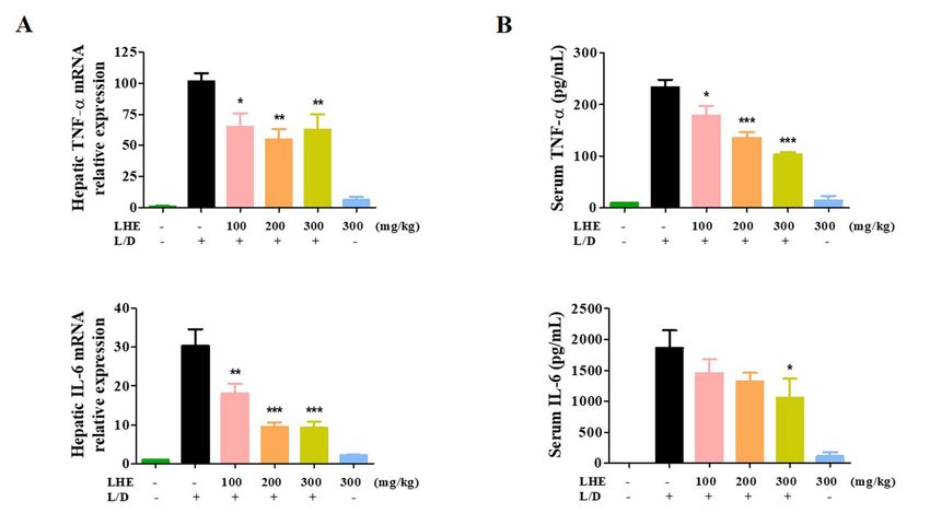
Antioxidants 2021, 10, 1387                                                                                           5 of 18

                              Table 2. Primers used for qPCR.

                                Target              Reference
                                                                                      Primer Sequence
                                Gene                Sequence
                                TNF-α             NM_013693.3     F: 50 -TTCTGTCTACTGAACTTCGGGGTGATCGGTCC-30
                                                                  R: 50 -GTATGAGATAGCAAATCGGCTGACGGTGTGGG-30
                                  IL-6            NM_031168.2                  F: 50 -TCCAGTTGCCTTCTTGGGAC-30
                                                                            R: 50 -GTGTAATTAAGCCTCCGACTTG-30
                                 IL-1β            NM_008361.4            F: 50 -ATGGCAACTGTTCCTGAACTCAACT-30
                                                                         R: 50 -CAGGACAGGTATAGATTCTTTCCTTT-30
                                 iNOS             NM_010927.4                 F: 50 -GGCAGCCTGTGAGACCTTTG-30
                                                                             R: 50 -GCATTGGAAGTGAAGCGTTTC-30
                                COX-2             NM_011198.4                F: 50 -TGAGTACCGCAAACGCTTCTC-30
                                                                             R: 50 -TGGACGAGGTTTTTCCACCAG-30
                                 HO-1             NM_010442.2               F: 50 -TGAAGGAGGCCACCAAGGAGG-30
                                                                            R: 50 -AGAGGTCACCCAGGTAGCGGG-30
                                β-actin           NM_007393.5                F: 50 -AGAGGGAAATCGTGCGTGAC-30
                                                                             R: 50 -CAATAGTGATGACCTGGCCGT-30
                              F, forward; R, reverse.

                              2.10. Animals Used for the Analysis of LPS/D-GalN-Induced Acute Hepatitis
                                   Six-week-old male imprinting control region (ICR) mice (30 ± 3 g) were purchased
                              from Samtako BioKorea (Osan, Korea). All animals were stored in a room with controlled
                              temperature under a 12 h light/12 h dark cycle, with food and water provided ad libitum.
                              Mice were fed gamma irradiation-sterilized LabDiet 5053 (Orient, Seongnam, Korea), which
                              included protein 20%, fat (ether extract) 4.5%, fat (acid hydrolysis) 5.4% crude fiber 4.7%,
                              ash 6%, calcium 0.8%, and phosphorus 0.62%. All mice were acclimatized for at least 7 days
                              prior to the experiments. All experimental procedures were performed in accordance
                              with the guidelines for the Animal Care and Use Committee of Korea Institute of Oriental
                              Medicine (reference number: #D-17-020).

                              2.11. Protocol for the LPS/D-GalN-Induced Hepatitis Mouse Model
                                   Mice were randomly divided into four groups, which are as follows: normal group
                              (vehicle, 0.5% carboxymethyl cellulose [CMC]), LPS/D-GalN group (50 µg LPS and 1 g
                              D-GalN/kg), LHE along group (300 mg/kg LHE), and test group (LPS/D-GalN + 100, 200,
                              or 300 mg/kg of LHE). The dose of LHE administered to mice was set with reference to a
                              previous study [32], and a group administered with 300 mg/kg of LHE alone was analyzed
                              to exclude potential toxicity caused by LHE administration. Briefly, three concentrations
                              of LHE were administered orally to mice for a total of 6 days once a day (diluted in a
                              volume of 10 mL/kg of 0.5% CMC). During this period, the normal and LPS/D-GalN
                              groups received the same amount of vehicle. One hour after the last injection, LPS/D-GalN
                              was administered intraperitoneally in each mouse. Next, the mice were sacrificed after 6 h.
                              Mouse liver tissues and serum samples were collected and used for hematoxylin and eosin
                              (H&E) staining, Western blotting, ELISA, and aminotransferase analysis.

                              2.12. Biochemical Analysis for the Evaluation of Serum Alanine Aminotransferase, Aspartate
                              Aminotransferase, Alkaline Phosphatase, and Liver Tissue Protein Levels in Mice
                                   All mice were euthanized 6 h after the administration of LPS/D-GalN. Then, liver tis-
                              sue and blood samples were collected for biochemical analysis. Mice serum was separated
                              from the blood via centrifugation at 2000× g for 15 min. The serum alanine aminotrans-
                              ferase (ALT), aspartate aminotransferase (AST), and alkaline phosphatase (ALP) levels
                              were assessed using an Erba XL-200 automated clinical chemistry analyzer (Mannheim,
                              Germany). To analyze the expression of different inflammatory proteins, the liver tissue
                              was homogenized and dissolved in radioimmunoprecipitation assay buffer. All protein
                              analysis results were normalized using the β-actin of each sample to make the total protein
                              amount constant.
Antioxidants 2021, 10, 1387                                                                                            6 of 18

                              2.13. Histopathological Examination
                                   Histological analysis of mice liver was performed to evaluate the inhibitory efficacy
                              of LHE on the histological changes in liver tissues caused by LPS/D-GalN. After the
                              mice were euthanized, the liver tissues were separated, fixed in 10% formaldehyde for
                              10 days, embedded in paraffin wax, and cut into sections with a thickness of 5 µM. Paraffin-
                              embedded sections were stained with H&E and were subjected to pathological analysis
                              under a microscope. Sections were assessed for liver injury with Axioskop 40 (Oberkochen,
                              Germany) and were photographed at 400× magnification.

                              2.14. HPLC Instruments
                                   HPLC analysis was conducted using the Dionex Ultimate 3000 system set up with
                              a column oven, auto sampler, binary pump, and diode assay UV/VIS detector (Dionex
                              Corp., Sunnyvale, CA, USA). Data acquisition and processing were performed using
                              Chromeleon 7 (Thermo Fisher, Counteaboeuf, France).

                              2.15. Preparation of Plant Material Sample and Standard
                                   In total, 100 mg of LHE was precisely weighted and dissolved in 1 mL of HPLC-
                              grade methanol using an ultrasonicator (JAC Ultrasonic JAC-3010) for 30 min. The LHE
                              solution sample at a concentration of 100 mg/mL was filtered using a 0.2 PETE membrane
                              syringe filter. After filtration, 10 µL of filtrate was injected for HPLC analysis. To confirm
                              each standard retention time using the established method, protocatechuic acid, catechin,
                              quercitrin, quercetin, and kaempferol standard solutions were prepared at 1.0 mg/mL with
                              methanol. Then, 10 µL of standard solution filtrate was analyzed using the HPLC system.
                              After analysis, the retention time of LHE and each standard compound were compared.
                              Moreover, to confirm the content of each compound in LHE, we prepared a standard curve
                              for several compounds, which were diluted with methanol at different concentrations.

                              2.16. HPLC Analysis
                                   To identify the content of five major compounds (protocatechuic acid, catechin,
                              quercitrin, quercetin, and kaempferol) in LHE, we conducted an HPLC analysis, which
                              was operated with C18 column with a C18 guard cartridge (4.0 × 3.0 mm). The mobile
                              phase was set up with A, 1.0% formic acid and B, acetonitrile was eluted at a flow rate
                              of 1 mL/min. The LHE and standard compound solution were analyzed using the estab-
                              lished gradient mobile phase method. The HPLC chromatogram of LHE and the standard
                              compound was detected under HPLC conditions, which included UV detection at 270 nm,
                              column oven temperature of 40 ◦ C, and injection volume of 10 µL (Table 3). Calibration
                              curves, assessed using the standard solution and the limits of detection and quantification
                              (LOQ) under chromatographic conditions, were determined by injecting a series of stan-
                              dard solutions. The result was processed using Chromeleon 7 (Thermo Fisher), and the
                              solutions were administered three times under the same condition.

                              Table 3. HPLC conditions for analysis.

                                                                       HPLC Conditions
                                            Detector                                         270 nm
                                            Column                           X-bridge C18 (250 mm × 4.6 mm, 5 µm)
                                     Column Temperature                                       40 ◦ C
                                       Injection Volume                                       10 µL
                                           Flow rate                                      1.0 mL/min
                                         Mobile phase                  Time (min)               A                 B
                                                                          0.0                  97                 3
                                 A: 1.0% Formic acid in Water             10.0                 85                 15
                                        B: Acetonitrile                   50.0                 50                 50
                                                                          80.0                  0                100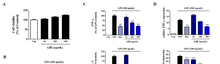
Antioxidants 2021, 10, 1387                                                                                                           7 of 18

                                   2.17. Statistical Analysis
                                        Data were expressed as means ± standard error of the mean (SEM) for all experiments,
                                   and all quantitative data were representative of at least three independent experiments.
                                   Statistical significance was determined via one-way analysis of variance, followed by the
                                   Dunnett’s test after comparing each treatment group and LPS or LPS/D-GalN. * p values
                                   of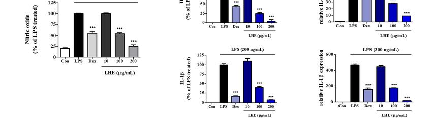
Antioxidants 2021, 10, 1387 8 of 18
for 24 h via LPS. Next, the levels of cytokines were analyzed using ELISA and qPCR. As
shown in Figure 1C, LHE strongly decreased LPS-induced TNF-α, IL-6, and IL-1β cytokine
secretion in a concentration-dependent manner. Moreover, the inhibitory rates were 54%,
96%, and 92% at a concentration of 200 µg/mL, respectively. Similarly, based on the qPCR
results, LHE significantly decreased the mRNA expression of TNF-α, IL-6, and IL-1β via
LHE treatment in a concentration-dependent manner (Figure 1D).
3.3. Effects of LHE on LPS-Induced iNOS and COX-2 Expression
NO and PGE2 expressions are involved in the regulation of iNOS and COX-2 levels in
RAW 264.7 cells, respectively. Thus, we assessed the inhibitory effects of LHE on the protein
and mRNA expression of iNOS and COX-2 via Western blotting and qPCR. As shown in
Figure 2A, compared with LPS treatment, pretreatment with LHE significantly reduced the
Antioxidants 2021, 10, x FOR PEER REVIEW
expression of protein and mRNA genes of iNOS and COX-2 in a concentration-dependent 9 of 19
manner.
Figure 2. Effects of LHE on (A) iNOS and COX-2 expression, (B) HO-1 induction, and (C) nuclear translocation of Nrf-2 in
Figure 2. Effects of LHE on (A) iNOS and COX-2 expression, (B) HO-1 induction, and (C) nuclear translocation of Nrf-2
RAW 264.7264.7
in RAW macrophages. (A) Cells
macrophages. werewere
(A) Cells incubated with with
incubated LHE LHE
for 1 h
forand
1 hthen
and stimulated with LPS
then stimulated withfor 24for
LPS h (protein) or 12 hor
24 h (protein)
(mRNA). Cells were incubated with LHE alone for (B) 6 h or (C) 3 h. The quantified number and histograms
12 h (mRNA). Cells were incubated with LHE alone for (B) 6 h or (C) 3 h. The quantified number and histograms showed showed the
expression levels of protein and mRNA relative to those of β-actin or TBP. ** p < 0.01 and *** p < 0.001 were calculated
the expression levels of protein and mRNA relative to those of β-actin or TBP. ** p < 0.01 and *** p < 0.001 were calculated by
by comparing
comparing LPS stimulation
LPS stimulation values.
values.
3.5.Effects
3.4. EffectsofofLHE
LHEononHO-1
LPS-Induced
ExpressionActivation
and Nrf-2of the MAPK
Nuclear Signaling Pathway
Translocation
Threeisfamilies
HO-1 of MAPK
an important including
enzyme, extracellular
and it plays signal-regulated
an essential kinase (ERK), p38,
role due to its anti-inflammatory
and c-Jun-NH
effects in RAW2-terminal
264.7 cellskinase (JNK) playthe
[34]. Moreover, an induction
important of role in LPS-induced
HO-1 is mediated inflamma-
by Nrf-2
tory responses
activation in macrophages.
via migration Thus,Thus,
into nucleus. the effects of LHE of
the influence onLHE
LPS-stimulated phosphoryla-
on HO-1 expression and
tion of Nrf-2
nuclear MAPK were assessed
accumulation wasviaexamined
Western using
blotting. Our data
Western showed
blotting and that theAs
qPCR. activation
shown inof
MAPK2B,C,
Figure increased after exposure
LHE treatment to LPS atenhanced
significantly a dose of the
200 expression
ng/mL. However,
of HO-1LHE andtreatment
nuclear
was exclusive,
Nrf-2 and it significantly
at a concentration of 200 µg/mL. inhibited the phosphorylation of ERK, p38, and JNK
MAPK in a dose-dependent manner (Figure 3A).
3.6. Effects of LHE on the Transcriptional Activity of NF-κB
NF-κB is a transcription factor that has a key role in inflammatory responses. To in-
vestigate whether the anti-inflammatory activity of LHE against the NF-κB signaling
pathway in RAW 264.7 cells, we evaluated whether LHE affects LPS-induced NF-κB acti-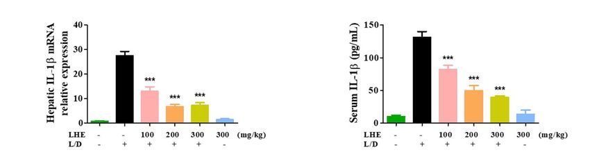
Antioxidants 2021, 10, 1387 9 of 18
3.5. Effects of LHE on LPS-Induced Activation of the MAPK Signaling Pathway
Three families of MAPK including extracellular signal-regulated kinase (ERK), p38,
and c-Jun-NH2 -terminal kinase (JNK) play an important role in LPS-induced inflammatory
responses in macrophages. Thus, the effects of LHE on LPS-stimulated phosphorylation of
MAPK were assessed via Western blotting. Our data showed that the activation of MAPK
increased after exposure to LPS at a dose of 200 ng/mL. However, LHE treatment was
Antioxidants 2021, 10, x FOR PEER REVIEW
exclusive, and it significantly inhibited the phosphorylation of ERK, p38, and JNK10MAPK
of 19
in a dose-dependent manner (Figure 3A).
Figure
Figure3.3. Effects
Effects of LHE onon (A)
(A)the
thephosphorylation
phosphorylationofofMAPK MAPK andand
(B)(B)
thethe activation
activation of NF-κB
of NF-κB in LPS-stimulated
in LPS-stimulated RAWRAW264.7
264.7 macrophages.
macrophages. CellsCells
werewere stimulated
stimulated withwith
LPSLPS for 30
for (A) (A)min
30 min or 1(B)
or (B) h.1The
h. The quantified
quantified number
number showed
showed thethe expres-
expression
sion levels
levels of protein
of protein relative
relative to those
to those of total-type
of total-type protein,
protein, β-actin,
β-actin, or TBP.
or TBP.
3.7.
3.6.Effects
EffectsofofLHE
LHEon onLPS/D-GalN-Induced Liver Injury
the Transcriptional Activity in Mice
of NF-κB
WeNF-κBexplored the hepatoprotective
is a transcription factor that effects
has aofkey
LHE viainLPS/D-GalN-induced
role inflammatory responses. hepatic
To
damage
investigatein vivo. The ICR
whether mice received different
the anti-inflammatory doses
activity of LHE,
of LHE including
against 100, signaling
the NF-κB 200, and
300 mg/kg in
pathway with
RAWLPS/D-GalN,
264.7 cells, orwe300evaluated
mg/kg of LHE alone.LHE
whether First,affects
we evaluated several
LPS-induced pro-
NF-κB
inflammatory cytokines in liver tissues and serum via qPCR and ELISA.
activation and inhibitor of NF-κB alpha (IκBα) phosphorylation. Western blotting revealed As shown in Fig-
ure 4, LHE could effectively inhibit the expression of TNF-α, IL-6, and
that LHE pretreatment significantly inhibited p65 nuclear localization (Figure 3B). That IL-1β cytokine andis,
their
it hasmRNA
a higher levels in liver
inhibitory tissues
effect than and
Dex serum.
when used Theaslevel of cytokine
a positive controlinat the 300 mg/kg
a concentration
LHE/no injectedConsistent
of 200 µg/mL. LPS/D-GaIN withgroup
thesewas closeLHE
results, to that
can of the normal
remarkably group. As
attenuate shown in
LPS-induced
Figure 5A, compared
degradation with the normalofgroup,
and the phosphorylation IκBα inthetheLPS/D-GalN
cytoplasm. group had severe liver
damage. However, the livers of the LHE group were morphologically comparable with
3.7. Effects
those of theofnormal
LHE ongroup
LPS/D-GalN-Induced
in a dose-dependent Liver Injury in Mice
manner. Moreover, the 300 mg/kg LHE
alone Wegroup was morphologically
explored the hepatoprotective similar to theofnormal
effects LHE via group. Hence, LHE at a dose
LPS/D-GalN-induced of
hepatic
up to 300 in
damage mg/kg
vivo.could
The ICR prevent
micehepatotoxicity,
received different anddoses
it hadofprotective effects against
LHE, including 100, 200,acute
and
300 mg/kg
hepatitis. with LPS/D-GalN,
Moreover, H&E staining orof300 mg/kg
liver tissuesofrevealed
LHE alone. thatFirst, we evaluated
the normal and 300 several
mg/kg
pro-inflammatory
LHE alone group had cytokines in liver tissues
no pathological and serum(Figure
abnormalities via qPCR 5B).and ELISA. As
However, theshown
LPS/D- in
Figure
GalN 4, LHE
group could effectively
resulted inhibit the expression
in severe histopathological of TNF-α,
changes in the IL-6,
liver,and
suchIL-1β cytokine
as extensive
and their mRNA
hemorrhage, levels
necrosis, in neutrophil
and liver tissuesinfiltration.
and serum.The The level
LHE of cytokine
group enhancedin the 300 mg/kg
LPS/D-GaIN-
LHE/noliver
induced injected
injuryLPS/D-GaIN group was
in a dose-dependent close (Figure
manner to that of the normal group. As shown
5B).
in Figure 5A, compared with the normal group, the LPS/D-GalN group had severe liver
damage. However, the livers of the LHE group were morphologically comparable with
those of the normal group in a dose-dependent manner. Moreover, the 300 mg/kg LHE
alone group was morphologically similar to the normal group. Hence, LHE at a dose
of up to 300 mg/kg could prevent hepatotoxicity, and it had protective effects against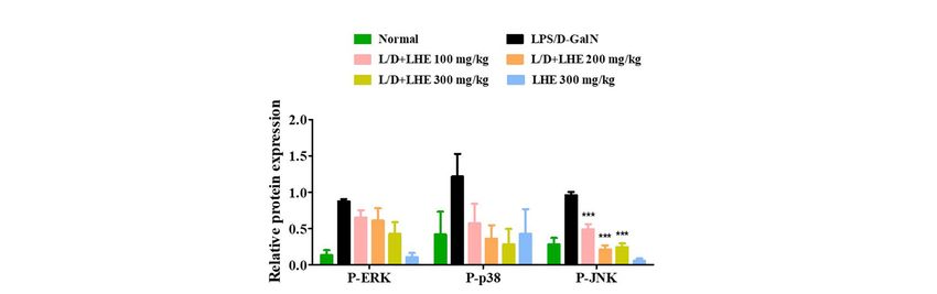
Antioxidants 2021, 10, 1387                                                                                                         10 of 18

                                    acute hepatitis. Moreover, H&E staining of liver tissues revealed that the normal and
                                    300 mg/kg LHE alone group had no pathological abnormalities (Figure 5B). However,
                                    the LPS/D-GalN group resulted in severe histopathological changes in the liver, such
 Antioxidants 2021, 10, x FOR PEER REVIEW
                                                                                                                         as
                                                                                                                     11 of 19
                                    extensive hemorrhage, necrosis, and neutrophil infiltration. The LHE group enhanced
                                    LPS/D-GaIN-induced liver injury in a dose-dependent manner (Figure 5B).

      Figure 4. Effects of LHE on (A) the expression of hepatic cytokine mRNA and (B) serum cytokine levels in LPS/D-GalN-
        Figure 4.
      induced      Effectsmouse
                hepatitis  of LHEmodel.
                                   on (A)Mice
                                          the expression  of hepatic
                                              were pretreated         cytokine
                                                               with LHE         mRNA
                                                                           or vehicle   andper
                                                                                      once   (B)day
                                                                                                 serum
                                                                                                    for 6cytokine levels
                                                                                                          days. Then,    in LPS/D-GalN-
                                                                                                                      LPS/D-GalN   was
        induced   hepatitis mouse   model. Mice  were pretreated   with  LHE   or vehicle  once  per day  for 6 days.
      injected 1 h after the last administration. After 6 h, blood samples were collected from the abdominal vena cavaThen,  LPS/D-GalN
                                                                                                                              puncture,
        was injected 1 h after the last administration. After 6 h, blood samples were collected from the abdominal vena cava
      and serum was prepared via centrifugation. (A) The mRNA levels of hepatic cytokine were analyzed via qPCR. (B) Serum
        puncture, and serum was prepared via centrifugation. (A) The mRNA levels of hepatic cytokine were analyzed via qPCR.
      cytokine levels were determined via ELISA. Data were expressed as mean ± SEM (n = 9). * p values of < 0.05, ** < 0.01, and
        (B) Serum cytokine levels were determined via ELISA. Data were expressed as mean ± SEM (n = 9). * p values of < 0.05, **
      *** < 0.001
        < 0.01,   (vs.
                and ***LPS/D-GalN)     were considered
                        < 0.001 (vs. LPS/D-GalN)  were statistically significant. significant.
                                                        considered statistically

                                   3.8.
                                     3.8.Effects
                                          EffectsofofLHE
                                                      LHEononLPS/D-GalN-Induced
                                                              LPS/D-GalN-InducedSerum
                                                                                    SerumALT,
                                                                                            ALT,AST,
                                                                                                  AST,and
                                                                                                       andALP
                                                                                                           ALPLevels
                                                                                                                 Levels
                                          Since
                                           SinceLHELHEhashasstrong anti-inflammatory
                                                              strong anti-inflammatory effects against
                                                                                          effects      LPS-induced
                                                                                                  against LPS-induced inflammation   in
                                                                                                                          inflammation
                                   macrophages,        we assessed whether    LHE could  effectively prevent  LPS/D-GalN–induced
                                     in macrophages, we assessed whether LHE could effectively prevent LPS/D-GalN–in-
                                   severe
                                     ducedhepatitis     in mice.in
                                             severe hepatitis     ALT, AST,
                                                                   mice.     andAST,
                                                                          ALT,   ALPand
                                                                                      are ALP
                                                                                           important   markers in
                                                                                                are important      evaluating
                                                                                                                markers         hepatic
                                                                                                                           in evaluating
                                   injury.  As  depicted   in Figure 5C, the serum ALT,  AST,  and ALP  levels significantly  increased
                                     hepatic injury. As depicted in Figure 5C, the serum ALT, AST, and ALP levels significantly
                                   inincreased
                                       the LPS/D-GalN       group compared
                                                 in the LPS/D-GalN     groupwith   the normal
                                                                               compared   with group.   However,
                                                                                                the normal  group.the   administration
                                                                                                                     However,    the ad-
                                    ministration of LHE can effectively inhibit the expression of ALT and AST in a dose-de-
                                    pendent manner (Figure 5C). At the ALP level, the inhibition rates were similar at 100 and
                                    200 mg/kg, and a more remarkable effect was detected at 300 mg/kg (Figure 5C).
Antioxidants 2021, 10, 1387 11 of 18
of LHE can effectively inhibit the expression of ALT and AST in a dose-dependent manner
Antioxidants 2021, 10, x FOR PEER REVIEW
(Figure 12 ofand
5C). At the ALP level, the inhibition rates were similar at 100 and 200 mg/kg, 19
a more remarkable effect was detected at 300 mg/kg (Figure 5C).
Effects of LHE on (A)
Figure 5. Effects
Figure (A) mouse
mouse liver
liver injury,
injury,(B)
(B)histopathological
histopathologicalchanges
changesofofmouse
mouseliver,
liver,and
and(C)
(C)serum
serumamino-
ami-
notransferase levels
transferase levels inin LPS/D-GalN-inducedhepatitis
LPS/D-GalN-induced hepatitismouse
mousemodel.
model. Mice
Mice were pretreated with LHE or vehicle once once per
per
day for 6 days. Then, LPS/D-GalN was injected 1 h after the last administration. After 6 h, mice were sacrificed,
day for 6 days. Then, LPS/D-GalN was injected 1 h after the last administration. After 6 h, mice were sacrificed, and liver and liver
tissue
tissue and
and blood
blood samples
samples were
were collected.
collected. Mouse
Mouse serum
serum was
was prepared
prepared via
via centrifugation.
centrifugation. (A)
(A) Images
Images ofof hepatitis
hepatitis lesions
lesions in
in
mice. (B) H&E staining of mice liver. Scale bars = 100 μm (200×) or 50 μm (400×). (C) Serum aminotransferase levels were
mice. (B) H&E staining of mice liver. Scale bars = 100 µm (200×) or 50 µm (400×). (C) Serum aminotransferase levels were
analyzed using an automated clinical chemistry analyzer. Data were expressed as means ± SEM (n = 9). *** p values of <
analyzed using an automated clinical chemistry analyzer. Data were expressed as means ± SEM (n = 9). *** p values of
0.001 (vs. LPS/D-GalN) were considered statistically significant.against LPS/D-GalN-induced liver failure (Figure 6A). In addition, LHE is effective in in-
Antioxidants 2021, 10, 1387
hibiting the expression of hepatic iNOS and COX-2 proteins in mice (Figure 6A).12 of 18
3.10. Effects of LHE on the Hepatic Activities of the NF-κB and MAPK Signaling Pathways
Our results in vitro showed that pretreatment with LHE had anti-inflammatory ef-
expression in the LPS/D-GalN-induced
fects by inhibiting the NF-κB and MAPK mouse model.
signaling As shown
pathways. in Figure
Therefore, 6A, thehepatitis
whether level
of was
Nrf-2 and HO-1 significantly decreased in the LPS/D-GalN group compared
inhibited via the regulation of NF-κB and MAPK in the LPS/D-GalN-induced mouse with the
normal group. In contrast, the administration of LHE could significantly increase
model was further assessed via Western blotting. As shown in Figure 6A,B, the activation the
expression
of NF-κBofandNrf-2 and HO-1
MAPK in a dose-dependent
was significantly induced manner, which has important
in the LPS/D-GalN protective
group compared with
effects against LPS/D-GalN-induced liver failure (Figure 6A). In addition,
the normal group. However, the phosphorylation of NF-κB p65 and three MAPK LHE is effective
as well
in as
inhibiting the expression
the degradation ofwere
of IκBα hepatic iNOS in
reduced and COX-2 proteins in
a dose-dependent mice (Figure
manner 6A). group.
in the LHE
Figure
Figure 6. Effects
6. Effects of LHE
of LHE on on
(A)(A)
the the expression
expression of iNOS/COX-2,
of iNOS/COX-2, induction
induction of HO-1/Nrf-2,
of HO-1/Nrf-2, activation
activation of of NF-κB,
NF-κB, andand
(B)(B)
phosphorylation of MAPK in LPS/D-GalN-induced hepatitis mouse model. Mice were pretreated
phosphorylation of MAPK in LPS/D-GalN-induced hepatitis mouse model. Mice were pretreated with LHE or vehicle with LHE or vehicle
once
once perper
daydayforfor 6 days.
6 days. Then,
Then, LPS/D-GalN
LPS/D-GalN waswas injected
injected 1 h1 after
h after
thethe
lastlast administration.
administration. After
After 6 h,
6 h, mice
mice were
were sacrificed,
sacrificed,
and liver tissue samples were collected. The expression of inflammatory synthetic enzyme, antioxidant molecules, and
inflammatory pathways were determined via Western blot analysis. The histograms showed the expression levels of protein
relative to those of β-actin. * p values of < 0.05, and *** < 0.001 (vs. LPS/D-GalN) were statistically significant.Antioxidants 2021, 10, 1387 13 of 18
3.10. Effects of LHE on the Hepatic Activities of the NF-κB and MAPK Signaling Pathways
Our results in vitro showed that pretreatment with LHE had anti-inflammatory effects
by inhibiting the NF-κB and MAPK signaling pathways. Therefore, whether hepatitis
was
Antioxidants 2021, 10,inhibited
x FOR PEERvia the regulation of NF-κB and MAPK in the LPS/D-GalN-induced mouse
REVIEW 14
model was further assessed via Western blotting. As shown in Figure 6A,B, the activation
of NF-κB and MAPK was significantly induced in the LPS/D-GalN group compared with
and liverthe normal
tissue group.
samples wereHowever, theexpression
collected. The phosphorylation of NF-κB
of inflammatory p65 and
synthetic threeantioxidant
enzyme, MAPK asmolecules,
well and
as the degradation of IκBα were reduced in a dose-dependent manner in the LHE group.
inflammatory pathways were determined via Western blot analysis. The histograms showed the expression levels of pro-
tein relative to those of β-actin. * p values of < 0.05, and *** < 0.001 (vs. LPS/D-GalN) were statistically significant.
3.11. Content of Major Compounds in LHE
3.11. Contentand
Five major compounds of Major
LHECompounds in LHE
were analyzed using the established HPLC system.
Thus, protocatechuicFiveacid,major compounds
catechin, and LHE
quercitrin, were analyzed
quercetin, using the were
and kaempferol established HPLC sys
succes-
Thus,
sively detected at protocatechuic
8.250, acid,
11.067, 20.453, catechin,
27.423, andquercitrin,
32.470 min,quercetin, and kaempferol
respectively (Figure 7).were suc
sively
A calibration curve of detected
the majorat compounds
8.250, 11.067, (protocatechuic
20.453, 27.423, and 32.470
acid, min, respectively
catechin, quercitrin,(Figure 7
calibration curve of the major compounds (protocatechuic
quercetin, and kaempferol) was prepared to determine the amount of each component acid, catechin, quercitrin, q
cetin, and kaempferol) was prepared to determine the amount
within LHE. The calibration curve linearity of the five major compounds was good at of each component wi
LHE. The calibration curve linearity of the five major compounds
the tested concentration range (Table 4). Each compound area mean value in LHE was was good at the te
concentration range (Table 4). Each compound area mean value
calculated using the calibration curve equation, which was prepared at the tested concen- in LHE was calcul
usingthe
tration range. Hence, thecontents
calibration
werecurve
0.03%equation, which was
protocatechuic acid,prepared at the tested
0.30% catechin, 0.04%concentra
range. Hence, the contents were 0.03% protocatechuic acid, 0.30% catechin, 0.04% q
quercitrin, 0.03% quercetin, and 0.04% kaempferol (Table 5).
citrin, 0.03% quercetin, and 0.04% kaempferol (Table 5).
Figure 7. HPLC chromatograms of standard solution and LHE at 270 nm.
Figure 7. HPLC chromatograms of standard solution and LHE at 270 nm.Antioxidants 2021, 10, 1387 14 of 18
Table 4. Calibration curves of compounds and LOD, LOQ.
Range Regression LOD LOQ
Compound r2
(µg/mL) Equation (µg/mL) (µg/mL)
1 10.0~50.0 y = 0.4829x + 0.2831 0.9997 0.0027 0.0083
2 80.0~400.0 y = 0.0430x + 0.1581 0.9991 0.0310 0.0939
3 10.0~50.0 y = 0.3330x + 0.2098 0.9998 0.0040 0.0121
4 10.0~50.0 y = 0.4722x + 0.0151 0.9996 0.0028 0.0085
5 10.0~50.0 y =0.5990x + 0.6334 0.9977 0.0022 0.0067
Protocatechuic acid (1); catechin (2); quercitrin (3); quercetin (4); kaempferol (5). LOD = 3.3 × σ/S. LOQ = 10 × σ/S.
σ is the standard deviation of the intercept from the regression equation and S is the slope of the calibration curve.
Table 5. The amount of constituents in LHE.
Compound Content (%)
Protocatechuic acid 0.03
Catechin 0.30
Quercitrin 0.04
Quercetin 0.03
Kaempferol 0.04
4. Discussion
The liver is a vital organ and is easily damaged by different factors, thereby leading to
liver failure. These factors include alcohol use and exposure to chemical substances, oxida-
tive products, and hepatitis virus [7]. Hepatitis is characterized by inflammatory conditions
in the liver, and it can be self-limiting or can lead to liver fibrosis, cirrhosis, and cancer. The
major causes of hepatitis are viruses, alcohol use, exposure to toxins, intake of certain drugs,
other infections, autoimmune diseases, and non-alcoholic steatohepatitis [35]. The role of
gut-derived LPS in the pathogenesis of hepatic injury has been widely revealed [36]. LPS
is the primary endogenous endotoxin of Gram-negative bacteria in the gut that induces a
strong inflammatory response, which can lead to liver tissue injury [37,38]. D-GaIN is a
specific hepatotoxicant that used to increase the sensitivity to the lethal effects of endotoxin
such as LPS [12]. Additionally, LPS/D-GalN exposure results in fulminant hepatitis due to
overgrowth of bacteria in the gut, disruption of intestinal barrier function, and an increase
in permeability to endotoxin and bacteria [13]. Thus, LPS/D-GalN-induced acute liver
injury is a well-established experimental model to investigate the underlying mechanisms
for fulminant hepatitis and screen potential therapeutic drugs [7]. Therefore, to investigate
the therapeutic effects of LHE on inflammatory symptoms in vivo, we determined the
hepatoprotective effects using LPS/D-GalN-induced acute hepatitis mice model.
Excessive production of inflammatory cytokines, such as TNF-α, IL-6, and IL-1β are
closely associated with LPS/D-GalN-induced liver injury. These inflammatory cytokines
have the ability to induce hepatocyte apoptosis and necrosis [39]. Thus, we examined the
inhibitory effects of LHE on inflammatory cytokine levels. The ICR mice received LHE at
different doses including 100, 200, and 300 mg/kg with LPS/D-GalN injection, or LHE at a
dose of 300 mg/kg alone. We found that injection of LPS/D-GalN dramatically induced
the expression of inflammatory cytokines. However, administration of LHE significantly
inhibited the production of inflammatory cytokines and their mRNA genes such as TNF-α,
IL-6, and IL-1β in a dose-dependent manner. Additionally, ALT and AST are known as
representative indicators of hepatocellular damage, indicating the degree of liver injury [40].
Thus, we determined the concentrations of ALT, AST, and ALP in the mouse serum of each
group. Our results indicated that LHE efficiently repressed the levels of ALT, AST, and ALP
in serum compared with the LPS/D-GalN group. In addition, LHE significantly decreased
LPS/D-GalN-triggered liver functional damage in a dose-dependent manner. Moreover,
the 300 mg/kg LHE/no injected LPS/D-GaIN group was morphologically comparable
with the normal group. Hence, LHE of a dose up to 300 mg/kg could not only prevent
hepatotoxicity but also protect the liver. These experimental data revealed that LHE exertsAntioxidants 2021, 10, 1387 15 of 18
protective effects against LPS/D-GalN-induced hepatic damage by inhibiting inflammatory
response.
The signal cascades that control pro-inflammatory cytokine expression is deeply
associated with the MAPK-mediated signaling pathway [21]. The MAPK signaling pathway
has been known to play a critical role in regulating inflammatory factors [41]. Several
studies have reported that the phosphorylation of MAPK by LPS-stimulation is involved
in the upregulation of pro-inflammatory factors via the activation of NF-κB [42,43]. NF-κB
is a transcription factor essential for the regulation of several pro-inflammatory enzymes
such as iNOS and COX-2 [44,45]. The p65 protein of NF-κB migrates into the nucleus via
LPS stimulation and facilitates the production of pro-inflammatory mediators associated
with liver inflammation [46,47]. Therefore, we investigated whether LHE was effective in
inhibiting MAPK/NF-κB activation in hepatic tissue lysate. In this study, Western blotting
revealed that LHE was significantly effective in inhibiting the phosphorylation of all types
of MAPK, including ERK, p38, and JNK, induced by LPS/D-GalN. Moreover, LHE could
inhibit the activation of NF-κB p65 via the inhibition of IκBα phosphorylation. These
results suggested that MAPK/NF-κB might be a pharmacological target of LHE against
LPS/D-GalN-induced hepatic damage.
Nrf-2 is an important transcription factor that can induce the production of antioxidant
enzymes in macrophages. Nrf-2 regulates the expression of HO-1, which is translocated
into the nucleus to exert its effect. As Nrf-2 activation is also involved in the expression of
iNOS and COX-2 [48], Nrf-2 plays an important role in the regulation of inflammation. Also,
previous studies reported that activating of Nrf-2 exerts protective effect against LPS/D-
GalN-induced liver injury [49,50]. Therefore, we investigated the effects of LHE treatment
on the expression of antioxidant-related proteins. LHE treatment was shown to markedly
increased expression of Nrf-2 and up-regulated the level of Nrf-2-depending signaling
including antioxidant protein HO-1. LHE also significantly inhibited the expression of
iNOS and COX-2 in mice liver upon LPS/D-GalN injection. These outcomes supported that
LHE improved LPS/D-GalN-induced hepatic liver injury via activating the Nrf-2/HO-1
signaling pathway.
Additionally, we determined how LHE affects the inflammatory reaction in endotoxin-
stimulated RAW 264.7 macrophages. LHE pretreatment at a non-toxic concentration
significantly suppressed the secretion of several inflammatory mediators, such as NO,
TNF-α, IL-6, and IL-1β in LPS-stimulated macrophage RAW 264.7 cells. Additionally, both
protein and mRNA levels of iNOS and COX-2 were inhibited in a concentration-dependent
manner by LHE. In addition, we showed that LHE dramatically increased the protein
and mRNA expression of HO-1. The cytosolic protein level of Nrf-2 decreased after LHE
treatment. Meanwhile, the nuclear protein level of Nrf-2 increased in a concentration-
dependent manner. Thus, LHE could promote the nuclear translocation of Nrf-2, leading
to HO-1 expression in RAW 264.7 cells. In addition, the activation of HO-1/Nrf-2 might
be mediated by the phosphorylation of MAPK [51]. Therefore, whether LHE prevents
MAPK phosphorylation and the nuclear translocation of NF-κB p65 in LPS-stimulated
macrophage RAW 264.7 cells was assessed. Our data show that pretreatment of LHE
remarkably inhibited the phosphorylation of MAPK and nuclear migration of NF-κB p65
in LPS-stimulated macrophages via repression of IκBα degradation. Taken together, LHE
exhibits strong hepatoprotective and anti-inflammatory effects through the regulation of
Nrf-2/HO-1 and MAPK/NF-κB signaling pathway.
To investigate the relationships between the physiological activities of LHE and its
components, we performed phytochemical analyses using HPLC. Moreover, five main
components, including protocatechuic acid, catechin, quercitrin, quercetin, and kaempferol,
were identified. Previous studies have shown that protocatechuic acid has protective effects
against menadione-induced liver damage by up-regulating Nrf-2 [52]. Moreover, it could
suppress airway inflammation via the inhibition of MAPK [53]. In addition, quercitrin
decreased the risk of acetaminophen-induced acute liver toxicity in mice [54]. Moreover,
quercitrin and quercetin reduced the occurrence of inflammatory response and oxidativeAntioxidants 2021, 10, 1387 16 of 18
stress in macrophages [55]. In addition, another recent study has shown that kaempferol
has mitigating effects against liver fibrosis in mice [56]. Based on the current HPLC
analysis and previous studies about these components, the anti-inflammatory properties
and hepatoprotective effects of LHE can likely reflect the presence of protocatechuic acid,
quercitrin, quercetin, and kaempferol.
5. Conclusions
LHE has hepatoprotective and anti-inflammatory effects in LPS/D-GaIN-induced
acute liver injury mouse model and in a LPS-stimulated macrophage RAW 264.7 cells.
The mechanisms underlying these effects include the inhibition of NF-κB activation via
IκBα stabilization and the reduction of MAPK phosphorylation, including ERK, P38, and
JNK. Moreover, it has an antioxidant effect by enhancing HO-1/Nrf-2 activation. This,
in turn, inhibits the production of inflammatory mediators, such as NO, iNOS, COX-2,
and pro-inflammatory cytokines in LPS/D-GaIN-induced liver injury mouse model and
the LPS-exposed macrophages. Histological evaluation showed that LHE is significantly
effective against LPS/D-GaIN-induced liver injury. Moreover, some LHE components, such
as protocatechuic acid, quercitrin, quercetin, and kaempferol, may be closely correlated
with the hepatoprotective and anti-inflammatory effects of LHE. These in vivo outcomes
indicated that LHE has anti-hepatotoxic and anti-inflammatory properties and that it has a
therapeutic potential against inflammatory-related hepatic disease.
Author Contributions: Conceptualization, Y.-C.O. and J.Y.M.; investigation, Y.H.J., T.I.K., and Y.-C.O.;
methodology, Y.H.J. and Y.-C.O.; validation, Y.H.J., T.I.K., and Y.-C.O.; writing—original draft, Y.H.J.;
writing—review and editing, Y.-C.O. and J.Y.M. All authors have read and agreed to the published
version of the manuscript.
Funding: This research was funded by Korea Institute of Oriental Medicine (KIOM), provided by
the Ministry of Science and ICT, Republic of Korea, grants number K17281 and KSN2021230.
Institutional Review Board Statement: The Animal Care and Use Committee of Korea Institute of
Oriental Medicine approved this animal study (#D-17-020). The study was conducted according to
the guidelines of the Declaration of Helsinki, and approved by the Institutional Review Board (or
Ethics Committee) of Korea Institute of Oriental Medicine (#D-17-020, approved on 3 July 2017).
Informed Consent Statement: Not applicable.
Data Availability Statement: The data are contained within the article.
Conflicts of Interest: The authors declare no conflict of interest.
References
1. China Pharmacopoeia Committee. Pharmacopoeia of the People’s Republic China 1st Division of 2010 Edition, 2010 ed.; China Medical
Science Press: Beijing, China, 2010; pp. 204–205.
2. Zhou, S.Y.; Yao, D.F.; Xu, C.F.; Huang, L.Q.; Zhang, S.P. Suppressive effect of Phyllanthus urinaria L. and Lysimachia christinae
Hance on hepatitis B surface antigen. Pract. J. Integr. Tradit. West Med. 1995, 8, 760–761.
3. Yeh, T.H.; Krauland, L.; Singh, V.; Zou, B.; Devaraj, P.; Stolz, D.B.; Franks, J.; Monga, S.P.; Sasatomi, E.; Behari, J. Liverspecific
β-catenin knockout mice have bile canalicular abnormalities, bile secretory defect, and intrahepatic cholestasis. Hepatology 2010,
52, 1410–1419. [CrossRef]
4. Zhao, J.; Davis, L.C.; Verpoorte, R. Elicitor signal transduction leading to production of plant secondary metabolites. Biotechnol.
Adv. 2005, 23, 283–333. [CrossRef] [PubMed]
5. Bhaduri, B.R.; Mieli-Vergani, G. Fulminant hepatic failure: Pediatric aspects. Semin. Liver Dis. 1996, 16, 349. [CrossRef]
6. O’Grady, J.G.; Schalm, S.W.; Williams, R. Acute liver failure: Redefining the syndromes. Lancet 1993, 342, 273.
7. Nakama, T.; Hirono, S.; Moriuchi, A.; Hasuike, S.; Nagata, K.; Hori, T.; Ido, A.; Hayashi, K.; Tsubouchi, H. Etoposide prevents
apoptosis in mouse liver with Dgalactosamine/lipopolysaccharide-induced fulminant hepatic failure resulting in reduction of
lethality. Hepatology 2001, 33, 1441–1450. [CrossRef] [PubMed]
8. Zhang, J.; Xu, L.; Zhang, L.; Ying, Z.; Su, W.; Wang, T. Curcumin attenuates D-galactosamine/lipopolysaccharide-induced liver
injury and mitochondrial dysfunction in mice. J. Nutr. 2014, 144, 1211–1218. [CrossRef]
9. Kim, S.J.; Cho, H.I.; Kim, S.J.; Park, J.H.; Kim, J.S.; Kim, Y.H.; Lee, S.K.; Kwak, J.H.; Lee, S.M. Protective effect of linarin against
D-galactosamine and lipopolysaccharide-induced fulminant hepatic failure. Eur. J. Pharmacol. 2014, 738, 66–73. [CrossRef]
[PubMed]Antioxidants 2021, 10, 1387 17 of 18
10. Peng, J.H.; Cui, T.; Sun, Z.L.; Huang, F.; Chen, L.; Xu, L.; Feng, Q.; Hu, Y.Y. Effects of Puerariae Radix Extract on Endotoxin
Receptors and TNF-alpha Expression Induced by Gut-Derived Endotoxin in Chronic Alcoholic Liver Injury. Evid. Based
Complement. Alternat. Med. 2012, 2012, 234987. [CrossRef]
11. Keppler, D.; Lesch, R.; Reutter, W.; Decker, K. Experimental hepatitis induced by D-galactosamine. Exp. Mol. Pathol. 1968, 9,
279–290. [CrossRef]
12. Xia, X.; Su, C.; Fu, J.; Zhang, P.; Jiang, X.; Xu, D.; Hu, L.; Song, E.; Song, Y. Role of alpha-lipoic acid in LPS/d-GalN induced
fulminant hepatic failure in mice: Studies on oxidative stress, inflammation and apoptosis. Int. Immunopharmacol. 2014, 22,
293–302. [CrossRef] [PubMed]
13. Rao, R.K.; Seth, A.; Sheth, P. Recent Advances in Alcoholic Liver Disease I. Role of intestinal permeability and endotoxemia in
alcoholic liver disease. Am. J. Physiol. Gastrointest. Liver Physiol. 2004, 286, G881–G884. [CrossRef] [PubMed]
14. Morita, T.; Jinno, K.; Kawagishi, H.; Arimoto, Y.; Suganuma, H.; Inakuma, T.; Sugiyama, K. Hepatoprotective effect of myristicin
from nutmeg (Myristica fragrans) on lipopolysaccharide/d-galactosamine-induced liver injury. J. Agric. Food Chem. 2003, 51,
1560–1565. [CrossRef]
15. Jiao, M.; Ren, F.; Zhou, L.; Zhang, X.; Zhang, L.; Wen, T.; Wei, L.; Wang, X.; Shi, H.; Bai, L.; et al. Peroxisome proliferator-activated
receptor alpha activation attenuates the inflammatory response to protect the liver from acute failure by promoting the autophagy
pathway. Cell Death Dis. 2014, 5, e1397. [CrossRef]
16. Li, J.; Ge, R.; Zhao, C.; Tang, L.; Li, J.; Li, Q. Farrerol regulates occludin expression in hydrogenperoxide-induced EA.hy926 cells
by modulating ERK1/2 activity. Eur. J. Pharmacol. 2014, 734, 9–14. [CrossRef] [PubMed]
17. Yoshinari, O.; Shiojima, Y.; Igarashi, K. Hepatoprotective effect of germaniumcontaining spirulina in rats with D-galactosamine-
and lipopolysaccharide-induced hepatitis. Brit. J. Nutr. 2014, 111, 135–140. [CrossRef] [PubMed]
18. Chatterjee, N.; Das, S.; Bose, D.; Banerjee, S.; Jha, T.; Saha, K.D. Leishmanial lipid affords protection against oxidative stress
induced hepatic injury by regulating inflammatory mediators and confining apoptosis progress. Toxicol. Lett. 2015, 232, 499–512.
[CrossRef]
19. Yang, Y.Q.; Yan, X.T.; Wang, K.; Tian, R.M.; Lu, Z.Y.; Wu, L.L.; Xu, H.T.; Wu, Y.S.; Liu, X.S.; Mao, W.; et al. Triptriolide alleviates
lipopolysaccharide-induced liver injury by Nrf2 and NF-kappaB signaling pathways. Front. Pharmacol. 2018, 9, 999. [CrossRef]
20. Yang, F.; Li, X.; Wang, L.K.; Wang, L.W.; Han, X.Q.; Zhang, H.; Gong, Z.J. Inhibitions of NF-κB and TNF-α result in differential
effects in rats with acute on chronic liver failure induced by d-Gal and LPS. Inflammation 2014, 37, 848–857. [CrossRef]
21. Guha, M.; Mackman, N. LPS induction of gene expression in human monocytes. Cell Signal. 2001, 13, 85–94. [CrossRef]
22. Lee, W.C.; Jung, H.A.; Choi, J.S.; Kim, Y.S.; Lee, S.M. Protective effects of luteolin against apoptotic liver damage induced by
D -galactosamine/lipopolysaccharide in mice. J. Nat. Prod. 2011, 74, 1916–1921. [CrossRef]
23. Yang, P.; Zhou, W.; Li, C.; Zhang, M.; Jiang, Y.; Jiang, R.; Ba, H.; Li, C.; Wang, J.; Yin, B.; et al. Kupffer-cell-expressed transmembrane
TNF-α is a major contributor to lipopolysaccharide and D-galactosamine-induced liver injury. Cell Tissue Res. 2016, 363, 371–383.
[CrossRef]
24. Shi, A.; Shi, H.; Wang, Y.; Liu, X.; Cheng, Y.; Li, H.; Zhao, H.; Wang, S.; Dong, L. Activation of Nrf2 pathway and inhibition of
NLRP3 inflammasome activation contribute to the protective effect of chlorogenic acid on acute liver injury. Int. Immunopharm.
2018, 54, 125–130. [CrossRef]
25. Anuja, G.; Shine, V.; Latha, P.; Suja, S. Protective effect of ethyl acetate fraction of Drynaria quercifolia against CCl4 induced rat
liver fibrosis via Nrf2/ARE and NFκB signalling pathway. J. Ethnopharmacol. 2018, 216, 79–88. [CrossRef] [PubMed]
26. Yan, H.; Huang, Z.; Bai, Q.; Sheng, Y.; Hao, Z.; Wang, Z.; Ji, L. Natural product andrographolide alleviated APAP-induced liver
fibrosis by activating Nrf2 antioxidant pathway. Toxicology 2018, 396, 1–12. [CrossRef] [PubMed]
27. Ehren, J.L.; Maher, P. Concurrent regulation of the transcription factors Nrf2 and ATF4 mediates the enhancement of glutathione
levels by the flavonoid fisetin. Biochem. Pharmacol. 2013, 85, 1816–1826.
28. Turkseven, S.; Kruger, A.; Mingone, C.J.; Kaminski, P.; Inaba, M.; Rodella, L.F.; Ikehara, S.; Wolin, M.S.; Abraham, N.G. Antioxidant
mechanism of heme oxygenase-1 involves an increase in superoxide dismutase and catalase in experimental diabetes. Am. J.
Physiol. Heart Circ. Physiol. 2005, 289, H701–H707. [CrossRef] [PubMed]
29. Nabila, V.K.; Putra, I.B. The effect of Aloe vera ethanol extract on the growth inhibition of Candida albicans. Med. Glas. 2020, 17,
485–489.
30. Do, H.J.; Oh, T.W.; Park, K.I. Ethanol Extract of Sesamum indicum Linn. Inhibits FcepsilonRI-Mediated Allergic Reaction via
Regulation of Lyn/Syk and Fyn Signaling Pathways in Rat Basophilic Leukemic RBL-2H3 Mast Cells. Mediat. Inflamm. 2019,
2019, 5914396. [CrossRef]
31. Jeong, Y.H.; Oh, Y.C.; Cho, W.K.; Yim, N.H.; Ma, J.Y. Hoveniae Semen Seu Fructus Ethanol Extract Exhibits Anti-Inflammatory
Activity via MAPK, AP-1, and STAT Signaling Pathways in LPS-Stimulated RAW 264.7 and Mouse Peritoneal Macrophages.
Mediat. Inflamm. 2019, 2019, 9184769. [CrossRef]
32. Deng, J.; Ren, M.; Dai, X.; Qu, D.; Yang, M.; Zhang, T.; Jiang, B. Lysimachia christinae Hance regresses preestablished cholesterol
gallstone in mice. J. Ethnopharmacol. 2015, 166, 102–108. [CrossRef]
33. Lee, C.W.; Yen, F.L.; Huang, H.W.; Wu, T.H.; Ko, H.H.; Tzeng, W.S.; Lin, C.C. Resveratrol nanoparticle system improves dissolution
properties and enhances the hepatoprotective effect of resveratrol through antioxidant and anti-inflammatory pathways. J. Agric.
Food Chem. 2012, 60, 4662–4671. [CrossRef]You can also read