MiRNA-584-3p inhibits gastric cancer progression by repressing Yin Yang 1- facilitated MMP-14 expression - Nature
←
→
Page content transcription
If your browser does not render page correctly, please read the page content below
www.nature.com/scientificreports

                         OPEN         miRNA-584-3p inhibits gastric
                                      cancer progression by repressing
                                      Yin Yang 1- facilitated MMP-14
Received: 27 March 2017
Accepted: 21 July 2017                expression
Published: xx xx xxxx
                                      Liduan Zheng1,2, Yajun Chen1, Lin Ye3, Wanju Jiao1, Huajie Song3, Hong Mei3, Dan Li3,
                                      Feng Yang3, Huanhuan Li3, Kai Huang2 & Qiangsong Tong2,3
                                      Recent evidence shows the emerging roles of promoter-targeting endogenous microRNAs
                                      (miRNAs) in regulating gene transcription. However, miRNAs affecting the transcription of matrix
                                      metalloproteinase 14 (MMP-14) in gastric cancer remain unknown. Herein, through integrative mining
                                      of public datasets, we identified the adjacent targeting sites of Yin Yang 1 (YY1) and miRNA-584-3p
                                      (miR-584-3p) within MMP-14 promoter. We demonstrated that YY1 directly targeted the MMP-14
                                      promoter to facilitate its expression in gastric cancer cells. In contrast, miR-584-3p recognized its
                                      complementary site within MMP-14 promoter to suppress its expression. Mechanistically, miR-
                                      584-3p interacted with Argonaute 2 to recruit enhancer of zeste homolog 2 and euchromatic histone
                                      lysine methyltransferase 2, resulting in enrichment of repressive epigenetic markers and decreased
                                      binding of YY1 to MMP-14 promoter. miR-584-3p inhibited the in vitro and in vivo tumorigenesis
                                      and aggressiveness of gastric cancer cells through repressing YY1-facilitated MMP-14 expression.
                                      In clinical gastric cancer tissues, the expression of YY1 and miR-584-3p was positively or negatively
                                      correlated with MMP-14 levels. In addition, miR-584-3p and YY1 were independent prognostic factors
                                      associated with favorable and unfavorable outcome of gastric cancer patients, respectively. These data
                                      demonstrate that miR-584-3p directly targets the MMP-14 promoter to repress YY1-facilitated MMP-14
                                      expression and inhibits the progression of gastric cancer.

                                      Gastric cancer is the third leading cause of cancer-related death in the world1. Despite achievement in surgery
                                      and chemotherapy, the clinical outcome of patients with advanced gastric cancer still remains poor, mainly due
                                      to tumor growth and progression1. Therefore, it is an urgent duty to elucidate the mechanisms underlying the
                                      tumorigenesis and aggressiveness of gastric cancer2. Matrix metalloproteinase 14 (MMP-14), also known as
                                      membrane type-1 MMP, plays a pivotal role in digesting the extracellular matrice (ECM) and activating MMP-2,
                                      thereby promoting tumor invasion and metastasis3. MMP-14 also facilitates tumor angiogenesis via increasing
                                      the expression of vascular endothelial growth factor (VEGF)4 and releasing bioactive ECM products3. MMP-14
                                      expression is elevated in most human cancers, and is associated with tumor invasion and metastasis5. It has been
                                      indicated that MMP-14 is highly expressed in gastric cancer and associated with poor survival of patients6, sug-
                                      gesting the importance of MMP-14 in the tumorigenesis and aggressiveness of gastric cancer.
                                          Human MMP-14 gene, consisting of 10 exons, locates at chromosome 14q11-12 and is regulated at the tran-
                                      scription level7. Previous studies have demonstrated that transcription factor early growth response 1 appears to
                                      be an essential regulator of MMP-14 transcription in endothelial cells8, while specificity protein 1 is the critical
                                      factor for MMP-14 promoter activity in fibrosarcoma cells7. The anti-tumorigenic effects of methylseleninic acid
                                      are mediated by the suppression of nuclear factor kappa B (NF-κB)-regulated MMP-14 levels in cancer cell lines9.

                                      1
                                       Department of Pathology, Union Hospital, Tongji Medical College, Huazhong University of Science and Technology,
                                      1277 Jiefang Avenue, Wuhan 430022, Hubei Province, P. R. China. 2Clinical Center of Human Genomic Research,
                                      Union Hospital, Tongji Medical College, Huazhong University of Science and Technology, 1277 Jiefang Avenue,
                                      Wuhan 430022, Hubei Province, P. R. China. 3Department of Surgery, Union Hospital, Tongji Medical College,
                                      Huazhong University of Science and Technology, 1277 Jiefang Avenue, Wuhan 430022, Hubei Province, P. R. China.
                                      Liduan Zheng, Yajun Chen and Lin Ye contributed equally to this work. Correspondence and requests for materials
                                      should be addressed to Q.T. (email: qs_tong@hotmail.com)

        SCienTifiC Reports | 7: 8967 | DOI:10.1038/s41598-017-09271-5                                                                                   1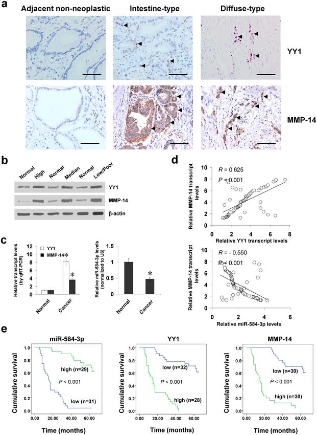
www.nature.com/scientificreports/

                              In addition, MMP-14 is directly regulated by homeobox D10 in glioma10, proto-oncogene FBI-1 in ovarian cancer
                              cells11, and hypoxia inducible factor 2 alpha in renal cancer12. However, the transcriptional regulators and under-
                              lying mechanisms essential for MMP-14 expression in gastric cancer still remain unclear.
                                  In this study, through an integrative approach to mine public datasets, we identified Yin Yang 1 (YY1) and
                              miRNA-584-3p (miR-584-3p) as crucial transcriptional regulators of MMP-14 expression in gastric cancer, with
                              their adjacent targeting sites within the MMP-14 promoter. We demonstrate, for the first time, that YY1 facili-
                              tates the expression of MMP-14 via directly binding to its promoter in gastric cancer. In contrast, miR-584-3p
                              is down-regulated and negatively correlated with MMP-14 levels in clinical gastric cancer tissues, and directly
                              targets the MMP-14 promoter to inhibit its expression. Mechanistically, miR-584-3p interacts with Argonaute
                              2 (AGO2) to recruit enhancer of zeste homolog 2 (EZH2) and euchromatic histone lysine methyltransferase 2
                              (EHMT2), which results in enrichment of repressive epigenetic markers and decreased binding of YY1 to MMP-
                              14 promoter, thus inhibiting the tumorigenesis and aggressiveness of gastric cancer.

                              Results
                              YY1 facilitates the expression of MMP-14 in gastric cancer cells. To explore the mechanisms crucial
                              for MMP-14 expression in gastric cancer, we analyzed the potential binding sites of transcription factors within
                              its promoter using computational algorithm programs. Over-lapping analysis of Genomatrix13, TFBIND14, and
                              PROMO15 revealed the potential binding sites of YY1, nuclear factor Y (NFY), and nuclear factor erythroid 2
                              like 2 (NFE2L2) within MMP-14 promoter region (chr14:23304582-23305827; Supplementary Fig. S1a), locat-
                              ing at bases −225/−219, −158/−144, and −179/−159 upstream the transcription start site (TSS) of MMP-14,
                              respectively. Further analysis of chromatin immunoprecipitation (ChIP) sequencing (ChIP-seq) data derived
                              from UCSC Genome Browser16 revealed YY1 as the only enriched transcription factor within MMP-14 promoter
                              (Supplementary Fig. S1a). Meanwhile, the targeting site of miR-584-3p with high complementarity was noted at
                              −275/−258 bp region surrounding that of YY1 (Fig. 1a). The expression levels of YY1 and MMP-14 were higher
                              in gastric cancer cell lines, when compared to those in normal gastric epithelial cells (Fig. 1b). Notably, mining of
                              publicly available Gene Expression Omnibus (GEO) datasets indicated that YY1 expression was positively corre-
                              lated with MMP-14 levels in different gastric cancer cohorts (Supplementary Fig. S1b)17–19.
                                  To address the hypothesis that YY1 might influence the MMP-14 expression, we performed the YY1
                              over-expression and knockdown experiments in gastric cancer cells. Stable transfection of YY1 or two clones
                              of short hairpin RNAs (shRNAs) targeting YY1 (sh-YY1 #1 and sh-YY1 #2) into SGC-7901, AGS, and MKN-
                              45 cells obviously up-regulated or decreased the protein and transcript levels of YY1 and MMP-14, than those
                              stably transfected with empty vector (mock) or scramble short hairpin RNA (sh-Scb), respectively (Fig. 1c,d,e,f).
                              Nuclear run-on assay demonstrated that stable over-expression or knockdown of YY1 increased and decreased
                              the nascent transcript levels of MMP-14 in gastric cancer cells, respectively (Fig. 1g,h). In addition, the
                              YY1-facilitated MMP-14 transcription was not abolished by treatment with C646, the inhibitor of histone acet-
                              yltransferase (Supplementary Fig. S1c). These results suggested that YY1 facilitated the expression of MMP-14 in
                              gastric cancer cells.

                              YY1 directly binds to MMP-14 promoter to increase its transcription. To investigate whether YY1
                              could target the MMP-14 promoter to increase its transcription, the luciferase reporter and its mutation vec-
                              tors of MMP-14 promoter (Fig. 2a) were transfected into gastric cancer cells SGC-7901, AGS, and MKN-45.
                              Dual-luciferase assay showed that ectopic expression or knockdown of YY1 enhanced and attenuated the MMP-
                              14 promoter activity, respectively (Fig. 2b,c), and mutation of YY1 binding site abolished these effects (Fig. 2b,c).
                              In addition, ChIP and real-time quantitative PCR (qPCR) indicated the enrichment of YY1 around its binding site
                              (−326/−130 bp relative to TSS) in MKN-45 and SGC-7901 cells (Fig. 2d). As controls, no obvious MMP-14 pro-
                              moter regions were immunoprecipitated with unspecific antibody (isotype IgG) or detected by qPCR with primer
                              set (−122/+69 bp) distal to the binding site of miR-584-3p (Fig. 2d). Stable over-expression of YY1 resulted in
                              enrichment of YY1 on the MMP-14 promoter in gastric cancer cells (Fig. 2d). Meanwhile, stable knockdown
                              of YY1 with shRNA constructs decreased the binding of YY1 to MMP-14 promoter in MKN-45 and AGS cells
                              (Fig. 2d). Moreover, knockdown of p65, a subunit of NF-κB crucial for YY1 expression20, reduced the YY1 lev-
                              els and prevented the gastric cancer cells from enhanced expression of YY1 and MMP-14, increased activity of
                              MMP-14 promoter, and increased binding of YY1 to MMP-14 promoter induced by ectopic expression of YY1
                              (Fig. 2e,f,g). These results indicated that YY1 directly bond to MMP-14 promoter to enhance its transcription.

                              miR-584-3p interacts with AGO2 to repress the YY1-facilitated MMP-14 transcription. Since
                              previous studies have indicated the roles of promoter-targeting miRNAs in regulating gene transcription21, 22,
                              we further observed the effects of miR-584-3p on the expression of MMP-14 in gastric cancer cells. Lower
                              miR-584-3p expression was observed in gastric cancer cell lines, when compared to that in normal gastric
                              epithelial cells (Fig. 3a). The miR-584-3p precursor was stably transfected into MKN-45 and AGS cells, result-
                              ing in increased miR-584-3p levels (Fig. 3b). Meanwhile, transfection of anti-miR-584-3p inhibitor obviously
                              decreased the miR-584-3p levels in SGC-7901 and AGS cells (Fig. 3c). In addition, over-expression or knock-
                              down of miR-584-3p decreased and increased the protein and transcript levels of MMP-14 in gastric cancer
                              cells, when compared to those transfected with empty vector (mock) or negative control (anti-NC) inhibi-
                              tor, respectively (Fig. 3d,e, Supplementary Fig. S2a, and Fig. S2b). Notably, ectopic expression or knockdown
                              of miR-584-3p reduced and enhanced the activity of MMP-14 promoter, respectively, which was abolished by
                              mutation of miR-584-3p or YY1 binding site (Fig. 3f,g). The expression of VEGF165, the most abundant VEGF
                              isoform4 and MMP-14 downstream target gene in gastric cancer23, was significantly decreased or enhanced in
                              miR-584-3p over-expressing and knockdown gastric cancer cells, respectively (Fig. 3d,e, Supplementary Fig. S2a
                              and b). The analysis of microPIR database24 revealed no miR-584-3p binding site within VEGF promoter, ruling

SCienTifiC Reports | 7: 8967 | DOI:10.1038/s41598-017-09271-5                                                                                    2
www.nature.com/scientificreports/

                              Figure 1. YY1 facilitates the expression of MMP-14 in gastric cancer cells. (a) Scheme of potential binding sites
                              of YY1 and miR-584-3p within the MMP-14 promoter, locating at bases −225/−219 and −167/−150 relative
                              to TSS. (b) Western blot showing the expression of YY1 and MMP-14 in normal gastric epithelial GES-1 cells
                              and gastric cancer cell lines SGC-7901, AGS, MKN-45, and MKN-28. (c and d) Western blot indicating the
                              expression of YY1 and MMP-14 in SGC-7901, AGS and MKN-45 cells stably transfected with empty vector
                              (mock), YY1, scramble shRNA (sh-Scb) or two YY1 shRNAs (sh-YY1 #1 and sh-YY1 #2). (e and f) Real-time
                              quantitative RT-PCR showing the transcript levels of YY1 and MMP-14 in gastric cancer cells stably transfected
                              with mock, YY1, sh-Scb or sh-YY1 (mean ± SD, n = 5). (g and h) Nuclear run-on assay indicating the
                              nascent MMP-14 transcript levels in gastric cancer cells stably transfected with mock, YY1, sh-Scb, or sh-YY1
                              (mean ± SD, n = 4). *P < 0.01 vs. GES-1, mock, or sh-Scb.

SCienTifiC Reports | 7: 8967 | DOI:10.1038/s41598-017-09271-5                                                                                 3
www.nature.com/scientificreports/
Figure 2. YY1 directly binds to MMP-14 promoter to increase its transcription. (a) Scheme and sequence of the
intact YY1 binding site (WT) and its mutation (Mut) within the MMP-14 promoter-luciferase reporter vectors.
(b) Dual-luciferase assay showing the activity (normalized to Mock + WT) of MMP-14 promoter reporter
[pGL3-MMP14 (-1246/ + 199)] and its mutant in SGC-7901 and AGS cells stably transfected with empty
vector (mock) or YY1 (mean ± SD, n = 5). (c) Dual-luciferase assay indicating the activity (normalized to sh-
Scb + WT) of MMP-14 promoter reporter [pGL3-MMP14 (−1246/+199)] and its mutant in MKN-45 and AGS
cells stably transfected with scramble shRNA (sh-Scb) or YY1 shRNA (sh-YY1) (mean ± SD, n = 4). (d) ChIP
(using YY1 antibody) and qPCR assay showing the enrichment of YY1 (normalized to 20% input DNA) on
the MMP-14 promoter in gastric cancer cells, and those stably transfected with mock, YY1, sh-Scb, or sh-YY1
(mean ± SD, n = 5). (e) Western blot indicating the expression of p65, YY1, and MMP-14 in SGC-7901 cells
transfected with mock or YY1, and those co-transfected with sh-Scb or sh-p65. (f and g) Dual-luciferase, ChIP
SCienTifiC Reports | 7: 8967 | DOI:10.1038/s41598-017-09271-5 4www.nature.com/scientificreports/
(using YY1 antibody) and qPCR assays showing the activity of MMP-14 promoter reporter [pGL3-MMP14
(-1246/ + 199)] and the binding of YY1 to MMP-14 promoter in gastric cancer cells transfected with mock or
YY1, and those co-transfected with sh-Scb or sh-p65 (mean ± SD, n = 5). *P < 0.01 vs. mock, sh-Scb, IgG, or
mock + sh-Scb. **P < 0.01 vs. WT.
out the possible roles of miR-584-3p in directly suppressing the VEGF transcription. Since AGO2 is involved
in miRNA-induced transcriptional repression21, 22, shRNAs specific for AGO2 were transfected into MKN-45
and AGS cells. Knockdown of AGO2 attenuated the transcriptional repression of MMP-14 induced by ectopic
expression of miR-584-3p in gastric cancer cells (Fig. 3h,i, Supplementary Fig. S2c). ChIP and real-time qPCR
assay indicated that in cultured SGC-7901 and AGS cells, the enrichment of AGO2 on MMP-14 promoter was
observed at the region (−326/−130 bp) around the targeting site of miR-584-3p (Fig. 3j). In addition, treatment
of gastric cancer cells with RNase H, but not with RNase A, abolished the enrichment of AGO2 on the MMP-14
promoter in (Fig. 3j). RNA immunoprecipitation (RIP) and co-immunoprecipitation (co-IP) assays indicated
that ectopic expression of miR-584-3p facilitated its interaction with AGO2, and increased the association of
AGO2 with histone methyltransferases EZH2 and EHMT2 (Supplementary Fig. S2d and Fig. 3k). However, YY1
was not associated with this multi-protein complex (Fig. 3k). Over-expression of miR-584-3p increased the levels
of histone H3 lysine 27 trimethylation (H3K27me3) and histone H3 lysine 9 dimethylation (H3K9me2), which
were abolished by knockdown of AGO2 or treatment with established specific inhibitors of EZH2 and EHMT2,
GSK34325 and A-36626, in gastric cancer cells (Supplementary Fig. S2e). Stable over-expression of miR-584-3p
resulted in increased binding of AGO2 and epigenetic markers EZH2, EHMT2, H3K27me3 and H3K9me2, and
decreased binding of YY1 to MMP-14 promoter in gastric cancer cells (Fig. 3l), which was attenuated by knock-
down of AGO2 (Fig. 3l). Meanwhile, ectopic expression of YY1 did not affect the enrichment of AGO2, EZH2,
EHMT2, H3K27me3, or H3K9me2 on MMP-14 promoter (Fig. 3l). Moreover, treatment with GSK343 and A-366
prevented the gastric cancer cells from increased enrichment of H3K27me3 and H3K9me2 and decreased bind-
ing of YY1 to MMP-14 promoter induced by miR-584-3p over-expression (Supplementary Fig. S2f). Collectively,
these data suggested that miR-584-3p interacted with AGO2 to repress the YY1-facilitated MMP-14 transcription
in gastric cancer cells.
miR-584-3p suppresses the tumorigenesis and aggressiveness of gastric cancer cells via
repressing YY1-facilitated MMP-14 expression in vitro. Since above evidence indicated that miR-
584-3p repressed the binding of YY1 to MMP-14 promoter, we further investigated the effects of miR-584-3p
over-expression on YY1-facilitated MMP-14 levels in cultured gastric cancer cells. Ectopic expression of miR-
584-3p did not affect the YY1 expression levels (Fig. 4a), but abolished the enhanced protein and transcript levels
of MMP-14 and increased levels of VEGF165 and active MMP-2 induced by stable transfection of YY1 (Fig. 4a,b,
Supplementary Fig. S3a). In addition, over-expression of miR-584-3p prevented the enhanced YY1 enrichment
on MMP-14 promoter induced by ectopic expression of YY1 (Fig. 4c). Notably, ectopic expression of miR-584-3p
abolished the increase in expression and promoter activity of MMP-14 and enrichment of YY1 induced by trans-
fection of YY1 in prostate cancer PC-3 cells and colon cancer LoVo cells (Supplementary Fig. S3b, Fig. S3c, and
Fig. S3d), suggesting the potential roles of miR-558-3p and YY1 in regulating MMP-14 expression in these types
of cancers. In soft agar assay, YY1 over-expression promoted the anchorage-independent growth of MKN-45
and AGS cells (Fig. 4d). In matrigel invasion assay, stable over-expression of YY1 increased the invasion capac-
ity of gastric cancer cells (Fig. 4e). The tube formation of endothelial cells was increased by treatment with the
medium preconditioned by gastric cancer cells stably transfected with YY1 (Fig. 4f). Moreover, transfection of
miR-584-3p rescued the MKN-45 and AGS cells from increased growth, invasion, and angiogenesis capability
induced by stable over-expression of YY1 (Fig. 4d,e,f). On the other hand, stable knockdown of YY1 resulted
in down-regulation of MMP-14 (Supplementary Fig. S4a) and decreased capability in growth (Supplementary
Fig. S4b,c), invasion (Supplementary Fig. S4d), and angiogenesis (Supplementary Fig. S4e) in SGC-7901 and
AGS cells. Down-regulation of miR-584-3p rescued the SGC-7901 and AGS cells from their changes in these
biological features induced by knockdown of YY1 (Supplementary Fig. S4,b,c,d,e). Meanwhile, ectopic expression
or knockdown of MMP-14 rescued the gastric cancer cells from alteration in the viability, invasion, and angiogen-
esis induced by over-expression of miR-584-3p or YY1, respectively (Supplementary Fig. S5). These results sug-
gested that miR-584-3p decreased the tumorigenesis and aggressiveness of gastric cancer cells through repressing
YY1-facilitated MMP-14 expression in vitro.
miR-584-3p attenuates the YY1-facilitated growth, metastasis, and angiogenesis of gastric
cancer cells in vivo. We further explored the effects of miR-584-3p on YY1-facilitated tumor growth and
metastasis in vivo. Stable over-expression of YY1 led to increased in vivo growth of SGC-7901 cells in athymic
nude mice and enhanced tumor weight of established subcutaneous xenograft tumors (Fig. 5a,b). The expression
of YY1, MMP-14, VEGF165, active MMP-2, but not of Snail27, was enhanced in xenograft tumors formed by SGC-
7901 cells stably transfected with YY1 (Fig. 5c). In addition, stable transfection of YY1 increased the Ki-67/CD31
ratio of microvessels within tumors and decreased the intratumoral necrosis area (Fig. 5d). Moreover, stable
transfection of YY1 into SGC-7901 cells established statistically more lung metastatic colonies and lower survival
probability than empty vector (mock) group (Fig. 5e). Meanwhile, stable over-expression of miR-584-3p precur-
sor led to increase in the miR-584-3p levels, and rescued the YY1-facilitated growth, gene expression, metastasis
and angiogenesis of SGC-7901 cells in athymic nude mice (Fig. 5a,b,c,d,e). These data indicated that miR-584-3p
could attenuate the YY1-facilitated growth, metastasis, and angiogenesis of gastric cancer cells in vivo.
SCienTifiC Reports | 7: 8967 | DOI:10.1038/s41598-017-09271-5 5www.nature.com/scientificreports/
Figure 3. miR-584-3p interacts with AGO2 to repress the YY1-facilitated MMP-14 transcription. (a) Real-
time quantitative RT-PCR assay showing the expression of miR-584-3p in normal gastric epithelial GES-1 cells
and gastric cancer cell lines SGC-7901, AGS, MKN-45, and MKN-28 (mean ± SD, n = 4). (b and c) Real-time
quantitative RT-PCR assays indicating the expression of miR-584-3p in gastric cancer cells transfected with
empty vector (mock), miR-584-3p precursor, negative control inhibitor (anti-NC, 100 nmol/L), or anti-miR-
584-3p inhibitor (100 nmol/L) (mean ± SD, n = 4). (d and e) Western blot assay showing the expression of
MMP-14 and VEGF165 in gastric cancer cells transfected with mock, miR-584-3p precursor, anti-NC (100
nmol/L), or anti-miR-584-3p inhibitor (100 nmol/L). (f and g) Dual-luciferase assay indicating the MMP-
14 promoter activity (normalized to pGL3-Basic) in gastric cancer cells transfected with mock, miR-584-3p
precursor, anti-NC (100 nmol/L), or anti-miR-584-3p inhibitor (100 nmol/L; mean ± SD, n = 4). (h and i)
Western blot and dual-luciferase assays showing the expression of AGO2 and MMP-14 and activity of MMP-
14 promoter reporter [pGL3-MMP14 (−1246/ + 199)] in gastric cancer cells stably transfected with mock or
SCienTifiC Reports | 7: 8967 | DOI:10.1038/s41598-017-09271-5 6www.nature.com/scientificreports/
miR-584-3p precursor, and those co-transfected with scramble shRNA (sh-Scb) or AGO2 shRNA (sh-AGO2).
(j) ChIP (using AGO2 antibody) and qPCR assay indicating the binding of AGO2 (normalized to 20% input
DNA) to MMP-14 promoter in gastric cancer cells treated with RNase H or RNase A (mean ± SD, n = 4). (k)
co-IP and western blot assays showing the interaction of AGO2 with EZH2, EHMT2, and YY1 in MKN-45 cells
stably transfected with mock or miR-584-3p precursor, running under the same experimental conditions (full-
length blots are presented in Supplementary Figure S7). (l) ChIP and qPCR assay indicating the enrichment
(normalized to mock + sh-Scb or mock + mock) of AGO2, EZH2, EHMT2, H3K27me3, H3K9me2, and YY1
on MMP-14 promoter in MKN-45 and AGS cells stably transfected with mock or miR-584-3p precursor, and
those co-transfected with sh-Scb, sh-AGO2, mock, or YY1 (mean ± SD, n = 5). *P < 0.01 vs. GES-1, mock,
anti-NC, pGL3-Basic, mock + sh-Scb, mock + mock, or IgG. **P < 0.01 vs. mock or anti-NC.
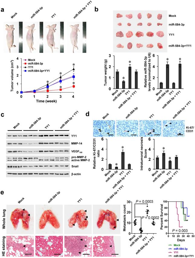
YY1 and miR-584-3p are positively or inversely correlated with MMP-14 levels in gastric can-
cer tissues. To reveal the expression of YY1 in gastric cancer tissues, immunohistochemical staining was
performed on paraffin-embedded sections from 60 well-established primary cases. The results indicated nuclear
YY1 expression in cancer cells (Fig. 6a), which was detected in 36/60 (60.0%) cases, with weak staining in 10,
moderate in 14, and intense in 12 (Supplementary Table S1). Higher YY1 expression was observed in gastric
cancer cases with deeper gastric wall invasion (P < 0.001), lymph node metastasis (P < 0.001), distant metastasis
(P = 0.005), or advanced tumor-node-metastasis (TNM) stage (P < 0.001) (Supplementary Table S1). Notably,
there was positive correlation between YY1 and MMP-14 immunoreactivity in gastric cancer cases (correla-
tion coefficient R = 0.802, P < 0.001, Fig. 6a and Supplementary Table S2). In 80 fresh gastric cancer specimens,
higher protein and transcript levels of YY1 or MMP-14 were observed than those in normal gastric mucosa
(Fig. 6b,c). In contrast, miR-584-3p was under-expressed in gastric cancer tissues than in normal gastric mucosa
(Fig. 6c and Supplementary Fig. S6a)28. The levels of SH3 domain and tetratricopeptide repeats 2 (SH3TC2), the
host gene of miR-584-3p29, were decreased in gastric cancer tissues derived from public datasets (Supplementary
Fig. S6b)19, 30. A positive correlation between YY1 and MMP-14 transcript levels was noted in gastric cancer tis-
sues (correlation coefficient R = 0.625, P < 0.001, Fig. 6d). Meanwhile, the miR-584-3p expression was inversely
correlated with MMP-14 transcript levels in gastric cancer tissues (R = −0.550, P < 0.001, Fig. 6d). The inverse
correlation between SH3TC2 and MMP-14 was also noted in gastric cancer specimens derived from public
datasets (Supplementary Fig. S6c)18, 31, 32. Kaplan–Meier survival analysis revealed that patients with low miR-
584-3p levels (P < 0.001), high YY1 levels (P < 0.001), or high MMP-14 expression (P < 0.001) had lower sur-
vival probability (Fig. 6e). Cox regression analysis of these gastric cancer cases indicated that distant metastasis
(hazard ratio HR = 2.126, P = 0.003), TNM stage (HR = 2.352, P = 0.001), miR-584-3p expression (HR = 0.312,
P = 0.018), and YY1 levels (HR = 2.743, P = 0.011) were independent prognostic factors for gastric cancer patients
(Supplementary Table S3). These results indicated the up-regulation of YY1 and under-expression of miR-584-3p
in gastric cancer tissues, which were positively and inversely correlated with the MMP-14 levels, respectively.
Discussion
YY1, one member of the GLI-Krüppel protein family, plays a fundamental role in embryogenesis, differentiation,
chromosomal dynamics, X-chromosome inactivation, and DNA repair33. Recent evidence shows the oncogenic
or tumor repressive roles of YY1 in tumorigenesis and aggressiveness34, 35. Ectopic expression of YY1 drives the
proliferation of cancer cells via increasing C-myc expression and decreasing P53 activity34. In addition, YY1 pro-
motes the cell invasion, angiogenesis, and metastasis of osteosarcoma36, and its high expression in osteosarcoma
tissues is associated with occurrence of metastasis and poor clinical outcome37. Depletion or knockdown of YY1
significantly decreases the tumorigenesis and aggressiveness of bone cancer cells36. Enhanced expression of YY1
has been documented in many types of human malignancies38, and is associated with poor prognosis36. On the
other hand, YY1 might exert tumor suppressive functions in certain types of cancers. High expression of YY1
predicts favorable outcome with longer survival in follicular lymphoma35. Previous studies have shown that YY1
is elevated in gastric cancer specimens, and knockdown of YY1 inhibits the proliferation of gastric cancer cells in
vitro39. However, the roles and underlying mechanisms of YY1 in the tumorigenesis and aggressiveness of gastric
cancer still remain largely unknown. In this study, our data showed that YY1 facilitated the in vitro and in vivo
growth, metastasis, and angiogenesis of gastric cancer cells, and patients with high expression of YY1 had lower
survival probability, indicating the oncogenic functions of YY1 in gastric cancer progression.
As a transcription factor, YY1 regulates gene transcription as an activator, initiator, or repressor in a cell type-
and sequence context-dependent manner38. YY1 exerts its oncogenic property through activating the expression
of c-Myc40 and epidermal growth factor receptor 241. YY1 binds to the promoter of prostate stem cell antigen
to facilitate the development of prostate cancer42. In melanoma cells, YY1 activates the transcription of Snail
through binding to the 3’ enhancer, suggesting its involvement in the epithelial-mesenchymal transition during
cancer metastasis27. In addition, YY1 also negatively regulates the expression of tumor suppressor genes p27, p16,
and p7338. It has been indicated that as a transcription factor, YY1 binds to specific DNA sequence and directs the
polycomb group (PRC) complexes to target loci, resulting in suppression of gene expression43. However, in this
study, we found that YY1 was not associated with PRC complex for MMP-14 expression in gastric cancer cells.
Our evidence indicated that YY1 expression was positively correlated with MMP-14 levels in gastric cancer spec-
imens and cell lines. Importantly, YY1 directly bond to MMP-14 promoter to increase its expression, resulting in
elevated levels of VEGF and active MMP-2, but not of Snail, in gastric cancer cells. Previous studies have shown
that NF-κB facilitates the expression of YY1 through the binding of p65/p50 subunits to YY1 promoter20. We
found that genetic ablation of the p65 subunit of NF-κB attenuated the expression of YY1 and downstream target
MMP-14, shedding light on a potential approach to regulate the MMP-14 expression in gastric cancer.
SCienTifiC Reports | 7: 8967 | DOI:10.1038/s41598-017-09271-5 7www.nature.com/scientificreports/
Figure 4. miR-584-3p suppresses the tumorigenesis and aggressiveness of gastric cancer cells via repressing
YY1-facilitated MMP-14 expression in vitro. (a and b) Western blot and dual-luciferase assays showing the
expression of YY1, MMP-14, VEGF165, and MMP-2 and activity of MMP-14 promoter reporter [pGL3-
MMP14 (-1246/ + 199)] in MKN-45 and AGS cells stably transfected with empty vector (mock) or miR-584-3p
precursor, and those co-transfected with YY1 (mean ± SD, n = 4). (c) ChIP (using YY1 antibody) and qPCR
assay indicating the enrichment of YY1 on MMP-14 promoter in gastric cancer cells stably transfected with
mock or miR-584-3p precursor, and those co-transfected with YY1 (mean ± SD, n = 4). (d) Representation (top)
and quantification (bottom) of soft agar assay showing the anchorage-independent growth of MKN-45 and AGS
cells stably transfected with mock or miR-584-3p precursor, and those co-transfected with YY1 (mean ± SD,
n = 5). (e) Representation (top) and quantification (bottom) of matrigel invasion assay indicating the invasion
capability of gastric cancer cells stably transfected with mock or miR-584-3p precursor, and those co-transfected
with YY1 (mean ± SD, n = 5). (f) Representation (top) and quantification (bottom) of tube formation assay
showing the angiogenic capability of gastric cancer cells stably transfected with mock or miR-584-3p precursor,
and those co-transfected with YY1 (mean ± SD, n = 5). *P < 0.01 vs. mock + mock.
SCienTifiC Reports | 7: 8967 | DOI:10.1038/s41598-017-09271-5 8www.nature.com/scientificreports/
Figure 5. miR-584-3p attenuates the YY1-facilitated growth, metastasis, and angiogenesis of gastric cancer
cells in vivo. (a) Tumor growth curve of SGC-7901 cells (1 × 10⁶) stably transfected with empty vector (mock)
or miR-584-3p precursor, and those co-transfected with YY1 in athymic nude mice (n = 5 for each group), after
hypodermic injection for 4 weeks. (b) Representative images (top), weight (bottom), and miR-584-3p levels
(bottom) of xenograft tumors formed by hypodermic injection of SGC-7901 cells stably transfected with mock
or miR-584-3p precursor, and those co-transfected with YY1 (mean ± SD, n = 5). (c) Western blot showing
the expression of YY1, MMP-14, VEGF165, and MMP-2 within tumors formed by hypodermic injection of
SGC-7901 cells stably transfected with mock or miR-584-3p precursor, and those co-transfected with YY1
(mean ± SD, n = 5). (d) Double immunohistochemical staining (top) and quantification (bottom) of Ki-67
(red, arrowheads)/CD31 (brown) expression and necrosis area within tumors formed by hypodermic injection
of SGC-7901 cells stably transfected with mock or miR-584-3p precursor, and those co-transfected with YY1
SCienTifiC Reports | 7: 8967 | DOI:10.1038/s41598-017-09271-5 9www.nature.com/scientificreports/
(mean ± SD, n = 5). Scale bars: 100 μm. (e) Representation (left, arrowheads) and quantification (middle)
of lung metastasis and Kaplan–Meier survival plots (right) of nude mice with injection of SGC-7901 cells
(0.4 × 10⁶) stably transfected with mock or miR-584-3p precursor, and those co-transfected with YY1 via the tail
vein (n = 5 for each group). Scale bars: 100 μm. *P < 0.001 vs. mock.
In this study, we noted the adjacent binding sites of miR-584-3p and YY1 within MMP-14 promoter. MiRNAs
are a class of small non-coding RNAs that mainly target binding sites in 3’-untranslated regions, and interact with
members of the AGO protein family to suppress translation or degrade mRNA44. Recent studies indicate that
endogenous miRNAs can recognize complementary genomic sites within human gene promoters, and participate
in the heterochromatin formation and regulation of gene transcription21, 22. For example, miR-423-5p recruits
AGO2 to repress the transcription of progesterone receptor via inducing DNA methylation and H3K9me2 enrich-
ment in breast cancer cells21. In human cells, let-7 forms complex with AGO2 to repress the transcription of ret-
inoblastoma 1/E2F downstream genes in senescence22. On the other hand, miR-373 functions in trans to recruit
RNA polymerase II at the E-cadherin promoter to activate its expression in prostate cancer cells45. It has been
indicated that miR-584, a recently identified tumor suppressive miRNA, is under-expressed in breast cancer46 and
renal cell carcinoma47. However, the functions of miR-584-3p in gastric cancer still remain to be elucidated. In the
current study, our findings showed that miR-584-3p recognized its binding site to repress the enrichment of YY1
on MMP-14 promoter, resulting in decreased expression of MMP-14 in gastric cancer cells. The fact that ectopic
expression of miR-584-3p was sufficient to prevent the gastric cancer cells from YY1-mediated biological behav-
iors indicates that the tumor suppressive functions of miR-584-3p are exerted, at least in part, through inhibiting
the YY1 activity in gastric cancer.
Since AGO2 is crucial for miRNA-induced silent-state epigenetic markers at target promoters21, 22, we further
observed the roles of AGO2 in miR-584-3p-mediated inhibition of MMP-14 expression in gastric cancer. We
found that AGO2 was enriched surrounding the binding site of miR-584-3p within MMP-14 promoter in gastric
cancer cells. In addition, treatment of gastric cancer cells with RNase H, an enzyme degrading the RNA within
RNA-DNA hybrid21, abolished the enrichment of AGO2 induced by miR-584-3p, indicating that miR-584-3p
directly interacted with MMP-14 promoter. Knockdown of AGO2 abolished the miR-584-3p-induced binding of
repressive epigenetic markers, accompanied by increased YY1 enrichment on MMP-14 promoter. Previous stud-
ies indicate that YY1 acts as novel critical interface between epigenetic code and miRNAs machinery in suppress-
ing gene expression48. However, in this study, our evidence indicated that YY1 served as a positive transcriptional
regulator of MMP-14 expression, and was not associated with AGO2/miR-584-3p for histone methylation. We
believe that miR-584-3p interacts with AGO2, which recruits EZH2 and EHMT2 to induce histone methylation
and decrease the binding of YY1 to MMP-14 promoter. Due to the efficiency of knockdown experiments, further
investigation is warranted to explore whether the functions of miR-584-3p are dependent on AGO2 by using
knockout cell lines.
In summary, we have shown that YY1 is highly expressed and directly binds to the MMP-14 promoter to
facilitate its transcription in gastric cancer. Furthermore, miR-584-3p is under-expressed in human gastric cancer,
and suppresses the transcription of MMP-14 via epigenetically suppressing the binding of YY1 to its promoter,
resulting in decreased growth, invasion, metastasis, and angiogenesis of gastric cancer cells in vitro and in vivo.
Our findings reveal the novel mechanisms of MMP-14 gene expression associated with gastric cancer progression,
and suggest that YY1 and miR-584-3p are potential targets for the therapeutics of gastric cancer. Meanwhile, the
roles of YY1 and miR-584-3p in regulating the MMP-14 expression warrant further investigation in other types
of cancers.
Methods
Cell line culture. Human gastric cancer cell lines [AGS (CRL-1739), SGC-7901, MKN-28, and MKN-45],
prostate cancer cell line PC-3 (CRL-1435), colon cancer cell line LoVo (CCL-229), normal gastric epithelial GES-1
cells, and human endothelial cell line HUVEC (CRL-1730) were purchased from the American Type Culture
Collection (Rockville, MD) and Type Culture Collection of Chinese Academy of Sciences (Shanghai, China). Cell
lines were authenticated by the provider, and used within 6 months after resuscitation of frozen aliquots. Cancer
cell lines were grown in RPMI1640 medium (Life Technologies, Inc., Gaithersburg, MD) containing 10% fetal
bovine serum (Life Technologies, Inc.), penicillin (100 U/ml), and streptomycin (100 μg/ml). The HUVEC cell
line was cultured in Ham’s F12K medium supplemented with 2 mmol/L L-glutamine, 0.1 mg/ml heparin, 0.03 mg/
ml endothelial cell growth supplement (Millipore, Billerica, MA), and 10% fetal bovine serum (Life Technologies,
Inc.), and used during passages 10 to 25. Cells were maintained at 37 °C in a humidified atmosphere of 5% CO2,
and applied for transfection or treatment with C646, GSK343, or A-366 (Sigma, St. Louis, MO) as indicated.
Over-expression and knockdown of genes. Human YY1 expression construct pcDNA3-YY1 was kindly
provided by Dr. Shourong Wu (Chongqing University, China)49. Human MMP-14 expression vector was previ-
ously described50. The oligonucleotides encoding sh-Scb and shRNA specific for YY1, p65, AGO2, and MMP-14
(Supplementary Table S4) were inserted into GV102 (Genechem Co., Ltd, Shanghai, China). Transfection of
these vectors was performed using Lipofectamine 2000 (Invitrogen, Carlsbad, CA). After selecting for puromycin
(Invitrogen) resistance, stable cell lines were obtained.
Western blotting. Protein from cell lines and tissues was extracted using 1× cell lysis buffer (Promega,
Madison, WI). The SDS-PAGE electrophoresis and immunoblotting were performed as previously described51–54,
with antibodies specific for MMP-2 (Millipore), YY1, MMP-14, VEGF165, Snail, AGO2, and β-actin (Santa Cruz
Biotechnology, Santa Cruz, CA).
SCienTifiC Reports | 7: 8967 | DOI:10.1038/s41598-017-09271-5 10www.nature.com/scientificreports/
Figure 6. YY1 and miR-584-3p are positively or inversely correlated with MMP-14 levels in gastric cancer
tissues. (a) Immunohistochemical staining showing the expression of YY1 and MMP-14 in the tumor cells of
gastric cancer specimens (arrowheads, brown). Scale bars: 100 μm. (b) Western blot assay indicating the protein
levels of YY1 and MMP-14 in gastric cancer tissues with different differentiation, and those in normal gastric
mucosa. (c) Real-time quantitative RT-PCR showing the transcript levels of YY1, MMP-14, and miR-584-3p
in normal gastric mucosa (n = 80) and gastric cancer tissues (n = 80). (d) The correlation between MMP-14
transcript levels and YY1 or miR-584-3p expression in gastric cancer tissues (n = 80). (e) Kaplan–Meier survival
plots of 60 well-defined gastric cancer cases with high or low expression of miR-584-3p, YY1, or MMP-14.
*P < 0.01 vs. normal.
SCienTifiC Reports | 7: 8967 | DOI:10.1038/s41598-017-09271-5 11www.nature.com/scientificreports/
Real-time RT-PCR. Isolation of total RNA from cell lines and tissues was performed using RNeasy Mini
Kit (Qiagen Inc., Valencia, CA). After the reverse transcription reactions with Transcriptor First Strand cDNA
Synthesis Kit (Roche, Indianapolis, IN), real-time PCR was conducted using primers (Supplementary Table S5)
and SYBR Green PCR Master Mix (Applied Biosystems, Foster City, CA). The transcript levels of genes were
analyzed by 2−ΔΔCt method.
Prediction and measurement of miRNA. The algorithm microPIR24 was applied to analyze the poten-
tial miRNA targeting sites within MMP-14 promoter. The miRNA-specific stem-loop primer, PCR primers
(Supplementary Table S5), and Bulge-LoopTM miRNAs qPCR Primer Set (RiboBio Co. Ltd, Guangzhou, China)
were used to synthesize the cDNA and measure the levels of mature miR-584-3p. The results were analyzed by
normalizing the miRNA levels to those of U6 snRNA.
Over-expression and knockdown of miRNA. Based on the sequence in the miRNA Registry data-
base55, the construct of miR-584-3p precursor was established by inserting the encoding oligonucleotides
(Supplementary Table S4) into pcDNA3.1(−) (Invitrogen). After selecting for neomycin (Invitrogen) resistance,
the miR-584-3p over-expressing stable cancer cell lines were obtained. To knockdown miR-584-3p, confluent
cells were transfected with negative control or anti-miR-584-3p inhibitors (RiboBio Co. Ltd) using Lipofectamine
2000 (Invitrogen).
Promoter activity assay. The luciferase reporter of human MMP-14 promoter was a kind gift from Dr.
Jouko Lohi7. The GeneTailorTM Site-Directed Mutagenesis System (Invitrogen) and PCR primers (Supplementary
Table S4) were applied to generate the constructs with mutant binding sites of YY1 or miR-584-3p. The activity of
MMP-14 promoter was measured by dual-luciferase assay53, 54, 56.
Nascent transcription detection. The nascent transcription of genes within cancer cells was meas-
ured by nuclear run-on assay56, 57. After incorporation of biotin-16-uridine-5′-triphosphate, the Trizol and
agarose-conjugated streptavidin beads (Invitrogen) were applied for extraction of total and biotinylated nascent
RNA. Real-time RT-PCR was performed with primers (Supplementary Table S5) as above described.
Chromatin immunoprecipitation assay. The EZ-ChIP kit (Upstate Biotechnology, Temacula, CA)
was applied in ChIP assay23, 53, 54, 56, with antibodies specific for YY1, AGO2, EZH2, EHMT2, H3K27me3, and
H3K9me2 (Upstate Biotechnology, Temacula, CA). Prior to immunoprecipitation, the RNase H (10 U) or RNase
A (20 μg) was used to treat the lysates. The SYBR Green PCR Master Mix and primer sets (Supplementary
Table S5) were applied for real-time qPCR. The input DNA, total amount of chromatin used in subsequent immu-
noprecipitation, was used as a control for normalization.
Co-immunoprecipitation. Co-immunoprecipitation (co-IP) was performed as previously described56, with
antibody specific for AGO2. The bead-bound protein was released by boiling the protein A-Sepharose beads
(Santa Cruz Biotechnology) in 1× SDS-PAGE loading buffer and analyzed by western blot.
Crosslink RNA immunoprecipitation (RIP). Cells were ultraviolet light crosslinked at 254 nm (200 J/
cm2) and collected by scraping. RIP assay was performed with AGO2 antibody as previously described54. The
co-precipitated RNAs were detected by RT-PCR with specific primers (Supplementary Table S5). Total RNAs
(input) and isotype antibody (IgG) were applied as controls.
Cell viability, growth, and invasion assay. The MTT (Sigma) colorimetric56, soft agar54, 58, and matrigel
invasion 23, 51–54, 59
assays were performed to measure the in vitro viability, growth, and invasion capabilities of
cancer cells.
In vitro angiogenesis assay. The HUVEC cells were starved for 24 hrs, suspended in medium precondi-
tioned with cancer cells, and added to the matrigel-coated 96-well plates (5 × 10⁴ cells/well). After incubation at
37 °C for 18 hrs, the angiogenic activity was detected and quantified51, 58.
In vivo growth and metastasis assay. All animal experiments were carried out in accordance with NIH
Guidelines for the Care and Use of Laboratory Animals, and approved by the Animal Care Committee of Tongji
Medical College (approval number: Y20080290). The blindly randomized four-week-old male BALB/c nude mice
(n = 5 for each group) were applied in the in vivo tumor growth and experimental metastasis studies51–53.
Clinical specimens and measurement. Approval to conduct this study was obtained from the
Institutional Review Board of Tongji Medical College (approval number: 2011-S085). All procedures were car-
ried out in accordance with the approved guidelines. Informed consent was obtained from all of the patients. The
fresh tumor and adjacent normal gastric specimens from 80 well-established primary gastric cancer cases were
collected at surgery, validated by pathological diagnosis, stored at −80 °C, and used for detection of gene expres-
sion by western blotting and real-time RT-PCR. The demographic and clinicopathological details of subtotal 60
patients were indicated in Supplementary Table S1.
Immunohistochemical staining. Immunohistochemical staining was undertaken as described previ-
ously51–53, with antibodies specific for YY1, MMP-14 (Santa Cruz Biotechnology; 1:200 dilutions), Ki-67, and
CD31 (R&D Systems, Inc., Minneapolis, MN; 1:200 dilutions). The intratumoral Ki-67/CD31 ratio and necrosis
score were evaluated as previously described60.
SCienTifiC Reports | 7: 8967 | DOI:10.1038/s41598-017-09271-5 12www.nature.com/scientificreports/
Data availability. The datasets analyzed during the current study were available in the Gene Expression
Omnibus (GEO; https://www.ncbi.nlm.nih.gov/geo/)16–19, 28, 31, 32 and ArrayExpress (http://www.ebi. ac.uk/array-
express/)30. All remaining data were contained within the article and supplementary information files or available
from the author on request.
Statistical analysis. All data were presented as mean ± standard error of the mean (SEM). To compare the
gene expression and analyze the relationship among gene expression, the χ2 analysis, Fisher exact probability
analysis, and Pearson’s coefficient correlation assay were applied. The log-rank test and Cox regression models
were used to assess survival difference and hazard ratios. The t test and analysis of variance (ANOVA) were used
to determine the difference of cancer cells.
References
1. Terry, M. B., Gaudet, M. M. & Gammon, M. D. The epidemiology of gastric cancer. Semin Radiat Oncol 12, 111–127 (2002).
2. Durães, C., Almeida, G., Seruca, R., Oliveira, C. & Carneiro, F. Biomarkers for gastric cancer: prognostic, predictive or targets of
therapy? Virchows Arch 464, 367–378 (2014).
3. Maruyama, Y. et al. Tumor growth suppression in pancreatic cancer by a putative metastasis suppressor gene Cap43/NDRG1/Drg-1
through modulation of angiogenesis. Cancer Res 66, 6233–6242 (2006).
4. Sounni, N. E. et al. Up-regulation of vascular endothelial growth factor-A by active membrane-type 1 matrix metalloproteinase
through activation of Src-tyrosine kinases. J Biol Chem 279, 13564–13574 (2004).
5. Imanishi, Y. et al. Clinical significance of expression of membrane type 1 matrix metalloproteinase and matrix metalloproteinase-2
in human head and neck squamous cell carcinoma. Hum Pathol 31, 895–904 (2000).
6. He, L. et al. Matrix metalloproteinase-14 is a negative prognostic marker for patients with gastric cancer. Dig Dis Sci 58, 1264–1270
(2013).
7. Lohi, J., Lehti, K., Valtanen, H., Parks, W. C. & Keski-Oja, J. Structural analysis and promoter characterization of the human
membrane-type matrix metalloproteinase-1 (MT1-MMP) gene. Gene 242, 75–86 (2000).
8. Haas, T. L., Stitelman, D., Davis, S. J., Apte, S. S. & Madri, J. A. Egr-1 mediates extracellular matrix-driven transcription of membrane
type 1 matrix metalloproteinase in endothelium. J Biol Chem 274, 22679–22685 (1999).
9. Park, J. M., Kim, A., Oh, J. H. & Chung, A. S. Methylseleninic acid inhibits PMA-stimulated pro-MMP-2 activation mediated by
MT1-MMP expression and further tumor invasion through suppression of NF-κB activation. Carcinogenesis 28, 837–847 (2006).
10. Sun, L. et al. MicroRNA-10b induces glioma cell invasion by modulating MMP-14 and uPAR expression via HOXD10. Brain Res
1389, 9–18 (2011).
11. Jiang, L. et al. Overexpression of proto-oncogene FBI-1 activates membrane type 1-matrix metalloproteinase in association with
adverse outcome in ovarian cancers. Mol Cancer 9, 318 (2010).
12. Petrella, B. L., Lohi, J. & Brinckerhoff, C. E. Identification of membrane type-1 matrix metalloproteinase as a target of hypoxia-
inducible factor-2 alpha in von Hippel-Lindau renal cell carcinoma. Oncogene 24, 1043–1052 (2005).
13. Cartharius, K. et al. MatInspector and beyond: promoter analysis based on transcription factor binding sites. Bioinformatics 21,
2933–2942 (2005).
14. Tsunoda, T. & Takagi, T. Estimating transcription factor bindability on DNA. Bioinformatics 15, 622–630 (1999).
15. Messeguer, X. et al. PROMO: detection of known transcription regulatory elements using species-tailored searches. Bioinformatics
18, 333–334 (2002).
16. Gertz, J. et al. Distinct properties of cell type-specific and shared transcription factor binding sites. Mol Cell 52, 25–36 (2013).
17. Cheng, L. et al. Identification of genes with a correlation between copy number and expression in gastric cancer. BMC Med Genomics
5, 14 (2012).
18. Qian, Z. et al. Whole genome gene copy number profiling of gastric cancer identifies PAK1 and KRAS gene amplification as therapy
targets. Genes Chromosomes Cancer 53, 883–894 (2014).
19. Wang, G. et al. Comparison of global gene expression of gastric cardia and noncardia cancers from a high-risk population in China.
PLoS One 8, e63826 (2013).
20. Wang, H. et al. NF-kappaB regulation of YY1 inhibits skeletal myogenesis through transcriptional silencing of myofibrillar genes.
Mol Cell Biol 27, 4374–4387 (2007).
21. Younger, S. T. & Corey, D. R. Transcriptional gene silencing in mammalian cells by miRNA mimics that target gene promoters.
Nucleic Acids Res 39, 5682–5691 (2011).
22. Benhamed, M., Herbig, U., Ye, T., Dejean, A. & Bischof, O. Senescence is an endogenous trigger for microRNA-directed
transcriptional gene silencing in human cells. Nat Cell Biol 14, 266–275 (2012).
23. Zheng, L. et al. Methyl jasmonate abolishes the migration, invasion and angiogenesis of gastric cancer cells through down-regulation
of matrix metalloproteinase 14. BMC Cancer 13, 74 (2013).
24. Piriyapongsa, J., Bootchai, C., Ngamphiw, C. & Tongsima, S. microPIR: an integrated database of microRNA target sites within
human promoter sequences. PLoS One 7 (2012).
25. Verma, S. K. et al. Identification of potent, selective, cell-active inhibitors of the histone lysine methyltransferase EZH2. ACS Med
Chem Lett 3, 1091–1096 (2012).
26. Pappano, W. N. et al. The histone methyltransferase inhibitor A-366 uncovers a role for G9a/GLP in the epigenetics of leukemia.
PLoS One 10, e0131716 (2015).
27. Palmer, M. B. et al. Yin Yang 1 regulates the expression of Snail through a distal enhancer. Mol Cancer Res 7, 221–229 (2009).
28. Oh, H. K. et al. Genomic loss of miR-486 regulates tumor progression and the OLFM4 antiapoptotic factor in gastric cancer. Clin
Cancer Res 17, 2657–2667 (2011).
29. White, N. M. A. et al. miRNA profiling for clear cell renal cell carcinoma: biomarker discovery and identification of potential
controls and consequences of miRNA dysregulation. J Urol 186, 1077–1083 (2011).
30. Eftang, L. L. et al. Up-regulation of CLDN1 in gastric cancer is correlated with reduced survival. BMC Cancer 13, 586–586 (2013).
31. D’Errico, M. et al. Genome-wide expression profile of sporadic gastric cancers with microsatellite instability. Eur J Cancer 45,
461–469 (2009).
32. Busuttil, R. A. et al. A signature predicting poor prognosis in gastric and ovarian cancer represents a coordinated macrophage and
stromal response. Clin Cancer Res 20, 2761–2772 (2014).
33. Atchison, M., Basu, A., Zaprazna, K. & Papasani, M. Mechanisms of Yin Yang 1 in oncogenesis: the importance of indirect effects.
Crit Rev Oncog 16, 143–161 (2011).
34. Nicholson, S., Whitehouse, H., Naidoo, K. & Byers, R. Yin Yang 1 in human cancer. Crit Rev Oncog 16, 245–260 (2011).
35. Naidoo, K. et al. YY1 expression predicts favourable outcome in follicular lymphoma. J Clin Pathol 64, 125–129 (2011).
36. de Nigris, F. et al. Deletion of Yin Yang 1 protein in osteosarcoma cells on cell invasion and CXCR4/angiogenesis and metastasis.
Cancer Res 68, 1797–1808 (2008).
37. de Nigris, F. et al. YY1 overexpression is associated with poor prognosis and metastasis- free survival in patients suffering
osteosarcoma. BMC Cancer 11, 472 (2011).
SCienTifiC Reports | 7: 8967 | DOI:10.1038/s41598-017-09271-5 13www.nature.com/scientificreports/
38. Shi, J. H. A., Zhang, Q. & Sui, G. The role of YY1 in oncogenesis and its potential as a drug target in cancer therapies. Curr Cancer
Drug Targets 15, 145–157 (2015).
39. Kang, W. et al. Yin Yang 1 contributes to gastric carcinogenesis and its nuclear expression correlates with shorter survival in patients
with early stage gastric adenocarcinoma. J Transl Med 12, 80–80 (2014).
40. Gordon, S., Akopyan, G., Garban, H. & Bonavida, B. Transcription factor YY1: structure, function, and therapeutic implications in
cancer biology. Oncogene 25, 1125–1142 (2005).
41. Begon, D. Y., Delacroix, L., Vernimmen, D., Jackers, P. & Winkler, R. Yin Yang 1 cooperates with activator protein 2 to stimulate
ERBB2 gene expression in mammary cancer cells. J Biol Chem 280, 24428–24434 (2005).
42. Tang, S. et al. Positive and negative regulation of prostate stem cell antigen expression by Yin Yang 1 in prostate epithelial cell lines.
PLoS One 7, e35570 (2012).
43. Wilkinson, F. H., Park, K. & Atchison, M. L. Polycomb recruitment to DNA in vivo by the YY1 REPO domain. Proc Natl Acad Sci
USA 103, 19296–19301 (2006).
44. Mei, H., Lin, Z. & Tong, Q. The roles of microRNAs in neuroblastoma. World J Pediatr 10, 10–16 (2014).
45. Place, R. F., Li, L. C., Pookot, D., Noonan, E. J. & Dahiya, R. MicroRNA-373 induces expression of genes with complementary
promoter sequences. Proc Natl Acad Sci USA 105, 1608–1613 (2008).
46. Fils-Aimé, N. et al. MicroRNA-584 and the protein phosphatase and actin regulator 1 (PHACTR1), a new signaling route through
which transforming growth factor-β Mediates the migration and actin dynamics of breast cancer cells. J Biol Chem 288, 11807–11823
(2013).
47. Ueno, K. et al. Tumour suppressor microRNA-584 directly targets oncogene Rock-1 and decreases invasion ability in human clear
cell renal cell carcinoma. Br J Cancer 104, 308–315 (2011).
48. Infante, T. et al. Polycomb YY1 is a critical interface between epigenetic code and miRNA machinery after exposure to hypoxia in
malignancy. Biochim Biophys Acta 1853, 975–986 (2015).
49. Wu, S. et al. Transcription factor YY1 contributes to tumor growth by stabilizing hypoxia factor HIF-1α in a p53-independent
manner. Cancer Res 73, 1787–1799 (2013).
50. Zhang, H. et al. microRNA-9 targets matrix metalloproteinase 14 to inhibit invasion, metastasis, and angiogenesis of neuroblastoma
cells. Mol Cancer Ther 11, 1454–1466 (2012).
51. Zheng, L. et al. miRNA-145 targets v-ets erythroblastosis virus E26 oncogene homolog 1 to suppress the invasion, metastasis, and
angiogenesis of gastric cancer cells. Mol Cancer Res 11, 182–193 (2013).
52. Zheng, L. et al. microRNA-9 suppresses the proliferation, invasion and metastasis of gastric cancer cells through targeting Cyclin D1
and Ets1. PLoS One 8, e55719 (2013).
53. Li, D. et al. Intelectin 1 suppresses tumor progression and is associated with improved survival in gastric cancer. Oncotarget 6,
16168–16182 (2015).
54. Zhao, X. et al. CTCF cooperates with noncoding RNA MYCNOS to promote neuroblastoma progression through facilitating MYCN
expression. Oncogene 35, 3565–3576 (2016).
55. Griffiths-Jones, S. The microRNA Registry. Nucleic Acids Res 32, D109–D111 (2004).
56. Jiang, G. et al. Small RNAs targeting transcription start site induce heparanase silencing through interference with transcription
initiation in human cancer cells. PLoS One 7, e31379 (2012).
57. Xiang, X. et al. miRNA-337-3p suppresses neuroblastoma progression by repressing the transcription of matrix metalloproteinase
14. Oncotarget 6, 22452–22466 (2015).
58. Li, D. et al. FOXD3 is a novel tumor suppressor that affects growth, invasion, metastasis and angiogenesis of neuroblastoma.
Oncotarget 4, 2021–2024 (2013).
59. Zheng, L. et al. Small RNA interference-mediated gene silencing of heparanase abolishes the invasion, metastasis and angiogenesis
of gastric cancer cells. BMC Cancer 10, 33 (2010).
60. Yazdani, S. et al. Proliferation and maturation of intratumoral blood vessels in non-small cell lung cancer. Hum Pathol 44, 1586–1596
(2013).
Acknowledgements
We are grateful for Drs. Shourong Wu and Jouko Lohi for providing vectors. This work was supported by the
National Natural Science Foundation of China (81672500, 81572423, 81402408, 81402301, 81472363, 81372401,
81372667, 81272779), Natural Science Foundation of Hubei Province (2014CFA012), and Fundamental Research
Funds for the Central Universities (01-18-530115, 01-18-530112, 2013ZHYX003, 2012QN224).
Author Contributions
L.Z. and Y.C. conceived and accomplished most of the experiments; L.Y., W.J., and H.S. performed some of in
vitro studies; D.L., F.Y., and H.L. performed in vivo experiments; H.M. performed the analysis of publicly available
clinical tumor datasets; K.H. critically reviewed the manuscript; L.Z. and Q.T. wrote the manuscript.
Additional Information
Supplementary information accompanies this paper at doi:10.1038/s41598-017-09271-5
Competing Interests: The authors declare that they have no competing interests.
Publisher's note: Springer Nature remains neutral with regard to jurisdictional claims in published maps and
institutional affiliations.
Open Access This article is licensed under a Creative Commons Attribution 4.0 International
License, which permits use, sharing, adaptation, distribution and reproduction in any medium or
format, as long as you give appropriate credit to the original author(s) and the source, provide a link to the Cre-
ative Commons license, and indicate if changes were made. The images or other third party material in this
article are included in the article’s Creative Commons license, unless indicated otherwise in a credit line to the
material. If material is not included in the article’s Creative Commons license and your intended use is not per-
mitted by statutory regulation or exceeds the permitted use, you will need to obtain permission directly from the
copyright holder. To view a copy of this license, visit http://creativecommons.org/licenses/by/4.0/.
© The Author(s) 2017
SCienTifiC Reports | 7: 8967 | DOI:10.1038/s41598-017-09271-5 14You can also read