New species of the ground sloth Parocnus from the late Pleistocene-early Holocene of Hispaniola
←
→
Page content transcription
If your browser does not render page correctly, please read the page content below
Vertebrate Anatomy Morphology Palaeontology 9:52–82                                                                                                                                                                    52
ISSN 2292-1389

New species of the ground sloth Parocnus from the
late Pleistocene–early Holocene of Hispaniola
Robert K. McAfee1, Sophia M. Beery2, Renato Rimoli3, Juan Almonte4,
Phillip Lehman5, and Siobhán B. Cooke6
1
  Department of Biomedical Sciences, Philadelphia College of Osteopathic Medicine – Georgia, Suwanee, GA, USA,
rkmcafee@gmail.com;
2
 Department of Biological Sciences, Ohio University, Athens, OH, USA, drbeercat@gmail.com;
3
 Museo del Hombre Dominicano, Plaza de la Cultura, Santo Domingo, D.N., Republica Dominicana, rimoli28@hotmail.com;
4
  Museo Nacional de Historia Natural “Prof. Eugenio de Jesús Marcano,” Santo Domingo, D.N., Republica Dominicana,
j.almonte@mnhn.gov.do;
5
  Dominican Republic Speleological Society, Republica Dominicana, philliplehman@mac.com;
6
  Center for Functional Morphology and Evolution, Johns Hopkins University School of Medicine, Baltimore, MD, USA,
scooke5@jhmi.edu

 Abstract: Parocnus dominicanus sp. nov. represents a new species of megalonychid ground sloth from the
Altagracia Province of southeastern Dominican Republic. Specimens of multiple individuals, including one
associated partial skeleton, were recovered from two separate underwater caves in the Parque Nacional del
Este through collaborations with museums and cave divers between 2009–2013. Parocnus dominicanus sp.
nov. is distinguished by its small size compared to that of P. serus, with percent differences in limb element
lengths ranging from 13−24%. Numerous cranial and post-cranial elements also exhibit morphological char-
acter states that are not attributable to size variations. The recovery of multiple individuals within each local-
ity demonstrates a size dimorphism, possibly sexual, which parallels patterns exhibited by P. serus. The two
species are also geographically distinct, with no examples of co-occurrence at any localities to date. Parocnus
dominicanus sp. nov. and P. serus share character states that are distinct from those of the Cuban species, P.
browni, and which suggest differential usage of the forelimb. The exact age of the specimens described here is
unknown, however, Parocnus has been dated to the Holocene in Haiti.
    http://zoobank.org/urn:lsid:zoobank.org:pub:12E495D3-E261-4522-9854-D3B4C2D5FFB8

INTRODUCTION                                                                                                     White and MacPhee 2001). Parocnus has also been con-
                                                                                                                 sidered to be a generic junior of Megalocnus (Miller 1922;
 Parocnus (Miller 1929) is currently known from Holocene
                                                                                                                 Mathew and Paula Couto 1959). In 2001, White and
deposits from two of the Greater Antilles islands, Cuba
                                                                                                                 McPhee established the validity and priority of Parocnus
and Hispaniola (Steadman et al. 2005). The history of
                                                                                                                 in the context of describing new fossil sloth material from
this genus is complex; various elements now attributed
                                                                                                                 Haiti but noted that because of a scarcity of specimens
to Parocnus have previously been assigned to a number of
                                                                                                                 and unexplored intraspecific variation patterns for Cuban
different genera and species. Initial descriptions of some
                                                                                                                 Parocnus, some specimens could still be valid as a unique
Cuban specimens used the name Mesocnus (Matthew
                                                                                                                 and separate genus, e.g., Mesocnus. Regardless, the presently
1931), which is now mostly viewed as a junior synonym
                                                                                                                 accepted taxonomy recognizes just two species of Parocnus:
(see White and MacPhee 2001). Some Parocnus elements
                                                                                                                 P. browni (Matthew 1931) of Cuba and P. serus (Miller
have been attributed to Neocnus comes (Paula Couto 1967;
                                                                                                                 1929) of Hispaniola.

Published August 31, 2021
© 2021 by the authors; submitted September 7, 2020; revisions received July 28, 2021; accepted August 19, 2021. Handling
editor: Robert Holmes. DOI 10.18435/vamp29369
Vertebrate Anatomy Morphology Palaeontology is an open access journal http://ejournals.library.ualberta.ca/index.php/VAMP Article copyright by the author(s). This open access work is distributed under a Creative
Commons Attribution 4.0 International (CC By 4.0) License, meaning you must give appropriate credit, provide a link to the license, and indicate if changes were made. You may do so in any reasonable manner, but not
in any way that suggests the licensor endorses you or your use. No additional restrictions — You may not apply legal terms or technological measures that legally restrict others from doing anything the license permits.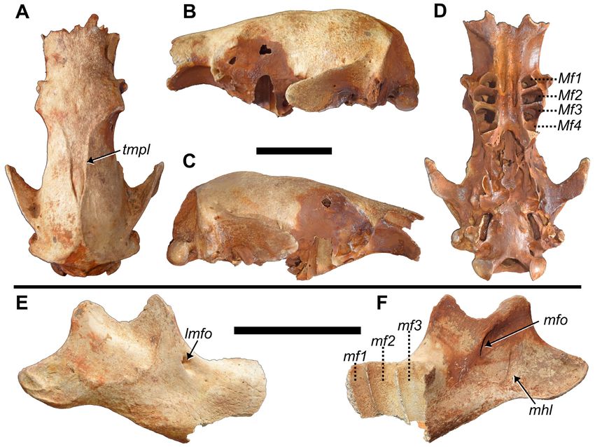
McAfee et al. — New species of the ground sloth Parocnus
 Parocnus has, until recently, been poorly represented in         Diagnosis: Distinguishable from the type specimens
paleontological sites and collections in Hispaniola, but         of Parocnus serus by the following characters: maximum
new sloth material from caves in the Dominican Republic          humeral length
Vertebrate Anatomy Morphology Palaeontology 9:52–82

extends from dorsal to palmar margins; Mt2 carina obscured         partial right scapula, two radii (left and right), left ulna,
in lateral view; Mt4 diaphysis narrow with sharp ridges and        two scaphoids (left and right), magnum (left), cuneiform
cuboid facet is rectilinear.                                       (left), two Mc3s (left and right), two right Mc4s, left femur,
 Etymology: Although never specified by Miller (1929),             two tibiae (left and right), left fibula, left calcaneus, right
we hypothesize Parocnus to have meant “other sloth”                Mt2, and left Mt4.
[para- (Greek) = other, besides; -ocnus (Greek) = sloth], as        Occurrence: Type locality is Padre Nuestro cave with-
a way to distinguish it from the other erected sloth taxa at       in the Nacional Parque del Este, Altagracia Province,
the time, many of which utilized size-based names (e.g.,           Dominican Republic. Additional referred material is from
Megalocnus, Microcnus). Para- has been used in a similar           the nearby cave (
McAfee et al. — New species of the ground sloth Parocnus

FIGURE 2. Map of Altagracia Province in southeastern Dominican Republic and the site localities in Nacional Parque del Este.

 Attempts to recover collagen from specimens from Padre         although the alveolae of the Mf1 and Mf2 are complete on
Nuestro have failed to produce any results, likely due to the   both sides. There is also damage to the basal portions of the
long-submerged status of the elements. Radiocarbon dating       cranium and attaching pterygoids; both jugals are also absent.
undertaken in 2009 on four sloth humeri from La Jeringa         The individual is considered a full adult because there are no
was unsuccessful. Sloth specimens from several caves in         signs of cranial sutures remaining (age class 3 sensu Anderson
Haiti (Steadman et al. 2005) have yielded Holocene dates.       and Handley 2001). Despite the incomplete condition of both
Cueva de Berna, a dry cave approximately 20 km east             this specimen and the paratypes, enough anatomy is preserved
of Padre Nuestro (the type site for Antillothrix bernensis)     to allow comparison with the limited cranial material of P.
has charcoal associated with faunal remains from which a        serus. The crania of both species are similar in morphology and
radiocarbon date of 3850 ±150 yr BP was obtained (Rímoli        relative dimensions, but P. dominicanus is smaller than P. serus
1977). The fauna present in Padre Nuestro and La Jeringa        in most dimensions (Tab. 1).
is similar to that found in these dated sites, though, the        In lateral views (Fig. 3B, C), the rostrum of the holotype
Haitian sites do contain a much greater diversity of rodents    is rather flat compared to that of the holotype for P. browni
from the genus Plagiodontia (Hansford et al. 2012).             (AMNH 16877), in which the anterior portion of the frontals
                                                                is quite bulged. This difference does not appear to be diagnos-
DESCRIPTION                                                     tic because the paratype crania for P. dominicanus (see below,
                                                                Fig. 11) also exhibit frontals bulged with respect to the nasals
Holotype - MHD 237                                              similar to that seen in P. browni. Variation in rostrum shape
 Cranium: This specimen is edentulous (like all the assigned    and/or doming of the cranium is common within Acratocnus,
crania); portions of most of the alveolar walls are damaged,    and also the extant sloths (pers. obs. RKM).

                                                                                                                             55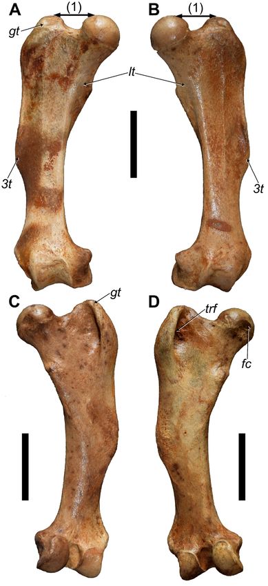
Vertebrate Anatomy Morphology Palaeontology 9:52–82

Table 1. Cranial measurements (in millimeters) for Parocnus dominicanus and P. serus.

          P. dominicanus (n=6*)                                                                             P. serus (n=4)
		MHD 237 Mean St Dev		                                                                                     Mean St Dev

Length of rostrum to occipital condyles		                                        179.3   ±1.8		             220.3    ±4.0
Nasal opening height                                               26.7          26.2    ±1.0		             30.1     ±2.2
Width across jugals/lacrimals                                      53.0          58.2    ±3.4		             68.0     ±4.9
Palate length                                                      67.6          71.6    ±3.2		             89.1     ±3.6
Toothrow length		                                                                90.7    ±3.3		             102.3    ±3.6
Breadth of rostrum across canines		                                              62.3    ±2.2		             68.8     ±5.7
Cf1 alveolar width		                                                             16.1    ±0.3		             16.2     ±0.7
Cf1 alveolar length		                                                            13.6    ±1.0		             15.7     ±0.2
Diastema length                                                    33.1          33.4    ±1.1		             38.9     ±0.8
Molariform toothrow length (Mf1-Mf4)                               41.3          42.9    ±1.4		             50.1     ±1.8
Mf1 alveolar width                                                 11.4          11.5    ±0.3		             13.3     ±1.5
Mf1 alveolar length                                                9.8           10.2    ±0.3		             12.5     ±0.9
Breadth of palate across Mf1                                       43.9          46.6    ±2.0		             44.1     ±2.3
Breadth of palate between Mf1                                      16.5          16.6    ±0.1		             17.6     ±1.0
Mf2 alveolar width                                                 15.1          14.9    ±0.1		             16.8     ±0.9
Mf2 alveolar length                                                10.5          10.5    ±0.2		             13.0     ±1.3
Mf3 alveolar width		                                                             13.5    ±0.2		             15.7     ±1.2
Mf3 alveolar length		                                                            9.2     ±0.1		             11.4     ±1.2
Mf4 alveolar width		                                                             11.5    ±0.2		             12.8     ±1.0
Mf4 alveolar length		                                                            7.2     --		               7.9      ±0.4
Breadth of postorbital processes at the root of the process        54.8          58.8    ±4.0		             74.2     ±4.3
Breadth at postorbital constriction                                39.4          45.0    ±4.2		             55.9     ±4.6
Breadth at posterior zygomatic roots (widest part of neurocranium) 52.2          56.5    ±4.6		             66.4     ±2.5
Width across the occiptal                                          71.5          76.8    ±2.8		             90.5     ±5.1
Width across the occipital condyles                                46.5          49.3    ±2.2		             58.0     ±3.3
Foramen magnum height                                              22.2          21.0    ±1.3		             20.0     ±1.5
Foramen magnum width                                               26.3          22.8    ±4.8		             28.8     ±3.3
Posterior cranium depth: sagittal crest to basisphenoid            51.5          51.5    --		               67.8     ±3.7

*includes holotype specimens; -- not enough specimens to calculate the value

 The nasals of both Hispaniolan species of Parocnus possess          tal crest. This point of union is posterior to that of other
an anterior extension along the lateral side, which with the         specimens of P. dominicanus as well as P. serus. Otherwise,
morphology of the maxilla makes the projections appear               the union point for P. dominicanus is just anterior to the
“prong-like.” Such anterior projections from the nasals              temporal process roots, which is still posterior to that of P.
are not seen in any species of the other Greater Antillean           serus where the union occurs midway between the postor-
genera. The characterization of this feature cannot be               bital process and the temporal process root. The holotype
determined for P. browni because the nasals of the holotype          specimen appears to have a bulbous portion of bone that
(AMNH 16877) are damaged and prevent direct compari-                 prevents an earlier union of the temporal lines (Fig. 3A).
son. Although the nasomaxillary sutures are absent in P.             This appears to be a feature unique to this individual and is
dominicanus types, their location can be determined from             not seen in any of the other cranial specimens.
inside the nasal cavity because both bones produce a short,           The rostrum of P. dominicanus is wider than that of P.
ventral projection that also serves as the lateral boundary to       browni, at least anterior to the root for the jugal. The
part of the vomer.                                                   palate between the molariform toothrows widens slightly
 In dorsal view (Figs. 3A, 11), the temporal lines merge to          posteriorly, is dotted with several smaller foramina, and
form a sagittal crest just posterior to a coronal plane con-         at the anterior portion exhibits two palatine sulci separ-
necting the anterior roots of the temporal processes. The            ated by the midline and each leading to a large, anterior
crest then diverges at the coronal plane across the posterior        palatine foramen (Fig 3D). The termination of the sulci
roots of the temporal processes to form two lines which              at each palatine foramen are unevenly positioned with the
deviate laterally before joining with the parieto-occipi-            left side extending to the level of the alveolae for Mf2-3

56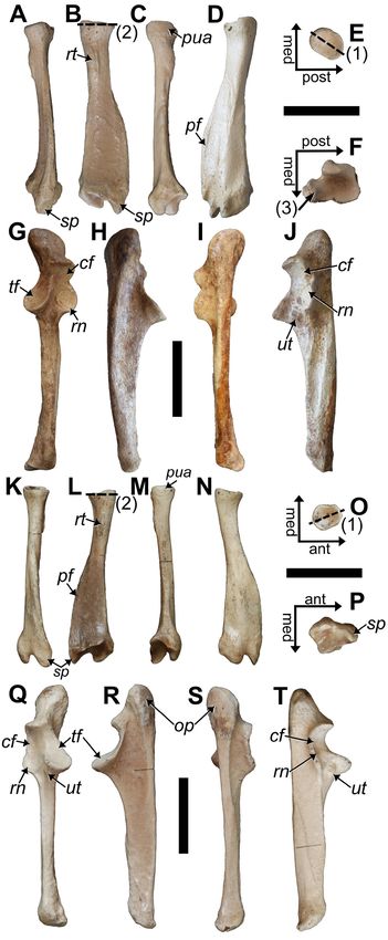
McAfee et al. — New species of the ground sloth Parocnus

Figure 3. Skull and right mandible of MHD 237 Parocnus dominicanus. Skull shown in A, dorsal, B, left lateral, C, right lateral,
and D, ventral views; right mandible shown in E, lateral and F, medialviews. Abbreviations: lmfo, lateral mandibular foramen;
Mf, upper molariform; mf, lower molariform; mhl, mylohyoid line; mfo, mandibular foramen; tmpl, temporal line. Scale bars
equal 5 cm.
while the right ends at the alveolae for Mf1-2. This is more       (Fig. 3E, F). Although the mandibular condyle is absent,
posterior than that exhibited by P. browni, in which the           its placement would have been elevated above the plane of
termination is in the middle of the diastema region but            the toothrow, as is also seen in P. serus. The posterior ramus
similar to that exhibited by Megalocnus at the anterior end        is complete and displays muscle scars for the various slips
of Mf1 (Matthew and Paula Couto 1959; Fischer 1971). In            of m. massetericus (Fig. 3E; Naples 1985, 1989; Naples
specimens of P. serus, the anterior palatine foramen occurs        and McAfee 2012, 2014). The posterior ramus is separ-
inconsistently along the palate, suggesting that this feature      ated from the body by a distinct notch due to the ventral
may not be taxonomically diagnostic. The lateral margin            bowing of the body to accommodate the molariforms. This
of the molariforms presents a convex arc with the Mf2 and          bowing and notching is more pronounced than in P. browni
Mf3 marking the widest points from the midline. Mf1                but is equal to that of P. serus. In lateral view, attachment
alveoli are subtriangular and smaller than the Mf2s, which         scarring for the zygomaticomandibularis and masseter is
are rectangular in outline and with the mediolateral long          evident. The scarring for zygomaticomandibularis extends
axis angled with respect to the palate.                            ventrally from the coronoid process in a straight line and
 Mandible: The right dentary, lacking the anterior half            begins to curve posteroventrally at the level of the lateral
and all the teeth, is preserved. The lateral aspects of the al-    mandibular foramen and toothrow but is well posterior
veoli for the molariforms (mf1-mf3) are partially preserved;       to the lateral mandibular foramen. This separation is the
both coronoid and condyloid processes are incomplete               same in P. serus but in P. browni, the vertical portion of

                                                                                                                               57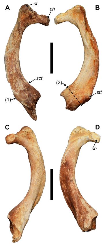
Vertebrate Anatomy Morphology Palaeontology 9:52–82

the attachment scar is just posterior to the foramen with          short, project dorsolateral from the laminar plane, and bear
almost no separation. The lateral mandibular foramen is            convexly rounded articular facets that in this thoracic series
positioned just posterior to the anterior margin of the base       are laterally directed. Each transverse process also bears a
of the coronoid process. On the medial surface, the mylo-          small cranially directed process along its cranial margin
hyoid groove is separate from the mandibular foramen and           that is separate from the rib articular facet. This process is
forms a ‘j’ shape (Fig. 3F); this condition is also the same in    likely associated with the intertransverse ligaments. The
P. browni and P. serus.                                            pedicles are strongly indented along their cranial margin
  Thoracic Vertebrae: In the skeletal specimen of P.               to form part of the articulation with the heads of the ribs.
browni described by Fischer (1971), the number of thor-            The caudodistally angled spinous processes are long with
acic vertebral elements was estimated to be 21 or 22,              an enlargement of the distal end. The pre- and postzygapo-
which is close to that of Choloepus and Hapalops (22–24:           physeal articular facets, located immediately medial to the
Scott 1903; Gaudin 1999; Hautier et al. 2010). However,            pedicels, are flat and within the same craniocaudal plane.
the number of thoracic vertebrae could be less given the             The lone vertebra from the posterior thoracic section (Fig.
variable ranges between 16–18 thoracics that have been             4I–K) has a range of morphological characters reminiscent
noted for other fossil sloths (Amson et al. 2015b). Of the         of both thoracic and lumbar vertebra, but is decidedly
five holotype vertebrae in Figure 4, four likely represent the     thoracic due to the presence of rib articulations. However,
middle thoracic region, whereas the final is certainly from        it differs from other thoracic vertebrae in several respects.
the posterior region, based on features described by Fischer       The spinous process has a distal enlargement but overall
(1971). There are no discernable xenarthrous articulations         is shorter and is less caudally projected. The transverse
on any of the vertebrae.                                           processes are a little longer but extend caudolaterally; the
  For the anterior to middle thoracic vertebrae (Fig. 4A–H),       costal facets are still convex. The zygapophyses are split into
the centra have the characteristic rounded, triangular shape       medial and lateral components or facets. The laminae cau-
when viewed cranially and caudally, and there are two              dal to the transverse processes are medially constricted and
small facets found on the dorsolateral margins of the caudal       bear more distinct medial postzygapophyses with convex
centrum surface. The vertebral canals are ovate, with the          facets. This is complementary to the cranial end of the next
long axis transversely oriented. The transverse processes are      posterior vertebra where the medial prezygapophyses are
                                                                   medial on the lamina with respect to the pedicles and are
                                                                   concave with a mediodorsal facing surface. The lateral pre-
                                                                   zygapophyses are lateral-to-even with the pedicles and are
                                                                   dorsolaterally facing, while the lateral postzygapophyses can
                                                                   be found on the ventral surface of the caudal portion of the
                                                                   transverse processes and facing ventromedial. The centrum
                                                                   is ovate instead of triangular and the small facets along the
                                                                   dorsolateral margin are no longer present. This matches
                                                                   with the descriptions given by Fischer (1971) for vertebrae
                                                                   of the posterior thoracic region of P. browni.
                                                                     Rib 1: The neck is short and is nearly as thick as the head,
                                                                   with a slight indentation on the dorsal margin that distin-
                                                                   guishes the costal tubercle from the head (Fig. 5). The articu-
                                                                   lation on the rib head is ovate but convexly folded along the
                                                                   middle to give the appearance of two continuous surfaces.
                                                                     As in P. serus and P. browni, a prominent lateral tubercle is
                                                                   present toward the point where the costal rib fuses with the
                                                                   ossified sternal cartilage, but in P. dominicanus this tubercle
                                                                   is reduced and gives the sternal end a more uniform width
                                                                   relative to that of the shaft. For P. dominicanus, the reduc-
Figure 4. Holotype vertebrae of MHD 237 Parocnus domin-
icanus from Padre Nuestro. Thoracic #1 in A, left lateral and      tion in the sternal tubercle also gives the diaphysis a bowed
dorsal E, views; thoracic #2 in B, left lateral and F, cranial     and twisted appearance when caudally viewed (Fig. 5C,
views; thoracic #3 in C, left lateral and G, dorsal views;         D), which is not exhibited by P. serus. The sternal tubercle
thoracic #4 in D, left lateral and H, cranial views; and           appears more caudally positioned than in P. serus where it is
Thoracic #5 in I, left lateral, J, dorsal, and K, cranial views.   instead moderately directed towards lateral. Across from the
Scale bars equal to 2 cm.                                          sternal tubercle on the medial margin is another smaller tu-

58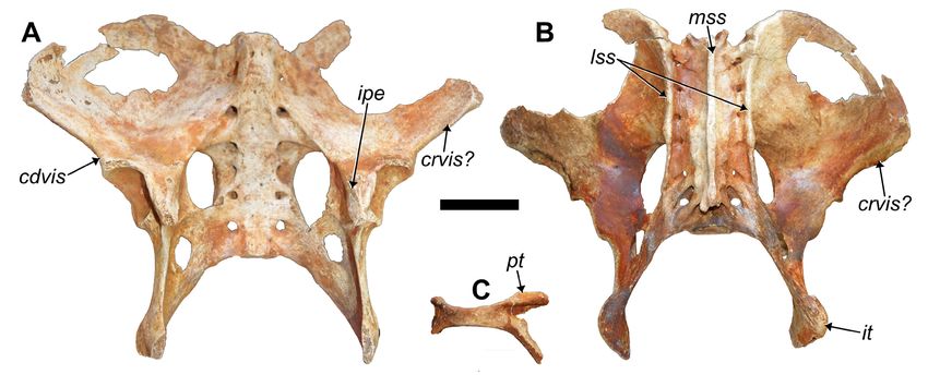
McAfee et al. — New species of the ground sloth Parocnus
                                                                     bercle (Fig. 5A), likely corresponding to the scalene tubercle,
                                                                     and which is reduced in size compared to that of P. serus.
                                                                       The inner surfaces of the distal ends exhibit two articu-
                                                                     lar surfaces for contact with the manubrium. The dorsal
                                                                     facet is a rounded depression, while the ventral is slightly
                                                                     concave, triangular, and extends onto the distal portion
                                                                     retained in the right first rib (Fig. 5C).
                                                                       Scapula: Both scapulae are incomplete but together
                                                                     account for the majority of aspects that each individually
                                                                     lacks. The left scapula is nearly complete (Fig. 6A) but lacks
                                                                     the anterior extension of the spine and the coraco-acrom-
                                                                     ial complex, which the right side retains (Fig. 6B, C). Part
                                                                     of the posterior border inferior to the spine and the fossae
                                                                     floors are also absent in both scapulae, but more is preserved
                                                                     in the paratype (MHD 350: see figure 4f in McAfee &
                                                                     Beery 2021). The secondary scapular spine is present, but
                                                                     not prominent, and the teres major fossa is visible but not
                                                                     strongly developed. The acromion and coracoid processes
                                                                     are fused into one complex, a feature also seen in the other
                                                                     Parocnus species, and sloths in general. The coraco-scapular
                                                                     foramen is incomplete in all the P. dominicanus specimens,
                                                                     but the anterior margins are relatively preserved to indicate
                                                                     its location just dorsal to the glenoid fossa (Fig. 6A, C).
                                                                       The glenoid fossa is ovate, with the inferior portion slight-
                                                                     ly wider than the superior, and the dimensions are smaller
                                                                     than those for P. serus (Tab. 2). The exception is UF-VP
                                                                     16997. This specimen of P. serus has glenoid values similar
                                                                     to those of P. dominicanus, but the shape of the scapular
                                                                     borders gives the impression that this specimen is younger
                                                                     and that the size similarities may be an ontogenetic artefact.
                                                                     The differences in the specimen likely fall within the vari-
                                                                     ability noted for P. serus by McAfee and Beery (2021).
                                                                       Humerus: Complete, paired humeri without an ente-
                                                                     picondylar foramen, characteristic of Parocnus, are pre-
                                                                     served. The diaphyses are medially bowed, such that when
                                                                     transected by a longitudinal plane, the humeral head is
                                                                     unevenly divided so that more of the humeral head is
                                                                     located on the medial side of the plane (Fig. 7D, E). This
                                                                     contrasts with P. serus and P. browni in which the diaphysis
                                                                     is straighter and so the head occupies both sides of the
                                                                     longitudinal plane with near equality (Fig. 7F). The bowed
                                                                     appearance is enhanced by the prominent narrowing of the
                                                                     diaphysis just distal to the tubercles. The most proximal
                                                                     margin of the lesser tubercle is situated distal to that of the
                                                                     greater tubercle in P. dominicanus (Fig. 7A, B), but the two
Figure 5. Right and left first ribs of MHD 237 Parocnus do-          are evenly aligned in P. serus (Fig. 7C).
minicanus from Padre Nuestro. Right rib in: A, cranial and C,          The deltopectoral shelf has a lateral extension at its
caudal views, and left rib in: B, cranial and D, caudal views.       midshaft termination, which exhibits two shallow fos-
Abbreviations: ch, costal rib head; ct, costal rib tubercle; sct,    sae separated by the brachiocephalicus crest (Amson et
scalene tubercle; stt, sternal tubercle. Diagnostic characters: 1,   al. 2015a). These fossae are related to equal sized muscle
sternal tubercle reduced, located more caudally, and deflected       attachment for m. pectoralis (medial) and m. deltoideus
away from the diaphysis, 2, sternal end of rib 1 is narrow. Scale    (lateral) (Toledo et al. 2013). In P. serus, the two fossae are
bars equal 5 cm.
                                                                                                                                 59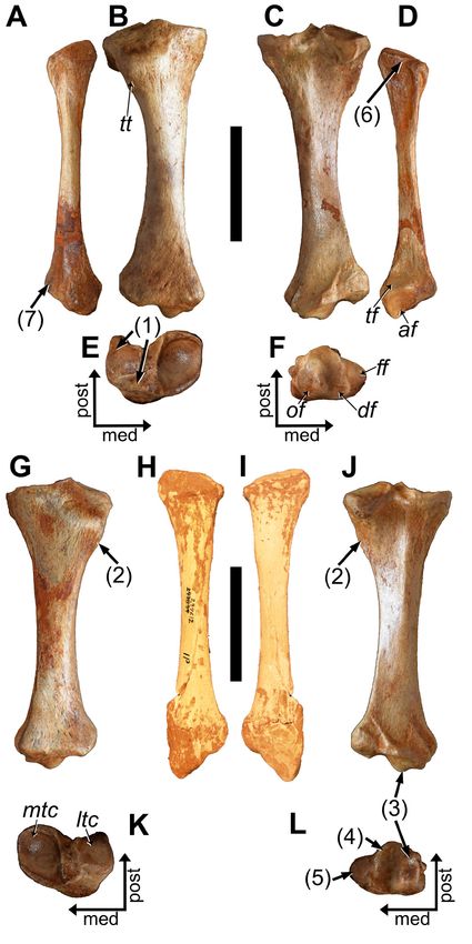
Vertebrate Anatomy Morphology Palaeontology 9:52–82

Figure 6. Right and left scapulae of MHD 237 Parocnus dominicanus from Padre Nuestro. Right scapula in: A, anterior/cranial
and B, lateral views, and C, left scapula in lateral view. Abbreviations: csf, coracoscapula foramen; gf, glenoid fossa; ssp, sec-
ondary scapular spine, tmf, teres major fossa.

present, but they are unequal with the medial larger than           from the tuber sacralis toward the (absent) dorsal spine,
the lateral fossa, as expected for a larger animal; this feature    which is consistent with the specimens described by Fischer
is broken in the holotype (Fig. 7C, F). The lateral border of       (1971) as P. browni and M. rodens. This morphology is also
the deltopectoral shelf in both P. serus and P. dominicanus         in contrast to the sharper, more angular features exhibited
(as well as Megalocnus rodens) occupies or nearly occupies          by Acratocnus and Neocnus (McAfee and Rimoli 2019). The
the same parasagittal plane as the lateral epicondyle, while        morphology of the ilium from the acetabulum to the cranial
in P. browni the crest is not as well-developed and does not        ventral iliac spine differs from the transverse lateral extension
extend laterally in the same plane.                                 exhibited by Megalocnus and Acratocnus by having a more
 In posterior view, the proximal half of the humerus in             dorsolateral extension, although it is more laterally angled
P. dominicanus does not exhibit strong muscle scars. The            than that exhibited by Neocnus (McAfee and Rimoli 2019).
pattern and location of theses scars is the same as in P. serus     The caudal ventral iliac spines are small tubercles situated just
and P. browni, with their reduced development, likely a re-         cranial to the acetabulum in the pelvises of Acratocnus (pers.
sult of the size difference between the species (Tab. 3). The       obs), Megalocnus (Fischer 1971), and Neocnus (McAfee and
overall anatomy of the distal half of the humerus does not          Rimoli 2019). The same positioning appears to be true for
significantly differ between the Parocnus species.                  the holotype and is confirmed by a more prominent occur-
 Pelvis: This specimen represents the most complete pelvis          rence in the paratype MHD 238.
known for any species of Parocnus. It is missing aspects from        The presence and location of the cranial ventral iliac spine
both iliac blades, and the pubic and ischial rami are broken        is somewhat uncertain because the lateral-most points of
but such that the pubic symphysis and part of those rami            the iliac blades are broken in all specimens, where it is
exist as a singular element, separate from the rest (Fig. 8C).      assumed to have occurred. The caudal margin of each iliac
Each iliac blade forms a rounded, convex arc going lateral          blade is worth noting because it does not present a straight-

Table 2. Scapulae measurements (in millimeters) for the holotype of Parocnus dominicanus versus P. serus.

  P. dominicanus		                                                                                           P. serus (n=5)

                                                            MHD 237 (L) MHD 237 (R)                          Mean     St Dev
Length of 2nd spine (to base of glenoid)				                                                                 116.6    ±4.3
Long axis of glenoid                                        28.9        27.9		                               36.5     ±4.2
Wide axis of glenoid                                        18.4        18.7		                               24.3     ±2.9
Length from Coraco-acromion to root of spine				                                                             165.6    ±3.5
Posterior width of infraspinous fossa                       45.8			                                          54.1     ±10.1
Maximum width of teres fossa				                                                                             15.3     ±2.4
Length: vertebral border to glenoid along supscapular ridge 95.9			                                          113.9    ±7.6
Width of infraglenoid tubercle                              5.5         6.3		                                9.3      ±1.4

60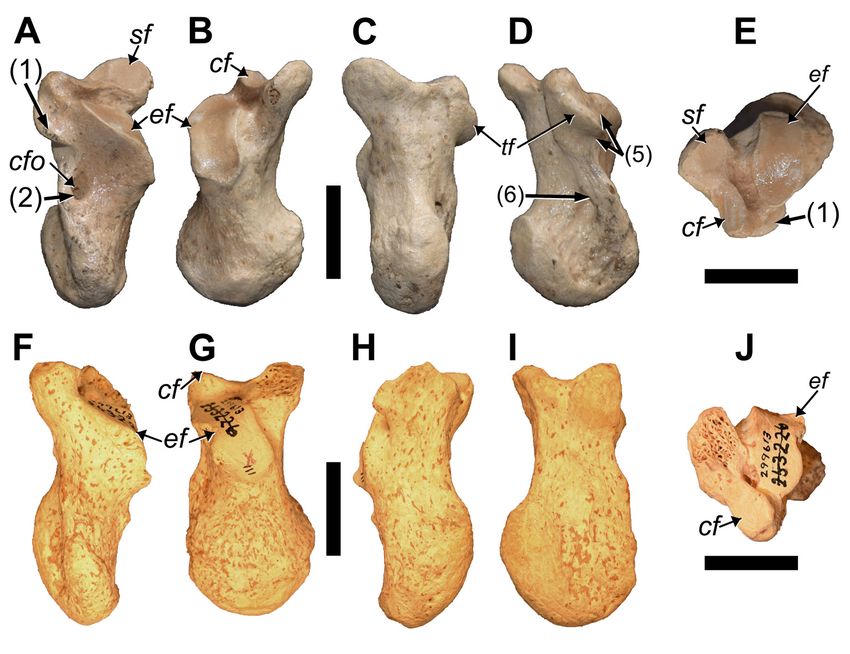
McAfee et al. — New species of the ground sloth Parocnus
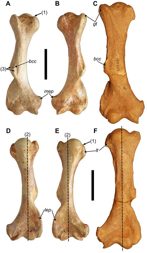
Figure 7. Right and left humeri of MHD 247 Parocnus dominicanus and comparison to the paratype right humerus of P. serus.
Right humerus of P. dominicanus in A, anterior and D, posterior views; left humerus of P. dominicanus in B, anterior and E, poster-
ior views; right humerus of P. serus (USNM PAL 700637) in C, anterior and F, posterior views. Abbreviations: bcc, brachiocephalic
crest; gt, greater tubercle; lep, lateral epicondyle; lt, lesser tubercle; mep, medial epicondyle. Diagnostic characters: 1, proximal
margin of the lesser tubercle is distal to that of the greater tubercle; 2, humeral head medially offset from longitudinal plane of
the diaphysis; 3, medial and lateral fossae of the deltopectoral crest are equal in size. Scale bars equal to 5 cm.
61Vertebrate Anatomy Morphology Palaeontology 9:52–82 Table 3. Humerus measurements (in millimeters) for Parocnus dominicanus and P. serus. MHD 237 (L) MHD 237 (R) P. dominicanus (n = 11*) P. serus (n = 7) Mean St. Dev Mean St. Dev Total Length 166.4 165.9 162.4 ±5.6 189.3 ±12.2 Max width across both tubercles 47.5 46.9 49.5 ±1.8 56.7 ±7.5 Width across lesser tubercle 24.7 23.6 23.1 ±1.8 24.0 ±2.9 Max epicondylar width 59.7 59.8 58.8 ±2.2 70.4 ±5.8 Medial Epicondyle height 22.2 22.3 24.2 ±2.5 26.8 ±3.2 Medial Epicondyle width 17.2 15.7 16.8 ±1.8 22.4 ±2.4 Max width across condyles (distal) 41.2 40.6 39.7 ±2.5 46.8 ±4.6 Anterior condyle width 19.4 21.1 20.3 ±1.5 36.2 ±8.6 *includes holotype specimens Figure 8. Pelvis and pubic symphysis of MHD 237 Parocnus dominicanus from Padre Nuestro. Pelvis shown in A, ventral/ cranial and B, dorsal/caudal views; C, associated pubic symphysis region shown in anterior/cranial view. Abbreviations: cdvis, caudal ventral iliac spine; crvis?, cranial ventral iliac spine (assumed); ipe, iliopectineal eminence; it, ischial tuberosity; lss, lateral sacral spines; mss, median sacral spine; pt, pubic tubercle. Scale bars equal 5 cm. line surface but has an anteriorly directed bulge about to Acratocnus (pers. obs.: RKM). The iliopectineal eminence midway between the acetabulum and the lateralmost point (pecten pubis), which represents the attachment of the m. (Fig. 8), which is a feature not evident in any of the other pectineus, is evident on both sides and sits medial to the Caribbean sloths. Damage to the lateralmost points of the acetabulum along the inner rim of the pelvis. Caudally, and iliac blades makes it impossible to rule out that the bulge along the innermost surface of the same region, there is a on the caudal margin may represent the cranial ventral iliac wide groove running between the ischiosciatic foramen and spine and the attachment of the rectus femoris muscle. the posterior margin of the obturator foramen, likely indicat- The acetabulum is subcircular and the lunate surface within ing the pathway of the obturator nerve and vessels. is ventrally separated by an incisive notch running between The ischial ramus is broadened by bony extensions to the the ischial and pubic cornuses. White and MacPhee (2001), terminal sacral vertebrae, and those extensions form the presumably using elements collected by Miller, listed the lack caudal margin of the ischiosciatic foramen. The holotype of such a gap in the acetabular rim as a feature of P. serus, exhibits damage in this area on both sides, giving the im- while Fischer (1971) noted the existence of the gap for P. pression that a foramen is present, but MHD 238 confirms browni. The variability in this feature may be age related, as that those are artificial features. The ischial rami terminate demonstrated by a greater range of pelvic specimens assigned as distinct ischial tuberosities for attachment of the poster- 62
McAfee et al. — New species of the ground sloth Parocnus
ior thigh muscles (Fig. 8B). A crest on the caudal margin
extending between the ischial tuberosity and the last/sev-
enth sacral vertebra is visible on the lateral surface, which
increases in craniocaudal height as it nears the sacrum and
the termination of the lateral sacral crest.
  The sacrum consists of seven fused vertebrae with lateral
alar extensions contacting the ilial and ischial elements,
resulting in six sets of sacral foramina. In MHD 238, there
are eight fused vertebrae, although the first in the series
occurs without alar extensions to the ilium and therefore
represents a fused lumbar or a pre-iliac synsacral vertebra
(sensu Galliari and Carlini 2019). In dorsal view, three
crests are clearly defined (one median sacral and two lateral
sacral crests). The median sacral crest (Fig. 8B) is distinct
and raised with respect to the laminae, which is more akin
to condition exhibited by Megalocnus (Fischer 1971), but
unlike the flattened condition in Acratocnus and Neocnus
(McAfee and Rimoli 2019). The lateral sacral crests are
most evident in the iliac region and are stronger than that
exhibited by Megalocnus; the crests are almost non-existent
in the iliac region in Acratocnus and Neocnus. The lateral
crest suddenly narrows posterior to the alar contribution
from the four sacrals. The caudal continuation of the crest
forms the mediodorsal margin of the ischiosciatic foramen
and then trends caudolateral into the caudal margin of the
expanded ischial ramus. Overall, the lateral sacral crests are
slightly more curved (laterally concave) than that exhibited
by Megalocnus and are distinct from Acratocnus and Neocnus
where they taper caudally from lateral to medial.
  Femur: The morphology of the paired femora is on par with
the characters attributed to Parocnus (see White and MacPhee
2001), but the lack of complete, adult, and accessible femora
for P. serus makes detailed comparisons impossible at this
time. Overall, there is little to distinguish the femora of P.
dominicanus from P. serus, and in most ways the morphology
is what is expected from a reduction in size (Tab. 4). The
greater trochanter is a little smaller, such that it is closer to
the same level as the femoral head rather than being above it
(Fig. 9A, B). The lesser trochanter, which is characteristically
underdeveloped in Parocnus overall, is less developed than that
of P. serus; it resembles the reduced development of P. browni
(specimen Ma. 5/67: Fischer 1971). The third trochanter is
prominent and confluent with the greater trochanter, although
the indentation created by the development of those trochant-       Figure 9. Right and left femora of MHD 247 Parocnus
ers is not as pronounced in P. dominicanus. The existence of a      dominicanus from Padre Nuestro. Right femur in A, anterior
trochanteric fossa (Fig. 9A, D) on the posterior surface has not    and C, posterior views, and left femur in B, anterior and
previously been recognized, but such a feature is evident in P.     D, posterior views. Abbreviations: 3t, third trochanter; fc,
serus and P. dominicanus, which serves as a unique character        fovea capitis; gt, greater trochanter; lt, lesser trochanter;
to distinguish these species from P. browni, as well as to all      trf, trochanteric fossa. Diagnostic characters: 1, femoral
other Caribbean sloth species. The fovea capitis is not centered    head and greater trochanter of equal height. Scale bars
on the femoral head but is posteriorly displaced, as in many        equal to 5 cm.
large-bodied ground sloths, and creates an indentation with

                                                                                                                               63
Vertebrate Anatomy Morphology Palaeontology 9:52–82
Table 4. Measurement (in millimeters) for the hind limb elements of Parocnus dominicanus and P. serus.
MHD 237 (L) MHD 237 (R) P. dominicanus (n=18*) P. serus MNHNSD
Femur Mean St. Dev FOS25.3353 (R)
Total length 200.4 200.3 197.9 ±10.9 240^
Circumference of head 86^ 96.0 100.2 ±17.2 118.0
AP depth of lesser trochanter 52.0 51.1 52.1 ±3.2 67.4
AP depth of 3rd trochanter 37.9 38.9 39.4 ±2.4 50.7
Epicondylar width 54.8 55.5 55.6 ±3.5 62^
Anterior condyle width 20.5 19.9 22.5 ±2.9 29.0
Posterior condyle width 44.7 44.8 44.3 ±2.6 53.4
Height of greater trochanter 56^ 53.3 58.3 ±4.1 64.5
Depth of greater trochanter 28.1 28.5 28.7 ±10.8 35.3
Depth of 3rd trochanter 10.0 10.2 10.1 ±1.2 13.7
Distance across trochanters 66.9 66.8 66.6 ±5.3 84.6
*includes Holotype specimens, ^ represents an estimated measurement
MHD 237 (L) MHD 237 (R) P. dominicanus (n=14*) P. serus (n=4)
Tibia Mean St. Dev Average St. Dev
Total length 125.8 126.4 120.6 ±5.1 138.2 ±13.9
Length of proximal fibular articular surface 6.2 5.0 5.7 ±0.6 18.6 --
Width of proximal fibular articular surface 11.9 17.2 15.6 ±2.5 9.5 ±2.5
Length of distal articular surface (astragular) 23.9 24.5 23.6 ±2.4 28.7 ±4.7
Width of distal articular surface (astragular) 22.7 22.7 22.6 ±2.3 30.2 ±3.2
Length of distal fibular articular surface 13.9 13.4 12.7 ±1.0 16.7 ±1.5
Width of distal fibular articular surface 9.7 11.3 8.9 ±1.5 10.7 ±0.9
Proximal epicondylar width 45.1 46.0 43.4 ±2.9 53.7 ±4.6
Distal epicondylar width 35.4 37.0 33.3 ±2.6 42.4 ±5.2
Medial condyle length 29.0 29.2 25.1 ±3.3 32.9 ±3.8
Medial condyle width 22.2 20.4 21.6 ±2.0 19.8 ±11.9
Lateral condyle length 20.6 24.4 19.8 ±3.1 22.5 ±4.0
Lateral condyle width 19.7 19.7 19.0 ±3.2 21.3 ±3.3
*includes Holotype specimens; -- not enough specimens to calculate the value
MHD 237 (R) P. dominicanus (n = 14*) P. serus
Fibula Mean St. Dev USNM 299612 (L)
Total length 119.3 117.6 ±4.1 133.8
Length of proximal articular surface 9.3 8.6 ±1.4 22.4
Width of proximal articular surface 21.8 17.5 ±2.8 9.8
Height of distal tibial articular facet 9.5 8.2 ±0.8 9.4
Length of distal tibial articular facet 12.7 11.4 ±1.8 14.2
Length of distal articular surface (astragular) 14.8 13.7 ±1.6 19.8
Height of distal articular surface (astragular) 16.6 18.6 ±1.4 19.4
Width of distal end 26.0 24.6 ±2.2 27.2
Depth of lateral malleolus 19.9 17.9 ±1.4 18.1
Width of proximal end 22.6 22.0 ±2.3 17.1
*includes Holotype specimens
64McAfee et al. — New species of the ground sloth Parocnus
the circumferential margin of the femoral head (Fig. 9A, C, Tibia: In proximal view (Fig. 10E, K), and compared to
D). The femoral shaft distal to the third trochanter strong- P. serus, the medial tibial condyle exhibits a greater exten-
ly narrows. The femoral condyles are continuous with the sion of the anterolateral region, and the anterior margin
articular surface for the patella, and as is typical for sloths, the of the lateral tibial condyle is positioned or begins more
medial condyle is larger than the lateral. posteriorly. In addition, the lateral tibial condyle slopes
distolaterally, rather than being flattened, and the posterior
surface from the lateral tibial condyle for the m. popliteus
and its sesamoid is less laterally positioned and angled
than in P. serus. In anterior view, the tibial tuberosity is
more distally positioned from the tibial plateau than in P.
serus. In both species, the lateral-most portion of the tibial
tuberosity presents as a small protrusion from the diaphysis
that can be seen in anterior and posterior views (Fig. 10G,
J), but the protrusion is smaller in P. dominicanus and does
not continue as a crest to the posterior surface in P. serus.
It is uncertain what muscle(s) attached to the crest, but the
distal-most portion remains visible posteriorly when the
fibula is in articulation.
At the distal end of the posterior tibia, the medial surface
exhibits the trochlear groove for muscle tendons but there
is also a groove on the lateral side. A lateral groove exists
in P. serus but the surrounding features (specifically the lat-
erally placed knob (tibial lateral malleolus) for articulation
with the distal fibula and the crest of bone medial it that
runs proximolateral from the posterior-most point of the
distal astragalar articular surfaces) are not as well developed
as those in P. dominicanus.
On the distal surface, the articular projection separating
the discoid (lateral) and odontoid (medial) surfaces for
the astragalus is more prominent in P. dominicanus (Fig.
10F, L). The discoid articular facet exhibits a greater degree
of concavity than in P. serus. Lateral to this surface, the
fibular articular surface is mediolaterally widened to give
the surface a more squared appearance, whereas it is more
rectilinear in P. serus.
Fibula: Unlike Acratocnus, the fibulae of the two
Hispaniola Parocnus species possess straight diaphyses (Fig.
10A, D, H, I). The proximal end with the articular surface
for the tibia is obliquely oriented along the anteroposterior
axis, with the slope trending posterodistal. The inclination
is steeper or more raised in P. dominicanus, which is similar
to that of P. browni. Both P. dominicanus and P. serus exhibit
an indentation along the medial margin, but the placement
Figure 10. Tibiae and right fibula of MHD 247 Parocnus dominicanus and comparison to the paratype left fibula of Parocnus serus.
For P. dominicanus: right fibula in A, medial and D, lateral views; right tibia in B, anterior, C, posterior, E, proximal, and F, distal views;
left tibia in G, anterior, J, posterior, K, proximal, and L, distal views. The holotype of P. serus (USNM PAL 299612) is a left fibula in
H, lateral and I, medial views. Abbreviations: af, astragular facet; df, discoid facet; ff, fibular facet; ltc, lateral tibial condyle; mtc,
medial tibial condyle; of, odontoid facet; tt, tibial tuberosity. Diagnostic characters: 1, lateral tibial condyle posteriorly displaced and
distolaterally sloped; 2, tibial tuberosity positioned more distal to the tibial plateau and with a more prominent lateral protrusion;
3, discoid and odontoid facets with a prominent separation; 4, discoid facet more concave; 5, distal fibular articular facet more
squared; 6, fibular head incline and its medial margin indented at the anteroposterior midpoint; 7, lateral malleolus of the fibula
with a prominent posterior flange bearing a tuberosity and distinct tendon groove. Scale bars equal to 5 cm.
65Vertebrate Anatomy Morphology Palaeontology 9:52–82
along the anteroposterior axis differs. In P. dominicanus, the Mf4 alveolus is mediolaterally narrower than the preced-
indentation occurs near the middle (Fig. 10D) while in P. ing alveolus and the lingual side is more anteroposteriorly
serus it is nearer to the posterior end. This affects the shape compressed than the labial side, giving it a subtriangular
of the articular surface, which is more uniform in P. serus appearance with unequal sides. The posterior border of the
as the surface does not extend much past the indentation, Mf4 alveolus also has a protrusion into the alveolar space.
but it does extend quite a bit further in P. dominicanus. The MHD 412 retains most of the anterior-most border of the
indentation continues distally as a wide groove along the maxilla that would have articulated with the absent pre-
medial surface of the fibula that is more prominent in P. maxillae. This border is U-shaped, although there may have
dominicanus. When viewed proximally, P. dominicanus has a been an anteriorly projecting spicule of bone from near
more prominent lateral bulge, and the proximal end is not the midline. The anterior palatine foramina are located at
as anteroposteriorly wide as the distal end. The bulge is not the anterior Mf1 alveolar margin in MHD 347, unequally
as evident in P. serus, and the overall proximal end is equal posterior and anterior to the Mf1 alveolus in MHD 351,
to or slightly larger than the distal end as it is not visible and in the diastema region of MHD 412.
from a distal view. The vomer is rather well preserved in MHD 347 and 412,
On the distal end, the lateral malleolus has a more and in anterior view it is T-shaped. From the dorsal part
prominent posterior flange than that of P. serus, which of the bone, the processes extend laterally to the ventrally
has a more developed lateral malleolus tuberosity and a directed nasomaxillary crests and then curve ventrally to
well-defined tendon groove along its posterior surface. The run along the medial margin of the nasomaxillary crests.
astragalar articulation on the medial surface consists of two The ventral part of the vomer reaches to the maxillary crest
facets, one facing proximally and the other distal to the that dorsally projects from the hard palate.
first that faces medially (Fig. 10D). The inclination of the Mandibulae (MHD 406, 408): Like the holotype,
proximal facet is the same in both taxa, but the distal facet neither mandible is complete, but each specimen retains
in P. dominicanus is not as vertically oriented so that the the teeth from the left molariform series; caniniforms are
angle between the two is nearly 90°. The angle between the absent (Fig. 11I−L). Of the measurements obtained, the
two astragular facets in P. serus is slightly obtuse. most significant differences between the two species are the
length of the diastema, total length of the dental series, and
Paratypes
width of the mandibular condyle (Tab. 5).
Crania (MHD 347, 351, 411, 412): The associat-
Although there may be dental characters unique to the
ed paratype cranial specimens exhibit different levels of
species of Parocnus from Hispaniola, the rarity of mandibu-
completeness (Fig. 11A−H), but overall conform to the
lar teeth for P. serus currently makes such determinations
morphology to be considered conspecific. The paratypes
impossible. However, P. dominicanus can be distinguished
all demonstrate a considerable degree of pneumatization
by the alveolar outlines in dorsal view, where the labial
of the cranium, consistent with that known for other fossil
alveolar margin of mf1 bulges past the same margin of mf2;
sloths (Boscaini et al. 2018). Jugals are also absent for these
these same labial margins in P. serus are in line with one an-
specimens, and any isolated jugals that were recovered
other. Additionally, the alveolae for the lower caniniforms
do not correspond to the morphology established with P.
indicate the teeth would be more anteriorly directed in P.
browni (Taboada et al. 2007). The pterygoid processes are
dominicanus than those of P. serus, which is slightly more
also broken/missing.
laterally projected. Like the upper caniniforms, the lower
The dental alveoli (Fig. 11B, F) match the morphology
alveoli are subtriangular and with slight protrusions along
described in the holotype for Mf1 and Mf2. The Cf1 al-
the inner surface of each alveolar margin.
veoli are subtriangular with equal sides like the Mf1 alveoli
The mf1 tooth is somewhat rectilinear but a protrusion
but are larger and with slight protrusions of the margins
on the anterior side of the alveolus gives the tooth an
that would impact tooth shape (i.e., create grooves). The
anterior indentation and groove (Fig. 11J, L). This, along
Mf3 alveolus is a little more squared than the Mf2. The
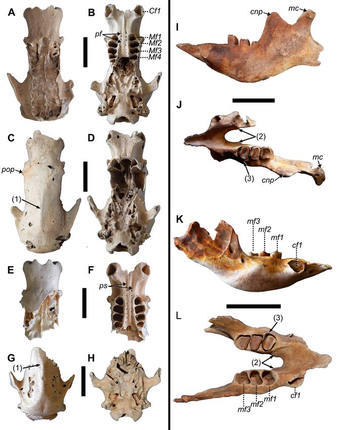
Figure 11 (opposite page). Paratype crania from La Jeringa and paratype mandibles from Padre Nuestro of Parocnus domin-
icanus. Crania: MHD 347 in A, dorsal and B, ventral view; MHD 351 in C, dorsal and D, ventral view; MHD 412 in E, dorsal and
F, ventral view; MHD 411 in G, dorsal and H, ventral view. Mandibles: MHD 406 in I, left lateral and J, dorsal view; MHD 408
in K, right lateral and L, dorsal views. Abbreviations: Cf/cf, upper caniniform/lower caniniform; cnp, coronoid process; mc,
mandibular condyle; Mf/mf, upper molariform/lower molariform; pf, palatine foramina; pop, postorbital process; ps, palatine
sulci. Diagnostic characters: 1, union of temporal lines into sagittal crest occurs anterior to the roots of the squamosal pro-
cesses of the temporal; 2, medioventral bulging of the cf1 roots creates a U-shape with posterior symphysis; 3, labial margin
of mf1 laterally bulges past the mf2 and mf3 labial margins. Scale bars equal to 5 cm.
66McAfee et al. — New species of the ground sloth Parocnus
67Vertebrate Anatomy Morphology Palaeontology 9:52–82
Table 5. Mandibulae measurement (in millimeters) of Parocnus dominicanus and P. serus.
P. dominicanus (n=13*) P. serus (n=2)
Mean St Dev Mean St Dev
Predental spout length 43.5 6.6 52.4 --
Predental spout width 19.5 2.6 18.6 --
Length of symphysis to level of canines (excluding projection) 29.9 2.7 51.6 --
Toothrow length 60.7 8.2 76.9 --
cf1 alveolar width 12.6 2.2 13.0 --
cf1 alveolar length 12.6 1.5 11.7 --
Diastema length (cf1-mf1) 15.6 2.1 26.0 --
Molariform toothrow length 36.9 1.9 40.9 --
mf1 alveolar width 14.5 1.3 15.0 --
mf1 alveolar length 13.1 0.3 10.2 --
mf2 alveolar width 15.1 1.5 14.8 --
mf2 alveolar length 11.5 1.5 10.5 --
mf3 alveolar width 13.0 1.6 13.7 --
mf3 alveolar length 12.6 1.3 11.9 --
Breadth of mandible across canines 49.5 3.3 57.7 --
Width between C1's 23.3 2.4 35.2 --
Width of mandible at mf1 20.7 1.7 23.0 --
Width of mandible at mf3 25.8 10.0 25.9 --
Depth at mf2 36.0 2.7 41.8 --
Ramus height from base to notch 37.9 3.5 54.0 --
Ramus height from base to condyle 51.1 0.0 57.6 --
*includes Holotype specimens; -- not enough specimens to calculate the value
with the morphology of the posterior margin, gives the underneath and lateral to the spout, with the standard sloth
tooth a mediolaterally curved appearance. The mf2 tooth pattern of at least one main foramen, which may or may
is more rectilinear but the lingual side is anteroposteriorly not be accompanied by a second smaller foramen per side.
compressed compared to the longer labial side. Individual MHD 406 preserves the nearly complete coronoid process
variation between specimens produces an indentation and and mandibular condyle on the left side (Fig. 11I). With
groove on the anterior side of this tooth in MHD 406 that the toothrow level in lateral view, the mandibular con-
is not as evident in MHD 408. The cusps for the first two dyle sits more dorsal than does the coronoid process. The
molariforms are most prominent at the anterolingual and mandibular condyle is transversely wide, with a greater
posterolabial corners of the teeth, such that the trough is lateral extent from the attachment to the mandibular neck.
oriented between them along an anterolabial to postero- Despite the seemingly flat nature of the glenoid fossa in
lingual axis. In mf3, the labial and lingual sides are higher the cranium, the mandibular condyle is arched with the
than the anterior and posterior sides, creating a shallow greatest dorsal extent in the middle and tapering ventrally
and uniform trough. The mf3 tooth morphology is sub- to the sides.
quandrangular with a pronounced indentation along the Atlas, C1 (MHD 836, 837): The ventral and dorsal
labial side, which with a shallow indentation of the anter- arches each lack tubercles for muscle tendon attachment
ior side forms a small lobe at the anterolabial corner. The (Fig. 12B, C, E, F), which may be related to their smaller
posterior margin is convex and curves strongly towards the size than P. serus. The cranial articular facets for the occipi-
lingual side such that a distinct posterolingual corner to the tal condyles are not very distinct. When viewed dorsally,
tooth is absent. these facets in P. serus project cranially from the dorsal arch
Predental spouts taper anteriorly but do not form a point and the cranial margins of the alar wings. Ventrally, there is
(Fig. 11J, L). The lateral margins in both specimens are not the same separation from the alar wings but there is the
abraded and incompletely preserved but are clearly raised distinct projection from the ventral arch.
to form a shallow trough for the spout. The length from Although the referred specimens bear damage to the
the mental symphysis is about the same as the dorsoven- alar wings, there is enough preserved to gain a complete
tral extent of the symphysis. Mental foramina are present picture of their morphology. The alar wings in P. serus and
68McAfee et al. — New species of the ground sloth Parocnus
Figure 12. Paratype atlas (C1) and axis (C2) vertebrae from La Jeringa of Parocnus dominicanus. Atlas: MHD 836 in A, dorsal,
B, cranial, and C, caudal views; MHD 837 in D, dorsal, E, cranial, and F, caudal views. Axis: MHD 909 in G, cranial, H, caudal,
and I, right lateral views; MHD 910 in J, cranial, K, caudal, and L, right lateral views. Abbreviations: cdaf, caudal facet; cd-axf,
caudal/axial facet; craf, cranial articular facet; da, dorsal arch; ocf, occipital fossa; odf, odontoid facet; odp, odontoid pro-
cess; tf, transverse foramen; va, ventral arch. Diagnostic characters: 1, C1 dorsal and ventral arch tubercles absent or weak; 2,
occipital condyles of C1 with minimal anterior projection from the ala and dorsal arch; 3, minimal extension of the caudal alar
wing margins and a small separation from the axial/caudal articular facets; 4, caudal alar tubercles are medially positioned;
5, C2 caudal articular facets with minimal caudal extension from the dorsal spinous process; 6, absent or weak depression at
the caudal base of the C2 spinous process; 7, odontoid process rounded and blunt. Scale bars equal to 2 cm.
P. dominicanus are rounded and exhibit a dorsoventral angle The odontoid process is rounded and blunt, anterodorsally
from the cranial to the caudal margin. The caudal margin angled, and bears a facet on the ventral surface for articu-
extends beyond the caudal facets to the axis vertebra in lation with the inner surface of the ventral arch of C1. The
P. serus but ends at nearly the same transverse plane in P. round, blunt shape contrasts with that of P. serus (MHD
dominicanus (Fig. 12A). This is also evident in caudal view 894), where the cranial tip is dorsoventrally flattened and
where the caudal alar tubercle sits more medial towards the more pointed. The caudal articular processes, retained only
caudal articular facet in P. dominicanus (Fig. 12F), while it in MHD 909 (Fig. 12I), are not as prominent nor as cau-
is more lateral in P. serus. Neither species exhibits connec- dally extended as those seen in P. serus.
tions between the caudal edge of the dorsal arch and the Radii (MHD 349, 521, 526): The radial head is more
dorsal edge of the caudal articular facets as exhibited by ovate than circular, and the overall rim is of equal height all
Acratocnus, as well as by some mylodontids (McAfee 2016) the way around the shallow articular depression, giving it a
and likely other sloth taxa. flat appearance. In P. serus, the height of the rims is unequal,
Axis, C2 (MHD 909, 910): None of the specimens as- with the posterior side that bears the ulnar articulation
signed to P. serus or P. dominicanus are complete, with most being taller and giving the proximal end an anteroposterior-
of the damage related to the spinous process, the transverse ly angled appearance; similar morphology is also seen in P.
process and foramina, and caudal portions of the centrum browni (Fischer 1971). The greater posterior height in P. serus
(Fig. 12G−L). The only noticeable difference lies in the also corresponds with a taller proximal ulnar articular surface
absence of a depression at the base of the spinous process than that of P. dominicanus. This also creates a defined inden-
in caudal view in P. dominicanus, whereas there is a distinct tation for a neck-like region in P. serus, whereas there is no
excavation in P. serus. obvious neck in P. dominicanus (Fig. 13).
69Vertebrate Anatomy Morphology Palaeontology 9:52–82
The diaphysis is slightly bowed, in a manner more akin to
that seen in Neocnus and Megalocnus, but the distal expan-
sion of the pronator flange is more pronounced than in
Neocnus and P. browni, and it begins more distally than in
Megalocnus. The radial tuberosity is near to the posterior
border but occupies a position on the medial surface where
it presents an ovate region, elongated in the proximodistal
plane. The medial surface is concave and slightly rugose,
while the lateral surface is convex, smooth, and mediolat-
erally separated by a small ridge. The posterior (inner)
surface of the diaphysis is rugose and distally broadens into
a triangular shape. The anterior (external) side bears the
pronator flange, which sometimes bears a distinct ridge.
The distal articular surface can be divided into anterior
and posterior surfaces. The anterior surface occurs along the
inside of the radial styloid process and is convex. In MHD
349 and 526 the surface has a squared appearance in distal
view (Fig. 13F), but in MHD 521 the anterior portion is
rounded and slightly tapered (Fig. 13P). The surface does
not appear to be heavily involved in articulation with the
scaphoid and serves more as a lateral boundary to carpal
abduction. The posterior surface is concave and deepened
by a bony ridge projected from the lateral surface. The
anterior point of this ridge joins with the proximodistal
ridge along the lateral radial surface, and overall serves as a
backstop that limits the degree of carpal extension by the
scaphoid. The medial margin of the distal articular surface
exhibits a slight indentation at the boundary between the
posterior and anterior portions.
Compared to other limb elements, the radii of P. domin-
icanus are significantly shorter than those of P. serus (Tab.
6). The percent difference in length for all the other limb
elements averages at 14.3% (12.1−17.5), while that of the
radius is 23.9%. Why this greater difference in length be-
tween the species occurs is uncertain, although it may relate
to sample size.
Ulnae (MHD 224, 225, 348, 512, 515): Similar to
the other Parocnus species, the ulnae have a pronounced
olecranon process, and their diaphysis is rectangular,
anteroposteriorly unbowed, and slightly tapered at the
distal end (Fig. 13). The olecranon is not as robust as in
Megalocnus but it exhibits a greater medial inclination than
seen in Megalocnus, which is also true for P. serus. At the
base of the olecranon in P. serus, just above the trochlear
notch, is a distinct tubercle that is not found in P. dominic-
Figure 13. Paratype radii and ulnae of Parocnus dominicanus from La Jeringa and Padre Nuestro. Radii: MHD 349 (left) and MHD
521 (right) in A & K, anterior, B & L, medial, C & M, posterior, D & N, lateral, E & O, proximal, and F & P, distal views. Ulnae: MHD
225 (left) and MHD 348 (right) in G & Q, anterior, H & R, medial, I & S, posterior, J & T, lateral views. Abbreviations: cf, capitular
facet; op, olecranon process; pua, proximal ulnar articulation; rn, radial notch; rt, radial tubercle; sp, styloid process; tf, trochlear
facet; ut, ulnar tuberosity. Diagnostic characters: 1, long axis of the radial head oriented anteromedial to posterolateral; 2, anter-
ior and posterior radial head margins less angled and closer to horizontal; 3, scaphoid facet along the styloid process is uniform-
ly wide and untapered. Scale bars equal to 5 cm.
70You can also read