Physicochemical and biological activities of the gamma-irradiated blended fibroin/aloe gel film - ScienceAsia
←
→
Page content transcription
If your browser does not render page correctly, please read the page content below
R ESEARCH         ARTICLE                                                           ScienceAsia 48 (2022): 278–286
doi: 10.2306/scienceasia1513-1874.2022.048

Physicochemical and biological activities of the gamma-
irradiated blended fibroin/aloe gel film
Preeyawass Phimnuana , Saran Worasakwutiphongb , Anuphan Sittichokechaiwutc ,
Francois Grandmottetd , Wongnapa Nakyaie , Kunlathida Luangpraditkuna , Céline Viennetf ,
Jarupa Viyocha,∗
a
     Department of Pharmaceutical Technology, Faculty of Pharmaceutical Sciences and Center of Excellence for
     Innovation in Chemistry, Naresuan University, Phitsanulok 65000 Thailand
b
     Division Plastic and Reconstructive Surgery, Department of Surgery, Faculty of Medicine, Naresuan University,
     Phitsanulok 65000 Thailand
c
     Department of Preventive Dentistry, Faculty of Dentistry, Naresuan University, Phitsanulok 65000 Thailand
d
     Department of Biochemistry, Faculty of Medical Science, Naresuan University, Phitsanulok 65000 Thailand
e
     Department of Chemistry, Faculty of Science, Ramkhamhaeng University, Bangkok 10240 Thailand
f
     UMR 1098 RIGHT INSERM EFS BFC, University of Bourgogne Franche-Comté, Besançon, 25000 France
∗
    Corresponding author, e-mail: jarupav@nu.ac.th
                                                                            Received 12 Jul 2021, Accepted 17 Dec 2021
                                                                                           Available online 28 Feb 2022

ABSTRACT: The physicochemical and biological properties of the blended fibroin/aloe gel film as a wound dressing
were investigated to support the wound healing efficacy of the film described in our previous study. In the current study,
protein content, molecular weight pattern, and chemical characteristics of the silk fibroin and the aloe gel extracts were
analyzed. The two extracts were then dissolved in lactic acid solution and casted to obtain the blended fibroin/aloe
gel film. We found that gamma irradiation did not affect any physicochemical properties of the film, i.e., the irradiated
and the non-sterilized films had similar physical appearance, surface morphology, mechanical properties, and chemical
characteristics. On normal human fibroblast cultures, the film induced non-cytotoxicity and stimulated the expression
of vascular epidermal growth factor. The film-treated cells were shown to proliferate by shifting from G0 /G1 phase
(76.26 ± 0.72%) to S phase (7.19 ± 0.23%) and G2 /M phase (16.09 ± 0.58%) which are higher than the untreated cells.
The film-treated cells provided a completely healed scratch at 36 h after scratch creation, while the created scratch of
the untreated cells was not healed, indicating that the biological activity of the film enhanced the proliferation and
the migration of fibroblast cells. We speculated that the prepared film might be able to use as wound dressing for the
diabetic foot ulcer.

KEYWORDS: silk fibroin, aloe gel, gamma-irradiation, wound dressing

INTRODUCTION                                                   nation, dermatologic conditions, and especially wound
                                                               healing properties [6, 7]. Many chemical compounds
Diabetic foot ulcer (DFU), defined as a foot affected by       are found in the Aloe vera leaf, including acetylated
ulceration, is one of the most serious complications of        mannans, polymannans, anthraquinone C-glycosides,
diabetes mellitus (DM) [1]. Several artificial DM poly-        anthrones, anthraquinones, and lectins [8]. Although
meric materials have been developed for application            it has been widely used as a folk treatment, few scien-
as wound dressings. Besides, the utilization of natural        tific studies have been reported on the incorporation of
biomaterials based on silk fibroin and aloe gel extracts,      Aloe vera with silk fibroin and the effects of the product’
shown to have wound healing properties in vitro, in            biological properties in wound healing [9].
vivo, and clinical trial, has also been reported [2, 3].             Our previous studies showed that a film prepared
     Silk fibroin, from the cocoons of Bombyx mori silk        from a blend of silk fibroin and aloe gel extracts
worm, has been highlighted for various applications            significantly accelerated the wound healing rate in
in the biomedical field due to its superior mechanical         streptozotocin-induced diabetic rats [2]. In addition,
properties, controllable biodegradability, hemostatic          the film rapidly attenuated the healing time and the
properties, non-cytotoxicity, and non-inflammatory             wound size in 5 DFU patients with complete healing
characteristics [4, 5]. Silk fibroin also exhibits ex-         within 4 weeks. In the current study, a blended
ceptional compatibility with a variety of cells and            fibroin/aloe gel film was prepared and sterilized by
tissues [2]. Because of its properties on enhancing the        gamma irradiation. Then, the physicochemical and
migration and proliferation of various cells, silk fibroin     biological properties of the film were analyzed, and the
has been considered as a potential biomaterial to be           effects on the wound healing efficacy of the film were
used as wound dressings with many formulations.                determined.
     Aloe vera has been traditionally used in diverse                For the biological effects, we focused on the ex-
cultures for its therapeutic properties including rejuve-      pression of growth factor, proliferation, and migra-

www.scienceasia.org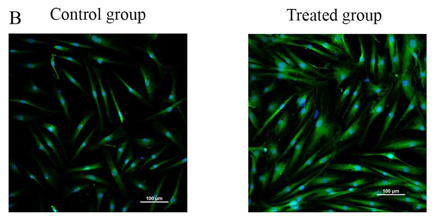
ScienceAsia 48 (2022)                                                                                             279

tion of skin fibroblast activities associated with wound   was kept in the desiccator at 25 ± 2 °C until further
granulation and subsequent wound closure. We also          use. The protein content and the molecular weight
expected to demonstrate that the physicochemical           pattern of the fibroin extract were determined using
properties of the sterilized film would not be altered     DC protein assay kit and sodium dodecyl sulfate poly-
by gamma irradiation process, and that the biologi-        acrylamide gel electrophoresis (SDS-PAGE) method,
cal activities of gamma-irradiated film would enhance      respectively [2]. The chemical characteristics of the
wound healing.                                             extract were analyzed using Fourier transform infrared
                                                           spectroscopy (FTIR spectrometer, Spectrum GX series,
MATERIALS AND METHODS                                      USA) [2].
Materials
                                                           Preparation and characteristic determination of
Yellow silkworm cocoons (Bombyx Mori, Nang-                the Aloe vera gel extract
Laai strain) were contributed by the Queen Sirikit
Sericulture Center, Chiang Mai Province, Thailand.         The extract of Aloe vera gel was prepared accord-
Aloe vera was cultured and collected from Phitsanulok      ing to the method described our previous study with
Province, Thailand.       Chemicals/materials were         some modifications [2]. Briefly, the colorless gel
purchased from different companies: calcium                part was collected, homogenized, and centrifuged at
chloride, sodium hydroxide, and ammonium sulfate           12 000 rpm at 4 °C for 15 min. The supernatant was
from RCI Labscan, Bangkok, Thailand; lactic acid           collected and precipitated by adding (NH4 )2 SO4 to get
solution (88%), sulfuric acid, and lipopolysaccharide      55% (w/v) of (NH4 )2 SO4 . The resulting precipitates
(LPS) from Sigma-Aldrich Chemie GmbH, Steinheim,           were isolated, dissolved in DI water. The obtained
Germany; dialysis membrane standard RC tubing              solution was dialyzed against 15 MΩ water using dial-
(MWCO: 6–8 kDa) from Spectrum Laboratories, Inc.,          ysis membrane standard RC tubing (MWCO 6–8 kDa)
California, USA; detergent compatible (DC) protein         at 23 ± 2 °C for 1 day with changes of water every
assay kit from BIO-RAD Laboratories, Philadelphia,         4–6 h. The desalted solution was lyophilized, and the
USA; phenol from AppliChem GmbH, Darmstadt,                lyophilized aloe gel extract was kept in the desiccator
Germany; Modified Eagle’s Medium (DMEM), fetal             at 25 ± 2 °C until further use. The protein content
bovine serum (FBS), and 0.25% trypsin/0.01M                and the molecular weight pattern of the extract were
EDTA from Sigma-Aldrich Co., Missouri, USA;                determined by DC protein assay kit and SDS-PAGE,
penicillin/streptomycin solution (10 000 U/ml)             respectively. Functional groups of the extract were
and amphotericin B (250 µg/ml) from Gibco,                 determined using FTIR spectrometry [2].
Invitrogen, Massachusetts, USA; cell proliferation
kit II (2,3-bis (2-methoxy-4-nitro-5-sulphophenyl)-5-      Preparation and characteristic determination of
[(phenylamino)carbonyl]-2H-tetrazoliumhydroxide,           the blended fibroin/aloe gel film
XTT) from Roche Diagnostics GmbH, Mannheim,                The blended fibroin/aloe gel film was prepared by
Germany; Mueller Hinton Agar from HiMedia,                 casting method described in our previous study with
Mumbai, India; and Muse™ Cell Cycle Assay Kit SDS          some modifications [2]. 540 mg of the fibroin extract
from MERCK, Darmstadt, Germany.                            and 15 mg of the aloe gel extract were separately
                                                           dissolved for 1 h in aqueous solution (with maintained
Preparation and characteristic determination of            pH 4.0 ± 0.2 using lactic acid) to a final volume of
the fibroin extract                                        15 ml. The mixture solution was then filtered and
The extraction of silk fibroin was performed according     subsequently cast in a square-shaped silicone mold
to the method described our previous study with some       (6 cm × 6 cm) under a dust-free condition and a main-
modifications [2]. Briefly, small pieces of silkworm       tained temperature of 47 ± 2 °C. The ratio of the fibroin
cocoons were treated with hot deionized (DI) water at      extract and the aloe gel extract in the film (36 cm2 ) was
85–90 °C for 2 h followed by 25 mM NaOH at 70 °C for       97.3% to 2.7% by weight. The physicochemical char-
30 min to remove silk gum protein. The degummed            acteristics of the films were done as follows; (1) scan-
fibers were washed with DI water and dried at 45 °C        ning electron microscopy (SEM, DAX®, LEO1455VP,
overnight. The dried samples were dissolved in 3 M         New Jersey, USA) for surface morphology observation;
CaCl2 solution (1 g of samples to 60 ml of CaCl2 ) at      (2) mechanical texture analysis (TA.XT Plus, Stable Mi-
85–90 °C for 4–6 h. The resulting solution was fil-        cro Systems, Ltd, Godalming, UK) including determi-
tered and dialyzed against 15 MΩ water using dialysis      nation of tensile strength and elongation at break; and
membrane standard RC tubing (MWCO 6–8 kDa) at              (3) Fourier transform infrared spectroscopy (FTIR) for
23 ± 2 °C for 2 days, with changes of water every 4–6 h,   chemical characteristics determination. The film was
until salts were completely removed. The desalted          sterilized by gamma irradiation technique (facilitated
solution was then centrifuged at 8000 rpm at 4 °C          by Thai Adhesive Tapes Industry Co., Ltd, Bangkok,
for 15 min. Finally, the supernatants were collected       Thailand), and the sterility of the irradiated film was
and lyophilized, and the lyophilized fibroin extract       confirmed by observing the appearance of bacteria

                                                                                                    www.scienceasia.org
280                                                                                           ScienceAsia 48 (2022)

growth using the agar plate culture technique [10].        3 min (5 times). Finally, the cells were mounted with
                                                           anti-fade mounting solution and observed under Laser
Determination of biological activities of the              confocal microscope (A1 HD25/A1R HD25, Nikon®,
blended fibroin/aloe gel film                              Tokyo, Japan).
Cytocompatibility and expression of growth factor
by skin fibroblast cells                                   Cell cycle
Normal human dermal fibroblast (NHDF) cells (Lot no.       NHDF cells (1 × 10⁵ cells/well, passage number 6)
C-12302, Promocell, Eppelheim, Germany) (1 × 10⁵           were seeded in a 24-well plate and incubated at 37 °C
cells/well, passage number 6) were seeded in a 24-         in 5% CO2 incubator for 24 h. The medium was
well plate, cultured in DMEM containing 10% FBS,           then replaced by DMEM serum-free. Trans-well inserts
and incubated at 37 °CC in a humidified 5% CO2             containing 6 mm diameter sterilized films were put
atmosphere for 24 h. The incubated medium was              into a 24-well plate with the adherent fibroblasts and
then replaced with serum-free medium. The sterilized       incubated for 24 h. After the incubation, cells were
films were cut into pieces in a circular shape (6 mm in    trypsinized using 0.25% trypsin/0.01 M EDTA. Cell
diameter and 4.56 mg in weight) and placed into trans-     suspensions (2 × 10⁵ cells/ml) were centrifuged, and
well inserts which were, then, put into individual wells   the cell pellet was then washed with PBS (pH 7.4)
of the 24-well plate containing fibroblast cells and       and fixed with 70% EtOH for 3 h. After the wash,
incubated for 24 h. After the incubation, the trans-well   cells were stained with 150 µl of Muse™ Cell Cycle
inserts were removed from the 24-well plate, and the       Assay Kit reagent at room temperature for 30 min
incubated serum-free medium was then discarded and         (protected from light). Cell cycle was then analyzed by
replaced with 250 µl of new serum-free medium plus         flow cytometry. The percentages of total cells for the
XTT reagent. The seeded cells were further incubated       cell cycle phases (G0 /G1 , S, and G2 /M) of the control
for 4 h, and the supernatant was then collected for        (untreated cells) and the film-treated sample were
absorbance measurement at 490 nm using microplate          calculated. The experiment was done in 3 replicates.
reader (Eon™, BioTek instrument, Vermont, USA). The
optical density of the control (untreated cells) was
adjusted to 100%, and the cell viability was thus shown    Migration of skin fibroblasts
in percentage. The experiment was performed in             The skin fibroblast migration was studied using the
triplicates.                                               scratch assay, which is typically utilized to quantify the
     Expression of vascular epidermal growth factor        migration of cells on two-dimensional (2-D) surfaces
(VEGF) was qualitatively determined using the Anti-        over time, following a modified method described in
VEGFA antibody (ab39250, Abcam, Massachusetts,             [11, 12]. NHDF cells (1×10⁵ cells/well, passage num-
USA). In brief, film extracts were prepared by incu-       ber 7) were seeded in a 24-well plate and incubated at
bating 1 × 1 cm2 sterilized films in 1 ml of DMEM          37 °C in 5% CO2 incubator for 48 h, during which cells
serum-free and incubated at 37 °C in 5% CO2 incubator      had grown to confluency in a monolayer. A scratch was
for 24 h. NHDF cells (2 × 10⁵ cells/well, passage          then made with a pipette tip by creating an incision-
number 11) were seeded in a cell culture slide with        like gap in the confluent monolayer of the fibroblasts in
DMEM containing 10% FBS and incubated at 37 °C             each well. The fibroblast scratches were washed twice
in a humidified 5% CO2 atmosphere for 24 h. The            with sterilized PBS (pH 7.4) followed by adding 800 µl
cultured medium was then replaced with the film ex-        of DMEM serum-free into each well. The trans-well
tracts and incubated for 24 h. Subsequently, cells were    inserts containing the sterilized films (circular shape,
fixed with 4% paraformaldehyde in PBS for 10 min           6 mm in diameter) were put into the 24-well plate
at room temperature, permeabilized by 0.1% Triton-         with fibroblast scratches, and the plate was further
X 100 at room temperature for 10 min, and washed           incubated at 37 °C in 5% CO2 incubator for 36 h.
3 times with PBS for 5 min. Cells were then incubated      The scratched gaps were photographed immediately
with 1% BSA, 22.52 mg/ml glycine in PBST to block          after scratching at defined time points (0, 12, 24,
unspecific binding of antibodies. At 30 min later, they    and 36 h). At 36 h, cell migration was observed as
were incubated with Anti-VEGFA antibody (diluted in        a completed closure of the scratched gaps under an
1% BSA in PBST) in a humidified chamber at 4 °C            inverted microscope [13]. The experiment was done
overnight. After washes with PBS for 3 min (5 times),      in three replicates.
cells were then incubated with Alexa Fluor®488 con-
jugated secondary antibody (diluted in 1% BSA in
PBST) at room temperature for 1 h in the dark. The         Statistical analysis
secondary antibody solution was discarded, and the         All values were expressed as mean ± SD. The student’s
cells were washed with PBS for 3 min (5 times). After      unpaired t-test was used to compare between the
the washes, the cells were incubated with 100 µl of        control and the sample. p < 0.01 was considered
DAPI (DNA stain) for 1 min and rinsed with PBS for         significant difference.

www.scienceasia.org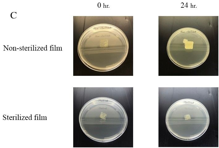
ScienceAsia 48 (2022) 281
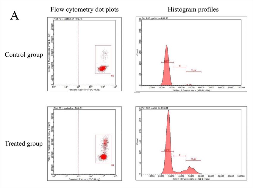
RESULTS stimulated the expression of VEGF. Compared with
Characteristics of the fibroin extract the control, the number and the size/shape of the
treated cells were increased (Fig. 4B). In comparison
The lyophilized fibroin extract prepared from silk- to the control, the treated cells showed a decrease in
worm cocoons (Nang-Laai strain) presented yellowish the percentage of total cells in the G0 /G1 phase; on
cotton-like characteristics. One gram of silk cocoons the other hand, increases in the percentages of total
yielded 0.58 g (58% w/w) of the extract. A DC protein cells of 7.19 ± 0.23% and 16.09 ± 0.58% were found in
assay showed a protein content of 97.43 ± 0.44% w/w the S and the G2 /M phases, respectively, which were
of the extract. Infrared spectra obtained using FTIR higher than those of the untreated cells (2.53 ± 0.92
spectroscopy showed the frequency peaks at 1634 and 4.67 ± 1.61%, respectively) (Fig. 5). Fig. 6 shows
(amide I), 1513 (amide II), and 1232 (amide III) cm−1 the result of cell migration, measured as the closure
(Fig. 1A). The molecular weight pattern of the extract, of the scratch gap at various times, indicating that at
as shown in Fig. 1B, indicated a specific band of L-chain 36 h after scratch creation, the treated cells provided
at approximately 25 kDa and a smear band of H-chain a completely healed scratch, while the scratch of the
in the range of 30 to 245 kDa. untreated cells was not healed.
Characteristics of the aloe gel extract
DISCUSSION
The lyophilized extract of the Aloe vera gel showed
white cotton-like characteristics, and 100 g of the gel In this study, Nang-Laai strain cocoons provided the
produced 6 g (0.06% w/w) of the extract. The protein percentage of yield and the protein content of the fi-
content in the extract was 6.86 ± 1.15% w/w of the broin extract corresponding to our previous report [2].
extract. Fig. 2A shows the IR spectra of the aloe gel The presence of amides I, II, and III and the random
extract indicating peak at 1731 (O-acetyl ester), 1238 coil groups in the FTIR spectrum confirmed that the
(O-acetyl ester), 1059 (glucan units), 955 (pyranoside extract structure consists of water-soluble random coil
ring), and 807 (mannose) cm−1 . The molecular weight conformation [14]. The appearance of the smeared
pattern of the extract showed a clear band at approxi- band in the analysis of protein, using SDS-PAGE tech-
mately 14 and 35 kDa (Fig. 2B). nique, might be a consequence of the degradation of
the heavy (H) chain (325–350 kDa) of silk fibroin
Characteristics of the blended fibroin/aloe gel protein during the extraction process. The clear band
extract at the range of 17–25 kDa is related to the light (L)
The physical appearance of the prepared film was flex- chain of fibroin [15].
ible, translucent, and yellowish with uniform thickness The percentage of yield and the protein content
of 50 µm. The SEM images showed a non-porous mor- of the aloe gel extract were similar to our previous
phology on the surface of the non-sterilized (Fig. 3A1) findings [2]. The FTIR spectrum showed the presence
as well as sterilized film (Fig. 3A2). For the mechan- of functional groups, including glucan units, pyra-
ical properties, the non-sterilized film provided the noside, and mannose relating to anti-inflammatory
breaking force and percentage of elongation at break and healing activities of the aloe gel extract [16, 17].
at 6.038 ± 0.746 N and 1.147 ± 0.119%, respectively, The SDS-PAGE molecular weight pattern was observed
which are not significantly different (p > 0.5) to the with two clear bands of mannose-binding lectin at
sterilized film (6.26 ± 0.44 N for the breaking force and approximately 14 and 35 kDa, also indicating the
1.20 ± 0.07% for percent elongation at break). The activities of anti-inflammatory [18], hemagglutinating,
FTIR spectroscopy presented infrared spectra of the and mitogenic activities [19] of the extract.
non-sterilized and sterilized film at 1633 and 1634 In our current study, the fibroin/aloe gel film
(amide I), 1514 and 1513 (amide II), 1231 and 1228 was sterilized by gamma irradiation which is one of
(amide III), 1055 and 1057 (glucan units), 1013 and the most common sterilization methods for health
1013 (pyranoside ring), 826 and 828 (mannose), re- care products including wound dressing [20] and
spectively, as shown in Fig. 3(B1,B2). Focusing on the temperature-sensitive materials [21]. However, some
sterility test using agar plate culture, the sterilized film studies indicated the adverse effects on molecular
showed no colonies of microbes or bacteria growth mechanisms involving gamma rays-induced cell dam-
(Fig. 3C). ages [22], microorganisms resistance [23], and struc-
tural changes in medical devices made of polymer [24].
Biological activities of the blended fibroin/aloe gel For these reasons, the physical characteristics, chem-
film ical, and sterility of the gamma-irradiated film were
For the cytocompatibility of the fibroin/aloe gel film, determined in the present study. The results showed
the viability percentage of the treated NHDF cells that gamma irradiation efficiently killed the microor-
was 145.95 ± 1.86, which is significantly higher than ganisms on the film; the physicochemical properties of
the control’s (100 ± 5.34) (p < 0.01) (Fig. 4A). In the the film were not affected; i.e. its physical appearance,
immunofluorescence experiment, the film significantly surface morphology, mechanical properties, and chem-
www.scienceasia.org282 ScienceAsia 48 (2022)
A
Fig. 1 (A) Infrared spectrum and (B) molecular weight pattern of the fibroin extract prepared from yellow silkworm cocoons
(Nang-Laai strain).
A
Fig. 2 (A) Infrared spectrum and (B) molecular weight pattern of the aloe gel extract prepared from the gel part of Aloe vera
leaves.
ical characteristics remained unchanged. in cell number, or the expression of VEGF, indicating
Results from our previous study showed that the the improvement of cell attachment and proliferation
fibroin/aloe gel film exerted potential healing effects, as well as the growth factor expression by the primary
in vitro and in vivo, and promoted wound closure skin fibroblasts seeded on the film [2]. There have
by 7 days compared with the untreated cells in been many studies supporting the potential of the silk
streptozotocin-induced diabetic rats [2]. In a pre- fibroin and aloe gel extracts on the acceleration of cell
liminary clinical study, the developed film accelerated proliferation and cell migration in the wound healing
the healing rate in 5 DFU patients within 4 weeks. process [2, 27, 28]. To determine the essential role
However, in the current study, we sought to empha- of the fibroin/aloe gel film in the migration and the
size the biological activities associated with healing proliferation of fibroblast cells on the wound healing
property to support the efficacy of the film sterilized process, we further performed the flow cytometry and
by gamma irradiation. The cytotoxicity results showed the scratch assay.
that the sterilized film did not affect the cell viability Focusing on cell proliferation relating to the cell
and morphology. An XTT assay demonstrated that cycle, which is the complex and orderly cellular process
the percentage of cell viability relating to the number through specific phases during the replication of DNA
of cells had a higher OD value, implying a higher into two daughter cells, our data revealed that the
proliferation rate. VEGF can be considered as a key film-treated cells shifted from G0 /G1 phase to S phase
angiogenesis regulator secreted by fibroblasts. It is and then G2 /M phase. Hence, the film promoted
also one of the most essential mediators associated the cells to enter into the S and G2 /M phases, which
with the wound healing process because it improves are essential stages for cell mitosis and cell growth,
the survival, the proliferation, and the migration of respectively [29]. Besides, Wei et al [30] showed that
endothelial cells [25, 26]. The results on the expres- an acemannan consisting of Aloe vera can stimulate
sion of VEGF indicated that the sterilized film did not cell proliferation by influencing the cyclin-dependent
induce any adverse effect, but stimulated an increase cell cycle through translational regulation of cyclin D1,
www.scienceasia.orgScienceAsia 48 (2022) 283
B1
B2
Fig. 3 (A1) SEM images of surface photomicrographs of the non-sterilized film and (A2) the sterilized film; (B1) infrared
spectra of the non-sterilized film and (B2) the sterilized film; and (C) sterility test of the non-sterilized and the sterilized films
after incubation for 24 h.
www.scienceasia.org284 ScienceAsia 48 (2022)
A *
160
140
120
100
% viability
80
60
40
20
0
Control group Treated group
Fig. 4 (A) Viability of NHDF cells treated with the blended fibroin/aloe gel film for 24 h. Data are expressed as percentage of
the control (untreated cells), and each column represents mean ± S.D. of triplicate study; * p < 0.01. (B) Immunofluorescence
for VEGF expression of the control (untreated NHDF cells) and the treated cells at 20 × magnification.
B 100.00
90.00
80.00
70.00
Percent of total cell (%)
60.00
50.00
40.00
30.00
20.00
10.00
0.00
Control group Treated group Control group Treated group Control group Treated group
G0/G1 phase S phase G2/M phase
Fig. 5 Cell cycle phases of NHDF cells treated with the blended fibroin/aloe gel film for 24 h compared with the control
(untreated cells). The figure shows the examples of cell cycle distribution in: (A) dot plots and histogram profiles;
(B) percentage of the total cell. Each column represents mean ± S.D. of triplicate study.
Fig. 6 Cell migration of NHDF cells treated with the blended fibroin/aloe gel film at 0, 12, 24, and 36 h compared with the
control (untreated cells) at 10x magnification.
www.scienceasia.orgScienceAsia 48 (2022) 285
which is the main alteration attributed to the transition sity for facility supports. We wish to thank Mr. Roy I. Morien
of G1 phase to S phase. of the Naresuan University Graduate School for his efforts in
Furthermore, it was found that the fibroin/aloe gel editing the English grammar and expression in this paper.
film exerted a beneficial effect by promoting the mi-
gration of fibroblasts and, thereby, stimulating wound REFERENCES
closure [31]. Additionally, cell proliferation and cell 1. Alexiadou K, Doupis J (2012) Management of diabetic
migration are correlated in the cell cycle, particularly foot ulcers. Diabetes ther 3, ID 4.
in mitosis phase (M phase), associated with the cell 2. Inpanya P, Faikrua A, Ounaroon A, Sittichokechaiwut,
division process when duplicated DNA and cytoplasm Viyoch J (2012) Effects of the blended fibroin/aloe
gel film on wound healing in streptozotocin-induced
were divided to create two identical cells [32]. Cell mi-
diabetic rats. Biomed Mater 7, ID 035008.
gration can be also correlated to cytoskeletal reorgani- 3. Zhang W, Chen L, Chen J, Wang L, Gui X, Ran J, Xu
zation and focal adhesion receptors [33]. Interestingly, G, Zhao H, et al (2017) Silk fibroin biomaterial shows
cell proliferation result described above showed that safe and effective wound healing in animal models and
the highest percentage of total cells in the fibroin/aloe a randomized controlled clinical trial. Adv Healthc Mater
gel film treated cells was found accumulated in the 6, ID 1700121.
G2 /M phase, implying the promotion of cell differ- 4. Mori H, Tsukada M (2000) New silk protein: modifica-
entiation and cell migration processes by the treated tion of silk protein by gene engineering for production
cells. The result was consistent with several prior of biomaterials. J Biotechnol 74, 95–103.
studies reporting that natural compounds promoted 5. Meinel L, Hofmann S, Karageorgiou V, Kirker-Head C,
McCool J, Gronowicz G, Zichner L, Langer R, et al
G2 /M phase and fibroblast migration [34, 35].
(2005) The inflammatory responses to silk films in vitro
The limitation of this study due to the use of and in vivo. Biomaterials 26, 147–155.
normal fibroblast cells should be noted. In our previous 6. Chithra P, Sajithlal GB, Chandrakasan G (1998) Influ-
study, the activities of the fibroin/aloe gel film were ence of Aloe vera on the glycosaminoglycans in the ma-
evaluated in the streptozotocin-induced diabetic rat trix of healing dermal wounds in rats. J Ethnopharmacol
and the diabetic fibroblast cells related to the DFU 59, 179–186.
patients [2]. In general, the growth factor functions 7. Maenthaisong R, Chaiyakunapruk N, Niruntraporn S,
of the diabetic fibroblast cells are impaired leading to Kongkaew C (2007) The efficacy of Aloe vera used for
the delayed cell proliferation and cell migration when burn wound healing: A systematic review. Burns 33,
compared with the normal fibroblast cells [36, 37]. 713–718.
8. Femenia A, Sánchez ES, Simal S, Rosselló C (1999)
However, the presence of functional groups in the fi-
Compositional features of polysaccharides from Aloe
broin/aloe gel film, including amides (I, II, III), glucan vera (Aloe barbadensis Miller) plant tissues. Carbohydr
units, pyranoside ring, and mannose, may enhance Polym 39, 109–117.
the proliferation and the migration of cells, leading 9. Sahu PK, Giri DD, Singh R, Pandey P, Gupta S, Shrivas-
to wound healing improvement in the diabetic foot tava AK, Kumar A, Pandey KD (2013) Therapeutic and
ulcers. Nevertheless, further studies on the expression medicinal uses of Aloe vera: A Review. Pharmacol Pharm
of growth factors and transcription factors of the film in 4, 599–610.
biomolecular level should be performed using human 10. Sanders ER (2012) Aseptic laboratory techniques: plat-
diabetic dermal fibroblast cells to investigate its wound ing methods. J Vis Exp 63, e3064.
healing efficacy. 11. Pinto Bi, Cruz ND, Lujan OR, Propper CR, Kellar RS
(2019) In vitro scratch assay to demonstrate effects of
CONCLUSION arsenic on skin cell migration. J Vis Exp 144, e58838.
12. He S, Shi D, Han Z, Dong Z, Xie Y, Zhang F, Zeng W, Yi Q
The physicochemical and biological activities of (2019) Heparinized silk fibroin hydrogels loading FGF1
gamma-irradiated fibroin/aloe gel film were deter- promote the wound healing in rats with full-thickness
mined to support the wound healing efficacy reported skin excision. Biomed Eng Online 18, ID 97.
in our previous study. The gamma irradiation was 13. Monsuur HN, Boink MA, Weijers EM, Roffel S, Breetveld
found to have no effects on the film’s physicochemical M, Gefen A, Broek LJ, Gibbs S (2016) Methods to study
properties (physical appearance, surface morphology, differences in cell mobility during skin wound healing in
mechanical properties, and chemical characteristics). vitro. J Biomech 49, 1381–1387.
14. Zhong J, Ma M, Li W, Zhou J, Yan Z, He D (2014) Self-
For biological activities, the blended fibroin/aloe gel
assembly of regenerated silk fibroin from random coil
film enhanced the proliferation and the migration of nanostructures to antiparallel β-sheet nanostructures.
the fibroblast cells and, thereby, might be able to help Biopolymers 101, 1181–1192.
in stimulating the DFU wound healing process. 15. Altman GH, Diaz F, Jakuba C, Calabro T, Horan RL,
Chen J, Lu H, Richmond J, et al (2003) Silk-based
Acknowledgements: We would like to thank Naresuan Uni- biomaterials. Biomaterials 24, 401–416.
versity for financial support for graduate student. We also 16. Esua MF, Rauwald J-W (2006) Novel bioactive maloyl
thank the Center of Excellence for Innovation in Chemistry glucans from Aloe vera gel: Isolation, structure elucida-
(PERCH-CIC), Office of the Higher Education Commission tion and in vitro bioassays. Carb Res 341, 355–364.
and the Faculty of Pharmaceutical Sciences, Naresuan Univer- 17. Kumar K, Bhowmik D, Bhattacharjee C, Biswajit (2010)
www.scienceasia.org286 ScienceAsia 48 (2022)
Aloe vera: a potential herb and its medicinal importance. 2956–2965.
J Chem Pharm Res 2, 21–29. 28. Teplicki E, Ma Q, Castillo DE, Zarei M, Hustad AP, Chen
18. Das S, Mishra B, Gill K, Ashraf MS, Singh AK, Sinha J, Li J (2018) The effects of Aloe vera on wound healing
M, Sharma S, Xess I, et al (2011) Isolation and char- in cell proliferation, migration, and viability. Wounds 30,
acterization of novel protein with anti-fungal and anti- 263–268.
inflammatory properties from Aloe vera leaf gel. Int J Biol 29. Hengst L, Nigg EA (2006) Cell cycle: Overview. In:
Macromol 48, 38–43. Encyclopedic Reference of Genomics and Proteomics in
19. Koike T, Beppu H, Kuzuya H, Maruta K, Shimpo K, Molecular Medicine, Springer, Berlin, Heidelberg, pp
Suzuki M, Titani K, Fujita K (1995) A 35 kDa mannose- 228–233.
binding lectin with hemagglutinating and mitogenic
30. Wei X, Guo W, Zou C-H, Fu T-T, Li X-Y, Zhu M, Qi J-H,
activities from “Kidachi Aloe” (Aloe arborescens Miller
Song J, et al (2015) Acemannan accelerates cell pro-
var. natalensis Berger). J Biochem 118, 1205–1210.
liferation and skin wound healing through AKT/mTOR
20. Fairand BP (2001) Radiation Sterilization for Health Care
signaling pathway. J Dermatol Sci 79, 101–109.
Products. X-Ray, Gamma, and Electron Beam, 1st edn,
CRC Press, Boca Raton, Florida, USA. 31. Bainbridge P (2013) Wound healing and the role of
21. Aquino K (2012) Sterilization by Gamma Irradiation, fibroblasts. J Wound Care 22, 407–408, 410–412.
Gamma Radiation, IntechOpen Limited, London, UK. 32. Yang VW (2012) Chapter 15: The cell cycle. In: Johnson
22. AlZahrani K, Al-Sewaidan HA (2017) Nanostructural LR, et al (eds) Physiology of the Gastrointestinal Tract,
changes in the cell membrane of gamma-irradiated 5th edn, Academic Press, Boston, pp 451–471.
red blood cells. Indian J Hematol Blood Transfus 33, 33. Kodama A, Lechler T, Fuchs E (2004) Coordinating
109–115. cytoskeletal tracks to polarize cellular movements. J Cell
23. Grieb TA, Forng R-Y, Stafford RE, Lin J, Almeida J, Biol 167, 203–207.
Bogdansky S, Ronholdt C, Drohan WN, et al (2005) 34. Harishkumar M, Masatoshi Y, Hiroshi S, Tsuyomu I,
Effective use of optimized, high-dose (50 kGy) gamma Masugi M (2013) Revealing the mechanism of in vitro
irradiation for pathogen inactivation of human bone wound healing properties of Citrus tamurana extract.
allografts. Biomaterials 26, 2033–2042. Biomed Res Int 2013, ID 963457.
24. Araújo ES, Khoury HJ, Silveira SV (1998) Effects of 35. Zhang S-L, Li B-L, Li W, Lu M, Ni L-Y, Ma H-L, Meng Q-G
gamma-irradiation on some properties of durolon poly- (2018) The Effects of ludartin on cell proliferation, cell
carbonate. Radiat Phys Chem 53, 79–84. migration, cell cycle arrest and apoptosis are associated
25. Khan S, Villalobos MA, Choron RL, Chang S, Brown SA, with upregulation of p21WAF1 in Saos-2 osteosarcoma
Carpenter JP, Tulenko TN, Zhang P (2017) Fibroblast cells in vitro. Med Sci Monit 16, 4926–4933.
growth factor and vascular endothelial growth factor 36. Lamers ML, Almeida ME, Vicente-Manzanares M, Hor-
play a critical role in endotheliogenesis from human witz AF, Santos MF (2011) High glucose-mediated ox-
adipose-derived stem cells. J Vasc Surg 65, 1483–1492. idative stress impairs cell migration. PLoS One 6, ID
26. Ferrara N (2000) VEGF: an update on biological and e22865.
therapeutic aspects. Curr Opin Biotechnol 11, 617–624. 37. Desta T, Li J, Chino T, Graves DT (2010) Altered fi-
27. Mandal BB, Kundu SC (2009) Cell proliferation and broblast proliferation and apoptosis in diabetic gingival
migration in silk fibroin 3D scaffolds. Biomaterials 30, wounds. J Dent Res 89, 609–614.
www.scienceasia.orgYou can also read