Regulation of human trophoblast gene expression by endogenous retroviruses - bioRxiv
←
→
Page content transcription
If your browser does not render page correctly, please read the page content below
bioRxiv preprint doi: https://doi.org/10.1101/2022.04.26.489485; this version posted April 26, 2022. The copyright holder for this preprint
(which was not certified by peer review) is the author/funder, who has granted bioRxiv a license to display the preprint in perpetuity. It is
                                          made available under aCC-BY 4.0 International license.

          Regulation of human trophoblast gene expression by
          endogenous retroviruses

          Jennifer M. Frost1,#, Samuele M. Amante1, Hiroaki Okae2, Eleri M. Jones1, Brogan
          Ashley3, Rohan M. Lewis3, Jane K. Cleal3, Matthew P. Caley1, Takahiro Arima2,
          Miguel R. Branco1,#

          1
            Blizard Institute, Barts and The London School of Medicine and Dentistry, QMUL,
          London E1 2AT, UK
          2
            Department of Informative Genetics, Environment and Genome Research Center,
          Tohoku University Graduate School of Medicine, Sendai 980-8575, Japan
          3
            The Institute of Developmental Sciences, Human Development and Health, Faculty
          of Medicine University of Southampton, Southampton O16 6YD, United Kingdom
          #
              Corresponding authors. Emails: j.frost@qmul.ac.uk, m.branco@qmul.ac.uk

          Abstract
          The placenta is a fast-evolving organ with large morphological and histological
          differences across eutherians, but the genetic changes driving placental evolution
          have not been fully elucidated. Transposable elements, through their capacity to
          quickly generate genetic variation and affect host gene regulation, may have helped
          to define species-specific trophoblast gene expression programmes. Here, we
          assessed the contribution of transposable elements to human trophoblast gene
          expression as enhancers or promoters. Using epigenomic data from primary human
          trophoblast and trophoblast stem cell lines, we identified multiple endogenous
          retrovirus families with regulatory potential that lie close to genes with preferential
          expression in trophoblast. These largely primate-specific elements are associated
          with inter-species gene expression differences, and are bound by transcription
          factors with key roles in placental development. Using genetic editing we
          demonstrated that several elements act as transcriptional enhancers of important
          placental genes, such as CSF1R and PSG5. We also identified an LTR10A element
          that regulates ENG expression, affecting secretion of soluble ENG, with potential
          implications for preeclampsia. Our data show that transposons have made important
          contributions to human trophoblast gene regulation, and suggest that their activity
          may affect pregnancy outcomes.

                                                                      1
bioRxiv preprint doi: https://doi.org/10.1101/2022.04.26.489485; this version posted April 26, 2022. The copyright holder for this preprint
(which was not certified by peer review) is the author/funder, who has granted bioRxiv a license to display the preprint in perpetuity. It is
                                          made available under aCC-BY 4.0 International license.

          Introduction
          The success of human pregnancy depends on the healthy development and function
          of the placenta. Establishment of appropriate blood flow and subsequent nutrient
          exchange is a carefully orchestrated balance between the requirements of the fetus
          and the mother, regulated by the interplay between immunological, genetic and
          hormonal systems at the feto-maternal interface. Following implantation, fetally
          derived trophoblast cells invade maternal tissues interstitially, remodelling uterine
          spiral arteries well into the myometrium (Turco and Moffett, 2019). Aberrations to this
          process result in serious pregnancy complications that cause maternal and fetal
          morbidity and mortality, including recurrent pregnancy loss, fetal growth restriction,
          preterm birth and preeclampsia in the case of too little invasion, or disorders of the
          placenta accreta spectrum where invasion is too extensive (Brosens et al., 2011).
          However, the genetic determinants of these disorders remain unclear, as genome-
          wide association studies have revealed very few candidates, with the notable
          exception of FLT1 in preeclampsia (McGinnis et al., 2017).
          Placental development and structure displays wide variation across eutherian
          species, even within the primate order (Carter and Enders, 2004). Notably, the deep
          interstitial trophoblast invasion observed in humans is unique to great apes (Carter,
          2021). Placentas differ in cellular composition, histological arrangement and gross
          morphology, as well as many molecular aspects, all of which shape interactions
          between conceptus and mother, and their outcomes. This striking variation reflects
          the myriad selective pressures associated with the feto-maternal conflicts that fuel
          fast evolution of this organ (Chuong et al., 2010). Yet, the genetic drivers of placental
          evolution remain to be fully elucidated.
          One important yet understudied source of genetic variation is transposable elements
          (TEs). These abundant repetitive elements, which include endogenous retroviruses
          (ERVs), have made major contributions to human evolution, helping to shape both
          the coding and regulatory (non-coding) landscape of the genome. Akin to the variable
          and species-specific development and structure of the placenta, TEs are highly
          species-specific, making them putative drivers of placental evolution. Indeed, multiple
          genes with key roles in placentation have been derived from TEs (Imakawa et al.,
          2015), most prominently the syncytin genes, whose products mediate cell-cell fusion
          to generate a syncytialised trophoblast layer that directly contacts maternal blood
          (Lavialle et al., 2013). Additionally, the non-coding portions of TEs (e.g., the LTRs –
          long terminal repeats – in ERVs) have the ability to regulate gene transcription, and
          through this action contribute to human embryonic development (Fuentes et al.,
          2018; Pontis et al., 2019, 2021), innate immunity (Chuong et al., 2016), the
          development of cancer (Jang et al., 2019), and evolution of the feto-maternal
          interface, amongst others (Chuong et al., 2017; Fueyo et al., 2022). TEs can recruit
          host transcription factors, often in a highly tissue-specific manner, and gain
          epigenetic hallmarks of gene regulatory activity (e.g., open chromatin, enrichment of
          H3K27ac), acting as transcriptional promoters or distal enhancer elements (Chuong
          et al., 2017; Fueyo et al., 2022; Sundaram et al., 2014). We and others have
          previously shown that in mouse trophoblast stem cells, several ERV families are
          enriched for binding of key stemness factors (CDX2, ELF5, EOMES) and can act as
          major enhancers of gene expression (Chuong et al., 2013; Todd et al., 2019). In
          humans, several examples of TE-encoded placenta-specific promoters have been
          uncovered throughout the years, such as those driving expression of CYP19A1,
          NOS3 and PTN genes (Cohen et al., 2009). A fascinating example of a human
          placental TE-derived enhancer has also been described, wherein a THE1B ERV
          regulates the expression of the corticotropin-releasing hormone, affecting gestational
          length when inserted into the mouse genome (Dunn-Fletcher et al., 2018). More

                                                                      2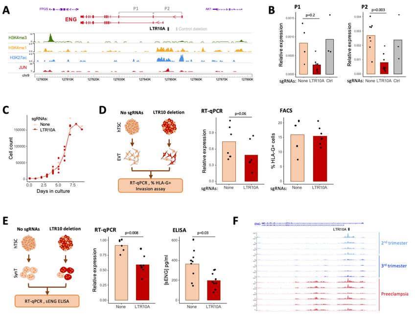
bioRxiv preprint doi: https://doi.org/10.1101/2022.04.26.489485; this version posted April 26, 2022. The copyright holder for this preprint
(which was not certified by peer review) is the author/funder, who has granted bioRxiv a license to display the preprint in perpetuity. It is
                                          made available under aCC-BY 4.0 International license.

          recently, the Macfarlan lab has used epigenomic data to identify a group of putative
          lineage-specific placental enhancers that are derived from ERVs (Sun et al., 2021).
          However, as the placenta is a heterogeneous tissue, it remains unclear whether all of
          these ERVs are active in trophoblast cells, and a genetic demonstration of their
          regulatory action is lacking.
          Here, we identified ERV families that exhibit hallmarks of gene regulatory activity in
          human trophoblast. We show that these ERVs bind transcription factors required for
          placental development, and lie close to genes with preferential trophoblast
          expression in a species-specific manner. Using genetic editing, we show examples of
          ERVs that act as gene enhancers in trophoblast, including an LTR10A element within
          the ENG gene that regulates the secretion of soluble ENG protein by the
          syncytiotrophoblast, which is both a marker for, and contributor to, the pathogenesis
          of preeclampsia.

          Results
          Primate-specific ERVs exhibit regulatory characteristics in human trophoblast
          To identify interspersed repetitive elements bearing hallmarks of activating regulatory
          potential in human trophoblast, we performed H3K27ac profiling using either ChIP-
          seq or CUT&Tag (Kaya-Okur et al., 2019). We analysed our previously published
          data from primary human cytotrophoblast (Ashley et al., 2022), as well as newly
          generated data from cytotrophoblast-like human trophoblast stem cells (hTSCs)
          (Okae et al., 2018), which can be differentiated in vitro and allow for easy genetic
          manipulation (Figure 1A). Using the Repeatmasker annotation (excluding simple and
          low complexity repeats), we determined the frequency of H3K27ac peaks per repeat
          family, and compared it to random controls using a permutation test (Figure 1A). This
          revealed 29 repeat families enriched for H3K27ac peaks in both primary
          cytotrophoblast and hTSCs, the vast majority of which were primate-specific ERVs
          (Figure 1B; Supplementary Table S1). For comparison, we performed the same
          analysis on published H3K27ac CUT&Tag data from human embryonic stem cells
          (hESCs) (Kaya-Okur et al., 2019). Most hTSC-enriched repeats displayed little to no
          enrichment in hESCs (Figure 1B), despite the fact that hESCs make use of a large
          set of TEs for gene regulatory purposes (Kunarso et al., 2010; Pontis et al., 2019).
          This suggests a divergence in TE-associated regulatory networks that is set up early
          in development, and that may help direct extraembryonic development. A more
          detailed analysis of H3K27ac signals across repeats belonging to each of the
          enriched families confirmed the asymmetry between hTSCs and hESCs (Figure 1C).
          HERVH-associated LTRs present an interesting case in which LTR7C elements are
          H3K27ac-enriched in hTSCs, whereas the related LTR7 family is H3K27ac-enriched
          in hESCs (Supplementary Figure S1A) – more specifically, it is the LTR7up subfamily
          that is active in hESCs (Carter et al., 2022).
          To further validate our findings and narrow down the list of candidate regulatory
          repeat families, we also analysed placental DNAse-seq data from ENCODE. We
          compared these data to DNAse-seq profiles from liver, lung and kidney, highlighting
          that most hTSC-associated families displayed tissue-specific enrichment of elements
          with accessible chromatin (Figure 1D; contrast with non-tissue-specific examples in
          Supplementary Figure 1B). Based on stringent criteria (see Methods), we decided to
          focus on 18 placenta-specific candidate regulatory repeat families, all of which were
          ERV-associated LTRs (Supplementary Table S1). This included families with known
          examples of elements bearing promoter activity in the placenta: LTR10A (NOS3
          gene), LTR2B (PTN gene), MER39 (PRL gene), MER39B (ENTPD1 gene) and
          MER21A (HSD17B1 and CYP19A1 genes) (Cohen et al., 2009).

                                                                      3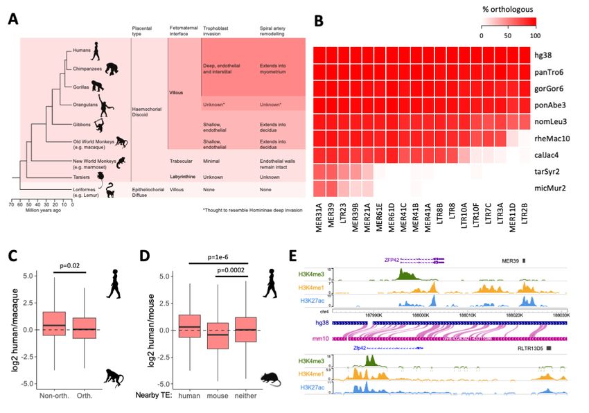
bioRxiv preprint doi: https://doi.org/10.1101/2022.04.26.489485; this version posted April 26, 2022. The copyright holder for this preprint
(which was not certified by peer review) is the author/funder, who has granted bioRxiv a license to display the preprint in perpetuity. It is
                                          made available under aCC-BY 4.0 International license.

          Figure 1 – Gene regulatory signatures of ERVs in human trophoblast. A) Primary cytotrophoblast and human
          trophoblast stem cells were profiled for H3K27ac to identify repeat families with putative gene regulatory potential. B)
          Enrichment for H3K27ac peaks for each repeat family in hTSCs or hESCs. Families with significant enrichment in
          hTSCs are highlighted. C) H3K27ac profiles of a subset of hTSC-enriched ERV families in hTSCs and hESCs. Each
          line represents an element in that family. D) Enrichment for DNase hypersensitive sites in the same ERV families, in
          kidney (K), liver (Li), lung (Lu) and placenta (P). Each datapoint represents a different ENCODE dataset. E)
          Proportion of elements from the same ERV families overlapping particular combinations of histone modifications.

          We then characterised in more detail the H3K27ac-marked elements from each of
          these families by performing CUT&Tag for H3K4me1, H3K4me3, H3K9me3 and
          H3K27me3 in hTSCs. This revealed that a large proportion of H3K27ac-marked
          elements across all families was also marked by H3K4me1 (median 72%, range 44-
          85%), a signature of active enhancers (Figure 1E). Only a small proportion (median
          3%, range 0-20%) was marked by H3K4me3, a signature of active promoters (Figure
          1E). There was also a large fraction of elements marked by H3K4me1 alone (median
          71% of all H3K4me1 elements, range 27-81%), which is normally associated with
          poised enhancers (Figure 1E), raising the possibility that this group of elements
          becomes active upon differentiation of hTSCs. To test this, we differentiated hTSCs
          into extravillous trophoblast (EVT; Supplementary Figure 1C) and performed
          CUT&Tag for H3K27ac. Half of the hTSC-active ERV families remained H3K27ac-
          enriched in EVT, whereas others displayed a clear specificity for the stem cell state
          (e.g., LTR10A, MER61E; Supplementary Figure 1D). In line with our hypothesis, a

                                                                      4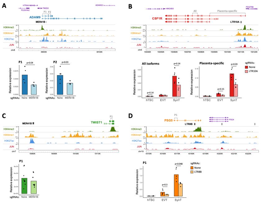
bioRxiv preprint doi: https://doi.org/10.1101/2022.04.26.489485; this version posted April 26, 2022. The copyright holder for this preprint
(which was not certified by peer review) is the author/funder, who has granted bioRxiv a license to display the preprint in perpetuity. It is
made available under aCC-BY 4.0 International license.
large proportion of ERVs that are active in EVT were found to be in a poised
enhancer state in hTSCs (Supplementary Figure 1E,F). It is possible that a different
set of poised ERV enhancers become active upon differentiation into
syncytiotrophoblast (SynT; Supplementary Figure 1C), but the multinucleated nature
of these cells seemingly interfered with our CUT&Tag attempts.
Our analyses suggest that a large number of ERVs (nearly all of which are primate-
specific) may act as gene regulatory elements in human trophoblast, showing
dynamic changes during differentiation.
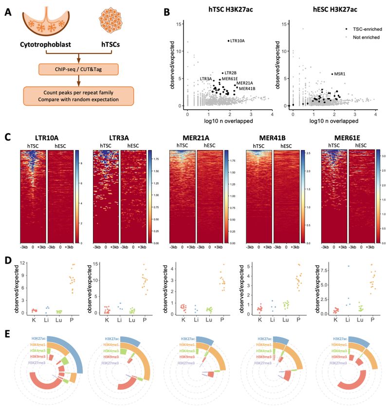
hTSC-active ERVs bind key placenta-associated transcription factors
Enhancers function to regulate gene expression through the binding of transcription
factors, providing exquisite tissue-specific control through sequential binding of
transcription factor combinations. We therefore identified transcription factor binding
motifs that were enriched within each hTSC-active ERV family, and focused on a
selection of transcription factors that are expressed in trophoblast. Reassuringly, we
identified previously described motifs on MER41B for STAT proteins and SRF
(Supplementary Figure 2A) (Chuong et al., 2016; Sun et al., 2021). We further
uncovered a large collection of motifs for transcription factors with known roles in
trophoblast development. Namely, multiple families bore motifs for key factors
involved in the maintenance of the stem cell state, such as ELF5, GATA3, TFAP2C,
TP63 and TEAD4 (Figure 2A; Supplementary Figure 2A). Additional transcription
factors with known roles in placental development and/or physiology included
JUN/FOS (He et al., 2019; Kubota et al., 2015), PPARG/RXRA (Kadam et al., 2015),
and FOXO3 (Chen et al., 2021). To validate the binding of some of these
transcription factors to hTSC-active TEs, we performed CUT&Tag or CUT&RUN
(Skene and Henikoff, 2017) for JUN, JUND, GATA3, TEAD4 and TFAP2C (Figure
2B). We applied the same peak enrichment pipeline as used above to identify
H3K27ac-enriched TE families and found that several of these families were enriched
for one or more of the evaluated transcription factors (Figure 2C; Supplementary
Figure 2B). In contrast, TE families active specifically in hESCs showed little to no
enrichment of these factors (Figure 2C; Supplementary Figure 2B). LTR10A and
LTR10F elements were strongly enriched for JUN binding, as predicted from our
motif analysis. Similarly, binding to motif-bearing ERVs was confirmed for GATA3
(e.g., LTR2B, MER11D, MER61E), TEAD4 (e.g., LTR3A, LTR7C, MER41C) and
TFAP2C (e.g., LTR23, MER21A). We also found instances of enriched transcription
factor binding to families that seemingly do not bear the corresponding motif, such as
in the case of GATA3 and TFAP2C binding at LTR3A elements. This could reflect
limitations of motif-finding approaches and/or suggest that interactions between
different transcription factors enable recruitment of large regulatory complexes based
on a small subset of motifs.
Given the striking enrichment for JUN and JUND binding over LTR10A/F elements
(Figure 2C; Supplementary Figure 2B), we further explored the corresponding motifs
in these families, as well as in LTR8B. JUN and JUND are two subunits of the AP-1
complex, which can heterodimerise with the FOS family of transcription factors. AP-1
plays important roles in cell proliferation and survival, and has been implicated in the
regulation of trophoblast differentiation and invasion (He et al., 2019; Kubota et al.,
2015). Both LTR10A and LTR10F active elements contained three AP-1 motifs, and
these were also present in the family-wide consensus sequence, whereas other
LTR10-related families lacked any such motifs (Figure 2D). In strict correspondence
with this, binding was observed for LTR10A/F, but not other LTR10 families (Figure
2E). In the case of LTR8B, only active elements contained one AP-1 motif,
suggesting divergence of a subset of elements after retroviral endogenization (Figure
5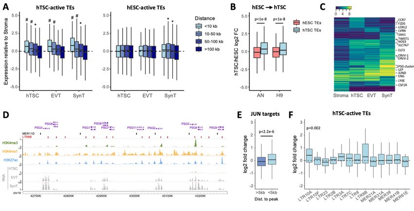
bioRxiv preprint doi: https://doi.org/10.1101/2022.04.26.489485; this version posted April 26, 2022. The copyright holder for this preprint
(which was not certified by peer review) is the author/funder, who has granted bioRxiv a license to display the preprint in perpetuity. It is
                                          made available under aCC-BY 4.0 International license.

          2D). CUT&Tag profiles confirmed that JUN/JUND bound LTR8B only, and not other
          LTR8-related families (Figure 2E).

          Figure 2 – Transcription factor repertoire at hTSC-active ERVs. A) Proportion of H3K27ac-marked elements from
          selected ERV families bearing motifs for the transcription factors on the y axis. B) Genome browser snapshots of
          transcription factor CUT&Tag/RUN data, showing examples of enrichment over ERVs. C) Repeat family-wide
          enrichment for peaks from transcription factor CUT&Tag/RUN data on hTSCs. H3K27ac-enriched families in hTSCs
          or hESCs are highlighted. D) Schematic and alignment of LTR10 and LTR8 subfamilies showing the presence of
          FOSB::JUNB (i.e., AP-1) motifs in the genome-wide consensus sequence and/or in a consensus of the H3K27ac-
          marked elements. E) Mean CUT&Tag profiles for JUN and JUND over LTR10 and LTR8 subfamilies.

          These results show that ERV families bearing regulatory potential in human
          trophoblast are bound by multiple transcription factors that play important gene
          regulatory roles in trophoblast.

          hTSC-active ERVs lie close to genes with preferential trophoblast expression
          To assess the potential of hTSC-active ERVs to drive trophoblast gene expression,
          we first asked whether some functioned as gene promoters. Using our primary
          cytotrophoblast RNA-seq data (Ashley et al., 2022), we performed de novo
          transcriptome assembly and extracted transcripts for which an ERV from the selected
          families overlapped the transcriptional start site. In line with the relative small
          proportion of H3K4me3-containing elements (Figure 1E), we identified few ERVs with
          apparent promoter activity (Supplementary Table S2). Reassuringly, this list included
          previously reported ERV-encoded promoters, such as those for CYP19A1 (van de

                                                                      6
bioRxiv preprint doi: https://doi.org/10.1101/2022.04.26.489485; this version posted April 26, 2022. The copyright holder for this preprint
(which was not certified by peer review) is the author/funder, who has granted bioRxiv a license to display the preprint in perpetuity. It is
made available under aCC-BY 4.0 International license.
Lagemaat et al., 2003), PTN (Schulte et al., 1996), PRL (Emera and Wagner, 2012)
and MID1 (Landry et al., 2002). Another notable gene was ACKR2, a chemokine
scavenger whose expression is driven by a MER39 element (Supplementary Figure
3A), and deficiency of which in mice leads to placental defects and pre/neo-natal
mortality (Teoh et al., 2014). Most other transcripts associated with ERV promoters
were lowly expressed (Supplementary Table S2).
We then sought to uncover correlations between candidate ERV-derived enhancers
and expression of nearby genes. We first associated each gene promoter to the
nearest H3K27ac-marked TE from the selected families. Using published RNA-seq
data (Okae et al., 2018), we then asked whether the distance to active ERVs
correlated with gene expression in trophoblast cells, by comparing it to the
expression in placental stroma (non-trophoblast, connective tissue containing
fibroblasts and macrophages). We found a strong association between gene-ERV
distance and preferential expression in both undifferentiated and differentiated
trophoblast (Figure 3A), and this was also seen in data from primary cells
(Supplementary Figure 3B). In contrast, genes proximal to H3K27ac-marked ERVs in
hESCs displayed no preferential expression in trophoblast (Figure 3A,
Supplementary Figure 3B). To further compare these two groups of genes, we
analysed RNA-seq data from transdifferentiation experiments of hESCs into hTSC-
like cells (Dong et al., 2020), which showed that genes lying within 50 kb of hTSC-
active ERVs displayed higher expression upon transdifferentiation than those close
to hESC-active ERVs (Figure 3B).
Figure 3 – Expression of genes close to regulatory ERVs. A) Gene expression in hTSCs and hTSC-derived EVT
and SynT relative to primary placental stroma, Genes are grouped based on their disrance to the nearest H3K27ac-
marked TE in hTSCs. * pbioRxiv preprint doi: https://doi.org/10.1101/2022.04.26.489485; this version posted April 26, 2022. The copyright holder for this preprint
(which was not certified by peer review) is the author/funder, who has granted bioRxiv a license to display the preprint in perpetuity. It is
made available under aCC-BY 4.0 International license.
highly expressed in SynT. In humans, PSGs are the most abundant conceptus-
derived proteins circulating in maternal blood (Lin et al., 1974). Their function in
pregnancy remains elusive, though they have been associated with immune
responses to pregnancy, and low levels of circulating PSGs are linked to recurrent
pregnancy loss, fetal growth restriction and preeclampsia (Arnold et al., 1999; Karg et
al., 1981; Towler et al., 1977). Within the PSG cluster, virtually every H3K27ac peak
overlaps either a MER11D or LTR8B element (Figure 3D), which presumably were
already present in the ancestral PSG gene before its duplication. These two ERV
families are associated with SynT-biased gene expression, and this is largely driven
by the expression of the PSG cluster (Supplementary Figure 3C). This is in contrast
with other families that are associated with genes with increased expression across
all three trophoblast cell types analysed when compared to placental stroma
(Supplementary Figure 3C).
Finally, we leveraged the information gained from our transcription factor binding
analysis to interfere with putative TE regulatory target genes. Given the enrichment in
JUN/JUND binding at LTR10A, LTR10F and LTR8B elements, we treated hTSCs
with SP600125, an inhibitor of c-Jun N-terminal kinases (JNKs) and performed RNA-
seq. JNK inhibition led to upregulation of genes involved in cell migration
(Supplementary Figure 4A), which is in agreement with past observations in human
trophoblast (He et al., 2019), but in contrast to what is commonly seen in most
cancer cells (e.g., (Chen and Zhao, 2020; Ou et al., 2021)). Unexpectedly, genes
lying within 5 kb of a JUN binding site were on average upregulated (Figure 3E),
including known JNK-dependent targets such as MMP14 (Supplementary Figure 4B).
This seemingly paradoxical result could be explained by increased JUN expression
(Supplementary Figure 4B) and/or the sometimes opposing roles of JNK1 and JNK2
(Sabapathy et al., 2004), which partly contribute to cell type-specific effects of JNK
signalling (Bubici and Papa, 2014). Irrespective of the mechanism, based on these
results we expected ERVs regulated by JUN to drive increased expression of their
target genes. Indeed, LTR10A (but not LTR10F) target genes were upregulated upon
SP600125 treatment (Figure 3F), including NOS3 (Supplementary Figure 4B), whose
placenta-specific gene expression is driven by an LTR10A-derived promoter (Huh et
al., 2008). LTR8B target genes were also upregulated (Figure 3F), although their low
number precluded robust statistical analysis. Notably, expression of all PSG genes
was increased by at least 2-fold, suggesting that JUN regulates this cluster via
LTR8B elements.
ERVs are associated with species-specific trophoblast gene expression
Primate evolution has involved dramatic divergence in placental phenotypes,
including differences in the cellular arrangement of the feto-maternal interface and
the extent of trophoblast invasion into the maternal decidua (Figure 4A). Namely,
great apes display a unique and deep form of trophoblast invasion, wherein EVT
migrate both into the uterine interstitium and endothelium, remodelling spiral arteries
as far as the inner third of the myometrium (Carter and Pijnenborg, 2011). The
integration of TEs with regulatory capacity in trophoblast may have helped to fuel
such fast placental evolution across primates. To test this, we first assessed how
divergent the landscape of hTSC-active ERV families was across primate species.
Using eight non-human primate genomes, we identified ERVs that were orthologous
to human integrants, showing inter-species differences that are in accordance with
the evolutionary age of the selected families (Figure 4B). We then took advantage of
published RNA-seq data from rhesus macaque TSCs (macTSCs), which were
recently derived using the same culture conditions as for hTSCs (Schmidt et al.,
2020). We extracted and normalised expression values for 1-to-1 gene orthologues
between human and macaque, and focused on genes within 100 kb of hTSC-active
ERVs. We found that genes close to human-specific ERVs displayed on average
8bioRxiv preprint doi: https://doi.org/10.1101/2022.04.26.489485; this version posted April 26, 2022. The copyright holder for this preprint
(which was not certified by peer review) is the author/funder, who has granted bioRxiv a license to display the preprint in perpetuity. It is
made available under aCC-BY 4.0 International license.
higher expression in hTSCs than in macTSCs, when compared to genes close to
conserved ERVs (Figure 4C). As expected, the majority of non-orthologous elements
were from the LTR2B family and included the previously characterised placenta-
specific promoter of PTN (Schulte et al., 1996), which we found to display human-
specific expression when compared to macaque. Additional LTR2B-associated
genes with human-specific expression included KCNE3, an estrogen receptor-
regulated potassium channel (Luo et al., 2013), and STOML2, which regulates
trophoblast proliferation and invasion (Zhang et al., 2020).
Figure 4 – Association of ERVs to species-specific gene expression. A) Primate phylogeny highlighting cross-
species differences in placental morphology and invasion. B) Proportion of human ERVs from each of the selected
families that contains orthologous elements in the non-human primates highlighted in A. C) Expression difference
between human and macaque TSCs for genes within 100kb of a H3K27ac-marked ERV from hTSC-active families,
depending on whether there is an orthologous element in macaque. P value is from a Wilcoxon test. D) Expression
difference between human and mouse TSCs for genes within 50kb of a H3K27ac-marked ERV in human, mouse
(RLTR13D5 and RLTR13B families), or neither species. P values are from an ANOVA with Tukey post-hoc test. E)
Genome browser snapshot showing a putateive example of a convergent evolution between mouse and human,
wherein different enhancer-like ERVs lie downstream of the the ZFP42/Zfp42 gene.
We and others have previously shown that Mus-specific ERVs also act as distal
enhancers in mouse TSCs (mTSCs) (Chuong et al., 2013; Todd et al., 2019). We
therefore extended our comparative expression analysis to Mus musculus, asking
whether human- and/or mouse-specific ERVs were associated with increased gene
expression in the respective species. Indeed, genes with active ERVs nearby in
human but not in mouse displayed higher expression in hTSCs, whereas those close
to active mouse ERVs had higher expression in mTSCs (Figure 4D). Mouse-specific
genes associated with active ERVs included a component of the FGF signalling
pathway (Fgfbp1), which maintains the stem cell state in mouse but not in human
trophoblast. Conversely, one human-specific ERV was associated with expression of
a Wnt signalling receptor (FZD5), a pathway that is important for hTSC derivation
(Okae et al., 2018). Other ERV-associated mouse-specific genes included Duox,
Duox2 and Nr0b1, which are mTSC markers (Kuales et al., 2015), and in human
MMP14, which is important for trophoblast invasion (Wang et al., 2014). We also
considered potential cases of convergent evolution, whereby the same gene may be
regulated by different ERVs in human and mouse. Out of the 12 genes that were
9bioRxiv preprint doi: https://doi.org/10.1101/2022.04.26.489485; this version posted April 26, 2022. The copyright holder for this preprint
(which was not certified by peer review) is the author/funder, who has granted bioRxiv a license to display the preprint in perpetuity. It is
made available under aCC-BY 4.0 International license.
close to active ERVs in both mouse and human, 10 were expressed in mTSCs and
hTSCs, including Zfp42/ZFP42 (Figure 4E). Despite being a well-known marker of
ESCs, Zfp42 is also expressed in mouse trophoblast, especially in early embryos
(Kim et al., 2011; Rogers et al., 1991), where it regulates the expression of some
imprinted genes (Kim et al., 2011). This raises the possibility that different ERVs
have convergently been co-opted to maintain the expression of Zfp42/ZFP42 in
mouse and human trophoblast, to support its essential roles therein.
These analyses suggest that some of the putative regulatory ERVs that we identified
help to drive species-specific expression of genes that are important for trophoblast
development and function.
ERVs regulate the expression of genes involved in human trophoblast function
Our data demonstrate that many primate specific ERVs exhibit epigenetic
characteristics of regulatory elements in human trophoblast. However, previous
observations have shown that these markers are not predictive of gene regulatory
activity (Todd et al., 2019). To test whether ERVs can act as enhancers in vivo, we
utilised CRISPR to genetically excise a subset of candidate regions, and then
measured nearby gene expression. Because the efficiency of growing clonal hTSCs
from single cells was extremely low after CRISPR, we employed a population-wide
lentiviral approach that we previously used (Deniz et al., 2020), achieving an average
of 49% deletion across different targets and experiments (Supplementary Table S3).
We first excised an enhancer-like MER41B element that is conserved from New
World monkeys to humans, and is located in the first intron of the ADAM9 gene
(Figure 5A), which encodes for a metalloproteinase. Genetic variants of ADAM9 are
implicated in preeclampsia (Ahmed et al., 2017), and its known substrates play roles
in inflammation, angiogenesis, cellular migration and proliferation (Chou et al., 2020).
Two independent MER41B excisions were derived (603 and 751bp), resulting in a
1.7-2 fold decrease in ADAM9 expression in hTSCs compared to no-sgRNA controls
(Figure 5A). The MER41B LTR is also 8 kb upstream of TM2D2 and 16 kb upstream
of HTRA4, a placenta-specific serine peptidase that is upregulated in early-onset
preeclampsia (Inagaki et al., 2012; Singh et al., 2015). Expression of these genes
was low and remained largely unchanged following MER41B excision
(Supplementary Figure 5A).
The CSF1R gene has a placenta-specific promoter (Visvader and Verma, 1989),
downstream of which lies an LTR10A that features enhancer-like chromatin features
and JUN binding in hTSC (Figure 5B), and is conserved from Old World monkeys to
humans. Both CSF1 and CSF1R expression increase in the placenta during
pregnancy (Kauma et al., 1991). CSF1 signalling via CSF1R promotes the growth,
proliferation and migration of trophoblasts in humans and mice (Ahmad et al., 2020;
Hamilton et al., 1998; Pollard et al., 1987), and high CSF1 levels are correlated with
preeclampsia development (Hayashi et al., 2003). Expression of both CSF1R
transcript variants was low in undifferentiated hTSCs, but increased following
differentiation to EVT, and was highest in SynT-differentiated hTSCs, particularly for
the placenta-specific CSF1R variant (Figure 5B). A 693 bp excision of the LTR10A
was derived in hTSCs, with CSF1R expression of the placenta-specific variant being
reduced by around 2-fold in both EVT and SynT cell pools (and to a lesser extend in
hTSCs), compared to no-sgRNA controls (Figure 5B). These differences were not
caused by an impairment in trophoblast differentiation efficiency, as judged by the
expression of key marker genes (Supplementary Figure 5C). The LTR10A element
therefore acts as an important enhancer of CSF1R in human trophoblast.
We excised a second MER41B element with enhancer-like chromatin conformation
(Figure 5C), deriving a 1,469 bp deletion in hTSCs. The TWIST1 (35 kb
10bioRxiv preprint doi: https://doi.org/10.1101/2022.04.26.489485; this version posted April 26, 2022. The copyright holder for this preprint
(which was not certified by peer review) is the author/funder, who has granted bioRxiv a license to display the preprint in perpetuity. It is
made available under aCC-BY 4.0 International license.
downstream), FERD3L (65 kb downstream) and HDAC9 (79 kb upstream) genes lie
in the vicinity of this LTR, but only TWIST1 is expressed in hTSCs. TWIST1 regulates
the syncytialisation of trophoblast, perhaps via GCM1 (Lu et al., 2016; Ng et al.,
2011) and promotes epithelial to mesenchymal transition – a key process in EVT
differentiation (Qin et al., 2012). We measured expression of TWIST1 in MER41B
excision hTSC pools, and found its expression to be unchanged (Figure 5C). Since
TWIST1 is important for trophoblast differentiation, we also differentiated hTSCs to
EVT and SynT, resulting in an 25-41 fold increase in TWIST1 expression, but there
was no difference in TWIST1 expression in excision versus no-sgRNA differentiated
cells (Supplementary Figure 5B). This particular MER41B element is therefore either
a redundant enhancer or does not regulate TWIST1, highlighting the importance of
these genetic experiments.
Figure 5 – Genetic excision of hTSC-active ERVs. A) Genome browser snapshot of the ADAM9 locus containing
an enhancer-like MER41B element, and ADAM9 expression (using primer pairs highlighted on the gene annotation)
in hTSC populations treated with lentiviral CRISPR constructs carrying either no sgRNAs or sgRNAs that excise the
MER41B element. B) As in A, but for CSF1R and its intronic LTR10A elements. CSF1R expression was measured in
hTSCs and hTSC-derived EVT and SynT. C) As in A, but for TWIST1 and a downstream MER41B element. D) As in
A, but for PSG5 and its intronic LTR8B elements. PSG5 expression was measured in hTSCs and hTSC-derived EVT
and SynT. P values throughout are from Wilcoxon tests with multiple comparisons correction.
As previously mentioned, the PSG cluster on chromosome 19 includes MER11D and
LTR8B elements at each tandemly repeated gene locus, all featuring enhancer-like
chromatin features in hTSCs (Figure 3D). In order to test LTR functionality, we
excised an LTR8B element, conserved only in apes, within the second intron of one
of the most highly expressed PSG in humans, PSG5 (Figure 5D) (Camolotto et al.,
2010), deriving two independent excisions (774 and 901 bp). PSG5 expression was
low in undifferentiated hTSCs and remained unchanged in LTR8B excision pools
compared to no sgRNA controls (Figure 5D). However, following differentiation to
EVT and SynT, PSG5 expression was increased by 15 and 81 fold respectively, and
was reduced in LTR8B excision pools compared to no-sgRNA controls by 2.3 fold in
EVT and 1.8 fold in SynT (Figure 5D), whilst differentiation efficiency was unaffected
11bioRxiv preprint doi: https://doi.org/10.1101/2022.04.26.489485; this version posted April 26, 2022. The copyright holder for this preprint
(which was not certified by peer review) is the author/funder, who has granted bioRxiv a license to display the preprint in perpetuity. It is
made available under aCC-BY 4.0 International license.
by the excision (Supplementary Figure 5D). These data indicate that the PSG5
LTR8B element acts an enhancer for PSG5 expression in human EVT and SynT.
The fact that the enhancer activity of this LTR8B-PSG5 element and the LTR10A-
CSF1R element are most strongly expressed after differentiation supports the notion
that some hTSC-active ERVs also play roles (and some may be poised to do so) in
differentiated trophoblast, as suggested by our epigenomic and transcriptomic
analyses above.
An LTR10A-derived enhancer promotes ENG expression, affecting sENG secretion
We were particularly interested in an enhancer-like (and JUN-bound) LTR10A
element within the first intron of the Endoglin gene (ENG/CD105) (Figure 6A). ENG is
a transforming growth factor-beta (TGF-ß) 1 and 3 co-receptor, with both membrane-
bound and soluble cleavage variants, highly expressed in the endothelium and SynT,
and involved in the pathogenesis of preeclampsia (Venkatesha et al., 2006). Indeed,
the serum levels of soluble ENG (sENG) are strongly correlated with the severity of
preeclampsia (Leaños-Miranda et al., 2019). Membrane-bound ENG is also
expressed in villous cell columns in the first trimester of pregnancy, regulating
trophoblast differentiation from proliferative cells to the migratory, invasive EVT that
colonise the uterus and remodel maternal spiral arteries (Caniggia et al., 1997; Mano
et al., 2011).
Figure 6 – Impact of an LTR10A element within the ENG gene. A) Genome browser snapshot of the ENG locus
containing an enhancer-like LTR10A element in its first intron. B) ENG expression (using primer pairs highlighted in
A) in hTSC populations treated with lentiviral CRISPR constructs carrying no sgRNAs, sgRNAs that excise the
LTR10A element, or sgRNAs that excise a control region highlighted in A. C) Growth curves for hTSC populations
carrying no sgRNAs or LTR10A-excising sgRNAS. D) The same hTSC populations were differentiated to EVT and
assays performed for ENG RNA levels and HLA-G cell surface expression. E) The same hTSC populations were
differentiated to SynT and assays performed for ENG RNA levels and secretion of soluble ENG protein. F) Genome
browser snapshot of the ENG locus with cytotrophoblast H3K27ac ChIP-seq data from second or third trimester
uncomplicated pregnancies, or third trimester severe preeclampsia placentas. P values throughout are from Wilcoxon
tests with multiple comparisons correction where relevant.
12bioRxiv preprint doi: https://doi.org/10.1101/2022.04.26.489485; this version posted April 26, 2022. The copyright holder for this preprint
(which was not certified by peer review) is the author/funder, who has granted bioRxiv a license to display the preprint in perpetuity. It is
made available under aCC-BY 4.0 International license.
We derived three independent excisions of the ENG LTR10A in hTSCs (508, 614
and 874 bp), which resulted in striking a decrease in ENG expression compared to
no-sgRNA controls (Figure 6B). Expression of neighbouring genes (AK1 and FPGS)
did not change (Supplementary Figure 6A). To confirm that loss of ENG expression
was strictly associated with deletion of the LTR10A element, and not transcriptional
interference by the CRISPR-Cas9 machinery, we also deleted a control region
upstream of the putative LTR10A enhancer that was devoid of active histone
modification marks in hTSCs (Figure 6A). Reassuringly, deletion of this region (1401
bp) in hTSCs had no effect on ENG expression (Figure 6B), confirming that the
LTR10A element is a bona fide enhancer. To assess the phenotypic impact of
LTR10A deletion, we first measured cell proliferation in hTSCs, finding no difference
when compared to no-sgRNA controls (Figure 6C). We then asked whether LTR10A
deletion affected trophoblast differentiation and/or phenotypes within differentiated
cell types. Differentiation to EVT was not affected by LTR10A deletion, as measured
by the percentage of HLA-G-positive cells (Figure 6D; Supplementary Figure 6B).
Notably, ENG expression remained lower (albeit variable) in LTR10A-deleted EVT,
when compared to no-sgRNA controls (Figure 6D). Differentiation into SynT was also
unaffected by deletion of the LTR10A element (Supplementary Figure 6C). Despite
the a large rise in ENG levels upon SynT differentiation, LTR10A-deleted cells
expressed less ENG than no-sgRNA controls (Figure 6E). We therefore tested
whether sENG protein levels were impacted by the LTR10A enhancer by performing
ELISA assays in the media of day 6 differentiated SynT cultures. We detected sENG
in the media at concentrations varying from ~100 pg/ml to 600 pg/ml, depending on
the experiment, and observed a significant decrease in sENG protein levels in ENG-
LTR10A excised SynT cultures when compared to no-sgRNA control (Figure 6E).
These experiments raise the possibility that deregulation of the ENG-LTR10A
element may be associated with elevated levels of ENG in preeclampsia. We
therefore leveraged recently published H3K27ac ChIP-seq data from cytotrophoblast
isolated from preeclamptic or uncomplicated pregnancies (Zhang et al., 2021). In
uncomplicated pregnancies, H3K27ac enrichment was clear over the ENG-LTR10A
element in second trimester placentas, but decreased in third trimester placentas
(Figure 6F). However, in third trimester placentas from severe preeclampsia,
H3K27ac levels remained high over the ENG-LTR10A enhancer (Figure 6F),
suggesting that this ERV may help to maintain ENG expression unduly high in
preeclampsia.
These data show that the ENG-LTR10A element acts as an enhancer in human
trophoblast, regulating ENG expression and sENG protein production, with potential
implications for preeclampsia.
Discussion
We have identified multiple ERV families that are enriched for elements bearing a
chromatin signature of cis regulatory elements in human trophoblast, most
resembling enhancers. Our stringent criteria ensured that the regulatory profiles of
these families reflect what is observed in vivo and are not driven by non-trophoblast
cell types present in the placenta. Indeed, we find that the activity of these families is
largely trophoblast-specific, with several being active and playing important roles in
both undifferentiated and differentiated trophoblast cell types. Our CRISPR genetic
editing experiments demonstrated that at least a subset of the ERVs we identified by
chromatin status act as bona fide enhancers of genes with important roles in
placentation. Notably, the fact that we were limited to performing CRISPR on a
13bioRxiv preprint doi: https://doi.org/10.1101/2022.04.26.489485; this version posted April 26, 2022. The copyright holder for this preprint
(which was not certified by peer review) is the author/funder, who has granted bioRxiv a license to display the preprint in perpetuity. It is
made available under aCC-BY 4.0 International license.
population scale implies that the effects we observed are actually an underestimation
of the true importance of those ERVs to gene expression.
We have already highlighted how many of the identified ERV families include
elements that act as placenta-specific promoters (Cohen et al., 2009). We also
compared our selected families with those identified by the Macfarlan lab as being
enriched for lineage-specific placental enhancers (Sun et al., 2021). Reassuringly,
we find multiple ERV families in both studies, including MER21A, MER41B, LTR8
and MER39 (Sun et al., 2021). On the other hand, the Sun et al. study did not list
other ERV families identified here, most prominently LTR10A, which has a strong
regulatory signature (Figure 1B), and copies of which we demonstrated act as
enhancers of important placental genes (Figure 5B and 6). Conversely, we find no
evidence of regulatory activity from the MaLR group of ERV families identified by Sun
et al. One possible reason for these discrepancies is that Sun et al. used whole
placental explants, leading to a mixed epigenomic profile from multiple cell types.
We also noted that a number of our trophoblast-active ERV families were recently
identified as active in several cancers, including LTR10A, LTR10F, LTR2B and
MER11D (Ivancevic et al., 2021). Multiple parallels have previously been drawn
between trophoblast and cancer cells, including an epigenetic landscape that is
strikingly different from other differentiated cell types (Smith et al., 2017). The
combination of an arguably permissive chromatin conformation and shared signalling
pathways may make the co-option of ERVs for both placental development/function
and cancer a frequent occurrence.
Several specific elements identified here suggested a potential important role in
placental evolution. This included the multiple LTR8B and MER11D copies
associated with the PSG gene cluster (Figure 3D). The PSG cluster is semi-
conserved in primates, with 6 to 24 genes found in Old World monkeys, 1 to 7 genes
in New World monkeys and none in more distantly related primates such as lemurs,
suggesting that the presence of PSG may correlate with haemochorial placentae,
since lemurs have an epitheliochorial placenta (Zimmermann and Kammerer, 2021).
Notably, PSG clusters are also present in mice, which also have a haemochorial
placenta and this region was expanded independently in mice and primates,
suggesting convergent evolution (Rudert et al., 1989). Our results suggest that the
integration of LTR8B elements ahead of PSG cluster expansion in humans was
important to ensure high trophoblast expression. MER11D elements may play a
similar role, and together with LTR8B be responsible for much of the transcriptional
regulation of this important locus. Similarly, MER61D/E retrotransposition may have
played a key role in setting the TP63 binding landscape, which in human trophoblast
supports cell proliferation and prevents cellular differentiation in trophoblast (Li et al.,
2014). TP63 belongs to the same family of transcription factors of TP53, sharing
many of its binding sites (Riege et al., 2020). It was previously shown that MER61
elements expanded the TP53 binding network in primates between 56 and 81 million
years ago, and a number of copies have been exapted to mediate cellular stress
response in lymphoblastoid cells (Su et al., 2015).
Other examples suggest a role of ERVs in coordinating the expression of genes from
the same pathway. MER21A elements were previously shown to act as promoters of
the steroidogenesis pathway genes CYP19A1 and HSD17B1, implicating these LTRs
in the regulation of steroidogenesis in human trophoblast. We also noted that both
NOS3 and ENG bear LTR10A elements as major transcriptional regulators (as
promoter and enhancer, respectively). This is interesting because the contribution of
sENG to vascular pathology in preeclampsia is partially due to effects on NOS3; in
concert with FLT1, ENG reduces placental angiogenesis and vasodilation of maternal
spiral arteries, and increases vessel permeability (Seligman et al., 1994; Venkatesha
et al., 2006).
14bioRxiv preprint doi: https://doi.org/10.1101/2022.04.26.489485; this version posted April 26, 2022. The copyright holder for this preprint
(which was not certified by peer review) is the author/funder, who has granted bioRxiv a license to display the preprint in perpetuity. It is
made available under aCC-BY 4.0 International license.
Our results implicate regulatory ERVs in pregnancy complications, prompting the
need for further investigation of their functional impact on pregnancy outcomes.
These ERVs may bear genetic variants that are difficult to investigate due to their
repetitive nature, and that may affect their regulatory activity in the placenta. Notably,
structural variants in LTR10A/F elements (in the form of variable number tandem
repeats), some potentially contributing to cancer, were recently described (Ivancevic
et al., 2021). Research into the effects of such variants in the placenta will benefit
from more complex models that can assess the impact of regulatory ERVs on cell-
cell interactions, such as those between the conceptus and the mother, that make
pregnancy so unique. Such experiments will be greatly supported by the recent
development of placental and endometrial organoids, as well as platforms that
support the study of trophoblast invasion (Abbas et al., 2020; Ojosnegros et al.,
2021).
Materials and Methods
Tissue culture
hTSCs were cultured according to (Okae et al., 2018), with modifications outlined in
(Takahashi et al., 2019). Briefly, ~1 x 10⁵ hTSC were seeded onto 6 well plates
coated with either 5–10 µg/mL collagen IV (10376931, Fisher Scientific) or 0.5 µg/mL
iMatrix 511 (NP892-011, Generon). Basal TS medium comprised DMEM/F12 (Gibco)
supplemented with 1% KnockOut Serum Replacement (KSR; Life Technologies Ltd
Invitrogen Division), 0.5% Penicillin-Streptomycin (Pen-Strep, Gibco), 0.15% BSA
(A9205, Sigma-Aldrich), 1% ITS-X supplement (Fisher Scientific) and 200 µM l-
ascorbic acid (Sigma-Aldrich). Complete media contained 2.5μm Rho-associated
protein kinase (ROCK) inhibitor Y27632, TGF-B inhibitors A83-01 (5uM), CHIR99021
(2uM) (all StemMACS) and epidermal growth factor (50ng/ml, E9644, Sigma-Aldrich),
with 0.8mM histone deacetylase inhibitor Valproic Acid (PHR1061, Sigma-Aldrich).
hTSC were passaged using TrypLE Express (Fisher Scientific) and split 1:3 to 1:5
every 2-3 days. All cells were cultured at 37°C in 5% CO2 and 20% O2. For JNK
inhibition assay, 1x10⁴ cells were seeded into wells of a 12-well plate and cultured for
24 hours in normal hTSC media before adding either 10 μM SP600125 (Cambridge
Bioscience) dissolved in DMSO, or the equivalent volume (1 μl) of pure DMSO.
Media containing inhibitor/DMSO was refreshed after 48 hours, and cells harvested
following a further 48 hours in culture.
Differentiation to EVT and SynT
Differentiation was performed as outlined in (Okae et al., 2018). For EVT, hTSC
were seeded in a 6-well plate coated with 1 μg/ml Col IV at a density of 0.75 x 10⁵
cells per well and cultured in 2 mL of EVT medium: DMEM/F12 supplemented with
0.1 mM 2-mercaptoethanol, 0.5% Pen-Strep, 0.3% BSA, 1% ITS-X, 100 ng/ml
Human Neuregulin-1 (NRG1; Cell Signalling), 7.5 μM A83-01, 2.5 μM Y27632, and
4% KSR. Cells were suspended in the medium, and Matrigel (LDEV-free, 354234,
Scientific Laboratory Supplies) added to a final concentration of 2%. On day 3,
medium was replaced with EVT medium without NRG1, and Matrigel added to a final
concentration of 0.5%. On day 6, medium was replaced with EVT medium without
NRG1 and KSR, and Matrigel added to a final concentration of 0.5%, with cells
grown for a further two days. Differentiation efficiency was measured using
Fluorescence Activated Cell Sorting (FACS) following staining with an APC
conjugated antibody to HLAG (APC anti-human HLA-G Antibody Clone: 87G,
BioLegend UK), in unmodified hTSC this was consistently between 40-70%, and in
lentivirus-infected CRISPR excision lines and controls, between 10-30 %. SynT (3D)
15bioRxiv preprint doi: https://doi.org/10.1101/2022.04.26.489485; this version posted April 26, 2022. The copyright holder for this preprint
(which was not certified by peer review) is the author/funder, who has granted bioRxiv a license to display the preprint in perpetuity. It is
made available under aCC-BY 4.0 International license.
differentiation was carried out by seeding 2.5 × 10⁵ hTSC in uncoated 6 well plates,
cultured in 2 mL of SynT medium: DMEM/F12 supplemented with 0.1 mM 2-
mercaptoethanol, 0.5% Pen-Strep, 0.3% BSA, 1% ITS-X, 2.5 μM Y27632, 50 ng/ml
EGF, 2 μM Forskolin (Cambridge Bioscience), and 4% KSR. 2 ml fresh ST(3D)
medium was added on day 3. Cells were passed through a 40 μm mesh strainer and
cells remaining on the strainer were collected and analysed. Differentiation was
assessed using RT-qPCR and immunostaining (Primers listed in Supplementary
Table S4)
FACS
Cells were single-cell dissociated using TrypLE Express and washed in FACS buffer
(PBS supplemented with 4% KSR). The cells were then resuspended in 500 μL fresh
FACS buffer, and 400 μL incubated with APC-HLAG for 15 minutes in the fridge, 100
μL were used as an unstained control. Following antibody incubation, the cells were
washed twice with FACS buffer, resuspended in fresh FACS buffer, and passed
through a 70 μm cell strainer. Flow cytometry was performed using a FACS Aria II,
and the data analyzed using the FlowJo software.
Immunostaining
5000 hTSC per well were grown on collagen IV coated glass coverslips in a 24 well
plate, and the differentiation protocol to EVT followed. On day 8 of EVT
differentiation, media was removed and cells washed with PBS x3, before fixing in
4% paraformaldehyde for 10 minutes before staining. Differentiated SynT cells were
centrifuged briefly (300g, 1 minute), resuspended gently in 500ul PBS + 4% KSR,
and added dropwise to poly-L-lysine coated glass coverslips. Once SynT cells had
gathered on the cover slip, the PBS+KSR was removed and cells were fixed in 4%
paraformaldehyde for 10 minutes. Both fixed EVT and SynT cells were permeabilised
with 0.1% Triton-X in blocking buffer (1% BSA (w/v) 2% FCS (v/v)) for 5 minutes,
followed by 1 hour incubation in blocking buffer. Incubation with the primary antibody
(SDC-1 (1:200, CD138 Mouse anti-Human, PE, Clone: MI15, Fisher Scientific, UK)
for SynT and HLAG (1:100, APC anti-human HLA-G Antibody Clone: 87G,
BioLegend UK) for EVT) diluted in blocking buffer was performed at 4°C overnight.
The secondary Alexa Fluor 488-green antibody was added at a 1:250 dilution for 1
hour at room temperature. DAPI (300nM) was used as a nuclear stain. Images were
collected using a Leica DM4000 epifluorescence microscope.
ChIP-seq
hTSC were dissociated using TrypLE express, pelleted and washed twice with PBS.
Cell pellets were fixed with 1% formaldehyde for 12 minutes in PBS, followed by
quenching with glycine (final concentration 0.125 M) and washing. Chromatin was
sonicated using a Bioruptor Pico (Diagenode) to an average size of 200–700 bp.
Immunoprecipitation was performed using 10 μg of chromatin and 2.5 μg of human
antibody (H3K4me3 [RRID:AB_2616052, Diagenode C15410003], H3K4me1
[RRID:AB_306847, Abcam ab8895], and H3K27ac [RRID:AB_2637079, Diagenode
C15410196]). Final DNA purification was performed using the GeneJET PCR
Purification Kit (Thermo Scientific, K0701) and eluted in 80 μL of elution buffer. ChIP-
seq libraries were prepared from 1 to 5 ng eluted DNA using NEBNext Ultra II DNA
library Prep Kit (New England Biolabs) with 12 cycles of library amplification.
CUT&Tag (Cleavage Under Targets and Tagmentation)
CUT&Tag was carried out as in (Kaya-Okur et al., 2019). 100,000 hTS or 50,000 EvT
cells per antibody were harvested fresh using TrypLE-express and centrifuged for
3 minutes at 600×g at room temperature (RT). Cells were washed twice in 1.5 mL
16bioRxiv preprint doi: https://doi.org/10.1101/2022.04.26.489485; this version posted April 26, 2022. The copyright holder for this preprint
(which was not certified by peer review) is the author/funder, who has granted bioRxiv a license to display the preprint in perpetuity. It is
made available under aCC-BY 4.0 International license.
Wash buffer (20 mM HEPES pH 7.5; 150 mM NaCl; 0.5 mM Spermidine; 1x Protease
inhibitor cocktail; Roche 11836170001 (PIC)) by gentle pipetting. BioMagPlus
Concanavalin A coated magnetic beads (Generon) were activated by washing and
resuspension in Binding buffer (20mM HEPES pH 7.5, 10 mM KCl, 1 mM CaCl2, 1
mM MnCl2). 10 µL activated beads were added per sample and incubated at RT for
15 minutes. A magnet stand was used to isolate bead-cell complexes (henceforth
‘cells’), the supernatant was removed and cells resuspended in 50–100 µL Antibody
buffer (20 mM HEPES pH 7.5; 150 mM NaCl; 0.5 mM Spermidine; 1× PIC; 0.05%
Digitonin, 2 mM EDTA, 0.1 % BSA) and a 1:50 dilution of primary antibody (H3K27Ac
(39034), Active Motif, H3K9me3 (C15410193) and H3K27me3 (C15410195),
Diagenode; c-Jun (60A8) – 9165T, and JunD (D17G2) – 5000S, Cell Signalling;
GATA3 sc-268 and TFAP2C sc-12762, Santa Cruz; TEAD4 CSB-PA618010LA01HU,
Stratech and negative control rabbit IgG, sc-2027, Santa Cruz). Primary antibody
incubation was performed on a nutator for 2 h at room temperature or overnight at
4 °C. Primary antibody was removed and secondary antibody (Guinea Pig anti-Rabbit
IgG (Heavy & Light Chain) ABIN101961) diluted 1:50 in 50–100 µL Dig-Wash buffer,
and added to the cells for 30 minutes incubation at RT. The supernatant was
removed and cells were washed 3x in Dig-Wash buffer. Protein A-Tn5 transposase
fusion protein (pA-Tn5) loaded with Illumina NEXTERA adapters (a kind gift from the
Henikoff lab, via Madapura lab, diluted 1:250, or Epicypher CUTANA® pAG-Tn5,
diluted 1:10) was prepared in Dig-300 Buffer (0.05% Digitonin, 20 mM HEPES, pH
7.5, 300 mM NaCl, 0.5 mM Spermidine, 1× PIC) and 50–100 µL was added to the
cells with gentle vortexing, followed by incubation at RT for 1 h. Cells were washed
3x in 800 μL Dig-300 buffer to remove unbound pA-Tn5. Next, cells were
resuspended in 50–100 µL Tagmentation buffer (10 mM MgCl2 in Dig-300 Buffer)
and incubated at 37 °C for 1 h. To stop tagmentation, 2.25 µL of 0.5 M EDTA, 2.75 µL
of 10% SDS and 0.5 µL of 20 mg/mL Proteinase K was added to the sample and
incubated overnight at 37 °C. To extract the DNA, 300 µL Phenol:Chloroform:Isoamyl
Alcohol (PCI 25:24:1, v/v; Sigma-Aldrich) was added to the sample and vortexed.
Samples were added to 5 PRIME™ Phase Lock Gel™ Light tubes and centrifuged
for 3 minutes at 16,000 x g. Samples were washed in chloroform, centrifuged for 3
minutes at 16,000 x g and supernatant added to 100 % ethanol, chilled on ice and
centrifuged 16,000 x g for 15 minutes. Pellets were washed in 100 % ethanol and
allowed to air-dry, followed by resuspension in 30 μL 10 mM Tris-HCl pH8 1 mM
EDTA containing 1/400 RNAse A. Libraries were indexed and amplified, using 21 µL
DNA per sample and adding 25 µL NEBNext HiFi 2x PCR Master mix + 2 µL
Universal i5 primer (10 µM) + 2 µL uniquely barcoded i7 primers (10 µM) in 0.2 ml
PCR tube strips, using a different barcode for each sample. Cycling parameters were
72 °C for 5 minutes, 98 °C for 30 s, then 12 cycles of 98 °C for 10 s, 63 °C for 10 s,
followed by 72°C for 1 minute followed by purification with 1X Agencourt AMPure XP
beads as per the manufacturer’s instructions (Beckman Coulter).
CUT&RUN (Cleavage Under Targets and Release Using Nuclease)
CUT&RUN was carried out as in (Skene and Henikoff, 2017). 500,000 hTSC were
washed and resuspended in Wash buffer (20 mM HEPES pH 7.5, 150 mM NaCl, 0.5
mM Spermidine, plus PIC). Following Concanavalin A bead preparation (as for
CUT&Tag), cells were incubated with 50 μL bead slurry for 15 minutes and
supernatant removed. Cell-bead complexes (hereafter, ‘cells’) were incubated
overnight with Antibody buffer (as for CUT&Tag) and primary antibodies (H3K27Ac
(39034), Active Motif, H3K9me3 (C15410193) and H3K27me3 (C15410195),
Diagenode; c-Jun (60A8) – 9165T, and JunD (D17G2) – 5000S, Cell Signalling;
GATA3 sc-268 and TFAP2C sc-12762, Santa Cruz; TEAD4 CSB-PA618010LA01HU,
Stratech and negative control rabbit IgG, sc-2027, Santa Cruz). Primary antibody
was removed and cells washed 3x in Dig-Wash buffer and incubated with 1:200 pA-
17You can also read