STRUCTURE GENOMICS OF SARS-COV-2 AND ITS OMICRON VARIANT: DRUG DESIGN TEMPLATES FOR COVID-19
←
→
Page content transcription
If your browser does not render page correctly, please read the page content below
www.nature.com/aps

                  REVIEW ARTICLE
                  Structure genomics of SARS-CoV-2 and its Omicron variant:
                  drug design templates for COVID-19
                  Can-rong Wu1, Wan-chao Yin1, Yi Jiang1 and H. Eric Xu1,2

                     Coronavirus disease 2019 (COVID-19), caused by severe acute respiratory syndrome coronavirus 2 (SARS-CoV-2), has brought an
                     unprecedented public health crisis and persistently threatens to humanity. With tireless efforts from scientists around the world,
                     understanding of the biology of coronavirus has been greatly enhanced over the past 2 years. Structural biology has demonstrated
                     its powerful impact on uncovering structures and functions for the vast majority of SARS-CoV-2 proteins and guided the
                     development of drugs and vaccines against COVID-19. In this review, we summarize current progress in the structural biology of
                     SARS-CoV-2 and discuss important biological issues that remain to be addressed. We present the examples of structure-based
                     design of Pfizer’s novel anti-SARS-CoV-2 drug PF-07321332 (Paxlovid), Merck’s nucleotide inhibitor molnupiravir (Lagevrio), and
                     VV116, an oral drug candidate for COVID-19. These examples highlight the importance of structure in drug discovery to combat
                     COVID-19. We also discussed the recent variants of Omicron and its implication in immunity escape from existing vaccines and
                     antibody therapies.
1234567890();,:

                     Keywords: COVID-19; SARS-CoV-2; structural biology; omicron; drug design; vaccine development

                     Acta Pharmacologica Sinica (2022) 0:1–13; https://doi.org/10.1038/s41401-021-00851-w

                  INTRODUCTION                                                                              Not long after the first SARS-CoV-2 genome sequence was
                  Coronavirus disease 2019 (COVID-19), caused by severe acute                            published [9], Rao et al. deposited the first structure of SARS-CoV-
                  respiratory syndrome coronavirus 2 (SARS-CoV-2), has led to                            2, Main protease (Mpro), into Protein Data Bank [13]. With the
                  more than 274 million infections and over 5.35 million deaths by                       tireless efforts of scientists worldwide, structures of SARS-CoV-2
                  December 20, 2021 [1–5]. SARS-CoV-2, an enveloped, single-                             proteins have burst and reached 1250 in the current moment.
                  stranded positive-sense RNA virus, belongs to beta-coronavirus                         These structures cover more than 90% of the SARS-CoV-2 coding
                  and is related to two highly pathogenic coronaviruses, SARS-CoV                        amino acids. The rapidly expanding repertoire of SARS-CoV-2
                  and Middle East respiratory syndrome coronavirus (MERS-CoV),                           structures has provided new insights into the viral life cycle and
                  which have caused over 8000 and 2500 confirmed cases,                                   facilitates drug and vaccine development.
                  with ~10% and 35% fatality rates, respectively [5–7]. Compared                            In this review, we provide an overview of the SARS-CoV-2
                  with SARS‐CoV and MERS‐CoV, SARS‐CoV‐2 is spreading more                               genome and its coded proteins. We then describe the structures
                  rapidly and causes much higher number of deaths. This                                  of SARS-CoV-2 proteins and mainly focus on RNA-dependent RNA
                  emerging virus has promoted massive hospitalizations, lockouts,                        polymerase and S protein. We provide insights into in situ
                  financial loss, unemployment, the closure of schools in nearly all                      structures of SARS-CoV-2 that have been solved by cryo-electron
                  countries [8].                                                                         tomography (cryo-ET). Finally, we discuss the remaining chal-
                     The SARS-CoV-2 genome is a non-segmented large positive-                            lenges and future perspectives on the structural biology of SARS-
                  sense stranded RNA with a length of about 30 kb. It contains a 5′-                     CoV-2 and highlight the recent success of the development of
                  cap structure and a 3′-poly-A tail [7, 9]. The viral genome encodes                    anti-COVID-19 drugs.
                  29 proteins, including 25 putative non-structural and accessory
                  proteins and four structural proteins [10]. Non-structural proteins
                  (NSPs) play crucial roles in viral RNA replication and immune                          THE GENOME OF THE SARS-COV-2 AND ITS CODED PROTEINS
                  evasion, while accessory proteins carry out multiple functions that                    The 5′-proximal two-thirds of the coronavirus genome contains
                  aid with viral infection, survival, and transmission in the host cells                 the replicase gene, which encodes two open reading frames,
                  [8, 11, 12]. Structural proteins are responsible for viral assembly                    ORF1a and ORF1b (Fig. 1a) [10]. The other one-third of the
                  and make up the mature viral particles. Elucidating the structure                      genome at the 3′-end encodes several ORFs. Among them, four
                  and function of these SARS-CoV-2 encoded proteins will deepen                          ORFs encode coronavirus structural proteins, which are spike
                  the understanding of the viral infection cycle and offer a new                         glycoprotein (S), membrane (M), envelope (E), and nucleocapsid
                  opportunity to develop effective vaccines and drugs to combat                          (N) proteins, where S, E, and M present on virion membrane
                  this global pandemic.                                                                  surfaces, with the N protein is involved in the binding and packing

                  1
                   The CAS Key Laboratory of Receptor Research, Shanghai Institute of Materia Medica, Chinese Academy of Sciences, Shanghai 201203, China and 2School of Life Science and
                  Technology, ShanghaiTech University, Shanghai 201210, China
                  Correspondence: Can-rong Wu (wucanrong@simm.ac.cn) or H. Eric Xu (eric.xu@simm.ac.cn)

                  Received: 19 October 2021 Accepted: 21 December 2021

                  © The Author(s), under exclusive licence to Shanghai Institute of Materia Medica, Chinese Academy of Sciences and Chinese Pharmacological Society 2022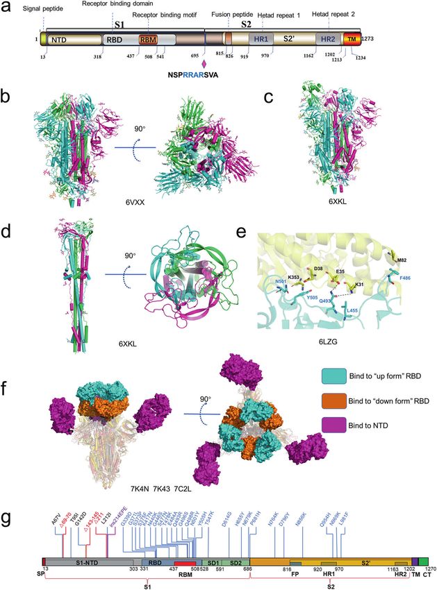
Structure genomics of SARS-CoV-2: drug design for COVID-19
                                                              CR Wu et al.
2

    Fig. 1 The genome of SARS-CoV-2 and its coded proteins. a The organization of SARS-CoV-2 genome. b Schematic illustrations of the
    secondary structure of the frameshift stimulation element −1 programmed ribosomal frameshifting, with different functional regions labeled
    and colored accordingly. c The rectangle depicts the nsps derived from processing of the pp1a and pp1ab polyproteins. Labels indicate
    protein names. The position of blue arrow indicates cleavage site of PLpro, and position of red arrow indicates the site of Mpro. d Domain
    architectures of SARS-CoV-2 genome coded proteins and summary of structural characterization of individual proteins. Bars above domain
    architectures indicate regions of the proteins for which high-resolution structures area are available. Ubl ubiquitin-like domain, MD
    macrodomain, SUD SARS unique domain, PLpro papain-like protease, NAB nucleic acid-binding domain, TM transmembrane domain, Y Y
    region, NTD N-terminal domain, CTD C-terminal domain.

    of the RNA genome. Other ORFs encode several accessory                   domain of nsp3 together with nsp4 and nsp6 plays a key role in
    proteins [14, 15] (Fig. 1a), with their functions less well-defined.      rearranging endoplasmic reticulum (ER) membranes, leading to
       ORF1a translation yields a replicase polyprotein 1a (ppla), and a     curvature of the ER membrane, which is adapted to form double-
    −1 ribosomal frameshift at the 3′-end of ORF1a, which facilitates        membrane vesicles (DMVs) and is essential for virus replication
    the translation of ORF1b to produce the replicase polyprotein 1ab        [27]. Nsp7 and nsp8 are assembled with nsp12 to enhance the
    (pp1ab) [16–18]. The −1 programmed ribosomal frameshifting               RNA polymerase activity.
    mechanism is stimulated by a three-stem pseudoknot at the 3′ of             Nsp7-nsp16 are involved in the formation of a large RTC to
    slippery sequence -UUUAAAC- named the frameshift stimulation             regulate the replication and transcription of SARS-CoV-2. Nsp9
    element (FSE) (Fig. 1b) [19]. When the frameshift does not occur,        inhibits the nucleotidyltransferase activity of nsp12 [28]; nsp10 is a
    the stop codon at the stem 1 induces translation termination             cofactor to nsp14 and nsp16 [29]; nsp12 functions as a RNA-
    (Fig. 1b). Conversely, when the −1 ribosomal frameshift occurs,          dependent RNA polymerase and nucleotidyltransferase [30];
    translation termination does not occur, which results in the             nsp13 acts as a helicase; nsp14 is exoribonuclease and N7-
    generation of a protein with additional ~2700 amino acids. The           guaninemethyltransferase [31]; nsp15 has uridine-specific endor-
    ribosomal frameshifting occurs at a frequency of 0.25–0.75 at this       ibonuclease activity [32]; nsp16 has 2′-O-methyltransferase activity
    site [19]. The structures of FSE alone and FSE complexed with            that mediates mRNA cap process [33].
    ribosomes were recently reported [19, 20].                                  Among four structural proteins, S, M and E are located on the
       In SARS-CoV-2, the polyprotein pp1a is proteolytically cleaved        viral membrane. S plays an essential role in the host receptor-
    into 11 functional NSPs, whereas pp1ab is cleaved into 15 NSPs           binding and membrane fusion [34, 35]. M protein is associated
    (Fig. 1c). Each NSP plays specific or multifaceted roles in the viral     with N protein and other viral structural proteins to facilitate the
    life cycle. Nsp1 interacts with the host ribosome to inhibit the         viral assembly and is involved in the pathogenesis process [36]. E
    synthesis of host proteins. Nsp2 may be involved in coupling viral       protein forms an ion channel, which promotes virus assembly and
    transcription with translation by interacting with both ribosome         pathogenesis [37, 38]. N protein participates in viral genome RNA
    and replication-transcription complexes (RTC) [21]. The papain-like      packaging and promotes virion assembly [39, 40].
    protease (PLpro) from nsp3 cleaves at three sites forming mature            Among nine accessory proteins, ORF3a, ORF7a and ORF7b are
    nsps 1–3, while the nsp5, also named main protease (Mpro),               transmembrane proteins. ORF3a forms a homo-dimer, with its
    cleaves pp1a and pp1ab at 11 sites, releasing the mature nsps            transmembrane domain forming an ion channel in the host cell
    4–16 [22, 23] (Fig. 1c). Considering their vital roles in NSPs           membrane. It participates in virus release, pathogenesis and host
    maturation, Mpro and PLpro have been considered attractive               cell apoptosis induced by the virus [41]. As a type I transmem-
    therapeutic targets for COVID-19 [24, 25]. In addition to the PLpro      brane protein, ORF7a is an immunomodulatory factor for immune
    domain, nsp3 contains several other domains: ubiquitin-like              cell binding and is involved in dramatic inflammatory responses.
    domain, macrodomain (also named “X domain”), nucleic acid-               ORF7b is a small, integrative membrane protein found in the host
    binding domain (NAB), SARS coronavirus-unique domain, trans-             cell Golgi, which may increase the virulence of SARS-CoV-2 [42]. It
    membrane domain and the Y1-3 domain [26]. The transmembrane              is coded by the same mRNA as ORF7a and expressed according to

                                                                                                          Acta Pharmacologica Sinica (2022) 0:1 – 13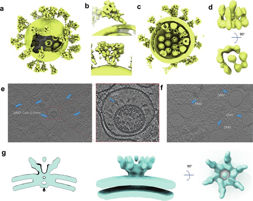
Structure genomics of SARS-CoV-2: drug design for COVID-19
                                                                          CR Wu et al.
                                                                                                                                                    3
the leaky-scanning mechanism of ribosomes [42]. ORF8 is one of            was developed and showed excellent efficacy in inhibiting SARS-
the newest genes and has low homology with SARS-CoV due to                CoV-2 replication in cell-based and animal models [64] (Fig. 2b).
deletion [43]. This protein interacts with major histocompatibility          Favipiravir, an orally administered nucleoside analog, also
complexes I, thus modulating their degradation in cell cultures,          showed a potential therapeutic effect on COVID-19 [65]. Unlike
thereby contributing to immune evasion [44]. ORF9b mediates               Remdesivir, Favipiravir showed a poor RNA replication termination
interferon response suppression by inhibiting the interaction             effect in the presence of natural nucleotides [66]. It mainly displays
between Hsp90 and TOM70 [45]. ORF10 is predicted to encode a              as a mutagenic agent toward viral genomic RNA in vivo [67, 68],
small protein. Pancer et al. suggest that this protein is not essential   different from Remdesivir, which impairs the elongation of RNA
for the viral life cycle in humans [46]. To date, only the structures     products. The mechanisms of binding of Favipiravir to RdRp and
of ORF3b, ORF6, ORF9c and ORF10 remain unsolved (Fig. 1d).                the low efficiency of incorporation of Favipiravir-RTP are revealed
                                                                          by the cryo-EM structure of the Favipiravir-bound SARS-CoV-2
                                                                          RdRp-RNA complex (Fig. 2b) [69]. A recent study reported similar
THE STRUCTURES OF RNA-DEPENDENT RNA POLYMERASE                            findings that Favipiravir-RTP complexed with SARS-CoV-2 poly-
AND REPLICATION-TRANSCRIPTION COMPLEX                                     merase in the pre-catalytic state [70].
Nsp12 functions as an RNA-dependent RNA polymerase and is a                  Molnupiravir, a novel RdRp inhibitor, has recently received
core component of the replication-transcription machinery [47].           much attention, as it showed very promising phase III study results
Auxiliary factors nsp7 and nsp8 are essential for enhancing the           [https://www.drugs.com/history/molnupiravir.htm]. Patients with
binding of nsp12 to the RNA template and its enzymatic activity           mild or moderate COVID-19 had a reduced risk of hospitalization
[48]. The association of the nsp7–nsp8–nsp12 and template-                or death by about 50% compared with placebo in a positive
primer RNA constitutes the minimum components of a functional             interim analysis of the oral antiviral drug Molnupiravir in a phase III
holo-RdRp, which is the main component of the SARS-CoV-2 RTC              study [71]. When Molnupiravir was incorporated into RNA
[49]. Several structures of holo-RdRp or holo-RdRp complexed              template by the RdRp, Molnupiravir directs incorporation of A or
with inhibitors have been determined by cryo-electron micro-              G, resulting in mutation of the viral RNA genome [59]. Recently,
scopy (cryo-EM) [30, 47, 50–53]. These structures provide insights        the first structure of SARS-CoV-2 RdRp complexed with the non-
into the molecular architecture of the holo-RdRp components.              nucleotide inhibitor, Suramin, was reported [30]. The RdRp harbors
The nsp12 subunit mainly contains several highly conserved                two independent Suramin binding sites. One suramin molecule
domains in coronavirus, including a nidovirus-specific extension           binds to the template RNA binding site, directly blocking the
domain (NiRAN), a C-terminal polymerase domain and an                     substrate binding. The other one binds to the site near the
interface domain in the middle (Fig. 2a). The β-hairpin (residues         catalytic site, thus blocking RNA primer binding by steric
V31 to K50), a unique conserved domain located upstream of the            hindrance (Fig. 2b). This unique binding pattern clarifies that
NiRAN domain, is sandwiched by the NiRAN and palm                         Suramin is at least 20-fold potent than the triphosphate form of
subdomain in the polymerase domain and stabilizes the overall             Remdesivir (RDV-TP) in polymerase activity inhibition assays
structure (Fig. 2a). Yan et al. disclosed that NiRAN catalyzes the        in vitro.
formation of cap core structure (GpppA) [28]. The C-terminal                 Promisingly, during the revision of this manuscript, Molnupiravir
polymerase core domain of nsp12 adopts a classic cupped right-            (Lagevrio) was approved by the Medicines and Healthcare
handed conformation, consisting of the finger, palm, and thumb             products Regulatory Agency (MHRA) and FDA for the treatment
subdomains and several conserved structural motifs. The SARS-             of established infections of COVID-19 [72]. Benefiting from recent
CoV-2 nsp7-nsp8 heterodimer shares significant structural                  progress in structural biology, the precise interaction between
similarity to that of SARS-CoV. The nsp7-nsp8 heterodimer binds           drugs or drug candidates and holo-RdRp has been elucidated, and
above the thumb subdomain and sandwiches the extended                     the mechanism of these drugs block virus replication by targeting
finger loops to stabilize nsp12 (Fig. 2a). Nsp7 contributes to most        RdRp has been revealed. The structural information will greatly
of the binding of the heterodimer to nsp12, while nsp8 sparsely           promote drug design through the structure-based approach and
contacts nsp12. The second nsp8 (nsp8-2) attaches to the top of           facilitate the development of the next generation of RdRp
the finger subdomain and forms additional interactions with the            inhibitors.
interface domain. The active sites of SARS-CoV-2 RdRp and RNA                Holo-RdRp is located in the central of the SARS-CoV-2 RTC [29].
recognition have been summarized and discussed in detail in the           In addition, a number of conserved accessory factors, which are
previous review [29].                                                     thought to coordinate with Holo-RdRp, are also required during
   To date, several antiviral drugs such as Remdesivir and                the viral life cycle [18]. One of these accessory proteins, nsp13, acts
Favipiravir that target the viral RdRp have been repurposed for           as an RNA helicase to unwind the RNA double strand. Chen et al.
COVID-19 therapy [54–59] (Fig. 2b). In April 2020, the first cryo-EM       reported the cryo-EM structure of two molecules of nsp13 in
structure of holo-RdRp in complexed with Remdesivir was                   complex with nsp7-nsp8-nsp12 containing the RNA template
reported. Biochemical studies showed that 1 mM Remdesivir                 products [73]. One of those interacted with nsp8-1 named nsp13-
triphosphate (RTP) displayed a complete inhibition of RdRp                1, the other named nsp13-2. The nsp13-1-ZBD interacts with
polymerization activity in the presence of 10 mM ATP, which               nsp12 thumb domain and nsp8-1. Meanwhile, nsp13-1 RecA1
mimicked the physiological concentrations of ATP. While 100 μM            domain abuts upon the nsp7 and the nsp8-1 head (Fig. 2c), while
RTP displayed a delayed chain termination mechanism [50].                 nsp13-2 binds less tightly in the complex than does nsp13-1. Aside
Kinetic studies on RdRp demonstrated that the polymerase can              from the interactions between the nsp13.2-ZBD and nsp8-2,
still add 2 or 3 nt after incorporating Remdesivir monophosphate          nsp13-2 only interacts with nsp13-1. The RdRp translocates in the
(RDV-MP) [60, 61]. This delayed chain termination model was               3′ → 5′ direction on the template RNA strand. Meanwhile, nsp13
further supported by recently solved structures of the SARS-CoV-2         translocates on this strand in the 5′ → 3′direction direction.
polymerase-RNA complex in its catalytic states incorporated with          Integrating structural analysis, unbiased molecular dynamics
RDV-MP [62, 63]. Upon incorporation of Remdesivir, RDV-MP                 simulations and RNA–protein cross-linking studies, Malone et al.
moved to position −4 of the RNA product strand results in a steric        proposed the backtracking mechanism [74]. The nsp13 facilitates
clash between the C1 cyano group of RDV-MP and the side chain             reverse RNA translocation, resulting in a single-stranded 3′ seg-
of nsp12 Ser861 (Fig. 2b), which may cause the delayed chain              ment of the product RNA extruding through the NTP entry tunnel.
termination of Remdesivir on RdRp. Because Remdesivir is                  This backtracking manner is similar to cellular DNA-dependent
intravenously administered nucleotide prodrug that is inconve-            RNA polymerases. Thus, nsp13 may play an important role in
nient to patients, VV116, an orally available Remdesivir derivative,      enhancing the fidelity of RdRp [75]. More recently, Yan et al. have

Acta Pharmacologica Sinica (2022) 0:1 – 13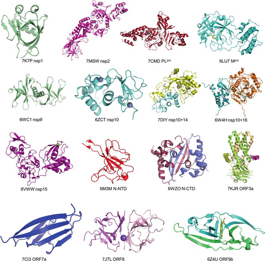
Structure genomics of SARS-CoV-2: drug design for COVID-19
CR Wu et al.
4
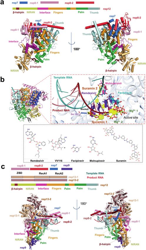
Fig. 2 Structures of the SARS-CoV-2 holo-RdRp and RTX. a The schematic diagram for the domain organization of the holo-RdRp, containing
nsp12, nsp7, nsp8-1 and nsp8-2 (PDB: 7C2K). Two views of the cartoon model of the cryo-EM structure of holo-RdRp. The subdomains and
components of the holo-RdRp are colored as follows: β-hairpin, chocolate; NiRAN, light orange; Interface, light magenta; Fingers, bright
orange; Palm, lime; Thumb, pale cyan; nsp7, marine; nsp8-1, pink; nsp8- 2, fire brick; template RNA, red; product RNA, deepteal. b Zoom in
views of holo-RdRp bound to inhibitors, Favipiravir at +1 (PDB: 7AAP, 7CTT), Remedisivir at +1 (PDB: 7BV2), Remedisivir at −1 (PDB: 7C2K),
Remedisivir at −3 (PDB: 7B3B), Remedisivir at +1, −1, −2, −3 (PDB: 7L1F), Suramin (PDB: 7D4F), Molnupiravir at template RNA, the four
inhibitors are indicated in different colors. Proteins and RNA are shown in cartoon representation, and inhibitors are shown as sticks. Nsp12 is
shown in transparent gray. Ligand atom color code: O atoms, red; N atoms, blue; P atoms, salmon; Mg2+ ion, green. 2D structures of five drugs
are presented. c The schematic diagram for the domain organization of the RTC, containing nsp12, nsp7, nsp8-1, nsp8-2, nsp9, nsp13-1 and
nsp13-2 (PDB: 7CYQ). Two views of the cartoon model of the cryo-EM structure of RTC. The subdomains and components of the holo-RdRp are
colored as follows: the subdomains and components of the RTC are colored as follows: β-hairpin, chocolate; NiRAN, light orange; Interface,
light magenta; Fingers, bright orange; Palm, lime; Thumb, pale cyan; nsp7, marine; nsp8-1, pink; nsp8-2, fire brick; nsp9, purple blue; nsp13-1,
wheat; nsp13-2, dark salmon; template RNA, red; product RNA, deep-teal.
Acta Pharmacologica Sinica (2022) 0:1 – 13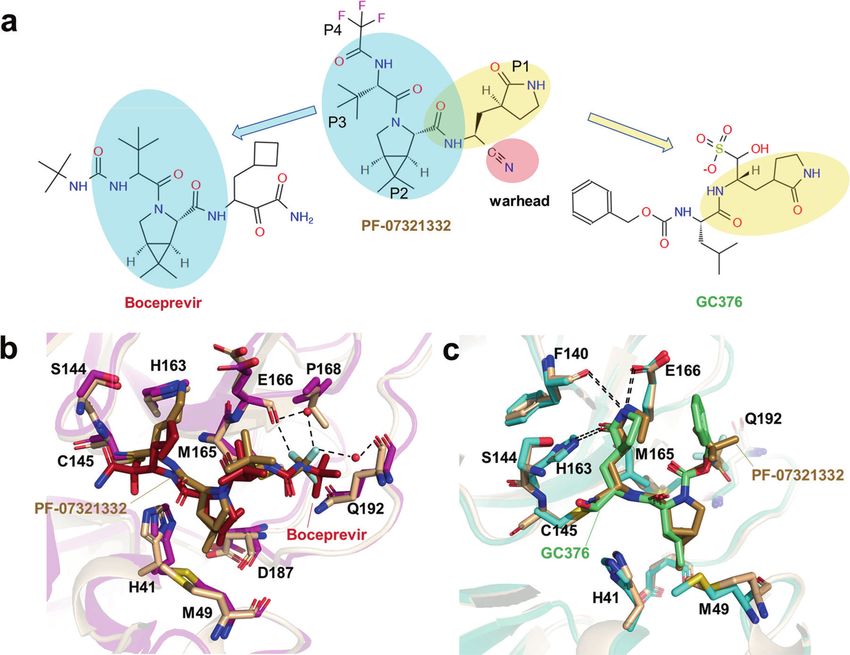
Structure genomics of SARS-CoV-2: drug design for COVID-19
                                                                          CR Wu et al.
                                                                                                                                                  5
presented a cryo-EM structure of the SARS-CoV-2 RTC that                  as K31 and E35 side chains, respectively [77, 78]. Intriguingly, the
includes an nsp9 [28]. The structure reveals that nsp9 binds              B.1.1.7 variant with N501Y substitution, which was originated in
tightly to the catalytic center of nsp12 NiRAN and inhibits the           the UK in September of 2020, displays higher transmissibility and
GTPase activity of nsp12 NiRAN (Fig. 2c). How the complete RTC            rapid spread globally, which may be caused by the enhanced ACE2
executes a series of complicated and fine processes in genome              affinity to the S variant of N501Y compared with the wild-type S
replication, such as the mRNA 7MeGpppA2′OMe cap process and the           protein [93–95].
mismatch repair process, remains to be addressed by continuous               As mentioned above, the SARS-CoV-2 S protein is proteolytically
and in-depth structural studies.                                          cleaved by furin during posttranslational maturation at ER to Golgi
                                                                          steps, leading to significantly decreased dependency of mem-
                                                                          brane protease. The non-covalently linked S protein is more
THE STRUCTURES OF S PROTEIN                                               flexible, which facilitates conformation change to the open state
The SARS-CoV-2 S protein belongs to the type I viral fusion protein       for receptor-binding and exposes the S2′ cleavage site to protease
and mainly comprises two functional subunits, S1 and S2 [76]. The         like TMPRSS2 [81, 96]. Cleavage of the S protein at the S2′ site by
S1 subunit is mainly responsible for receptor-binding, and the            cell surface protease facilitates membrane fusion for viral cell
S2 subunit mediates membrane fusion following proteolytic                 entry. Mutations promoting the cleavage efficiency on S1/S2 or S2′
activation [77–79]. The S1 subunit can be further subdivided into         site lead to higher transmissibility. By early April of 2020, a SARS-
the N-terminal domain (S1-NTD), two subdomains (SD1 and SD2)              CoV-2 variant containing a spike D614G mutation is spread rapidly
and C-terminal domain (S1-CTD), which is also known as the                and become the dominant form in the pandemic. Korber et al.
receptor-binding domain (RBD) [79] (Fig. 3a). The SARS-CoV-2 S            found that the D614G variant is closely related to greater
RBD is responsible for recognizing host receptor ACE2 and                 infectivity and increased viral load [97]. Yan et al. reported the
contains two subdomains, a receptor-binding motif (RBM) and a             cryo-EM structure of the D614G variant S protein and found that
core structure (Fig. 3a). The S2 subunit consists of multi-structural     this mutation makes S protein more flexible and is susceptible to
components, including hydrophobic fusion peptide (FP), heptad             protease cleavage [83]. Another study demonstrated that the
repeats (HR1 and HR2), transmembrane domain (TM). Unlike SARS-            D614G mutation disrupted a salt bridge between D614 and K854,
CoV and other beta-CoVs in subtype B, SARS-CoV-2 S protein                thereby attenuating the interaction between S1 and S2 subunits
contains a furin cleavage site between S1 and S2 subunits [80]            [84]. In the spring of 2021, the B.1.617 lineage (Delta) containing a
(Fig. 3a). In host cells, S protein is proteolytically cleaved at the     P681R mutation in S protein emerged from India and became the
PRRAR motif (residues 681–685) by furin into S1 and S2 to form a          dominant strain globally [98]. Saito et al. suggested that P681R
S1/S2 heterodimer, which was further assembled into the final              mutation in the spike protein facilitates spike protein cleavage and
trimeric spike complex [76, 81]. The S2 subunit contains an S’            enhances viral fusogenicity [99].
cleavage site, which is cleaved by host proteases like transmem-             In addition to higher affinity to ACE2 and more accessible to be
brane protease serine 2 (TMPRSS2) to expose the fusion peptide            digested by host proteases, S protein binds to several other host
for membrane fusion [79]. To date, numerous SARS-CoV-2 S                  receptors or co-receptors to facilitate the entry of SARS-CoV-2 into
protein structures in both prefusion and post-fusion forms have           host cells. The N-terminal domain of S protein specifically interacts
been reported [77, 78, 80, 82–84]. In the prefusion state, the SARS-      with tyrosine-protein kinase receptor UFO (AXL) in the human cell
CoV-2 trimeric S protein primarily adopts two conformations, a            surface [100]. It was reported that other proteins like neuropilin-1
close form with all RBD domains in “down” conformation and an             and asialoglycoprotein receptors also facilitate SARS-CoV-2 cell
open form with one or multiple RBDs in the “up” conformation              entry and infectivity [101, 102].
(Fig. 3b, c). In a closed form, the receptor-binding site was                The S protein determines the viral infection efficiency of human
enshrouded by S1-NTD, thereby impeding the binding of the                 cells and is pursued as a prominent anti-virus drug target. Due to
receptor ACE2, whereas, in the open conformation, one or                  the large interaction interface between S protein and ACE2, it is
multiple RBDs expose the receptor-binding site for ACE2 binding           hard to block their interaction by small-molecule drugs. SARS-CoV-
[83]. Ke et al. determined the S protein structure of intact SARS-        2 S neutralizing antibodies (nAbs) may be the preferred
CoV-2 virion by cryo-EM and cryo-ET and found that among all              therapeutics. Recently, a number of broad and potent SARS-CoV-
prefusion state S protein, fully closed trimers account for 31%, one      2 nAbs have been identified, including nAbs which were also
RBD opened or two RBDs opened trimers account for 55% and                 identified from convalescent COVID-19 patients and nanobodies
14%, respectively [83]. ACE2 binding destabilizes the prefusion           screened in the surface-displayed library. A number of structures
structure and facilitates the S1 subunit dissociates, thereby             of the SARS-CoV-2 S protein or RBD in complexed with antibody
enhancing the prefusion to the post-fusion transition of the S            have been determined by cryo-EM and X-ray crystallography
protein [85]. Fan et al. solved the cryo-EM structure of the post-        [83, 95, 103–110]. Most nAbs bind to the RBD, which is in the “up”
fusion state of SARS-CoV-2 S and showed that S2 forms a tightly           conformation resembling the conformation when interacting with
bound six-helix bundle by rotating HR1-HR2 and the linker region          ACE2. Conversely, only a few nAbs can bind to the “down”
at the upstream of the HR2 motif [86] (Fig. 3d). The mechanism of         conformation and thus impede the conformational switching
S protein-mediated coronavirus attachment and fusion has been             required for viral entry [105, 111] (Fig. 3f). Besides anti-RBD
described in detail in previous reviews [87–90].                          antibodies, an antibody named 4A8 responds to S1-NTD, which
   SARS-CoV-2 displays higher transmissibility than other human           has also been identified from convalescent COVID-19 patients
coronaviruses, mainly attributing to the following factors: a, higher     [112](Fig. 3f). Recently, two reviews have conducted a compre-
receptor-binding affinity; b, more susceptibility to protease; c,          hensive analysis on epitopes on S recognized by various
existing more alternative host cell receptors. Walls et al. found that    antibodies [113, 114]. Currently, more than 50 nAbs-related
SARS-CoV-2 S is bound to ACE2 with significantly higher affinity            clinical trials have been conducted for patients with COVID-19.
compared to SARS-CoV S [76]. Sequence alignment showed that four          Among them, eight RBD-specific nAbs have been authorized by
of five key residues at the interface between SARS-CoV-S RBD and           the Food and Drug Administration (FDA) for emergency use [115].
host ACE2 are non-conserved, including Y422SARS-CoV/L455SARS-CoV-2,          Unfortunately, the continuously evolving SARS-CoV-2 has led to
L472SARS-CoV/F486SARS-CoV-2, N479SARS-CoV/Q493SARS-CoV-2, T487SARS-CoV/   vaccine and nAbs resistance. Several variants of SARS-CoV-2 with
N501SARS-CoV-2 and Y491SARS-CoV/Y505SARS-CoV-2 [84, 90–92]. Shang         certain mutations have been designated as a variant of concern
et al. demonstrated that the N501 and Q493 contribute to the              (VOC) by the World Health Organization (WHO) [98]. On 26
enhanced ACE2-binding affinity of SARS-CoV-2 relative to SAR-CoV           November 2021, the WHO designated a new variant B1.1.1.529
by forming additional hydrogen bonds with RBM main chain, as well         (Omicron) as a VOC only 2 days after it was reported [98]. The

Acta Pharmacologica Sinica (2022) 0:1 – 13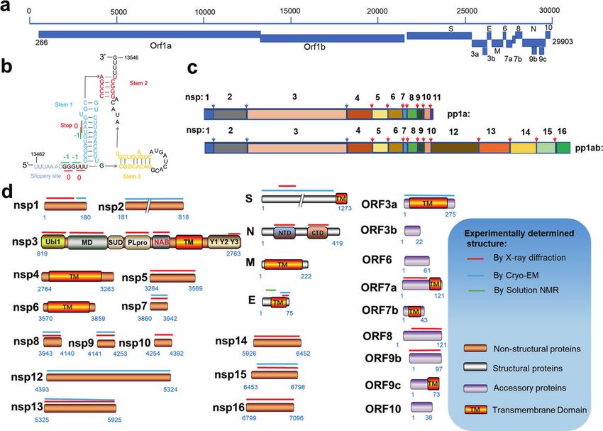
Structure genomics of SARS-CoV-2: drug design for COVID-19
                                                              CR Wu et al.
6

    Fig. 3 The structures of S protein. a Schematic of SARS-CoV-2 S protein domain architecture. The S1 and S2 subunits are indicated, with
    diamond representing the locations of furin cleavage site. SP signal peptide, RBD receptor-binding domain, RBM receptor-binding motif,
    SD1 subdomain 1, SD2 subdomain 2, FP fusion peptide, HR1 heptad repeat 1, HR2 heptad repeat 2, TM transmembrane region, CT
    cytoplasmic tail. b Side and top views of the prefusion structure of the SARS-CoV-2 S protein with all RBD in the “down” conformation (PDB:
    6VXX). Protein is shown in cartoon representation, and glycosyls are shown as sticks. c Side view of the prefusion structure of the SARS-CoV-2
    S protein with one RBD in the “up” conformation (PDB: 6XKL). d Side and top views of the post-fusion structure of the SARS-CoV-2 S protein
    (PDB: 6M3W). e Structure of the SARS-CoV-2 RBD complexed with ACE2 (PDB: 6LZG). ACE2 is shown in yellow. The RBD is shown in cyan. Key
    contacting residues are shown as sticks at the SARS-CoV-2 RBD–ACE2 interfaces. f Side and top views of superimposed three cryo-EM
    structures of SARS-CoV-2 S in complex with nAbs. S2E12 (represented as a cyan surface) binds to the “up” conformation of SARS-CoV-2 S RBD
    (PDB: 7K4N); S2M11 (represented as a brown surface) binds to the “down” conformation of SARS-CoV-2 S RBD (PDB: 7K43); 4A8 (represented as
    a magento surface) binds to the NTD of SARS-CoV-2 S (PDB: 7C2L). g Amino acids mutated in the Omicron variant S protein.

                                                                                                          Acta Pharmacologica Sinica (2022) 0:1 – 13
Structure genomics of SARS-CoV-2: drug design for COVID-19
CR Wu et al.
7
Fig. 4 In situ structures of SARS-CoV-2 virions. a Cryo-EM map of SARS-CoV-2 virion structure. The S proteins with different conformations
are distributed over the virion surface and can be tilted to different directions (EMD-30430). b A tilted conformation of S trimer is presented in
the upper plots, while a Y-shaped spike pairs having two heads and one combined stem are presented in the lower plots. c Side view of cryo-
EM map of SARS-CoV-2 virion structure, displaying RNPs assembled in the virus envelope. d Side and top views of in situ structure of SARS-
CoV-2 RNP (EMD-30429). e Tomograms showing SARS-CoV-2 virions in VeroE6 cells (EMD-11865). f Tomograms showing DMVs in SARS-CoV-2
affected VeroE6 cells (EMD-11866). g Different views of the EM structure of the MHV-induced pore complex embedded in the DMVs
membranes (EMD-11514).
Omicron variant accumulated a total of 60 mutations compared to average virion diameter of 91 ± 11 nm [118]. The virions may
the original SARS-CoV-2 variant [98]. There are 15 mutations on become less spherical after being concentrated by ultracentrifu-
the RBD of S protein, which has more than 30 mutations in total gation through a sucrose cushion [118]. Yao et al. measured the
(Fig. 3g). Recently, Xie et al. determined the RBD escaping virion diameters for the short, medium, and the long axis of the
mutation profiles for a total of 247 anti-RBD NAbs by yeast display envelope are 64.8 ± 11.8, 85.9 ± 9.4, and 96.6 ± 11.8 nm, respec-
screening [116]. Among all these tested nAbs, more than 85% are tively [119]. The S trimers are randomly distributed on the surface
escaped by Omicron. For instance, nAbs whose epitope included of the envelope (Fig. 4a). In three independent studies, the
in the RBM are escaped mainly by six mutations (K417N, N440K, average number of S trimers resided on the virion surface was
G446S, E484A, Q493K, and G496S). Combination therapy may calculated to be between 20–40, with the reported statistical
greatly reduce the probability of immune escape of mutant results being 40 [120], 24 ± 9 [118] and 26 ± 15 [119], respectively.
viruses caused by single-antibody treatment. High-resolution Surprisingly, the S protein in the virus is not perpendicular to the
structures of nAbs and S protein are of vital importance in viral membrane but tilted at different angles, which is significantly
revealing their epitope distribution and understanding the different from other enveloped viruses possessing class I fusion
neutralization mechanism. The structural information of neutraliz- proteins (Fig. 4b). A small part of the S trimer even tilted by over
ing antibodies to different epitopes will be critical to accelerate 90° toward the membrane [118]. The tilt of S protein is thought to
the development of broadly protective nAb cocktail therapies for be attributed to the flexibility of the hinge region in proximity to
COVID-19. Moreover, structure-based design of nAb mutants help the membrane. Interestingly, besides tilting from the membrane, S
to generate novel nAbs with improved potency and efficacy [117]. trimers can also move freely on the viral envelope. Yao et al. even
observed two S trimers are combined together with heads and
stalks forming a Y-shaped spike [119] (Fig. 4b). The flexibility of S
IN SITU STRUCTURE OF SARS-COV-2 VIRIONS protein both in direction and location may facilitate the virus
Coronavirus belongs to the envelope virus. Its shape is relatively sensing and binding to ACE2, allowing one S trimer to bind with
variable, and each virus has a unique structure. Therefore, it two or three ACE2s, or two S proteins with one ACE2 dimer.
cannot be solved by the single-particle cryo-EM, which limits the Moreover, compared with recombinant S proteins, the enriched
acquisition of high-resolution entire virus structure. With the N-linked glycans present on the native spike are more compli-
improvement of the cryo-EM facility and the optimization of the cated. These enriched glycosylation shields S from host protease
image processing algorithm in recent years, it is possible to obtain digestion and antibody recognition.
the in situ structure with a near-atomic resolution by cryo-ET. In The coronavirus has the largest genome across all RNA viruses
the autumn of 2020, three research groups reported the overall but a smaller size (Ø = ∼90 nm) compared with other RNA viruses
structure of purified SARS-CoV-2 virions, which presents an like human immunodeficiency virus (Ø = 120–170 nm) [121],
approximately spherical conformation. Ke et al. measured the and human respiratory syncytial virus (Ø = 150–250 nm) [122].
Acta Pharmacologica Sinica (2022) 0:1 – 13Structure genomics of SARS-CoV-2: drug design for COVID-19
CR Wu et al.
8
Considering that coronaviruses pack their large genomes to form inhibitors, Pfizer has developed the second-generation orally
a supercoiled dense structure into a relatively small viral particle SARS-CoV-2 Mpro inhibitor, PF-07321332. It showed positive phase
[123], it seems that the RNPs of SARS-CoV-2 inside the envelope III results for the treatment of COVID-19 in combination with
are more tightly packed relative to other coronaviruses. Ritonavir, which maintains higher circulating concentrations of PF-
The crystal structures of the nuclear acid-binding domain (NTD 07321332 by inhibiting cytochrome enzymes [142].
and CTD) of nucleocapsid protein have recently been solved PF-07321332 shows an overall structural similarity to the anti-
[124, 125]. However, the lack of the native structure of the HCV drug Boceprevir, which harbored a rigid P2 dimethylcyclo-
ribonucleoprotein complex limits our understanding of the propylproline residue and a hydrophobic P3 residue (Fig. 6a) [143].
assembly and function of coronavirus RNP. Yao et al. find that The P2 residue fits well to the S2 subsite and hydrophobically
each SARS-CoV-2 virion contains an average of more than 26 interacts with Met149 and Asp187, while the P3 residue interacts
RNPs, of which the membrane-proximal RNPs assemble as “hexon” with Met165 in S3 subsite (Fig. 6b). The P1 c-Bua residue of
and the membrane-free assemble as “tetrahedron” (Fig. 4c). They Boceprevir does not directly contact the SARS-CoV-2 Mpro
proposed an RNP assembly model in which the native RNPs S1 subsite, thus limiting its affinity [144]. The P1 residue of PF-
interact with the RNA in a “beads on a string” pattern, similar to 07321332 is a glutamine surrogate that mimics the equivalent
the mechanism of the chromatin forming in the eukaryotic cell residue of GC376. As a broad-spectrum cysteine protease covalent
[119]. Through the sub-tomogram averaging method, Yao et al. inhibitor, GC376 has shown promise in treating cats with fatal
solved a 13.1 Å resolution structure of RNPs, which revealed their feline infectious peritonitis (FIP) caused by FIPV and is being
reverse G-shaped architecture, with its diameter 15 nm and height investigated as a treatment for COVID-19 [144]. The glutamine
16 nm (Fig. 4d). This is similar to the 30 Å-resolution RNPs structure surrogate ring of both GC376 and PF-07321332 fits into the S1
in the intracellular virions, which is solved by Klein et al. [126]. This pocket and forms three stable hydrogen bonds with Phe140,
size is different from the reported architecture of SARS-CoV N His163, and Glu166 (Fig. 6c). Unlike other Mpro inhibitors, PF-
protein, which is assembled by crystal packing of 24-mer CTD 07321332 contains a nitrile warhead in P1’ residue, forming a
domain [127, 128]. The shape is also distinct from the released reversible covalent thioimidate adduct with the catalytic Cys145. A
MHV RNP structure [129]. Limited by the resolution, it is hard to trifluoroacetyl capping group was applied to the P4 residue to
clarify the precise mechanism of RNP assembly. This structure also improve the oral delivery efficiency. As shown in the SARS-CoV-2
cannot provide more information on the interaction between N Mpro-PF-07321332 complex structure, the trifluoromethyl group
protein and other structural proteins like M and E [127]. Recently, a forms hydrogen bonds with Gln192 and two ordered water
4.3 Å cryo-EM map of full-length SARS-CoV-2 N protein has been molecules, providing stronger interactions to the S4 subsite
released in the Electron Microscopy Data Bank, but the relative to Boceprevir (Fig. 6b). Collectively, through the structure-
corresponding structural model is still unavailable [130]. This N based approach, the affinity and physicochemical properties were
protein structure of moderate resolution may provide valuable optimized from initial leads in a short time. On November 16, an
insights into RNP assembly of SARS-CoV-2. application was submitted to the U.S. FDA for PF-07321332 in
To better understand the life cycle of the SARS-CoV-2 virus in combination with Ritonavir (brand name: Paxlovid) for emergency
host cells, Steffen et al. used the cryo-ET method to structurally treatment of COVID-19. The advent of this promising new agent
characterize the near-native state of virus assembly, DMVs has offered much hope to people worldwide. In addition to SARS-
morphology, and extracellular virions [126] (Fig. 4e). DMVs inside CoV-2 Mpro, RdRp and spike protein, several other promising
the SARS-CoV-2 infected cells show an average diameter of 338 therapeutic targets have been identified in the laboratory, such as
nm, which approaches the size of DMVs in SARS-CoV-1-infected PLpro and helicase; structure-based antiviral drugs development
VeroE6 cells [131]. The inner and outer membranes of DMVs are targeting these proteins are ongoing [145, 146].
separated by 5–10 nm but are clamped together in several sites So far, there are only a few SARS-CoV-2 proteins whose
(Fig. 4f). These connected sites may be the proteinaceous pore structures have not been characterized, and most of them are
complex formed by NSPs. This proteinaceous pore complex is transmembrane proteins (Table 1). These studies shed light on the
analogous to the hexameric assembly crown-shaped complex, biological functions of SARS-CoV-2 proteins and offer new
which is disclosed in murine hepatitis coronavirus-induced DMVs opportunities to develop vaccines and drugs. Nevertheless, there
in infected cells [132]. This DMVs pore complex is a well- are still some important issues waiting for elucidation from a
developed pathway for the transport of coronaviral RNA products structural perspective.
out of the DMVs [132] (Fig. 4g). However, the exact component At present, the largest available RTC structure only contains
and the function of this molecular pore remain to be elucidated by nsp7-8-9-12-13 [28]. Although the structures of other potential
high-resolution structures. components such as nsp10, nsp14, nsp15 and nsp16 have been
solved, it remains unclear how these subunits participate in the
assembly of the complete RTC. As we know, coronavirus contains
SUMMARY AND PERSPECTIVES the largest single-stranded RNA genome among all RNA viruses.
As a result of the rapid development of structural biology How the complete RTC executes a series of complicated and fine
techniques and the efficient investment of global researchers processes in genome replication, including the mRNA 7MeGpppA2′
over recent years, remarkable progress has been made on OMe cap process and the mismatch repair process, remains to be
structural studies of SARS-CoV-2. In addition to the RTC and S revealed by the structural study. For biosafety reasons, the current
protein, the structures of most SARS-CoV-2 proteins have been structural studies of RTC are carried out through in vitro
resolved in the past 2 years. These proteins include (1) NSPs Mpro recombinant expression and assembly, which hampers the
[13], PLpro [22, 133], nsp1 [134, 135], nsp9 [136], nsp10-nsp14 acquisition of stable RTC. Overcoming the biosafety issues of
complex [31], nsp10-nsp16 complex [33] and nsp15 [32], (2) SARS-CoV-2, or alternatively to culture low pathogenicity corona-
structural protein N [125, 137] and E proteins [138], (3) and viruses like MHV to isolate and purify natural RTC may be a
accessory proteins such as orf8 [43] (Fig. 5). Recently, the feasible strategy to solve the complete RTC structure.
structures of ORF3a [41], ORF7a [139], ORF9b [45], and Nsp2 [21] Although the high-resolution crystal structures of NTD and CTD
have also been reported (Fig. 5). of N protein have been solved, and a low resolution full-length N
A better understanding of these viral protein structures has protein structure has been available recently [124, 125, 130],
accelerated the structure-based development of novel drugs, the structural basis for the assembly of RNPs has not been
including anti-influenza drugs Zanamivir [140] and Oseltamivir clarified. It has been proposed that the assembly of RNPs enables
[141]. Based on the structure of SARS-CoV-2 Mpro in complex with efficient genome packaging in a nucleosome-like manner [126].
Acta Pharmacologica Sinica (2022) 0:1 – 13Structure genomics of SARS-CoV-2: drug design for COVID-19
CR Wu et al.
9
Fig. 5 High-resolution structures of SARS-CoV-2 proteins. Crystal structure of SARS-CoV-2 nsp1 (PDB: 7K7P); Cryo-EM structure of the SARS-
CoV-2 nsp2 (PDB: 7MSW); crystal structure of the SARS-CoV-2 PLpro with GRL0617 (PDB: 7CMD); crystal structure of the SARS-CoV-2 Mpro in
complex with an inhibitor N3 (PDB: 6LU7); crystal structure of the SARS-CoV-2 Nsp9 in complex with a peptide (PDB: 6WC1); crystal structure
of the SARS-CoV-2 nsp10 (PDB: 6ZCT); crystal structure of SARS-CoV-2 nsp10 bound to nsp14-exoribonuclease domain (PDB: 7DIY); crystal
structure of SARS-CoV-2 nsp10 in complex with nsp16 (PDB: 6W4H); crystal structure of the SARS-CoV-2 nsp15 endoribonuclease (PDB:
6VWW); crystal structure of SARS-CoV-2 nucleocapsid protein N-terminal RNA binding domain (PDB: 6M3M); crystal structure of SARS-CoV-2
nucleocapsid protein C-terminal RNA binding domain (PDB: 6WZO); Cryo-EM structure of SARS-CoV-2 ORF3a (PDB: 7KJR); the crystal structure
of the SARS-CoV-2 ORF7a ectodomain (PDB: 7CI3); the crystal structure of SARS-CoV-2 ORF8 accessory protein (PDB: 7JTL); the crystal structure
of SARS-CoV-2 ORF9b accessory protein (PDB: 6Z4U).
High-resolution of assembled RNP structure is needed to explain making it difficult for in vitro assembly of the recombinant N protein
how many N proteins assembled in the natural state and how the complex. Alternatively, virus-like particles (VLPs), which can be
RNA wounds around them. N protein plays a very important role spontaneously assembled by co-expressing these structural proteins,
in the process of virus assembly. Relevant studies have proven could be a potential resource of the N-M-E protein complex samples,
that N protein can interact not only with RNA but also with M and since VLPs are viral genome-free and are considered biologically safe
E proteins [147]. As described in the cryo-ET virion structure, the [147]. Nevertheless, obtaining the homogeneous, soluble N-M-E
RNP is close to the inner membrane of the virus capsule, protein complexes required for detailed structural studies is still a
indicating that it is feasible to interact with membrane proteins huge challenge, because detergents can hamper the assembly of
like M and E proteins in terms of space proximity [119]. Elucidating such membrane protein complexes. Extensive efforts to improve
the interaction mode of N protein with M protein and E protein the stability of these complexes, such as developing specific Fabs
would help understand the viral assembly process and provide a or nanobodies, should be made for high-resolution structure
new opportunity for designing antiviral drugs that interfere with determination.
viral assembly. The mechanism of DMVs formation in the life process of
Structurally, N proteins consist of two ordered domains: the coronavirus has not been clearly explained. Previous studies have
N-terminal domain (NTD), and C-terminal domain (CTD), connected shown that nsp3, nsp4 are mainly involved in the DMVs formation
by flexible LKR linker regions and two disordered regions, N-arm and process [148]. Interestingly, a hexameric assembly pore complex
C-tail. These disordered regions of the N protein make it extremely that spans both membranes of the MHV-induced DMVs was
difficult for high-resolution structure determination. In the SARS- visualized by the cryo-ET method [132]. Nsp3 was identified as a
CoV-2 virions, the N protein may be stabilized by interacting with major constituent of the pore complex, and nsp4, nsp6 and some
several molecules, such as viral genome RNA, M protein, E protein other unknown proteins may also be involved in this process. It is
and S protein, with an unknown assembly ratio. Besides, the N speculated that the pore complex can export synthesized RNA to
protein binds nonspecifically with the RNA phosphate backbone, the cytosol. In a sense, DMVs are somewhat similar to the nucleus
Acta Pharmacologica Sinica (2022) 0:1 – 13Structure genomics of SARS-CoV-2: drug design for COVID-19
CR Wu et al.
10
Fig. 6 The structures of SARS-CoV-2 Mpro in complex with inhibitors. a Chemical structure of PF-07321332, Boceprevir and GC376. b The
comparison of SARS-CoV-2 Mpro-PF-07321332 complex structure with that of SARS-CoV-2 Mpro-boceprevir. c The comparison of SARS-CoV-2
Mpro-PF-07321332 complex structure with that of SARS-CoV-2 Mpro-GC376.
understanding the mechanism of DMVs formation and material
Table 1. A summary of SARS-CoV-2 protein structures that have not transport. Moreover, it can also provide a structural basis for the
been resolved. design of antiviral drugs through intervening viral replication.
With the advances in recent cryo-EM methods, it is practicable
Proteins name Sequence range Transmembrane to solve the structure of protein-free RNA. Recently, Wah Chiu
protein or not
group and Rhiju Das group developed a set of methods for RNA
Nsp3 379–742, Yes structure study by cryo-EM [149]. These two groups solved several
1410–1842 near-atomic resolution RNA structures [150, 151], including a 3.1 Å
Nsp4 1–500 Yes
cryo-EM structure of the full-length Tetrahymena ribozyme. To
date, 3D structures of key RNA elements in the SARS-CoV-2
Nsp6 1–290 Yes genome remain poorly characterized. Only the structure of FSE
S 1209–1273 Yes was solved [20]. There remain dozens of highly conserved RNA
M 1–222 Yes segments in the SARS-CoV-2 genome. Targeting these segments
N 1–43, 181–254, No by antisense RNA is a good strategy for therapeutic agent
364–419 development [152–154]. Applying the cryo-EM method to study
ORF3b 1–22 No these RNA structures is of great significance for the elucidation of
SARS-CoV-2 RNA biology, hopefully, which would facilitate the
ORF6 1–61 No
development of genome-disrupting antiviral agents.
ORF7a 83–121 Yes
ORF7b 1–43 Yes
ORF9c 1–73 Yes
ACKNOWLEDGEMENTS
ORF10 1–38 No This work was partially supported by Ministry of Science and Technology (China)
grants (2018YFA0507002 to HEX); the Shanghai Municipal Science and Technology
Major Project (2019SHZDZX02 to HEX); the CAS Strategic Priority Research Program
of eukaryotic cells. As an independent platform of mRNA and (XDB08020303 to HEX); the National Natural Science Foundation of China (31770796
genome replication, it is continuously exported to the cytoplasm. to YJ); the Youth Innovation Promotion Association of CAS (No. 2021278 to WCY).
Similarly, the novel pore complex is analogous to a nuclear pore
and also spans double membranes. The pore complex may finely
control the inlet and outlet of raw materials and products in the ADDITIONAL INFORMATION
DMVs. The structure of the pore complex is important for Competing interests: The authors declare no competing interests.
Acta Pharmacologica Sinica (2022) 0:1 – 13Structure genomics of SARS-CoV-2: drug design for COVID-19
CR Wu et al.
11
REFERENCES 28. Yan L, Ge J, Zheng L, Zhang Y, Gao Y, Wang T, et al. Cryo-EM structure of an
1. Chen N, Zhou M, Dong X, Qu J, Gong F, Han Y, et al. Epidemiological and clinical extended SARS-CoV-2 replication and transcription complex reveals an inter-
characteristics of 99 cases of 2019 novel coronavirus pneumonia in Wuhan, mediate state in cap synthesis. Cell. 2021;184:184–93.
China: a descriptive study. Lancet. 2020;395:507–13. 29. Jiang Y, Yin W, Xu HE. RNA-dependent RNA polymerase: structure, mechanism,
2. Chan JF, Yuan S, Kok KH, To KK, Chu H, Yang J, et al. A familial cluster of and drug discovery for COVID-19. Biochem Biophys Res Commun.
pneumonia associated with the 2019 novel coronavirus indicating person-to- 2021;538:47–53.
person transmission: a study of a family cluster. Lancet. 2020;395:514–23. 30. Yin W, Luan X, Li Z, Zhou Z, Wang Q, Gao M, et al. Structural basis for inhibition
3. Li Q, Guan X, Wu P, Wang X, Zhou L, Tong Y, et al. Early transmission dynamics in of the SARS-CoV-2 RNA polymerase by suramin. Nat Struct Mol Biol.
Wuhan, China, of novel Coronavirus-infected pneumonia. N Engl J Med. 2021;28:319–25.
2020;382:1199–207. 31. Lin S, Chen H, Chen Z, Yang F, Ye F, Zheng Y, et al. Crystal structure of SARS-CoV-
4. Dong E, Du H, Gardner L. An interactive web-based dashboard to track COVID- 2 nsp10 bound to nsp14-ExoN domain reveals an exoribonuclease with both
19 in real time. Lancet Infect Dis. 2020;20:533–4. structural and functional integrity. Nucleic Acids Res. 2021;49:5382–92.
5. Johns Hopkins Coronavirus Resource Center. COVID-19 dashboard. JHU. 2021. 32. Kim Y, Jedrzejczak R, Maltseva NI, Wilamowski M, Endres M, Godzik A, et al.
https://coronavirus.jhu.edu/map.html. Crystal structure of Nsp15 endoribonuclease NendoU from SARS-CoV-2. Protein
6. WHO | Middle East respiratory syndrome coronavirus (MERS-CoV). 2021. https:// Sci. 2020;29:1596–605.
www.who.int/emergencies/mers-cov/en/. 33. Rosas-Lemus M, Minasov G, Shuvalova L, Inniss NL, Kiryukhina O, Brunzelle J,
7. Zhu N, Zhang D, Wang W, Li X, Yang B, Song J.China Novel Coronavirus et al. High-resolution structures of the SARS-CoV-2 2′-O-methyltransferase reveal
Investigating and Research Team, et al. A novel Coronavirus from patients with strategies for structure-based inhibitor design. Sci Signal. 2020;13:eabe1202.
pneumonia in China, 2019. N Engl J Med. 2020;382:727–33. 34. Moreira RA, Guzman HV, Boopathi S, Baker JL, Poma AB. Characterization of
8. Thomas S. Mapping the nonstructural transmembrane proteins of severe acute structural and energetic differences between conformations of the SARS-CoV-2
respiratory syndrome Coronavirus 2. J Comput Biol. 2021;28:909–21. spike protein. Materials. 2020;13:5362.
9. Wu F, Zhao S, Yu B, Chen YM, Wang W, Song ZG, et al. A new coronavirus 35. Huang Y, Yang C, Xu XF, Xu W, Liu SW. Structural and functional properties of
associated with human respiratory disease in China. Nature. 2020;579:265–9. SARS-CoV-2 spike protein: potential antivirus drug development for COVID-19.
10. Zhou P, Yang XL, Wang XG, Hu B, Zhang L, Zhang W, et al. A pneumonia Acta Pharmacol Sin. 2020;41:1141–9.
outbreak associated with a new coronavirus of probable bat origin. Nature. 36. Fu YZ, Wang SY, Zheng ZQ, Yi H, Li WW, Xu ZS, et al. SARS-CoV-2 membrane
2020;579:270–3. glycoprotein M antagonizes the MAVS-mediated innate antiviral response. Cell
11. Yoshimoto FK. The proteins of severe acute respiratory syndrome coronavirus-2 Mol Immunol. 2021;18:613–20.
(SARS CoV-2 or n-COV19), the cause of COVID-19. Protein J. 2020;39:198–216. 37. Nieto-Torres JL, DeDiego ML, Verdiá-Báguena C, Jimenez-Guardeño JM, Regla-
12. Gordon DE, Jang GM, Bouhaddou M, Xu J, Obernier K, White KM, et al. A SARS- Nava JA, Fernandez-Delgado R, et al. Severe acute respiratory syndrome cor-
CoV-2 protein interaction map reveals targets for drug repurposing. Nature. onavirus envelope protein ion channel activity promotes virus fitness and
2020;583:459–68. pathogenesis. PLoS Pathog. 2014;10:e1004077.
13. Jin Z, Du X, Xu Y, Deng Y, Liu M, Zhao Y, et al. Structure of Mpro from SARS-CoV- 38. Schoeman D, Fielding BC. Coronavirus envelope protein: current knowledge.
2 and discovery of its inhibitors. Nature. 2020;582:289–93. Virol J. 2019;16:69.
14. Poran A, Harjanto D, Malloy M, Arieta CM, Rothenberg DA, Lenkala D, et al. 39. Sheikh A, Al-Taher A, Al-Nazawi M, Al-Mubarak AI, Kandeel M. Analysis of pre-
Sequence-based prediction of SARS-CoV-2 vaccine targets using a mass ferred codon usage in the coronavirus N genes and their implications for
spectrometry-based bioinformatics predictor identifies immunogenic T cell genome evolution and vaccine design. J Virol Methods. 2020;277:113806.
epitopes. Genome Med. 2020;12:70. 40. Zeng W, Liu G, Ma H, Zhao D, Yang Y, Liu M, et al. Biochemical characterization
15. Lowery SA, Sariol A, Perlman S. Innate immune and inflammatory responses to of SARS-CoV-2 nucleocapsid protein. Biochem Biophys Res Commun.
SARS-CoV-2: implications for COVID-19. Cell Host Microbe. 2021;29:1052–62. 2020;527:618–23.
16. Jungreis I, Sealfon R, Kellis M. SARS-CoV-2 gene content and COVID-19 muta- 41. Kern DM, Sorum B, Mali SS, Hoel CM, Sridharan S, Remis JP, et al. Cryo-EM
tion impact by comparing 44 Sarbecovirus genomes. Nat Commun. 2021;12: structure of SARS-CoV-2 ORF3a in lipid nanodiscs. Nat Struct Mol Biol.
2642. 2021;28:573–82.
17. Chiara M, D’Erchia AM, Gissi C, Manzari C, Parisi A, Resta N, et al. Next generation 42. McBride R, Fielding BC. The role of severe acute respiratory syndrome (SARS)-
sequencing of SARS-CoV-2 genomes: challenges, applications and opportu- coronavirus accessory proteins in virus pathogenesis. Viruses. 2012;4:2902–23.
nities. Brief Bioinform. 2021;22:616–30. 43. Flower TG, Buffalo CZ, Hooy RM, Allaire M, Ren X, Hurley JH. Structure of SARS-
18. Sola I, Almazán F, Zúñiga S, Enjuanes L. Continuous and discontinuous RNA CoV-2 ORF8, a rapidly evolving immune evasion protein. Proc Natl Acad Sci USA.
synthesis in coronaviruses. Annu Rev Virol. 2015;2:265–88. 2021;118:e2021785118.
19. Bhatt PR, Scaiola A, Loughran G, Leibundgut M, Kratzel A, Meurs R, et al. 44. Zhang Y, Chen Y, Li Y, Huang F, Luo B, Yuan Y, et al. The ORF8 protein of SARS-
Structural basis of ribosomal frameshifting during translation of the SARS-CoV-2 CoV-2 mediates immune evasion through down-regulating MHC-Ι. Proc Natl
RNA genome. Science. 2021;372:1306–13. Acad Sci USA. 2021;118:e2024202118.
20. Zhang K, Zheludev IN, Hagey RJ, Haslecker R, Hou YJ, Kretsch R, et al. Cryo-EM 45. Gao X, Zhu K, Qin B, Olieric V, Wang M, Cui S. Crystal structure of SARS-CoV-2
and antisense targeting of the 28-kDa frameshift stimulation element from the Orf9b in complex with human TOM70 suggests unusual virus-host interactions.
SARS-CoV-2 RNA genome. Nat Struct Mol Biol. 2021;28:747–54. Nat Commun. 2021;12:2843.
21. Gupta M, Azumaya CM, Moritz M, Pourmal S, Diallo A, Merz GE, et al. CryoEM 46. Pancer K, Milewska A, Owczarek K, Dabrowska A, Kowalski M, Łabaj PP, et al. The
and AI reveal a structure of SARS-CoV-2 Nsp2, a multifunctional protein involved SARS-CoV-2 ORF10 is not essential in vitro or in vivo in humans. PLoS Pathog.
in key host processes. bioRxiv [Preprint]. 2021. Available from: https://doi.org/ 2020;16:e1008959.
10.1101/2021.05.10.443524. 47. Hillen HS, Kokic G, Farnung L, Dienemann C, Tegunov D, Cramer P. Structure of
22. Shin D, Mukherjee R, Grewe D, Bojkova D, Baek K, Bhattacharya A, et al. Papain- replicating SARS-CoV-2 polymerase. Nature. 2020;584:154–6.
like protease regulates SARS-CoV-2 viral spread and innate immunity. Nature. 48. Kirchdoerfer RN, Ward AB. Structure of the SARS-CoV nsp12 polymerase bound
2020;587:657–62. to nsp7 and nsp8 co-factors. Nat Commun. 2019;10:2342.
23. Dai W, Zhang B, Jiang XM, Su H, Li J, Zhao Y, et al. Structure-based design of 49. Subissi L, Posthuma CC, Collet A, Zevenhoven-Dobbe JC, Gorbalenya AE, Decroly
antiviral drug candidates targeting the SARS-CoV-2 main protease. Science. E, et al. One severe acute respiratory syndrome coronavirus protein complex
2020;368:1331–5. integrates processive RNA polymerase and exonuclease activities. Proc Natl
24. Yang H, Xie W, Xue X, Yang K, Ma J, Liang W, et al. Design of wide-spectrum Acad Sci USA. 2014;111:E3900–9.
inhibitors targeting coronavirus main proteases. PLoS Biol. 2005;3:e324. 50. Yin W, Mao C, Luan X, Shen DD, Shen Q, Su H, et al. Structural basis for inhibition
25. Anand K, Ziebuhr J, Wadhwani P, Mesters JR, Hilgenfeld R. Coronavirus main of the RNA-dependent RNA polymerase from SARS-CoV-2 by remdesivir. Sci-
proteinase (3CLpro) structure: basis for design of anti-SARS drugs. Science. ence. 2020;368:1499–504.
2003;300:1763–7. 51. Wang Q, Wu J, Wang H, Gao Y, Liu Q, Mu A, et al. Structural basis for RNA
26. Lei J, Kusov Y, Hilgenfeld R. Nsp3 of coronaviruses: structures and functions of a replication by the SARS-CoV-2 polymerase. Cell. 2020;182:417–28.e13.
large multi-domain protein. Antivir Res. 2018;149:58–74. 52. Gao Y, Yan L, Huang Y, Liu F, Zhao Y, Cao L, et al. Structure of the RNA-
27. Oostra M, Hagemeijer MC, van Gent M, Bekker CP, te Lintelo EG, Rottier PJ, et al. dependent RNA polymerase from COVID-19 virus. Science. 2020;368:779–82.
Topology and membrane anchoring of the coronavirus replication complex: not 53. Kabinger F, Stiller C, Schmitzová J, Dienemann C, Kokic G, Hillen HS, et al.
all hydrophobic domains of nsp3 and nsp6 are membrane spanning. J Virol. Mechanism of molnupiravir-induced SARS-CoV-2 mutagenesis. Nat Struct Mol
2008;82:12392–405. Biol. 2021;28:740–6.
Acta Pharmacologica Sinica (2022) 0:1 – 13You can also read