Structural basis for zinc-induced activation of a zinc uptake transcriptional regulator
←
→
Page content transcription
If your browser does not render page correctly, please read the page content below
Published online 28 May 2021                                                              Nucleic Acids Research, 2021, Vol. 49, No. 11 6511–6528
                                                                                                                https://doi.org/10.1093/nar/gkab432

Structural basis for zinc-induced activation of a zinc
uptake transcriptional regulator
Fenmei Liu1,† , Zihui Su1,† , Peng Chen1 , Xiaolin Tian2 , Lijie Wu3 , Dong-Jie Tang1 , Peifang Li1 ,
Haiteng Deng2 , Pengfei Ding4 , Qiang Fu1 , Ji-Liang Tang1,* and Zhenhua Ming 1,*
1
  State Key Laboratory for Conservation and Utilization of Subtropical Agro-bioresources, College of Life Science and
Technology, Guangxi Key Laboratory for Sugarcane Biology, Guangxi University, Nanning 530004, China, 2 Protein
Chemistry and Proteomics Facility, Protein Research Technology Center, Tsinghua University, Beijing 100084, China,
3
  iHuman Institute, ShanghaiTech University, Shanghai 201210, China and 4 Department of Chemistry and

                                                                                                                                                                  Downloaded from https://academic.oup.com/nar/article/49/11/6511/6287845 by guest on 29 December 2021
Biochemistry, University of Maryland, Baltimore County, Baltimore, MD 21250, USA

Received November 15, 2020; Revised April 29, 2021; Editorial Decision April 30, 2021; Accepted May 06, 2021

ABSTRACT                                                                                 GRAPHICAL ABSTRACT
The zinc uptake regulator (Zur) is a member of the
Fur (ferric uptake regulator) family transcriptional
regulators that plays important roles in zinc home-
ostasis and virulence of bacteria. Upon zinc per-
ception, Zur binds to the promoters of zinc respon-
sive genes and controls their transcription. However,
the mechanism underlying zinc-mediated Zur activa-
tion remains unclear. Here we report a 2.2-Å crystal
structure of apo Zur from the phytopathogen Xan-
thomonas campestris pv. campestris (XcZur), which
reveals the molecular mechanism that XcZur exists in
a closed inactive state before regulatory zinc binding.
Subsequently, we present a 1.9-Å crystal structure
of holo XcZur, which, by contrast, adopts an open
state that has enough capacity to bind DNA. Struc-
tural comparison and hydrogen deuterium exchange                                         INTRODUCTION
mass spectrometry (HDX-MS) analyses uncover that
binding of a zinc atom in the regulatory site, formed                                    As a chemically stable trace element, zinc is essential for al-
by the hinge region, the dimerization domain and the                                     most all living organisms and required for the structural sta-
                                                                                         bility and/or catalytic activity of many zinc-dependent met-
DNA binding domain, drives a closed-to-open con-
                                                                                         alloproteins (1,2). However, zinc in excess inhibits NADH
formational change that is essential for XcZur acti-                                     oxidase activity of the respiratory chain and is toxic to cells
vation. Moreover, key residues responsible for DNA                                       (3,4). Consequently, to survive, cells must develop mecha-
recognition are identified by site-directed mutagene-                                    nisms to sense this metal ion and tightly regulate its intracel-
sis. This work provides important insights into zinc-                                    lular level (5). In bacteria, maintenance of zinc homeostasis
induced XcZur activation and valuable discussions                                        depends primarily on the zinc uptake regulator (Zur) and
on the mechanism of DNA recognition.                                                     the MerR family and ArsR/SmtB family regulators (ZntR,
                                                                                         SmtB, CzrA) (6,7). Zur is a zinc sensor that not only re-
                                                                                         presses the zinc-uptake systems but also activates the zinc-
                                                                                         export systems, in a zinc-dependent manner (8,9). Zinc de-
                                                                                         pletion leads to inactivation of Zur and de-repression of
                                                                                         zinc responsive genes encoding the zinc importer system
                                                                                         (6). Corresponding to its critical role in regulation of zinc

* To
   whom correspondence should be addressed. Tel: +86 0771 3237873; Fax: +86 0771 3237873; Email: zhming@gxu.edu.cn
Correspondence may also be addressed to Ji-Liang Tang. Email: jltang@gxu.edu.cn
†
    The authors wish it to be known that, in their opinion, the first two authors should be regarded as Joint First Authors.

C The Author(s) 2021. Published by Oxford University Press on behalf of Nucleic Acids Research.
This is an Open Access article distributed under the terms of the Creative Commons Attribution-NonCommercial License
(http://creativecommons.org/licenses/by-nc/4.0/), which permits non-commercial re-use, distribution, and reproduction in any medium, provided the original work
is properly cited. For commercial re-use, please contact journals.permissions@oup.com
6512 Nucleic Acids Research, 2021, Vol. 49, No. 11
homeostasis, Zur homologs have been identified in a num- (MtZur from M. tuberculosis, ScZur from S. coelicolor and
ber of bacteria, including Escherichia coli (10), Bacillus sub- EcZur from E. coli) and they all contain two or three zinc
tilis (11), Listeria monocytogenes (12), Staphylococcus au- ions (36,37,43). EcZur binds to its cognate DNA as a dimer
reus (13), Xanthomonas campestris (8), Mycobacterium tu- of dimers, which is stabilized by a pair of asymmetric salt
berculosis (14), Streptomyces coelicolor (15), Streptococcus bridges between D49 and R52 (37). Consequently, it is en-
suis (16), Yersinia pestis (17), Corynebacterium glutamicum tirely unknown which conformation apo Zur is most likely
(18), Pseudomonas aeruginosa (19) and Salmonella enterica to adopt and what structural changes are associated with
(20). zinc binding.
Zur belongs to the Fur (ferric uptake regulator) family of Our previous studies characterized a Zur protein (XcZur)
transcriptional regulators that modulate the expression of in Xanthomonas campestris pv. campestris (Xcc), the causal
metal responsive genes, in response to the fluxes of a specific agent of black rot disease of cruciferous crops worldwide
metal. Since the first discovery of Fur in E. coli, >16 000 (8). The studies demonstrated that XcZur is essential for
members have been documented in Pfam (PF01475) (21). zinc homeostasis and full virulence of Xcc (8). To regu-
Downloaded from https://academic.oup.com/nar/article/49/11/6511/6287845 by guest on 29 December 2021
The Fur family regulators are typically divided into three late the intracellular zinc level, XcZur acts as an activa-
functional groups, i.e. the Fur group members that sense tor for the expression of the zinc-export gene, XC2976,
ferric and participate in iron homeostasis and virulence (22– and a repressor for the expression of the zinc-uptake genes,
24), the Zur group members that sense zinc and are involved XC0267, XC2472 and XC3788 (52). Particularly, the pro-
in zinc uptake, virulence and rearrangement of ribosomal teins XC0267 and XC2472 are components of an YciABC-
proteins (6,25), and the PerR group members that sense per- like low-affinity zinc-uptake system; XC3788 exhibits 38%
oxides and play important roles in regulating the peroxide similarity to the ZnuC, a component of a high-affinity zinc-
stress response (26,27). In addition, Fur family regulators uptake system; while XC2976 displays 67% similarity to the
that respond to other metals include sensors of manganese zinc efflux pump CzcD of Ralstonia metallidurans. In ad-
(Mur) (28,29) and nickel (Nur) (30,31). To couple metal per- dition, XcZur is involved in hypersensitive response (HR)
ception and DNA binding, Fur family proteins conserva- and positively regulates the transcription of genes associ-
tively fold into a two-domain architecture and frequently ated with HR and pathogenicity (53). Among Zur group
utilize two or more metal binding sites for metal-dependent members, XcZur shares 42% sequence identity with EcZur
conformational regulation (32). Available Fur family struc- (8), indicating a similar structure and biochemical function.
tures reveal a dimeric assembly and a modular domain orga- However, sequence alignment between XcZur and MtZur
nization for each monomer, which contains an N-terminal or ScZur reveals limited conservation (∼30% identity only).
DNA binding domain (DBD), a C-terminal dimerization To understand the fundamental mechanisms of Zur acti-
domain (DD), and a hinge region linking the two domains vation and zinc perception, we determined the crystal struc-
(32). The capability for a Fur protein to bind its target DNA tures of an inactive apo XcZur bound to a single zinc ion
depends on the exact conformation of the protein, which in and an active holo XcZur in complex with two zinc ions.
turn depends on the type and number of binding metals as These structures reveal drastic conformational changes as-
well as where the metals bind to. sociated with XcZur bound to a zinc ion in the regula-
The molecular mechanisms by which metal binding in- tory site. In conjunction with results from hydrogen deu-
duces conformational changes of Fur proteins have been terium exchange mass spectrometry (HDX-MS) analyses of
studied in some details. Evolutionary pressure seems to XcZur in different conformational states, our work provides
have forced Fur proteins to adopt similar active sates so a mechanism and structural basis for zinc-induced XcZur
that structural elements required for DNA binding, espe- activation as well as DNA recognition.
cially the recognition helix and the -hairpin wing in the
DBD, are precisely positioned (24,31,33–40). Conversely, MATERIALS AND METHODS
inactive Fur proteins can adopt very distinct conformations
Sequence alignment and phylogenetic analysis
and often vary between individual proteins and their dif-
ferent metal-binding states (38,41–48). Structural compar- The primary sequences of XcZur homologs were ac-
isons between inactive and active states reveal an allosteric quired by a three-iteration PSI-BLAST search (54) from
mechanism for activation of Fur and PerR group members, the PDB (protein data bank) database. A total of 15
in which binding with appropriate metal ion in the correct non-redundant sequences from 21 structures were iden-
metal binding site is the requisite (31,34,38,47). Typically, tified and their NCBI accession numbers are as follows:
Fur family proteins possess two or three metal binding sites. EcZur, 4MTD A; MtZur, 2O03 A; ScZur, 3MWM A; Sc-
Site 1 is a common structural site with four conserved cys- Nur, 3EYY A; CjPerR, 6DK4 A; LiPerR, 5NL9 A; Sp-
teines for zinc coordination; site 2 is thought to be the ma- PerR, 4I7H A; BsPerR, 2FE3 A; CjFur, 4ETS A; Hp-
jor metal sensor located between DBD and DD; while the Fur, 2XIG A; RlMur, 5FD6 A; MgFur, 4RAY A; FtFur,
function of DD-located site 3 is less clear (49–51). It appears 5NBC A; VcFur, 2W57 A; PaFur, 1MZB A.
that metal binding in site 2 may affect the mutual orienta- The T-Coffee server (55) was used to perform the
tion of the two domains and regulate DNA binding. De- multiple-sequence alignment (MSA). Then, MEGA version
spite these advances, the mechanism and structural basis of 7 was applied to conduct phylogenetic analysis for the MSA
zinc-induced Zur activation remains unclear, due partly to using the bootstrap neighbor joining method (56). For test
lack of structural information on the inactive state at which of phylogeny, 10 000 replications were used in the bootstrap
only one zinc is bound (apo state). Only three structures test (57). The Poisson correction method (58) was used to
to date have been determined for the Zur group proteins calculate evolutionary distances. Results derived from MSA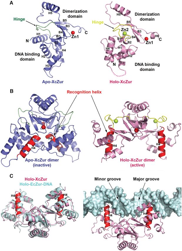
Nucleic Acids Research, 2021, Vol. 49, No. 11 6513
were also subjected to figure production of sequence align- NCS of 4 molecules in an asymmetric unit was used for
ment by ESPript version 3.0 (59). density improvement. Native data in higher resolution was
then used for model building in Phenix.autobuild (63), re-
finement and validation. The whole model of the structure
Cloning and expression
was manually built in COOT (64) and iteratively refined
The coding sequence of XcZur was amplified by stan- by Phenix.refine (65). In the final model, more than 96.4%
dard polymerase chain reaction (PCR) from the genomic residues fall in the favored region in the Ramachandran plot
DNA of Xcc strain 8004. The amplified DNA fragment was and the final Rwork /Rfree are 0.219/0.276.
inserted into the pRSFDuet-1 vector (Novagen) between The SAD dataset of holo XcZur was collected on beam-
BamHI and XhoI restriction sites. After confirmation by line BL17U1 at SSRF (66), processed and scaled using XDS
DNA sequencing, the recombinant plasmid was introduced (61). The initial phase was determined by Phenix.autosol
into E. coli strain BL21(DE3) for protein expression. Trans- (62) through the SAD method and the initial model was
formed cells were grown at 37◦ C in Luria-Bertani medium built by Phenix.autobuild (63). The whole model was
Downloaded from https://academic.oup.com/nar/article/49/11/6511/6287845 by guest on 29 December 2021
containing kanamycin. When the OD600 reached 0.6–0.8, manually built in COOT (64) and iteratively refined by
protein expression was induced by adding 0.5 mM Iso- Phenix.refine (65). In the final model, more than 96.4%
propyl -D-1-thiogalactopyranoside (IPTG) followed by residues fall in the favored region in the Ramachandran plot
18–20 h incubation at 16◦ C. The construct of XcZur-16 and the final Rwork /Rfree are 0.213/0.252.
was generated and expressed in a similar way. The final pro- Data collection and refinement statistics are summarized
tein product from the recombinant pRSFDuet-1 vector is in Table 1. Structural figures were produced by PyMOL
expected to contain a 6× His tag at the N-terminus. (The PyMOL Molecular Graphics System, Schrödinger,
LLC).
Protein purification
Site-specific mutagenesis
The cells were harvested by centrifugation at 4200 rpm for
15 min, resuspended with 30 ml lysis buffer containing 20 The 19 mutant clones were constructed by a PCR-based
mM Tris–HCl (pH 8.0), 150 mM NaCl, 30 mM imidazole, strategy using the Fast Mutagenesis System (TransGen
and then homogenized using a low-temperature ultra-high- Biotech, Beijing, China). The corresponding primers were
pressure cell disrupter (JNBIO, Guangzhou, China). After listed in Supplementary Table S1. Each mutant clone, veri-
centrifugation at 12 000 rpm for 1 h, the resulting super- fied by DNA sequencing, was transferred into BL21(DE3)
natant was loaded onto a Ni-NTA agarose column (GE and expressed using the same procedure for the wild type
Healthcare). The eluted protein was subjected onto a Hi- clone. The mutant protein was purified tandemly by using
Trap Heparin column (GE Healthcare) equilibrated with the Ni-NTA agarose column (GE Healthcare), the HiTrap
20 mM Tris–HCl (pH 8.0), 75 mM NaCl. The protein was Q column (GE Healthcare) and the Superdex 200 Increase
eluted by a linear NaCl gradient and further purified by prepacked column (GE Healthcare).
size exclusion chromatography using Superdex 200 columns
(GE Healthcare) in a buffer containing 20 mM Tris (pH 8.0) Analytical ultracentrifugation (AUC)
and 150 mM NaCl. The protein was concentrated to ap- Sedimentation Velocity Analytical Ultracentrifugation (SV-
proximate 10 mg/ml and stored at −80◦ C. AUC) experiments were carried out at 20◦ C in an XL-I
analytical ultracentrifuge (Beckman Coulter). Absorbance
Crystallization and structure determination (XcZur and XcZur-DNA complexes: 280 nm; DNA: 260
nm) and Raleigh interference optics were used to simultane-
Crystals were obtained by the hanging drop vapor diffu- ously record the radial concentration as a function of time
sion method, by mixing 1 l of protein sample with 1 l of until the lightest sedimenting component cleared the op-
reservoir solution at 16◦ C. Best crystals of apo XcZur grew tical window (7 h). All samples (400 l) were centrifuged
in a reservoir solution containing 0.2 M calcium acetate at 50 000 rpm for 8 h in an An50Ti rotor using 12 mm
hydrate, 0.1 M sodium cacodylate trihydrate pH 6.5, 18% double-sector aluminum centerpieces. Apo XcZur was pre-
(w/v) polyethylene glycol 8000, 0.3 M glycyl-glycyl-glycine. pared in the interaction buffer A (20 mM HEPES pH 7.4,
To avoid zinc precipitation in basic conditions, holo XcZur 150 mM NaCl), other samples were in the interaction buffer
was buffer exchanged into a solution containing 100 mM B (20 mM HEPES pH 7.4, 150 mM NaCl, 0.18 mM EDTA,
MES (pH 6.0) and 150mM NaCl and then incubated with 0.2 mM Zn). Interference profiles were recorded every 6
excess zinc acetate (molar ratio of metal to protein equals to min. Data analysis was conducted by the software SEDFIT
1.2:1). Best crystals of holo XcZur grew in a reservoir solu- 11.7 (67), GUSSI and SEDPHAT. Theoretical sedimenta-
tion containing 0.2 M sodium malonate pH 5.0, 20% (w/v) tion coefficients were calculated by using HydroPro 7c (68)
polyethylene glycol 3,350. with a hydrated radius of 3.1 Å for the atomic elements.
The native and single-wavelength anomalous dispersion
(SAD) datasets of apo XcZur were collected on beam-
Electrophoretic mobility shift assay (EMSA)
line BL19U1 (60) at the Shanghai Synchrotron Radiation
Facility (SSRF). Datasets were processed and scaled us- DNA oligonucleotides (Supplementary Table S2) were
ing XDS (61). Two sets of data were merged to improve synthesized by Shanghai mapbioo (China) with the for-
the anomalous signal. The initial phase was determined by ward strand labelled by FAM (fluorophore 6-carboxy-
Phenix.autosol (62) using the SAD method, during which fluorescein) and annealed in a resolving buffer containing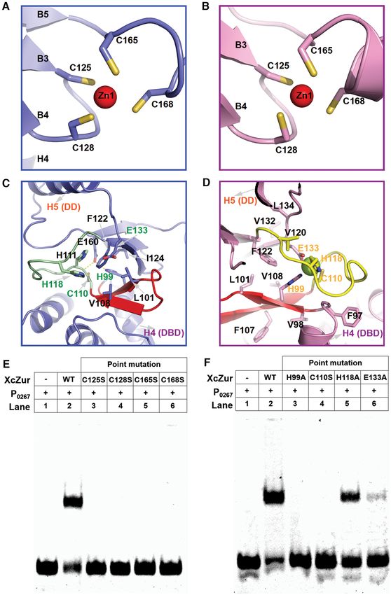
6514 Nucleic Acids Research, 2021, Vol. 49, No. 11
Table 1. Data collection and structure refinement statistics
Apo XcZur Holo XcZur
Parameters Zn-SAD Native Zn-SAD
Data collection statistics
Cell parameters
a (Å) 153.43 153.64 34.56
b (Å) 77.91 78.05 122.9
c (Å) 73.09 73.12 86.89
α, β, γ (◦ ) 90,0 103.08, 90.0 90,0 103,06 90.0 90, 90, 90
Space group C2 C2 C2221
Wavelength used (Å) 0.9785 0.9785 0.9792
Resolution (Å) 50.0–2.40 (2.49–2.40)c 50.00–2.20 (2.34–2.20)c 22.57–1.85 (1.92–1.85)c
No. of all reflections 413 164 141 407 186 417
Downloaded from https://academic.oup.com/nar/article/49/11/6511/6287845 by guest on 29 December 2021
No. of unique reflections 32 172 42 394 15 694
Completeness (%) 99.7 (99.6) 98.7 (96.9) 95.5 (86.7)
Average I/σ (I) 17.4 (4.93) 13.01 (2.26) 15.35 (1.69)
Rmerge a (%) 6.2 (20.5) 4.8 (38.7) 4.0 (52.9)
Refinement statistics
No. of reflections used (σ (F) > 0) 42 394 15 557
Rwork b (%) 21.94 21.26
Rfree b (%) 27.55 25.17
r.m.s.d. bond distance (Å) 0.008 0.015
r.m.s.d. bond angle (◦ ) 1.430 1.315
Average B-value (Å2 )
Average B-value for protein atoms 40.06 50.14
Average B-value for solvent atoms 41.18 46.17
No. of atoms
No. of protein atoms 4550 1086
No. of solvent atoms 364 70
Ramachandran plot
Res. in favored regions (%) 96.43 96.35
Res. in outlier regions (%) 0 0
merge = h i |Ih,i −Ih |/ h i Ih,i , where Ih is the mean
aR intensity of the i observations of symmetry related reflections of h.
work =
bR (||Fp (obs)|−|Fp (calc)||)/ |Fp (obs)|; Rfree is an R factor for a pre-selected subset (5%) of reflections that was not included in refinement. Fp ,
structure factor of protein.
c Numbers in parentheses are corresponding values for the highest resolution shell.
50 mM Tris–HCl (pH 7.5) and 250 mM KCl. For each as- Exactive Orbitrap mass spectrometer (Thermo, CA). Data
say, FAM-labelled DNA (10 nM) was incubated with pro- analysis was performed by the HDExaminer (Sierra Ana-
tein in a reaction buffer containing 10 mM Tris–HCl (pH lytics) and Thermo Xcalibur Qual Browser (Thermo, CA).
7.5), 50 mM KCl, 5 mM MgCl2 , 1 mM DTT, 0.1% NP-40, According to HDX-MS analyses, the peptides are listed
10% glycerol at room temperature for 30 min. Then the sam- by the amplitude of rate changes caused by zinc or DNA
ples were subjected to a native 6% polyacrylamide gel at 150 binding, and eight peptides with most significant changes
V and 4◦ C in a running buffer of 40 mM Tris–HCl pH 8.0. are selected and mapped onto the crystal structure of holo
The gel was scanned using the PharosFX Personal Molecu- XcZur. On the other hand, eight peptides with most signif-
lar Image (Bio-Rad). All assays were performed in triplicate. icant changes of solvent accessibility are also mapped onto
the structure of holo XcZur.
Hydrogen/deuterium exchange mass spectrometry (HDX-
MS) Metal content analysis by ICP-MS
For HDX-MS, 8 g/l of XcZur in the presence and ab- The metal content of protein samples was determined by
sence of ligand was prepared. For each sample, deuterium inductively coupled plasma mass spectrometry (ICP-MS)
labeling was carried out for 1 min and 5 min to capture dif- using Plasma Quant MS from Analytik Jena AG. All pro-
ferent exchange times, in a procedure described previously tein samples were soaked in 65% HNO3 and incubated
(69). To increase peptide coverage of XcZur (Supplemen- overnight at room temperature. Then the protein samples
tary Figure S1), protein digestion was accomplished by sep- dissolved in 65% HNO3 were digested in Graphite furnace
arately adding 6 l of 1 M pepsin or protease from As- at a temperature of up to 140◦ C. The final reaction was di-
pergillus saitoi Type XIII on ice for 10 min. After centrifu- luted to 4% HNO3 , and the metal concentration of the pro-
gation, the resulting soluble peptides were mixed and ana- tein was determined by ICP-MS. The protein samples for
lyzed by the ACQUITY UPLC 1.7 m BEH C18 1.0 m metal content determination include XcZur, EDTA treated
× 50 mm column (Waters) in a 0–100% gradient of acetoni- XcZur, XcZur-16, EDTA treated XcZur-16, XcZurH99A
trile, supplemented with 1% (v/v) formic acid, over a course and XcZurC125S , and all samples were measured in three
of 20 min. Mass spectrometry analysis was performed on Q replicates.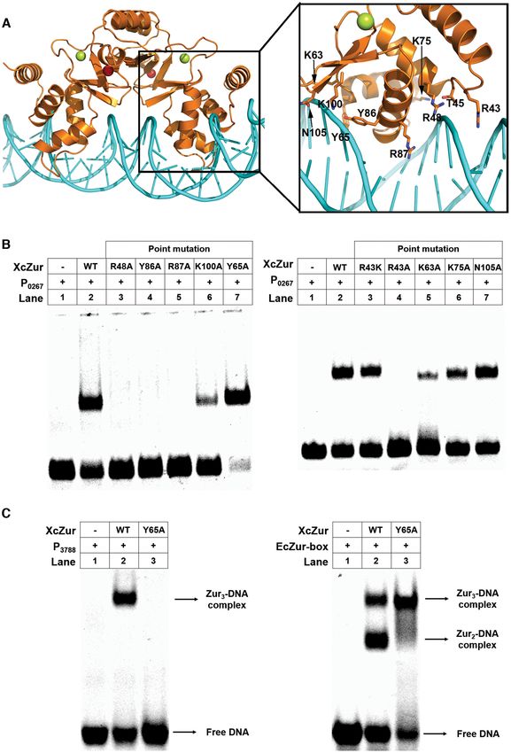
Nucleic Acids Research, 2021, Vol. 49, No. 11 6515
Biolayer interferometry (BLI) revealed that E. coli Fur exists in solution under several
oligomeric states, including both dimeric and monomeric
The DNA binding affinity of XcZur was determined by
states (41). To investigate the oligomeric state of XcZur in
biolayer interferometry (BLI) on ForteBio Octet K2 two-
solution, purified XcZur was subjected to SV-AUC analy-
channel system by using the streptavidin (SA) sensors. Bind-
ses. Purified XcZur behaved mainly as monomers (60.5%)
ing study was performed in 96-well microplates in a volume
and dimers (32.9%), with a trace amount of trimers (5.8%)
of 200 l per well at 25◦ C with constant shaking at 1000
(Figure 2A). Zinc addition altered the oligomeric states to
rpm. The assay buffer contained 20 mM HEPES pH 7.4,
60.3% of dimers, 28.9% of monomers and 4.9% of tetramers
150 mM NaCl, 0.18 mM EDTA, 0.2 mM Zn and 0.02%
(Figure 2B). These results demonstrate that XcZur can form
Tween-20. The analysis was carried out using a set of eight
both monomers and dimers in solution and zinc percep-
appropriate XcZur concentrations (P0267 and P2472 : 0, 9.4–
tion appears to be able to significantly stabilize the dimeric
600 nM; P3788 : 0, 15.6 to 1000 nM). The sequence for each
form.
run was as follows: SA sensors prewet (10 min), equilibra-
Previous DNaseI footprinting studies revealed that
Downloaded from https://academic.oup.com/nar/article/49/11/6511/6287845 by guest on 29 December 2021
tion (180 s), binding of 10 nM biotinylated–DNA (90 s),
XcZur binds with specific DNA fragments (referred to as
baseline stabilization (180 s), XcZur association (300 s) and
P0267 , P2472 , P3788 and P2976 ) from the promoters of four
XcZur dissociation (500 s). For each run a new biosensor
zinc homeostasis genes (52). According to the results de-
was used. Global fitting was used for data analysis by the
rived from EMSA, XcZur bound to three of the four DNA
ForteBio software, specifying the 1:1 kinetic model and cor-
fragments, P0267 , P2472 and P3788 , with the former two as the
recting both association and dissociation curves.
most preferred targets (Figure 2C). These three DNA frag-
ments are well conserved with a pseudo-palindromic core
RESULTS
sequence (Supplementary Figure S2B). To demonstrate the
XcZur is a Zur group regulator with specific sequence fea- specificity of XcZur–DNA interaction, FAM-labeled P0267
tures and XcZur were incubated with varying amounts of un-
labeled DNA (cold DNA). Competitive binding of excess
The amino acid sequence of XcZur was subjected to a three-
cold P0267 , but not that of a control DNA with a random
iteration PSI-BLAST search (54) and led to the identifica-
sequence, to XcZur confirmed the binding specificity (Fig-
tion of 15 sequences with significant regions of similarity
ure 2D). Surprisingly, no stable association was detected
from PDB. As can be expected, all identified proteins be-
between XcZur and P2976 , suggesting requirement of other
long to the Fur family of transcriptional regulators. The
nucleotides in the promoter for protein-DNA interaction
neighbor-joining method (56) was used to perform phylo-
(Supplementary Figure S3). The DNA binding affinity of
genetic analysis shown in Supplementary Figure S2A. The
XcZur was determined by BLI (Figure 2E and Supplemen-
phylogenetic tree includes three main clades, correspond-
tary Figure S4). The resulting dissociation constants (KD )
ing exactly to the three functional groups of Fur family
of XcZur for P0267 , P2472 and P3788 were approximate 22,
proteins. Notably, the four selected Zur proteins (XcZur,
38 and 63 nM, respectively. These dissociation constants are
EcZur, MtZur and ScZur) and a nickel uptake regulator
in a similar range with the DNA-binding affinities of many
(Nur) are located in the same clade (referred to as Zur
Fur family regulators and consistent with our EMSA re-
group) but separated from other Fur family regulators (Fur
sults (Figure 2C). To determine the stoichiometries of the
group and PerR group) on the evolutionary tree. Sequence
three XcZur-DNA complexes, we subjected them to SV-
alignment of the 16 Fur family proteins were accomplished
AUC analyses. The stoichiometries for the XcZur-P0267 ,
by combining multiple methods and shown in Figure 1.The
XcZur-P2472 and XcZur-P3788 complexes were 2:1, 2:1 and
Fur family regulators possess a distinctive core domain with
3:1, respectively (Supplementary Figure S5).
∼130 residues in length, corresponding to a fragment of
To test whether zinc perception is required for the DNA
XcZur encompassing residues C37 to C165. Nevertheless,
binding activity of XcZur, we titrated the regulator with
the overall amino acid sequence identity among the Fur
P0267 in the presence of the zinc chelator EDTA by EMSA.
family members is not extensive and there is only one in-
Addition of 3 mM of EDTA abrogated DNA binding (Fig-
variant residue existing in the family (R48, numbered in
ure 2F), suggesting that a metal ion is required for the DNA
XcZur sequence) (Figure 1). Limited overall sequence iden-
binding activity of XcZur. Importantly, further addition of
tity can be also found in the Zur group members (Figure 1),
zinc to a concentration of 0.3 mM in the presence of 3 mM
although within which XcZur displays a sequence identity
EDTA almost completely restored the DNA binding capac-
of 42% with EcZur (8). XcZur is unique in that it has an
ity of XcZur (Figure 2F). Since there are 5 mM of magne-
additional extension N-terminal to the relatively conserved
sium ions in the binding buffer, most of the EDTA can be
core domain. While the overall sequence identities of Fur
used by magnesium ions and 0.3 mM of zinc ions can be
family proteins are relatively low, three of the four ligands
sufficient to activate the DNA binding activity of XcZur.
in the structural metal binding site (C125, C128, C165) and
In sharp contrast, however, addition of the other 10 diva-
three ligands in the regulatory site (C110, H118, E133) are
lent metal ions, including the most common Fur ligands
well conserved (Figure 1).
nickel, ferric and manganese, could not restore the DNA
binding ability (Figure 2F). Consistently, ICP-MS revealed
Zinc perception is essentially required for the DNA binding
that XcZur can bind zinc ions but not nickel ions (Sup-
capacity of XcZur
plementary Table S3). These observations strongly indicate
It is reported that Fur family proteins behave typically as that zinc binding is essentially required for the DNA bind-
homodimers in solution, however, previous NMR study ing capability of XcZur.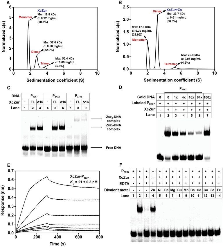
6516 Nucleic Acids Research, 2021, Vol. 49, No. 11

                                                                                                                                                        Downloaded from https://academic.oup.com/nar/article/49/11/6511/6287845 by guest on 29 December 2021

Figure 1. Structural elements and conservation of Fur family proteins. Sequence alignment of XcZur with fifteen representative Fur family proteins.
Conserved amino acids are highlighted in blue box. Secondary structure elements of apo and holo XcZur are shown above the alignment. The position
and frequency of metal ligands are indicated by red (Zur group), blue (Fur group) and green (PerR group) boxes, under the alignment. The key residues
for DNA binding, zinc coordination in site 1 and in site 2 are displayed as red arrow, gray stars and green stars, respectively.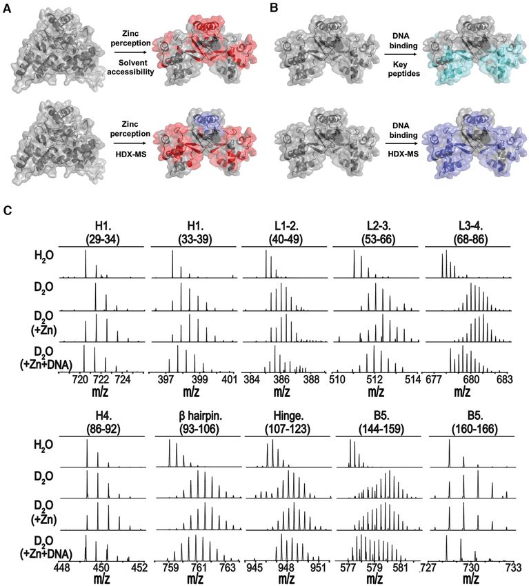
Nucleic Acids Research, 2021, Vol. 49, No. 11 6517

                                                                                                                                                               Downloaded from https://academic.oup.com/nar/article/49/11/6511/6287845 by guest on 29 December 2021

Figure 2. Biochemical characterization of XcZur. (A) Sedimentation coefficient distribution c(s) of XcZur determined by SV-AUC. The calculated value
of the protein oligomeric state of XcZur is indicated. (B) Sedimentation coefficient distribution c(s) of XcZur+Zn determined by SV-AUC. The calculated
value of the protein oligomeric state of XcZur is indicated. (C) EMSA for XcZur and XcZur-16 with P0267 , P2472 and P3788 . (D) Competitive binding
assay. The FAM-labelled P0267 probe and XcZur were incubated with varying amounts of unlabeled DNA. Lane1, DNA probe; lane 2, standard binding
reaction; lanes 3–6, standard binding reaction plus 1- to 64-fold (4-fold increase) molar excess of unlabeled P0267 , lane 7, standard binding reaction plus
100-fold molar excess irrelevant unlabeled DNA. (E) Affinity determination of XcZur binding with P0267 by BLI. The BLI responses for association
and dissociation, and the calculated KD are shown. (F) Effect of EDTA, zinc and other divalent metals on the binding of XcZur to P0267 . Each EMSA
experiment was repeated three times and similar results were obtained.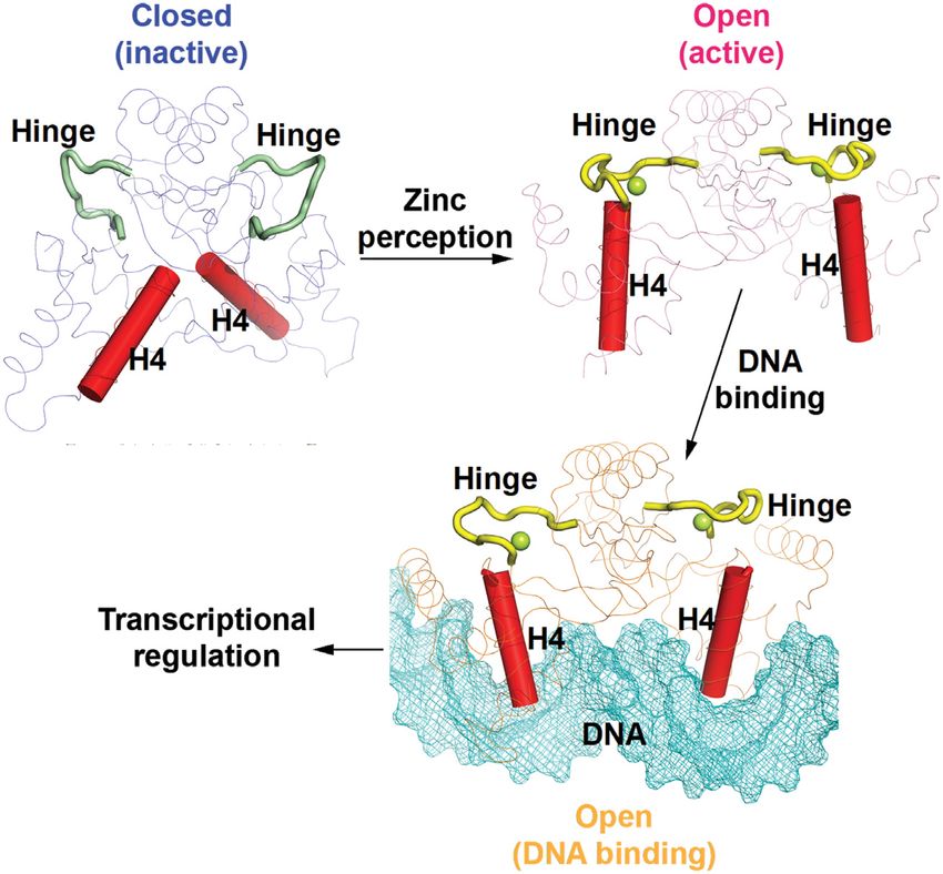
6518 Nucleic Acids Research, 2021, Vol. 49, No. 11
Apo- and holo-XcZur structures reveal distinct competencies Zinc perception induces an inactive-to-active conformational
for DNA binding transition of XcZur
To facilitate structural studies, we employed an approach To accurately located the site of the conformational rear-
combining disorder prediction, sequence alignment with rangement, we compared the results of two structural align-
other Fur family proteins and DNA binding assay to define ments between the apo- and holo-XcZur monomers. The
the domain boundaries in XcZur. The flexible N-terminal first alignment was based on the individual DNA binding
16 residues unique to XcZur are dispensable for DNA bind- domain of XcZur whereas the other alignment focused on
ing (Figure 2C). We thus purified the N-terminal extension the individual dimerization domain. Intriguingly, the com-
deleted XcZur (T17-G172, hereafter referred to as XcZur) parison revealed that the DBD structures of apo and holo
and crystallized this core domain in its apo form. To assess XcZur are very similar, with a r.m.s.d. of 1.1 Å for 69 aligned
possible conformational changes associated with zinc bind- C␣ atoms, and their DDs share moderate structural simi-
ing, we also crystallized XcZur in complex with addition- larity (2.9 Å r.m.s.d. for 44 aligned C␣ atoms) (Figure 4A).
Downloaded from https://academic.oup.com/nar/article/49/11/6511/6287845 by guest on 29 December 2021
ally supplemented zinc (holo form). Both structures were These observations indicate a rigid body rotation of one
determined by the SAD method, and the final atomic mod- domain related to the other upon zinc binding. When the
els were refined at 2.2 and 1.9 Å resolutions, respectively, for DDs of apo and holo XcZur were aligned, the DBDs ex-
the apo and holo form of XcZur (Table 1). hibited a rotation of 132◦ accompanied by a translation of
Similar to the reported structures of Fur family proteins, 1.9 Å along the rotation axis (Figure 4A). In this process, the
XcZur adopts a two-domain architecture connected by a hinge region, where zinc perception takes place, is proposed
partially flexible hinge, with its N-terminal DBD contain- to be the critical site responsible for the dramatic conforma-
ing four ␣ helices and a  hairpin and its C-terminal DD tional transition. Obviously, zinc perception in the hinge re-
comprising a three-stranded  sheet and a long ␣ helix (Fig- gion caused an approximate 180◦ rotation of this structural
ures 1 and 3A). Although both apo and holo XcZur fold element, especially at its N-terminal portion (residues C110
into a conserved two-domain structure and consist of al- to Q117) (Figure 4B), which is expected to subsequently al-
most identical secondary structure elements (Figures 1 and ter the orientation of the DBD with respect to the DD (Fig-
3A), structural comparison reveals significant conforma- ure 4A).
tional variation (Figure 3A). Evidently, apo XcZur binds The results from previous structural studies on Fur
only one zinc ion (Zn1) while holo XcZur binds two (Zn1 family regulators support an ‘open-to-closed’ conforma-
and Zn2). In addition, zinc binding to the regulatory site, tional change mechanism upon metal-dependent activation
formed by the hinge region and the two domains, results in (24,31,33–37,41–46,70), however, our structural observa-
drastic structural changes of the connecting hinge and com- tions reveal a completely different ‘closed-to-open’ transi-
pletely different orientations of the two domains. tion model. As a result of this dramatic change, the C-
Examination of crystal packing identified a dimeric as- termini (residue N94) of the recognition helices are now 39.8
sembly of both apo and holo XcZur structures, consistent Å apart from each other in the open holo XcZur dimer,
with the observations that XcZur can exist as homodimers compared to 8.1 Å in the closed apo XcZur dimer (Fig-
in solution (Figure 2A and 2B) and that most available ure 4C). Ultimately, this closed-to-open transition of the
structures of the Fur family proteins are in a dimeric form. DNA binding groove exposed the H4 recognition helices,
Notably, the apo and holo XcZur dimers represent two dif- the C-terminal portions of H1 and the -hairpin wings (B1
ferent conformations, termed respectively the ‘closed’ and and B2) in the DBD (Figures 1 and 4C). Importantly, these
‘open’ conformations, according to different size of the structural elements were reported to contribute to major
DNA binding groove and accessibility (Figure 3B). In par- groove, minor groove and nonspecific DNA contacts, re-
ticular, apo-XcZur dimer is in a closed and inactive state in spectively (37,38). These observations indicate that zinc per-
which the recognition helices cannot accommodate DNA ception of XcZur in the hinge region induces an inactive-
binding. On the contrary, holo-XcZur dimer is in an open to-active conformational transition that precisely rear-
and active state, and the recognition helices of holo XcZur ranges the structural elements responsible for target DNA
are precisely positioned and poised to contact DNA. Sup- binding.
porting this notion, structural comparisons reveal that the
overall structure of EcZur in complex with DNA (37) is
most similar to that of holo XcZur (Figure 3C), but to- Structural basis for the zinc perception of XcZur
tally different from that of apo XcZur. Superimposition of Similar to most Fur family metalloregulators, XcZur bears
the EcZur dimer with holo- and apo-XcZur dimer gives two types of zinc binding sites, a structural site (site 1) and
a root mean square deviation (r.m.s.d.) of 1.713 Å (229 a regulatory site (site 2). The structural zinc binding site lo-
aligned C␣ atoms) and 9.873 Å (246 aligned C␣ atoms), re- cates in the C-terminal DD whereas the regulatory site re-
spectively. Moreover, holo-XcZur dimer can be positioned sides between the two domains (Figures 3A and 5). At site 1,
into the E. coli Zur-box in an orientation similar but not the structural zinc ion (Zn1) is coordinated by four cysteines
identical to EcZur, with limited steric clashes (Figure 3C). (C125, C128, C165 and C168) from two CXXC sequence
These findings indicate that holo XcZur adopts a confor- motifs. The C125-XX-C128 motif is derived from the loop
mation intermediate between that of apo XcZur and that between  strands B3 and B4 and the C165-XX-C168 mo-
of the DNA bound EcZur. In other words, zinc percep- tif comes from helix H6 and its preceding N-terminal loop
tion is able to activate XcZur for the subsequent DNA (Figures 5A and B). Obviously, Zn1 functions as an or-
binding. ganizing center for packing of almost all structural ele-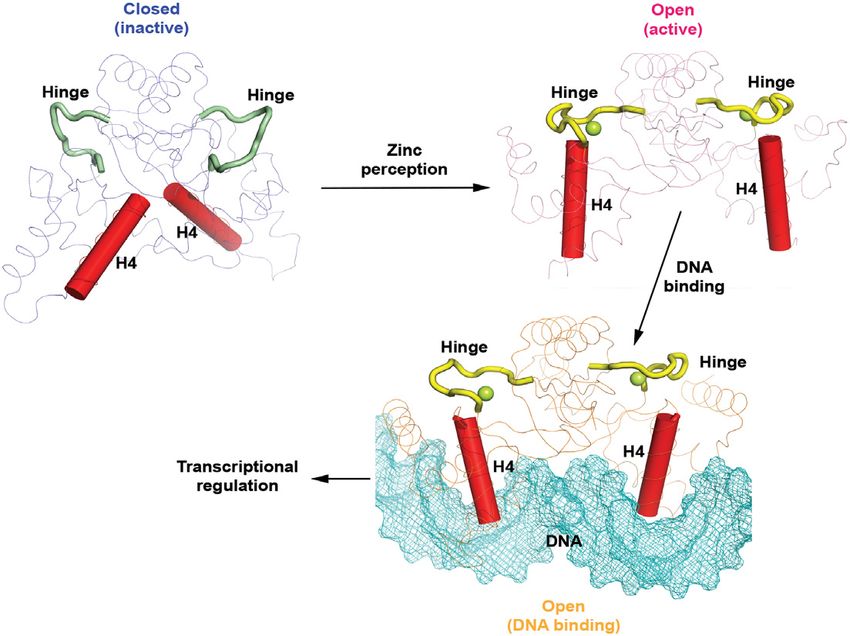
Nucleic Acids Research, 2021, Vol. 49, No. 11 6519

Figure 3. Apo- and holo-XcZur structures reveal distinct competencies for DNA binding. (A) Cartoon representation of the apo- and holo-XcZur                     Downloaded from https://academic.oup.com/nar/article/49/11/6511/6287845 by guest on 29 December 2021
monomers. The apo-XcZur structure is shown in blue, with the hinge region highlighted in green (left panel). The holo-XcZur structure is shown in
pink, with the hinge highlighted in yellow (right panel). Zn1 and Zn2 are shown in red and limon spheres, respectively. Secondary structural elements are
labeled. (B) Cartoon representation of the apo- (left panel) and holo-XcZur (right panel) dimers. Recognition helix (H4) is highlighted in red. (C) Structural
comparison of the holo-XcZur dimer with the holo-EcZur structure from the EcZur-DNA complex. Left panel shows a superimposition of holo XcZur
(pink) with holo EcZur (cyan). Right panel displays a docking of holo XcZur into the EcZur-box (cyan surface) in the same orientation of EcZur. Minor
and major grooves of the DNA are indicated.
6520 Nucleic Acids Research, 2021, Vol. 49, No. 11

                                                                                                                                                              Downloaded from https://academic.oup.com/nar/article/49/11/6511/6287845 by guest on 29 December 2021

Figure 4. Zinc binding induces an inactive-to-active conformational transition of XcZur. (A) Structural comparison of apo (blue) and holo XcZur (pink) on
the whole monomer (left panel), the DNA binding domain (middle panel), and the dimerization domain (right panel). The representation and color scheme
are the same as in Figure 3A. (B) Structural comparison of the apo-XcZur hinge (green) and holo-XcZur hinge (yellow). Left and middle panels show
the 2Fo -Fc electron density maps, contoured at 1σ , of the apo- and holo-XcZur hinges, respectively. Right panel displays the overall structure comparison
of apo- and holo-XcZur hinges. Residues in the hinge are shown as colored sticks. The bound Zn2 atom in the hinge is shown as a limon sphere. (C)
The inactive-to-active conformational transition of XcZur induced by zinc binding. Upper panel shows the cartoon representation of the apo- (blue) and
holo-XcZur (pink) dimers in which the recognition helix (H4) is highlighted in red. Structural elements responsible for DNA binding are indicated. Lower
panel displays the surface representation of apo- and holo-XcZur dimers in which the recognition helix is highlighted in red. The putative DNA binding
groove is indicated.
Nucleic Acids Research, 2021, Vol. 49, No. 11 6521
Downloaded from https://academic.oup.com/nar/article/49/11/6511/6287845 by guest on 29 December 2021
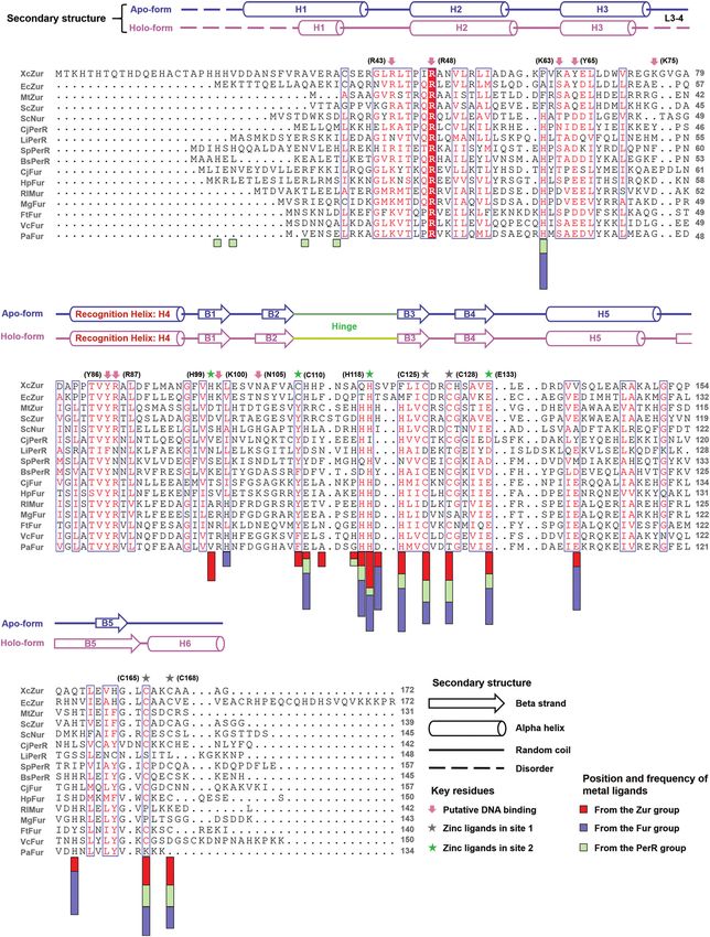
Figure 5. The structural and regulatory zinc binding sites of XcZur. (A) Close-up view of the structural zinc binding site (site 1) of apo XcZur. The zinc
atom is represented by a red sphere and the corresponding coordination ligands are shown in colored sticks. (B) Close-up view of the structural zinc binding
site of holo XcZur. (C) Close-up view of the regulatory zinc binding site (site 2) of apo XcZur. Key residues in this site are shown in colored sticks. The
characteristic  hairpin in DBD are shown in red. (D) Close-up view of the regulatory zinc binding site of holo XcZur. (E) Binding activity of wild type
and site 1-related mutants of XcZur with P0267 . (F) Binding activity of wild type and site 2-related mutants of XcZur with P0267 .6522 Nucleic Acids Research, 2021, Vol. 49, No. 11
ments in DD (B3-B5 and H6) and zinc perception in site Putative DNA-binding mode of XcZur
2 has no detectable effect on Zn1 coordination (Figures
To characterize the DNA-binding mode of XcZur, we con-
5A and B). Coordination of the structural zinc seems to
structed a model for the complex between XcZur and the
be extremely stable, as evidenced by the result from ICP-
P0267 DNA by the NPdock server (73). In the model, the
MS that treatment of EDTA (10 mM) along cannot re-
caliper-like XcZur dimer employs two recognition helices,
move this zinc (Supplementary Table S3). Previous study
each from one monomer, to contact DNA in the major
revealed that the structural zinc can only be removed by
groove, by principally using residues Y86 and R87 (Fig-
protein denature in addition to EDTA (71). In agreement
ure 6A). Residues in the L1-2 loop (R43 and T45), the
with its critical structural role in maintaining the correct
N-terminal portion of H2 (R48), N- and C-termini of H3
DD conformation, substitution of any of the four zinc lig-
(K63, Y65 and K75) and the DBD  hairpin (K100 and
ands in site 1 led to complete loss of DNA binding of XcZur
N105) also appear to contribute to DNA recognition (Fig-
(Figure 5E).
ure 6A). We next used mutagenesis to test whether these
In apo XcZur, the hinge packs against a highly curved
Downloaded from https://academic.oup.com/nar/article/49/11/6511/6287845 by guest on 29 December 2021
residues play key roles in DNA binding. All mutants, ex-
ten-stranded  sheet, with five  strands from each
cept for T45A, were successfully expressed and purified
monomer (Figure 5C). Despite the fact that this large 
to homogeneity. As shown in Figure 6B, the Y86A and
sheet brings the XcZur dimer into an intact entity, in-
R87A mutations at the recognition helix essentially abol-
teractions between the hinge and the  sheet are exten-
ished DNA binding, whereas the K75A and N105A mu-
sive but suboptimal, because the mixed polar and nonpo-
tations had little or no effect on formation of the protein-
lar residues at the interface have not made effective con-
DNA complex. Intriguingly, replacement of Y65 by ala-
tacts (Figure 5C). In fact, zinc ion (Zn2) perception in site
nine in H3, corresponding to a base-contacting residue
2 optimizes these interactions. In holo XcZur, binding of
Y45 in EcZur (37), greatly enhanced DNA binding. Con-
the regulatory zinc is coupled to disruption of the ten-
sistent with the observation that MgFur and EcZur ap-
stranded  sheet and formation of two  sandwiches in-
ply a positive-charged residue for minor groove contact
stead between the  hairpin from the DBD and the three
(37,38), the corresponding R43A mutation failed to bind
 strands from the DD (Figure 5D). Most of the hydropho-
DNA while the R43K mutation had no detectable effect.
bic residues are buried in the core of the new  sand-
Notably, alanine substitution of the only invariant residue
wich to link together the hydrophobic cores of the two do-
R48 among Fur family proteins resulted in a complete loss
mains, whereas polar residues are either involved in Zn2
of DNA recognition (Figures 1 and 6B), revealing an indis-
perception or exposed to the solvent (Figure 5D). At one
pensable role of the protein-backbone interactions. In sup-
end of the  sandwich, the regulatory zinc ion is coor-
port, the K63A and K100A mutations, which might also
dinated by four ligands, H99 from the hairpin of DBD,
impact the interactions between XcZur and the phosphate
E133 from strand B4 of DD, C110 and H118 from the
backbone, significantly impaired DNA binding. Taken to-
hinge region (Figures 1 and 5D). These contacts sequester
gether, contacts from major groove, minor groove and DNA
the hinge and contribute to formation and stabilization
backbone all play important roles in XcZur-DNA recogni-
of the holo conformation. To confirm the importance of
tion.
Zn2 perception in site 2, we generated four missense muta-
As described above, XcZur prefers to bind its target DNA
tions in XcZur and individually examined their interactions
in a XcZur2 –DNA mode, resembling that of MgFur (38).
with DNA by EMSA. Consistent with our structural ob-
This result is in contrast to a recent report in which EcZur
servation, the H99A and C110S mutations resulted in com-
mainly forms a high affinity (EcZur2 )2 –DNA complex in a
plete abrogation of DNA binding whereas the E133A mu-
polymeric mode (37). The discrepancy may be in part due
tation substantially reduced interactions with DNA (Figure
to different DNA fragments used, since we observed that
5F). Notably, the H118A mutation gave a moderate effect
XcZur could also forms a XcZur3 –DNA complex with the
on the DNA binding ability of XcZur (Figure 5F), which
Zur-box of EcZur and with P3788 (Figures 2C and 6C). In-
might be explained by the existence of possible zinc ligands
terestingly, XcZur could also forms a XcZur2 –DNA com-
nearby in the hinge (C110-HHPNSAQ-H118-SV-P121,
plex with the EcZur-box (Figure 6C).
underlined) (72).
To map the position and frequency of the metal ligands in
In solution evidence for zinc-mediated conformational
Fur family metalloregulators, we examined and counted on
changes of XcZur
23 structures from the aforementioned 15 XcZur homologs
and XcZur (Figure 1, Supplementary Tables S4 and S5). To further investigate conformational changes of XcZur
Our study revealed that the metal ligands locate primarily upon zinc and DNA binding in solution, we performed
at the hinge region and three  strands in its close vicinity, HDX-MS for apo XcZur, holo XcZur and holo-XcZur–
i.e. B3 and B4 in the DD and B1 in the DBD. Other ligands DNA complex (Supplementary Table S6). HDX-MS re-
reside in H5, H6 and B5 from DD, and in the L2-3 loop vealed that zinc perception increased deuterium exchange
and H1 from DBD. Notably, most members from three Fur of DBD, especially H1, the L1-2 and L2-3 loops, H4, the
groups utilize the structural site to bind a zinc ion. By con-  hairpin and hinge region (Figures 7A and C), suggesting
trast, H99 and C110 of site 2, which are essentially required obvious conformational alterations which might contribute
for the DNA binding activity, are distinctive to XcZur and to exposure of key structural elements for subsequent DNA
EcZur, confirming the importance of this regulatory site in binding. Mapping peptides with significant changes of deu-
specific zinc perception and functional activation of XcZur terium exchange or solvent accessibility to XcZur struc-
and EcZur. tures revealed a very similar ‘exposed’ pattern in DBD, butNucleic Acids Research, 2021, Vol. 49, No. 11 6523
Downloaded from https://academic.oup.com/nar/article/49/11/6511/6287845 by guest on 29 December 2021
Figure 6. Putative DNA-binding residues of XcZur. (A) A structure model for the XcZur-DNA complex and a close-up view of putative DNA binding
residues. XcZur and DNA are colored in orange and cyan, respectively. The Zn1 and Zn2 atoms are shown as red and limon spheres, respectively. Putative
DNA contacting residues are highlighted as colored sticks. (B) Mutational effects of putative DNA binding residues on the DNA binding activity of XcZur.
(C) EMSA for XcZur and XcZurY65A with P3788 and EcZur-box.6524 Nucleic Acids Research, 2021, Vol. 49, No. 11
Downloaded from https://academic.oup.com/nar/article/49/11/6511/6287845 by guest on 29 December 2021
Figure 7. In solution evidence for zinc-mediated conformational changes of XcZur. (A) Peptides with significant changes in solvent accessibility (upper
panel) or deuterium exchange (lower panel) upon zinc perception are mapped onto the crystal structures. (B) Peptides that contain residues important to
DNA binding are mapped onto the holo-XcZur structure (upper panel). Peptides with remarkable changes in deuterium exchange during DNA binding
are mapped onto the holo-XcZur structure (lower panel). For (A) and (B), the more exposed and hidden peptides are colored in red and blue, respectively.
Key peptides for DNA recognition are colored in cyan. (C) Representative mass spectra for XcZur peptides from HDX-MS.
distinct patterns in DD, especially the last  strand (Fig- ticularly, peptides with significantly decreased deuterium
ure 7A, Supplementary Table S6). This may be explained exchange in DBD matched nicely with those involved in
by the fact that, besides the closed-to-open conformational DNA recognition (Figure 6).
change, zinc perception of XcZur in solution involves an ad-
ditional monomer-to-dimer transition to severely decrease
DISCUSSION
the deuterium exchange of DD and create the ‘hidden’ pat-
tern in HDX-MS (Figures 2A, B and 7A). XcZur is a global transcriptional regulator essential for zinc
Notably, target DNA binding decreased deuterium ex- homeostasis and full virulence of Xanthomonas (8,53). Our
change in XcZur encompassing nearly the whole protein se- biochemical studies reveal that zinc perception is necessary
quence (Figures 7B and C, Supplementary Figure S6), indi- and sufficient for the DNA binding capacity of XcZur. De-
cating the formation of a stable protein-DNA complex. Par- tailed structural analyses demonstrate that XcZur utilizesNucleic Acids Research, 2021, Vol. 49, No. 11 6525
two sites for zinc perception and conformational activation. tively sequesters the protein in a linear inactive conforma-
The Zn1 atom bound in the structural site 1 plays an indis- tion (46). This indicates that the metal coordinating residues
pensable role in stabilizing the proper fold of the dimeriza- used in the regulatory site play important roles in regula-
tion domain. The Zn2 atom bound in the regulatory site tion of Fur protein conformations. Given that the unique
2 functions in productive positioning of the DNA bind- N-terminal extension of XcZur is rich in metal-ligand can-
ing domain into an active conformation, which is capable didates (7 histidines, 1 cysteine, 1 glutamate and 1 aspartate
for DNA binding. It is generally accepted that, similar to in a 23-residue fragment), this region is highly possible to
BsPerR (Supplementary Figure S7A), Fur family proteins participate in metal coordination and conformational reg-
employ an open-to-closed mechanism for conformational ulation, as observed in SpRerR (46). We further discovered
activation (24,31,33–37,41–46,70). However, by comparing that an acidic residue corresponding to E133 in  strand
the XcZur structures in the apo form and the holo form, we B4 of XcZur is required for maintenance of the active state
discovered that Zn2 perception in site 2 induces a closed-to- in Fur family proteins. Particularly, this highly conserved
open conformational change to activate the transcriptional residue (with only aspartate and glutamate being tolerated,
Downloaded from https://academic.oup.com/nar/article/49/11/6511/6287845 by guest on 29 December 2021
regulator (Supplementary Figure S7A). Figure 1) is used for metal coordination in the regulatory
Unlike BsPerR and XcZur, most Fur family proteins have site of all active Fur structures, and the E133A mutation
only the inactive- or active-state structure determined. To almost completely abrogates DNA binding (Figure 5F). In
explore the possible mechanisms that underlie Fur fam- keeping with this, the MtZur structure with zinc occupation
ily protein activation, we compared structures in the inac- in the regulatory site but not involved the glutamate ligand
tive state and the active state, respectively. Structure com- folds into a linear inactive conformation (43). In this regard,
parisons reveal that Fur family proteins appear to employ involvement of this acidic residue in metal perception of the
a conserved caliper-like active conformation (Supplemen- regulatory site can be used as a hallmark of the active state.
tary Figure S7B). In contrast, inactive conformations in Fur Our previous study found that XcZur binding might in-
family members are diverse (Supplementary Figure S7C) duce local distortions corresponding to DNase I hypersen-
and can be categorized into at least three forms, the closed sitive nucleotides in the XcZur-protected regions of target
form, the linear form, and the intermediate form between DNAs (52). Consistently, MgFur and EcZur use a combi-
the two. Notably, the recognition helices of Fur family reg- nation of base readout (major groove contacts) and DNA-
ulators in the all three forms cannot mediate DNA con- shape readout (minor groove contacts) mechanisms to rec-
tact without a pronounced conformational change of the ognize the distorted DNA (37,38). On the other hand, for-
dimeric structure. The closed form can be also termed as the mation of the XcZur-DNA complex may in turn result
V-shaped form because the dimeric structure resembles a V- in certain conformational alterations of the protein (Fig-
shaped letter (45,48). Obviously, apo XcZur belongs to the ures 7B and C, Supplementary Figure S6). As mentioned
closed form, although it cannot be reasonably aligned with earlier, XcZur utilizes respectively the dimeric mode, the
the other two structures (Supplementary Figure S7C). Half polymeric mode and mixed mode (with both dimers and
of the inactive structures, such as apo BsPerR (42), MtZur polymers) to recognize P0267 /P2472 , P3788 and the EcZur-
(43), oxidized BsPerR (44) and SpPerR (46), exhibits a lin- box (Figures 2C and 6C). Despite that XcZur can form
ear form with an extended conformation. The intermediate XcZur3 -DNA complex with both P3788 and EcZur-box, the
form, which brings a closed-like monomer and a linear-like structures of XcZur in these two complexes are likely to be
monomer together in a homodimer, possesses a chimeric distinct. According to our mutagenesis studies, the Y65A
character from both forms. In particular, apo MgFur (38) mutation, which affects a tyrosine residue important for
and LiPerR (47) are included in the intermediate form. With base contact in EcZur (37), eliminates P3788 binding but
regard to the three inactive forms, it can be safely assumed retains EcZur-box recognition in the XcZur3 -DNA com-
that only Fur proteins with a linear or intermediate inactive plex mode (Figure 6C). According to the results from the
form could experience an open-to-closed conformational present and previous studies (37,38,52), DNA binding of
activation process upon metal binding. On the other hand, XcZur is likely to be a process of induced fit. The mech-
the closed-to-open activation mechanism of XcZur is likely anism of induced fit can be used to explain why XcZur
applicable to other Fur family proteins with a similar closed is able to bind different target DNAs with such diverse
inactive conformation, although their structures of active modes. In fact, XcZur prefers binding P0267 and P2472 ,
conformation are yet to be determined. the promoters of low-affinity zinc-uptake systems, to bind-
Our structural observations suggest that appropriate ing P3788 , the promoter of high-affinity zinc-uptake system
binding of a specific metal in the regulatory site, which (Figure 2C and Supplementary Figure S8). This indicates
is formed by residues from the hinge region and the two that the P0267 /P2472 and P3788 might serve as a tuner and
domains, is a key determinant underlying the conforma- an on-off switch, respectively, for the control of zinc up-
tional activation of Fur family proteins. Supporting this take by XcZur (Supplementary Figure S8). Our previous
notion, most of the inactive dimer structures have at least in vivo and in vitro assays showed that XcZur represses
one monomer in the apo state in which only the structural transcription from the promoters of XC0267, XC2472 and
site is occupied by a metal (38,42,44,45,47,48), and the ab- XC3788, but activates transcription from the XC2976 pro-
sence of a metal in the regulatory site probably renders moter (52). The current EMSA results may provide a pos-
the two recognition helices of a Fur dimer unable to con- sible explanation for the distinct functions of XcZur. Un-
tact DNA. SpPerR does bear a nickel atom bound in the like binding with promoter sequences between the -10 and
hinge region, however, coordination of the nickel atom by -35 regions of XC0267/XC2472/XC3788 to possibly com-
additional residues from the most N-terminal helix effec- pete RNA polymerase contact, the binding of XcZur to the6526 Nucleic Acids Research, 2021, Vol. 49, No. 11
Downloaded from https://academic.oup.com/nar/article/49/11/6511/6287845 by guest on 29 December 2021
Figure 8. A proposed model of zinc-perception-induced DNA binding for XcZur. In the model, apo XcZur is in a closed inactive state. Zinc perception
in the regulatory zinc binding site induces a 180◦ rotation of the hinge region to drive a closed-to-open conformational change of the XcZur dimer. Holo
XcZur thus bears an active state, in which the putative DNA binding elements, especially the recognition helix (H4), are precisely positioned and poised
for DNA binding. Subsequently, holo XcZur binds with DNA in a mechanism of mutual induced fit and regulates DNA transcription.
P2976 -containing fragment requires DNA sequences outside University) for assistance with AUC data analysis; Dr Jix-
of the promoter region and may facilitate the recruitment ing Xia (College of Life Science and Technology) and Jun
of RNA polymerase to transcriptionally activate the zinc- Zhang (School of Marine Sciences) at Guangxi University
export gene. for help with ICP-MS analysis; Wei Hu at Guangxi Univer-
In summary, our study provides important insights into sity for assistance in HDX-MS analysis.
zinc-mediated conformational activation of a zinc uptake
transcriptional regulator in Xanthomonas (Figure 8). Our FUNDING
structural and biophysical analyses reveal that zinc percep-
tion in the regulator site of XcZur induces a closed-to-open National Natural Science Foundation of China [31700052];
conformational change to activate the transcriptional regu- Ba Gui Scholar Program of Guangxi Zhuang Autonomous
lator, and to which subsequent DNA binding likely employs Region of China [2014A002]; Guangxi Natural Science
a mechanism of induced fit. These mechanisms may also ap- Foundation [2020GXNSFFA297007]; State Key Labora-
ply to other Fur family regulators. tory for Conservation and Utilization of Subtropical Agro-
bioresources [SKLCUSA-a201806]; Guangxi Key Labora-
tory for Sugarcane Biology [GXKLSCB-20190304]. Fund-
DATA AVAILABILITY ing for open access charge: National Natural Science Foun-
Atomic coordinates and structure factors for the present dation of China.
crystal structures have been deposited to the Protein Data Conflict of interest statement. None declared.
Bank under accession codes 7DH7 and 7DH8.
REFERENCES
SUPPLEMENTARY DATA 1. Berg,J.M. and Shi,Y. (1996) The galvanization of biology: a growing
appreciation for the roles of zinc. Science, 271, 1081–1085.
Supplementary Data are available at NAR Online. 2. Lu,D., Boyd,B. and Lingwood,C.A. (1997) Identification of the key
protein for zinc uptake in Hemophilus influenzae. J. Biol. Chem., 272,
29033–29038.
ACKNOWLEDGEMENTS 3. Kasahara,M. and Anraku,Y. (1974) Succinate- and NADH oxidase
systems of Escherichia coli membrane vesicles. Mechanism of selective
The authors thank the staffs from beamlines BL17U1 and inhibition of the systems by zinc ions. J. Biochem., 76, 967–976.
BL19U1 at SSRF, and Dr Liming Yan from Tsinghua Uni- 4. Beard,S.J., Hughes,M.N. and Poole,R.K. (1995) Inhibition of the
versity, for their assistance with data collection. We also cytochrome bd-terminated NADH oxidase system in Escherichia coli
K-12 by divalent metal cations. FEMS Microbiol. Lett., 131, 205–210.
thank Dr Hai-Bo Liu (Guangxi University) and Dr Feng 5. Outten,C.E. and O’Halloran,T.V. (2001) Femtomolar sensitivity of
Yang (Guangxi Normal University) for assistance with de- metalloregulatory proteins controlling zinc homeostasis. Science, 292,
termination of binding affinity; Cuiyan Zhou (Tsinghua 2488–2492.You can also read