The morphology, biomechanics, and physiological function of the suboccipital myodural connections
←
→
Page content transcription
If your browser does not render page correctly, please read the page content below
www.nature.com/scientificreports
OPEN The morphology, biomechanics,
and physiological function
of the suboccipital myodural
connections
Yue Ma1, Wei Tang2, De‑Zheng Gong3, Xing‑Yi Li4, Jing‑Hui Zhang2, Jia‑Hui Sun5, Bing Wang4,
Ying Zhang1, Yu‑Xiao Chen4, Zhi‑Hong Zhang2, Nan Zheng2, Chukwuemeka Samuel Okoye2,
Yan‑Yan Chi2, Cheng‑Wei Wu6, Sheng‑Bo Yu2,7* & Hong‑Jin Sui2,7*
The myodural bridge (MDB) connects the suboccipital musculature to the spinal dura mater (SDM)
as it passed through the posterior atlanto-occipital and the atlanto-axial interspaces. Although the
actual function of the MDB is not understood at this time, it has recently been proposed that head
movement may assist in powering the movement of cerebrospinal fluid (CSF) via muscular tension
transmitted to the SDM via the MDB. But there is little information about it. The present study
utilized dogs as the experimental model to explore the MDB’s effects on the CSF pressure (CSFP)
during stimulated contractions of the suboccipital muscles as well as during manipulated movements
of the atlanto-occiptal and atlanto-axial joints. The morphology of MDB was investigated by gross
anatomic dissection and by histological observation utilizing both light microscopy and scanning
electron microscopy. Additionally biomechanical tensile strength tests were conducted. Functionally,
the CSFP was analyzed during passive head movements and electrical stimulation of the suboccipital
muscles, respectively. The MDB was observed passing through both the dorsal atlanto-occipital and
the atlanto-axial interspaces of the canine and consisted of collagenous fibers. The tensile strength
of the collagenous fibers passing through the dorsal atlanto-occipital and atlanto-axial interspaces
were 0.16 ± 0.04 MPa and 0.82 ± 0.57 MPa, respectively. Passive head movement, including lateral
flexion, rotation, as well as flexion–extension, all significantly increased CSFP. Furthermore, the
CSFP was significantly raised from 12.41 ± 4.58 to 13.45 ± 5.16 mmHg when the obliques capitis
inferior (OCI) muscles of the examined specimens were electrically stimulated. This stimulatory
effect was completely eliminated by severing the myodural bridge attachments to the OCI muscle.
Head movements appeared to be an important factor affecting CSF pressure, with the MDB of the
suboccipital muscles playing a key role this process. The present study provides direct evidence to
support the hypothesis that the MDB may be a previously unappreciated significant power source
(pump) for CSF circulation.
The MDB is a complex anatomical structure that physically connects the cervical spinal dura mater (SDM) to the
suboccipital muscles, including the rectus capitis posterior minor (RCPmi), the rectus capitis posterior major
(RCPma), and the obliquus capitis inferior (OCI) while passing through the posterior atlanto-axial and atlanto-
occipital interspaces1–5. Recently, several published studies have identified the existence of the MDB in mammals
(porpoises, macaques, dogs, cats, mice, and rabbits)6, birds7,8 and reptiles9, that the MDB is an evolutionary
conserved physical structure that is involved in important physiological functions.
1
The Second Affiliated Hospital of Dalian Medical University, Dalian 116027, Liaoning, People’s Republic of
China. 2Department of Anatomy, College of Basic Medical Science, Dalian Medical University, 9 West Section,
Lushun South Road, Dalian 116044, People’s Republic of China. 3Medical Foundation Experiment Teaching Center,
College of Basic Medical Science, Dalian, Liaoning 116011, People’s Republic of China. 4The First Affiliated Hospital
of Dalian Medical University, Dalian 116011, Liaoning, People’s Republic of China. 5Department of Anesthesiology,
Dalian Medical University, Dalian 116044, Liaoning, People’s Republic of China. 6Department of Engineering
Mechanics, State Key Lab of Structural Analysis for Industrial Equipment, Faculty of Vehicle Engineering and
Mechanics, Dalian University of Technology, Dalian 116024, Liaoning, People’s Republic of China. 7These authors
contributed equally: Hong-Jin Sui and Sheng-Bo Yu. *email: ysbdmu@126.com; suihj@hotmail.com
Scientific Reports | (2021) 11:8064 | https://doi.org/10.1038/s41598-021-86934-4 1
Vol.:(0123456789)
www.nature.com/scientificreports/
Concerning the functionality of the MDB, it has recently been proposed that contraction of the suboc-
cipital muscles, produced during head movement, might assist in resisting SDM in-folding1. Moreover, that
the suboccipital muscles, along with MDB might play an essential role in transmitting cervical proprioception
by monitoring tensional changes in the cervical SDM and effecting a rapid adjustment of the head p osition10.
Furthermore, as the MDB might prevent the cervical SDM from folding inward the spinal cord during head and
neck movements thus preventing the impedance of cerebrospinal fluid (CSF) flow in the subarachnoid space and
cisterna magna2,4,11. Other recent studies have postulated that during head movement, the MDB would pull on
the cervical SDM producing changes in the volume and pressure of the CSF located in the cisterna magna and
subarachnoid space of the upper cervical spine, consequently promoting the circulation of the cerebrospinal fluid
at the occipito-cervical junction12–14. A recent study using the PC cine of MRI evidenced a significant increase
in the net flow of cerebrospinal fluid in the cephalic direction after head r otation15, providing indirect evidence
for the hypothesis that the MDB serves as one of the power sources for CSF circulation.
Clinically, dysfunctional suboccipital musculature was observed to be related to various types of
headaches16–20. However, its mechanism is still unknown up to now. The basic research on the function of the
myodural bridge is very important for the study of the mechanism of clinical disease.
To test the proposed “cerebrospinal fluid circulation dynamic hypothesis” relating to the myodural bridge,
the present study explored the effects of suboccipital muscles contractions and head movements on the CSF
pressure and the potential role of MDB.
Materials and methods
Experimental subjects. Seventy-three healthy adult Beagle dogs (Both male and female, body weight
10–12 kg, body length 650–770 mm) were provided after experimental teaching of surgery for medical students
by the Experimental Animal Center of Dalian Medical University. All animal experiments conformed to ethical
requirements of the school.
Investigations of the morphology of MDB. Gross anatomic dissection in the nuchal regions. Ten Bea-
gle dogs were euthanized. Afterwards, the nuchal region of each dog was dissected, layer by layer, to explore the
subocciptal musculature.
Dissection at the level of the dorsal atlanto‑occipital interspace. The rectus capitis dorsal minor (RCDmi)
muscle was fully exposed. Fibrous connections between the RCDmi and the dorsal atlanto-occipital membrane
(DAOM) were observed as well as fibrous connections between the DAOM and the upper cervical SDM.
Dissection at the level of the dorsal atlanto‑axial interspace. The short head of the rectus capitis dorsal major
(RCDma) and the obliquus capitis inferior (OCI) muscles were exposed integrally. Fibrous connections between
these two muscles and the dorsal atlanto-axial membrane (DAAM) were observed as well fibrous connections
between the DAAM and the upper cervical SDM.
Histological study of connections between the dorsal occipital muscles and the SDM. Utilizing six of the eutha-
nized Beagle dogs, tissue-blocks from the sub occipital region were prepared, which included the suboccipital
muscles, the cervical dura mater, and tissues between them at the level of the dorsal atlanto-occipital interspace
or dorsal atlanto-axial interspace. These tissue blocks were conventionally sectioned and stained with Masson
trichrome and HE. The fibrous connections among the suboccipital muscles, DAOM (or DAAM) and SDM were
observed and photographed under the light microscope.
Ultrastructural investigation of connections between the dorsal occipital muscles and SDM under the scanning elec-
tron microscope (SEM). Tissue blocks taken from eight of the euthanized Beagle dogs at the level of the dorsal
atlanto-occipital interspace, consisted of the RCDmi, the DAOM and the SDM were dissected out.
At the level of the dorsal atlanto-axial interspace, tissue blocks consisting of the OCI, the short head of the
RCDma, the DAAM, and the SDM were obtained.
These specimens all underwent conventional preparation for SEM evaluations. The fibrous connections
between the DAOM (or DAOM) and the SDM at the dorsal atlanto-occipital and atlanto-axial interspaces were
observed and photographed under the SEM.
Biomechanical investigation of connections between the dorsal occipital muscles and the
SDM. Specimen preparation. Tissue blocks were taken from the euthanized the Beagle dogs (N = 13) were
utilized for this experiment. In the dorsal atlanto-occipital interspace, the specimens were intact tissue blocks
containing the RCDmi, the DAOM, the SDM, and the fibrous connections (Fig. 1A,a). The dorsal atlanto-axial
interspace, test samples were prepared containing the short head of the RCDma, the OCI, the DAAM, the SDM,
and fibrous connections among them (Fig. 1B,b).
Measurement of the connected areas between the MDB and SDM. The SDM, the DAOM (or DAAM), and the
muscles (RCDmi and OCI) were placed on a plastic film while maintaining their original in-vivo relationships.
Ink (Hero 234 Advanced Carbon Ink, Shanghai Ink Factory, China) was then dropped into the narrow gap
between the SDM and the other tissues to evidence the contour of the connected area between the MDB and the
SDM (Fig. 1). After staining the specimens the specimens were photographed and the connected areas between
Scientific Reports | (2021) 11:8064 | https://doi.org/10.1038/s41598-021-86934-4 2
Vol:.(1234567890)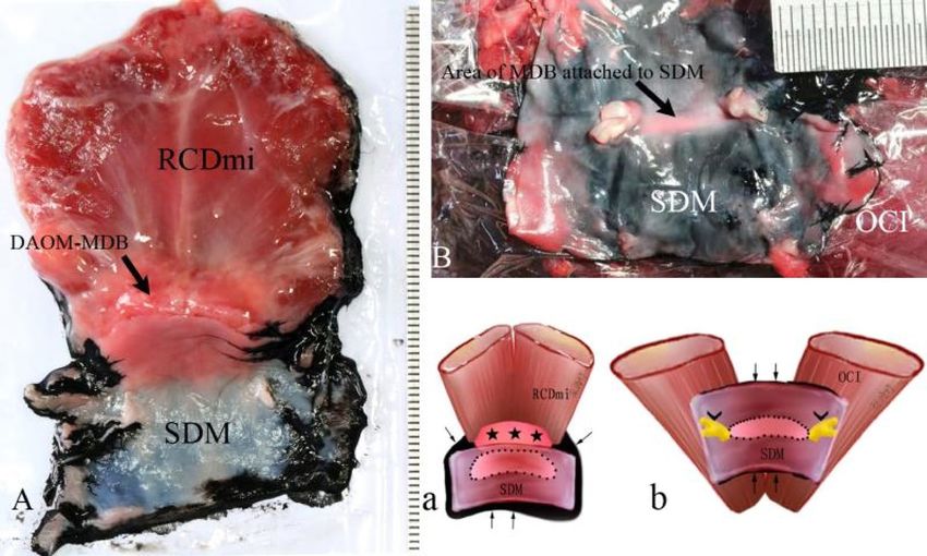
www.nature.com/scientificreports/
Figure 1. Diagram of the method for showing the connected area between the MDB and the SDM. The contour
of the area of the MDB attached to the SDM was showed by infiltrated black ink. Figure 1A: The complex of
RCDmi-MDB-SDM located at the dorsal atlanto-occipital interspace; Fig. 1B: The complex of OCI-MDB-SDM
located at the dorsal atlanto-axial interspace; Fig. 1a: Illustration for Fig A; Fig. 1b: Illustration for Fig. 1B. Thin
black rrows: black ink infiltrated into the narrow gap between the SDM and muscles. Dot line: the connected
area between the MDB and the SDM showed up by black ink infiltrating. Stars: DAOM and MDB. Arrowheads:
Roots of the spinal nerve.
Figure 2. Illustration of preparation (A) and fixation (B) of the test specimen.
the MDB and the SDM were measured using Adobe Photoshop (URL:http://m.xitongtiandi.net/soft_tx/5244.
html, version number:13.0 (13.0 20120315.r.428 2012/03/15:21:00:00) × 64) software.
Tensile testing. The fresh specimens were sealed in airtight bags and stored at 4 degree (C). Before measure-
ment the specimens were left at room temperature for half an hour. Uniaxial tension experiments were per-
formed at room temperature within the post mortem time of 10hrs. The specimen was fixed on the test rig as
shown in Fig. 2. The surface of muscle end of the specimen was glued (Cyanoacrylate Super Glue, Deli Group
Co., Ltd., China) onto a bottom platform, and the dural end of the specimen was glued in a folded sandpaper and
Scientific Reports | (2021) 11:8064 | https://doi.org/10.1038/s41598-021-86934-4 3
Vol.:(0123456789)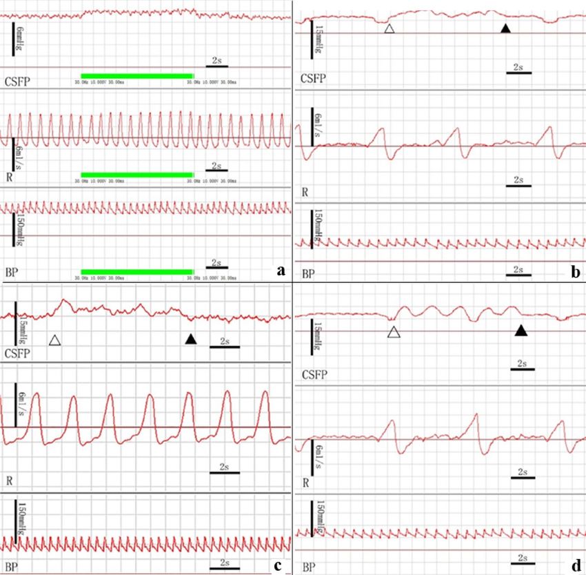
www.nature.com/scientificreports/

                                            clamped into the top clamp, which mounted on to a 50 N capacity load cell with sensitivity of 1mN (BAB S type
                                            aluminum weighing sensor, BAB-XS-5 M, Transcell Technology Inc., USA), which was attached to the actua-
                                            tor of a 20 kN CMT-4204 electronic universal testing machine (New Sansi Material Testing Co., Ltd, China).
                                            No preconditioning was conducted and only one loading cycle was executed on each specimen. The sampling
                                            frequency for both the force and displacement data was 100 Hz. Following this, the force was set at zero. The top
                                            clamp was raised slowly so as to just stretch the dura mater tight. Then the displacement was set at zero. After
                                            this, the top clamp moved with a pre-set velocity (10 mm/min) and the force–displacement curve until failure
                                            was recorded. The ultimate tensile strength of the MDB was calculated utilizing the ratio of the fracture force
                                            with the area of the connection between the MDB and the SDM.

                                            Cerebrospinal fluid pressure measurement during head movements or electrical stimulation
                                            of the suboccipital muscles. Pre-operation preparation: 33 animals were used for this aspect of the study.
                                            3% Nembutal was injected into the animal intraperitoneally for anesthesia with tracheal intubation.
                                               Cerebrospinal fluid pressure monitoring: The puncture needle was inserted into the subarachnoid space
                                            through the dorsal space between L4 and L5. When cerebrospinal fluid (CSF) appeared at the opening of the
                                            puncture needle, a catheter was inserted 24 cm cranially into the spinal canal and then connected to a blood
                                            pressure sensor (PT-102, Sensitivity:100 μV/mmHg, Resolution: 200 mmHg/(65536/2) = 0.006 mmHg) to record
                                            the CSF pressure by BL-420F biological data acquisition and analysis system (ChengDu Techman SoftWare CO.,
                                            LTD. China) during the experiment.
                                               Blood pressure monitoring: An arterial intubation was performed into the femoral artery and was monitored
                                            with a blood pressure sensor (PT-102) to record the blood pressure and the heart rate by the BL-420F system.
                                               Respiration monitoring: A bandage type respiratory sensor (HX200) was fixed to the chest of the dogs, and
                                            the frequency and amplitude of their respiration were recorded with the BL-420F system.
                                               Exposure of suboccipital muscles: The superficial structures were reflected layer by layer until the suboccipital
                                            muscles were exposed.

                                            Electrical stimulation of the superficial lay of the sub occipital muscles (RCDma, OCI). The BL-420F biological
                                            function laboratory system was used for this aspect of the study. Electrical stimulation was set at an output volt-
                                            age of 10 V, wavelength of 30 ms and 30 Hz, and the electrical stimulation was performed thrice for 10 s in each
                                            side of the long and short heads of the RCDma and the OCI muscles, respectively. The CSF pressure, respiration,
                                            and blood pressure were all recorded simultaneously.

                                            Passive movements of head. The spinous process of the axis (C2) was fixed with a custom made cervical fixa-
                                            tion device to stabilize the neck during passive movements of the head. Lateral flexion, flexion–extension and
                                            rotations of the head at the atlanto-occipital and atlanto-axial joints were respectively performed 10 times, with
                                            the frequency of motion being once per second. The CSF pressure, respiration,and blood pressures were moni-
                                            tored and recorded.

                                            Electrical stimulation of the RCDmi muscle. The stimulating electrode was inserted into the left and right side
                                            of the RCDmi respectively, and then electrical stimulations were given with a stimulus intensity of 10 V for 10 s,
                                            thrice for each side. The CSF pressure, respiration, and blood pressure were observed and recorded simultane-
                                            ously.

                                            Electrical stimulation in the OCI muscle after severing of its underlying connective fibers. In 6 animals, the
                                            left OCI muscle was disconnected from its deep structures and the right side was not treated as a control group.
                                            Subsequently the OCI was electrically stimulated on the left and right side respectively, as the CSF pressure,
                                            respiration, and blood pressures were monitored and recorded.

                                            Euthanizing the animals. The experimental dogs were euthanized by air embolism under anesthesia (3%
                                            pentobarbital sodium, 1 ml/kg).

                                            Collection and analysis of data. The experimental data was recorded before and after the electrical stimulations
                                            by using the Biological function laboratory system machine, and peak value of pressure wave was collected. The
                                            data were recorded as: Mean ± SD and analyzed by paired samples t-test and rank sum test (Wilcoxon signed-
                                            rank), P < 0.05 was chosen to be a statistically significant result.

                                            Ethics approval and consent to participate. All animal experiments conformed to ethical require-
                                            ments of the school.

                                            Limitations. The experiment was performed under anesthesia, which may be different from the physiologi-
                                            cal condition; Because of the great difficulty of surgery, the number of cases of myodural bridge disconnection
                                            was relatively small, so statistical analysis cannot be realized.

                                            Approval for animal experiments. All of the described experiments were approved by the Animal
                                            Experimental Center of Dalian Medical University.The present study was designed, performed, and reported
                                            according to the principles of the ARRIVE guidelines.

          Scientific Reports |   (2021) 11:8064 |                  https://doi.org/10.1038/s41598-021-86934-4                                                 4

Vol:.(1234567890)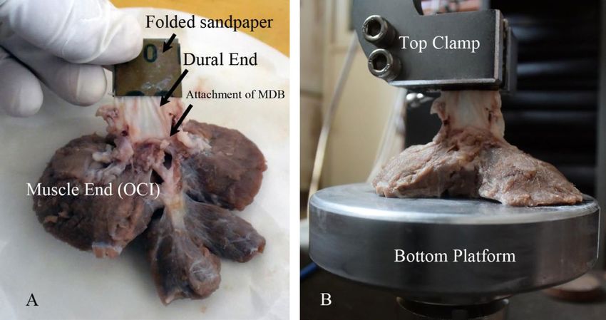
www.nature.com/scientificreports/

                                  Figure 3.  The MDB at the dorsal atlanto-occipital or atlanto-axial interspaces. The RCDmi overlay the dorsal
                                  atlanto-occipital interspace (Fig. 3a). When the RCDmi was reflected and stretched caudally the DAOM was
                                  severed along the posterior margin of occipital bone, and a fibrous connection was observed between the
                                  RCDmi and the DAOM (trigeminy stars), and also between the DAOM and the SDM (double arrowheads)
                                  (Fig. 3b). After the DM was cut along the margin of foramen magnum, and the RCDmi was reflected caudally,
                                  it was found that the DM was stretched dorsally via the MDB (enclosed by the dotted line) (Fig. 3c). On the
                                  other hand, the connective tissues (enclosed by the dotted line) among the RCDmi, the DAOM, and the DM
                                  were observed from the caudal side when the posterior arch of the atlas and the vertebral lamina were removed
                                  and the RCDmi was reflected to the cephalic side (Fig. 3d). At the lateral side of the dorsal atlanto-occipital
                                  interspace, the posterior arch of the atlas and the vertebral lamina of the axis were removed and the RCDmi
                                  was reflected laterally, and the connective tissues (enclosed by the dotted line) among the RCDmi, the DAOM,
                                  and the DM were again observed (Fig. 3e). Between the atlas and axis, the short head of the RCDma (RCDmaS)
                                  and the OCI passed over the dorsal atlanto-axial interspace (Fig. 3f). The fibrous connections between the OCI
                                  and the DAAM (white arrow), and between the short head of the RCDma and the DAAM (black arrow) were
                                  exposed in cranial aspect (Fig. 3g) and in the caudal aspect (double arrowheads) (Fig. 3h). After the lamina of
                                  the axis was removed and the OCI was reflected cranially, the dense connective tissue (enclosed by the dotted
                                  line) among the OCI, DAAM, and DM was observed (Fig. 3i). OCC: Occipital bone, RCDmi: Rectus capitis
                                  dorsal minor, DAOM: Dorsal atlanto-occipital membrane, DM: Dura mater, SC: Spinal cord, MDB: Myodural
                                  bridge, OC: Occipitalcondyle. OCI: Obliquus capitis inferior, RCPma S: Short head of the rectus capitis dorsal
                                  major, DAAM: Dorsal atlanto-axial membrane.

                                  Statement.      All methods were carried out in accordance with relevant guidelines and regulations. All exper-
                                  imental protocolswere approved by Dalian medical university.

                                  Results
                                  The results of anatomical dissection of the suboccipital region. In the suboccipital region, three
                                  suboccipital muscles were identified, the RCDma and RCDmi, and OCI. The RCDma was observed to have long
                                  and short heads (Fig. 3a,f).

                                  MDB in the dorsal atlanto‑occipital interspace. The RCDmi was observed to cross over the dorsal atlanto-occip-
                                  ital interspace (Fig. 3a), giving off dense connective tissues ventrally that inserted into the DAOM. The DAOM
                                  was observed to be tightly fused to the SDM via dense connective tissues fibers (Fig. 3b-e).

Scientific Reports |   (2021) 11:8064 |               https://doi.org/10.1038/s41598-021-86934-4                                                5

                                                                                                                                           Vol.:(0123456789)
www.nature.com/scientificreports/
Figure 4. The MDB showed in sagittal section of the dorsal atlanto-occipital or atlanto-axial regions, with
H&E stained (a, c, e) and Masson stained (figure b, d, f, g). Abundant dense connective tissues (white arrows
in Fig. 4d) were found to be continued with the muscular bundles of the RCDmi and inserted into the DAOM.
And then dense fibrous tracks (thick arrows in Fig. 4c,d) from the ventral side of the DAOM connected with the
SDM. Meantime, abundant dense connective tissues (white stars in Fig. 4f) were found to be originated from the
caudal end of the short head of RCDma and the ventral side of the OCI and inserted into the DAOM. And then
the ventral fibers of the dense part of the DAAM are connected to the SDM (black arrow in Fig. 4f,g), where
the SDM appeared multilayer structure and became thick caudally (arrowhead in Fig. 4f,g). OCC: the occiput,
C1: the atlas, C2: the axis; DAOM: Dorsal atlanto-occipital membrane; DAAM: Dorsal atlanto-axial membrane;
RCDmi: Rectus capitis dorsal minor; OCI: Obliquus capitis inferior; RCPma S: Short head of the rectus capitis
dorsal major; MDB: the myodural bridge; DM: the spinal dura mater.
MDB in the dorsal atlanto‑axis interspace. The short head of the RCDma as well as the OCI were both observed
to pass over the dorsal atlanto-axial interspace. The short head of the RCDma was found to cover the small cen-
tral part of this space and the OCI covered the large lateral part of this space (Fig. 3f). Deep to these two muscles,
dense connections were found between the short head of the RCDma and the central part of the DAAM and
between the OCI and the lateral part of the DAAM (Fig. 3g,h). Subsequently, when the dorsal arch of atlas was
removed to expose the SDM, dense connective tissue bridge was observed extending from the OCI to the SDM
via the DAAM (Fig. 3i).
The histological results of connections between the dorsal occipital muscles and the
SDM. Histological staining results. At the level of the dorsal atlanto-occipital interspace, it was found that
Scientific Reports | (2021) 11:8064 | https://doi.org/10.1038/s41598-021-86934-4 6
Vol:.(1234567890)www.nature.com/scientificreports/
Figure 5. Connections between the DAOM and SDM or between the DAAM and SDM showed under the
SEM. Figure (b) and (c): Magnification of the boxes in figure (a). Figure (e): Magnification of the box (e) in
figure (d). Figure (f): Magnification of the box (e) in figure (e). Abundant cord-like tissues were a terminal part
of the MDB, connecting the DAOM to the SDM (Fig. 5a). These connective cords were fused compactly with
the DAOM (Fig. 5b) and the SDM (Fig. 5c) respectively. The DAAM gave off bundle of plexiform collagen fibers
(Fig. 5d), which were woven into the collagen fibers array of SDM (Fig. 5e,f), thus anchoring the SDM to the
DAAM. DAOM: Dorsal atlanto-occipital membrane; DAAM: Dorsal atlanto-axial membrane; MDB: Myodural
bridge; SDM: Spinal dura mater.
abundant dense collagen fibers from the ventral side of the RCDmi were inserted into the DAOM. The DAOM
subsequently gave off collagen fibrous clusters from its ventral side and connected to the SDM (Fig. 4a-d).
At the dorsal atlanto-axial interspace, the cranial part of the DAAM was composed of abundant concentrated
dense connective fibers, whereas the caudal part of the DAAM evidenced loose fibers, having scattered parallel
running collagen fibrous bundles (Fig. 4e-g).
Dorsal to the DAAM, numerous fibrous collagen bundles were found originating from the ventral aspect of
the OCI and dorsal end of the short head of the RCDma and inserted into the cranial dense part of the DAAM,
and continuously the DAAM gave off abundant collagen fibers and then fused with the SDM. (Fig. 4e-g).
The results of observation under the SEM. For convenience of observation, the DAOM and SDM were separated
a little apart and then lots of cord-like structures were found connecting them. These plexiform cords were origi-
nated from the ventral side of the DAOM and then fused together with the SDM (Fig. 5a-c). Between the DAAM
and the SDM, masses of fibrous clusters were observed. They were given off by the DAAM and then interwoven
into the collagen fibrous array of the SDM (Fig. 5d-f).
Results of the tensile experiment. The RCDmi’s MDB was inserted into the SDM covering about
47.58 ± 10.80 mm2. The fracture force of the MDB was 7.55 ± 1.74 N, and the ultimate tensile strength amounts to
0.16 ± 0.04 MPa. The RCDma and OCI’s MDB was insert into the SDM covering 18.60 ± 11.62 mm2. The fracture
force of the MDB was 13.80 ± 10.43 N, and the ultimate tensile strength 0.82 ± 0.57 MPa.
Results of CSF pressure monitoring. CSF pressure changes during the electrical stimulation of the selected
suboccipital muscles. The results showed that the CSF pressure of the specimens were significantly increased
from 12.41 ± 4.58 mmHg to 13.45 ± 5.16 mmHg during electrical stimulation of the OCI (Fig. 6a, Table 1). But
when the OCI was detached from the DAAM as a negative control group, CSF pressure of this group was not
changed obviously during the electrical stimulation (Table 2). Furthermore, the long and short heads of RCDma,
and the RCDmi were stimulated electrically. There were no significant changes in CSF pressure during their
electrical stimulation.
Scientific Reports | (2021) 11:8064 | https://doi.org/10.1038/s41598-021-86934-4 7
Vol.:(0123456789)www.nature.com/scientificreports/
Figure 6. Measurements of CSF pressure during electrical stimulation of the suboccipital muscles or during
passive head movements. The CSF pressure was significantly increased when the OCI muscle was stimulated
but no change occurred in the blood pressure and respiration rate (Fig. 6a). And a rise and waveform change of
CSFP appeared during the lateral head flexion (Fig. 6b), head rotation (Fig. 6c), and head flexion and extension
(Fig. 6d), without changes in the respiration and blood pressure. CSFP: Cerebrospinal fluid pressure, R:
Respiration, BP: Blood pressure, Hollow Triangle: the beginning of passive motion of the head, Filled Triangle:
the end of passive motion of the head.
CSF pressure (mmHg)
N Before stimulations During stimulations T value P value (both sides α = 0.05)
OCI 26 12.41 ± 4.58 13.45 ± 5.16 3.589 0.001
Long head of RCDma 25 12.59 ± 4.61 12.93 ± 4.50 2.042 0.052
Short head of RCDma 9 11.18 ± 5.36 11.68 ± 5.81 1.981 0.083
RCDmi 8 14.33 ± 5.06 14.56 ± 5.56 0.636 0.545
Table 1. The effects of electrical stimulation of muscles on the CSF pressure (CSFP).
CSF pressure changes during passive movements of head. It was found that the CSF pressures were increased from
11.08 ± 4.21 to 12.14 ± 4.11 mmHg during the lateral flexion, from 10.73 ± 3.83 mmHg to 11.83 ± 3.88 mmHg
during the head rotation, and from 11.25 ± 3.94 mmHg to 12.48 ± 4.08 mmHg during the head flexion–exten-
sion respectively. A significantly increasing pressure of CSF appeared during each of the movements (Fig. 6b-d,
Table 3).
Scientific Reports | (2021) 11:8064 | https://doi.org/10.1038/s41598-021-86934-4 8
Vol:.(1234567890)www.nature.com/scientificreports/
CSF pressure (mmHg)
Before stimulations During stimulations Increase value (mmHg) P value
Before detached from the DAAM 12.14 ± 4.11 14.92 ± 5.18 2.10 ± 1.58 0.028
After detached from the DAAM 12.37 ± 3.41 12.34 ± 3.51 − 0.03 ± 0.58 0.917
P value – – 0.028 –
Table 2. The role of the myodural bridge in OCI affecting the CSF pressure (n = 6).
CSFP(mmHg)
N Before motion During motion T value P value
Head rotation 12 10.73 ± 3.83 11.83 ± 3.88 6.577 0.000
Flexion–Extension 14 11.23 ± 3.94 12.48 ± 4.08 3.912 0.002
Lateral Flexion 14 11.08 ± 4.21 12.14 ± 4.11 4.578 0.001
Table 3. The effect of passive head movement on CSF pressure (CSFP).
Discussion
In the recent 30 years, the MDBs have been discovered constantly between the suboccipital muscles and the
upper cervical spinal dura mater (SDM) in the occipitocervical junction in human. The MDB of RCPmi is located
in the posterior atlanto-occipital interspace1,11,13,14,21–25. And the MDBs of the RCPma and OCI are connected
with the spinal dura mater through the posterior atlanto-axial interspace2,3,13,22. Further more the nuchal liga-
ment (NL) was also found to participate the MDB at the posterior atlanto-axial and atlanto-occipital interspaces
respectively12,26–29. Up to now, the MDB has been proved to be a constant anatomical structure in the occipito-
cervical junction. But its function is still unclear. Recently, a new functional hypothesis was proposed that the
MDB complex might be one of the sources of power for CSF circulation12–15,22.
In this study, combining the multi-methods of gross dissection, histology, and scanning electoral microscope,
it was provided that the MDB was compose of abundant dense connective fibers existing between the dorsal
occipital muscles (the RCDmi, the short head of RCDma, and OCI ) and the SDM. Recently, Zheng et al.30 sug-
gested that the MDB might be a common existence and a normal anatomical structure in mammals. The present
animal study provides further evidence for the ubiquity of MDB in mammals.
Based on the MDB structure, the tensile strength of the MDB was managed to be measured. The rupture of the
myodural bridge complex (MDBc) occurred at site between the DAOM (or DAAM) and the SDM, which may be
the weakest part of the MDBc. The ultimate tensile strength (UTS) of the MDB at the dorsal atlanto-occipital and
atlanto-axial interspaces are 0.16 ± 0.04 MPa and 0.82 ± 0.57 MPa respectively, and the latter is obviously stronger
than the former. It has been reported that the UTS of the arterial wall is 0.5–1.72 MPa31. Although the MDBs are
different from the arterioles in structure, the UTS of the MDB is similar to that of the arteriole. This allows us
easily to perceive the strength of the dural bridge by comparing the MDB with the arterioles in the epidural space.
The dural is a membranous structure, with cerebrospinal fluid in the dural sac, venous plexus and loose con-
nective tissue in the epidural space. The resistance of the dura movement would be small, and the UTS of the
MDB attached to the dura mater may be enough to complete the bridge function of pulling the dura mater. And
the strength of the dura mater is within the range of 3.28–7.86 MPa32, with one order of magnitude stronger than
that of the MDB, and this may accord with the primary and secondary relationship between structures and is
also necessary to ensure the structural safety of the dura mater. In addition, tendons and ligaments are tough soft
tissue structures33, and the UTS of the MDB attached to the dura mater is 40 to 2000 times weaker than tendons
or ligaments. However, their physiological functions are also significantly different.
This study defines the mechanical properties of the MDB and provides data and method for further investiga-
tion on the biomechanical characteristics of the MDB.
In this study, we demonstrated the effect of stimulated contractions of the suboccipital muscles and passive
movements of the atlanto-occiptal and atlanto-axial joints on the CSF pressure. The results showed that the
electrical stimulation of the OCI caused a significant increase in the CSF pressure, while this effect was elimi-
nated by cutting the myodural bridge of OCI in advance. Also in vivo remaining dorsal occipital muscles intact,
there was an obvious rise in the CSF pressure during passive head rotations. Head rotation mainly occurs at
the atlanto-axial joint, where the OCI muscle is involved. Combining anatomical and physiological results, it
was proved that, during head rotation, the contraction of the OCI is probably transferred to the SDM via the
MDB, and repeated traction on the spinal dural during head rotations might lead to reciprocating changes of
geometrical shape of the upper cervical spinal dura capsule and then result in the subarachnoid space pressure
increase with some kind of dynamic mechanism.
Moreover, the present study found that passive head movements of lateral flexion and flexion–extension all
made significant increase of the CSF pressure. During these kinds of head motion, one or more dorsal occipital
muscles were involved at the occipito-atlantal joint, and the short head of RCDma and the RCDmi might pull
the spinal dura capsule via their MDBs, and then cause a rise in the CSF pressure. Nevertheless, there was no
obvious change in the CSF pressure when active electrical stimulation of the short head of RCDma or the RCDmi
Scientific Reports | (2021) 11:8064 | https://doi.org/10.1038/s41598-021-86934-4 9
Vol.:(0123456789)www.nature.com/scientificreports/
was performed. The RCDmi and the short head of RCDma are small muscles, and they could not move the the
occipito-atlantal joint alone. So during their active electrical stimulation, they produced only isometric contrac-
tions and thus a negligible pull on the SDM was present without effect on the CSF pressure.
Generally, the present results provide direct evidence that the MDB complex is one of the power sources for
CSF circulation. Additional in clinic, it also sheds light on the possible relationship between pathologic suboccipi-
tal muscles and chronic headache. Clinical studies have shown that the dysfunction of the suboccipital muscles
under various kinds of pathologic status might be correlated with chronic cervical headache14,17–20. The role of
the suboccipital muscles via the MDB in CSF circulation might be its possible mechanism.
Conclusion
The myodural bridge exists in the suboccipital region of dogs and has a tensile strength similar to the walls of
small artery. The head movements are an important factor affecting CSF pressure, and the MDB of the suboccipi-
tal muscles may play a key role this process. The present study provides direct evidence to support the hypothesis
that the MDB may be an important power source for CSF circulation.
Data availability
All data generated or analyzed during this study are included in this article. All the data was obtained and docu-
mented via the described experiments.
Received: 18 September 2020; Accepted: 16 March 2021
References
1. Alix, M. E. & Bates, D. K. A proposed etiology of cervicogenic headache: The neurophysiologic basis and anatomic relationship
between the dura mater and the rectus capitis posterior minor muscle. J. Man. Manip. Ther. 22, 534–539 (1999).
2. DeanNA, Mitchell BS. Anatomic relation between the nuchal ligament (1igamentum nuchae) and the spinal dura mater in the
craniocervical region. Clin. Anat. 15, 182–185 (2002).
3. Dou, Y. R. et al. Existence and features of the myodural bridge in Gallus domesticus: Indication of its important physiological
function. Anat. Sci. Int. 94(2), 184–191 (2019).
4. Fernhndez-de-las-Penas, C., Cuadrado, M. L., Arendt-Nielsen, L. & Ge, H. Y. Pareja JA 2008 Association of cross-sectional area of
the rectus capitis posterior minor muscle with active trigger points in chronic tension-type headache: A pilot study. Am. J. Phys.
Med. Rehabil. 87(3), 197–203 (2008).
5. Hack, G. D. & Hallgren, R. C. Chronic headache relief after section of suboccipital muscle dural connections: A case report.
Headache 44(1), 84–89 (2004).
6. McPartland, J. M., Brodeur, R. R. & Hatlgren, R. C. Chronic neck pain, standing balance, and suboccipital muscle atrophy–a pilot
study. J Manipul. Physiol. Ther. 20(1), 24–29 (1997).
7. Mitchell, B. S., Humphreys, B. K. & O’Sullivan, E. Attachments of the ligamentum nuchae to cervical posterior spinal dura and
the lateral part of the occipital bone. J. Manip. Physiol. Ther. 21, 145–148 (1998).
8. Nash, L., Nicholson, H., Lee, A. S., Johnson, G. M. & Zhang, M. Configuration of the connective tissue in the posterior atlanto-
occipital interspace: A sheet plastination and confocal microscopy study. Spine 30(12), 1359–1366 (2005).
9. Okoye, C. S., Zheng, N., Yu, S. B. & Sui, H. J. The myodural bridge in the common rock pigeon (Columbia livia): Morphology and
possible physiological implications. J. Morphol. 279(10), 1524–1531 (2018).
10. Peck, D., Buxton, D. F. & Nitz, A. A comparison of spindle concentrations in large and small muscles acting in parallel combina-
tions. J. Morphol. 180, 243–252 (1984).
11. Pontell, M. E., Scali, F., Enix, D. E., Battaglia, P. J. & Marshall, E. Histological examination of the human obliquus capitis inferior
myodural bridge. Ann. Anat. 195, 522–526 (2013).
12. Humphreys, B. K., Kenin, S., Hubbard, B. & Cramer, G. D. Investigation of connective tissue attachments to the cervical spinal
dura mater. Clin. Anat. 16(2), 152–159 (2003).
13. Pontell, M. E., Scali, F., Marshall, E. & Enix, D. E. The obliquus capitis inferior myodural bridge. Clin. Anat. 26, 450–454 (2013).
14. Rutten, H. P., Szpak, K., van Mameren, H., Ten Holter, J. & de Jong, J. C. Anatomic relation between the rectus capitis posterior
minor muscle and the dura mater. Spine (Phila Pa 1976) 22, 924–926 (1997).
15. Scali, F., Marsili, E. S. & Pontell, M. E. Anatomical connection between the rectus capitis posterior major and the dura mater. Spine
36, E1612-1614 (2011).
16. Scali, F., Pontell, M. E., Enix, D. E. & Marshall, E. Histological analysis of the rectus capitis posterior major’s myodural bridge.
Spine J. 13, 558–563 (2013).
17. Venne, G., Rasquinha, B. J., Kunz, M. & Ellis, R. E. Rectus capitis posterior minor: Histological and biomechanical links to the
spinal dura mater. Spine (Phila Pa 1976) 42(8), E466–E473 (2017).
18. Xu, Q. et al. Head movement, an important contributor to human cerebrospinal fluid circulation. Sci. Rep. 6, 31787 (2016).
19. Yuan, X. Y. et al. Patterns of attachment of the myodural bridge by the rectus capitis posterior minor muscle. Anat. Sci. Int. 91(2),
175–179 (2016).
20. Yuan, X. Y. et al. Correlation between chronic headaches and the rectus capitis posterior minor muscle: A comparative analysis of
cross-sectional trail. Cephalalgia 37(11), 1051–1056 (2017).
21. Kumar, A., Ghosh, S. N. & Sadique, S. I. Clinicoradiological study of adult Chiari malformation type I patients with emphasis on
cerebrospinal fluid peak flow velocity at foramen magnum level. Neurol. India 67(3), 744–748 (2019).
22. Zhang, J. H. et al. Connection of the Posterior Occipital Muscle and Dura Mater of the Siamese Crocodile. Anat. Rec. (Hoboken)
299, 1402–1408 (2016).
23. Zheng, N. et al. Orientation and property of fibers of the myodural bridge in humans. Spine 18(6), 1081–1087 (2018).
24. Zheng, N. et al. The myodural bridge complex defined as a new functional structure. Surg. Radiol. Anat. 42, 143–153 (2020).
25. Zumpano, M. P., Hartwell, S. & Jagos, C. S. Soft tissue connection between rectus capitis posterior minor and the posterior atlan-
tooccipital membrane: A cadaveric study. Clin. Anat. 19, 522–527 (2006).
26. Hack, G. D., Koritzer, R. T., Robinson, W. L., Hallgren, R. C. & Greenman, P. E. Anatomic relation between the rectus capitis
posterior minor muscle and the dura mater. Spine (Phila Pa 1976) 20, 2484–2486 (1995).
27. Hentschel, S., Mardal, K. A., Lovgren, A. E., Linge, S. & Haughton, V. Characterization of cyclic CSF flow in the foramen and upper
cervical spinal canal with MR flow imaging and computational fluid dynamics. Am. J. Neuroradiol. 31, 997–1002 (2010).
28. Hofkes, S. K. et al. Differentiation between symptomatic Chiari I malformation and asymptomatic tonsilar ectopia by using cer-
ebrospinal fluid flow imaging: Initial estimate of imaging accuracy. Radiology 245(2), 532–540 (2007).
Scientific Reports | (2021) 11:8064 | https://doi.org/10.1038/s41598-021-86934-4 10
Vol:.(1234567890)www.nature.com/scientificreports/
29. Zheng, N. et al. Definition of the to be named ligament and vertebrodural ligament and their possible effects on the circulation of
CSF. PLoS ONE 9(8), e103451 (2014).
30. Zheng, N. et al. The universal existence of myodural bridge in mammals: An indication of a necessary function. Sci. Rep. 7(1),
8248 (2017).
31. Silver F.H., Christiansen D.L.: Mechanical properties of tissues. In Biomaterials Science and Biocompatibility (Springer, New York,
NY, 1999). https://doi.org/10.1007/978-1-4612-0557-9_7
32. van Noort, R., Black, M. M., Martin, T. R. & Meanley, S. A study of the uniaxial mechanical properties of human dura mater
preserved in glycerol. Biomaterials 2(1), 41–45 (1981).
33. Bowser, J. E., Elder, S. H., Rashmir-Raven, A. M. & Swiderski, C. E. A cryogenic clamping technique that facilitates ultimate tensile
strength determinations in tendons and ligaments. Vet. Comp. Orthop. Traumatol. 24(5), 370–373 (2011).
Acknowledgements
The authors thank Dr. Gary D. Hack from the University of Maryland School of Dentistry, and Ms. Xue-Jun
Sui from the University of Rochester for their polishing the language and thank Miss Shi-Zhu Sun from Dalian
Medical University College of Basic Medical Sciences for her drawing pattern diagrams.
Author contributions
S.H.J. and Y.S.B. were in charge of the overall design of the project, the overall planning of the operation of the
project, the formulation of the implementation plan of the project, and the writing of proofreading papers. M.Y.
participated in the experiment design, organized and implemented the experiment, collated the data, and wrote
the paper. T.W. participated in the experiment and was responsible for the tensile test. G.D.Z. participated in the
experiment design and physiological function experiment. L.X.Y.participated in all experiments, data sorting and
writing papers. Zhang J.H. participated in the experiment implementation and tensile test. S.J.H. participated in
most experiments, collated data and wrote papers. W.B. was responsible for the gross anatomy and tensile test
of the dural bridge of atlanto-occipital muscle. Z.Y. and C.Y.X. were responsible for histological staining. Z.Z.H.
was responsible for electron microscopy experiment. Z.N. participated in the electron microscope experiment.
O.C.S. wrote the paper. C.Y.Y. participated in the experimental management. W.C.W. participated in the tensile
test.All authors reviewed the manuscript.
Funding
This study was supported by National Natural Science Foundation of China: NSFC 31871213 awarded to Hong-
Jin Sui and NSFC31600972 awarded to Nan Zheng.
Competing interests
The authors declare no competing interests.
Additional information
Correspondence and requests for materials should be addressed to S.-B.Y. or H.-J.S.
Reprints and permissions information is available at www.nature.com/reprints.
Publisher’s note Springer Nature remains neutral with regard to jurisdictional claims in published maps and
institutional affiliations.
Open Access This article is licensed under a Creative Commons Attribution 4.0 International
License, which permits use, sharing, adaptation, distribution and reproduction in any medium or
format, as long as you give appropriate credit to the original author(s) and the source, provide a link to the
Creative Commons licence, and indicate if changes were made. The images or other third party material in this
article are included in the article’s Creative Commons licence, unless indicated otherwise in a credit line to the
material. If material is not included in the article’s Creative Commons licence and your intended use is not
permitted by statutory regulation or exceeds the permitted use, you will need to obtain permission directly from
the copyright holder. To view a copy of this licence, visit http://creativecommons.org/licenses/by/4.0/.
© The Author(s) 2021
Scientific Reports | (2021) 11:8064 | https://doi.org/10.1038/s41598-021-86934-4 11
Vol.:(0123456789)You can also read