Transcription factor HOXC10 activates the expression of MTFR2 to regulate the proliferation, invasion and migration of colorectal cancer cells
←
→
Page content transcription
If your browser does not render page correctly, please read the page content below
MOLECULAR MEDICINE REPORTS 24: 797, 2021

            Transcription factor HOXC10 activates the expression
              of MTFR2 to regulate the proliferation, invasion
                   and migration of colorectal cancer cells
                                     YING XIE, RAN CHEN, LIUJIA YAN, ZHANGJUN JIA,
                                            GUANGSHU LIANG and QIN WANG

                Department of Clinical Laboratory, Jiangsu Cancer Hospital, Nanjing, Jiangsu 210009, P.R. China

                                           Received May 27, 2021; Accepted June 25, 2021

                                                        DOI: 10.3892/mmr.2021.12437

Abstract. HOXC10 and mitochondrial fission regulator 2                    proliferation, clone formation, invasion and migration of CRC
(MTFR2) have been reported to be abnormally expressed                     cells.
in multiple types of cancer tissues. However, the effects of
HOXC10 and MTFR2 on colorectal cancer (CRC) remain                        Introduction
poorly understood. Therefore, the present study aimed to inves‑
tigate the expression of HOXC10 and MTFR2 in CRC tissues                  Colorectal cancer (CRC) is one of the most common malignant
and cells, and analyze their effects on CRC cell proliferation,           tumors. The incidence rate of CRC in women ranks third only
invasion and migration. Reverse transcription‑quantitative                to lung cancer and breast cancer, and ranks fourth only to lung
PCR and western blotting were used to detect the expression               cancer, stomach cancer and liver cancer in men (1). According
levels of MTFR2 and HOXC10 in tissues and cells. To inves‑                to the World Health Organization's International Agency for
tigate the association between MTFR2 and HOXC10, short                    Research on Cancer, in 2018 there were ~1.8 million new CRC
hairpin RNA‑MTFR2 and overexpression vector‑HOXC10                        diagnoses, accounting for 10% of all patients with cancer,
were transfected into the cells, respectively. Furthermore,               and ~860,000 CRC related‑deaths, accounting for 9.9% of
western blotting was performed to detect the expression                   all cancer‑related mortalities (1). The prevalence of CRC has
levels of invasion‑associated proteins. The proliferation, clone          been increasing over the last 5‑10 years, with an estimated
formation, invasion and migration of colorectal cancer cells              2.2 million new CRC cases and 1.1 million deaths worldwide
were in turn analyzed by the Cell Counting Kit‑8, clone forma‑            predicted by 2030 (2). In 1990, CRC was ranked 21st among the
tion, wound healing and Transwell assays. Japan Automotive                leading causes of death in China, but in 2017 it rose to the 11th,
Software Platform and Architecture software predicted the                 with a CRC mortality rate of 13.24 per 100,000 people (3).
binding sites between HOXC10 and MTFR2, which was                         In 2017, CRC was therefore accountable for 1.79% of all
confirmed by the dual‑luciferase reporter assay and chromatin             deaths, seriously threatening human health and imposing a
immunoprecipitation. The present study demonstrated that                  huge economic burden on society (3). Thus, it is important to
HOXC10 and MTFR2 mRNA and protein expression levels                       find a new, safe and effective treatment for CRC.
were significantly upregulated in CRC tissues and cells.                      According to the Gene Expression Omnibus database
MTFR2 knockdown significantly inhibited CRC cell prolif‑                  (dataset, GSE10950), the expression of mitochondrial fission
eration, clone formation, invasion and migration. Furthermore,            regulator (MTFR) 2 was significantly upregulated in patients
HOXC10 was shown to interact with MTFR2. HOXC10 overex‑                   with CRC (4). MTFR2 is a gene that encodes mitochondrial
pression was able to significantly reverse the inhibitory effects         proteins and belongs to the MTFR1/family with sequence
of MTFR2 knockdown on CRC cells. In conclusion, HOXC10                    similarity 54 member A family. MTFR2 induces fission of
overexpression activated MTFR2 expression to enhance the                  mitochondria in eukaryotic cells and provides energy for
                                                                          aerobic respiration (5). Wang et al (6) demonstrated that
                                                                          MTFR2 is highly expressed in glioblastoma and is associated
                                                                          with a poor prognosis (6). MTFR2 has also been reported to
                                                                          be highly expressed in oral squamous cell carcinoma tissues
Correspondence to: Dr Ying Xie, Department of Clinical
Laboratory, Jiangsu Cancer Hospital, 42 Baiziting, Xuanwu,                and to be negatively related to the overall survival (OS)
Nanjing, Jiangsu 210009, P.R. China                                       of patients (7). MTFR2 may also aggravate proliferation,
E‑mail: xieying122@126.com                                                migration and invasion of oral squamous carcinoma cells
                                                                          by switching oxidative phosphorylation to glycolysis (7). A
Key    words: HOXC10, mitochondrial fission              regulator   2,   previous study revealed that high MTFR2 expression is related
proliferation, invasion, migration, colorectal cancer                     to poor prognosis in patients with breast cancer (BC) and is
                                                                          more prevalent in patients with aggressive tumors (8). The
                                                                          invasion, migration and epithelial‑mesenchymal transition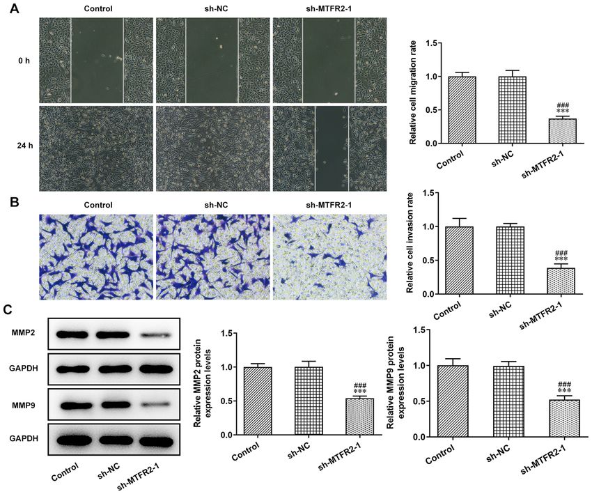
2 XIE et al: ROLE OF HOXC10/MTFR2 IN COLORECTAL CANCER
of BC cells has also been shown to be suppressed by the to the manufacturer's protocol. Total RNA (1 µg) was reverse
knockdown of MTFR2 (9). However, the biological function transcribed into cDNA at 42˚C for 30 min, according to the
of MTFR2 in CRC remains unclear. manufacturer's protocol of the PrimeScript RT Reagent Kit
A previous study revealed that HOXC10 expression is (cat. no. RR037A; Takara Biotechnology Co., Ltd.). qPCR was
significantly higher in CRC samples than in normal adjacent performed using SYBR Premix Ex Taq (Takara Biotechnology
tissues, and may be used as a biomarker for the diagnosis of Co., Ltd.) on an Applied Biosystems 7900 PCR system
CRC and as a potential therapeutic target (10). HOXC10 is also (Thermo Fisher Scientific, Inc.). The thermocycling conditions
considered a key modulator in ovarian cancer (OC), which is were used: Initial denaturation at 95˚C for 10 min; followed
associated with poor prognosis in patients with OC, actively by 40 cycles of denaturation at 95˚C for 15 sec and annealing
regulating Slug transcription to promote OC metastasis (11). at 60˚C for 1 min; and a final extension of 10 min at 72˚C. The
Furthermore, knockdown of HOXC10 has been reported to following primer pairs were used for qPCR: MTFR2 forward,
suppress the proliferation, migration and invasion of gastric 5'‑AGGG CTACGG GCCAAT TTGA‑3' and reverse, 5'‑TTC
cancer (GC) cells in vitro, and to suppress tumor growth and CTA A AT A AA G TT T GG T CC AC‑3'; HOXC10 forward,
induce apoptosis in vivo (12). HOXC10 expression is also mark‑ 5'‑CTCGGATAACGAAGCGAAAG‑3' and reverse, 5'‑CGC
edly upregulated in glioblastoma tissues and cells, resulting in TCTCGCGTCAAATACAT‑3'; and GAPDH forward, 5'‑TGT
a poor OS in patients with glioblastoma. Notably, inhibition of GGGCATCAATGGATTTGG‑3' and reverse, 5'‑ACACCA
HOXC10 could inhibit the proliferation, migration and inva‑ TGTATTCCG G GTCAAT‑3'. MTFR2 and HOXC10 mRNA
sion of glioblastoma cells (13). Therefore, it is speculated that expression levels were quantified using the 2‑ΔΔCq method and
HOXC10 could also regulate CRC progression. normalized to the internal reference gene GAPDH (14).
The aim of the present study was to investigate whether
HOXC10 binding to MTFR2 could regulate the proliferation, Western blotting. Total protein was extracted from cells
invasion and migration of CRC cells. using cold RIPA buffer (cat. no. P0013C; Beyotime Institute
of Biotechnology), and were centrifuged at 3,000 x g for
Materials and methods 15 min at 4˚C. The non‑transfected cells were used as the
control group. Total protein was quantified using a BCA kit
Human tumor samples. The present study was approved by (Beyotime Institute of Biotechnology) and 20 µg protein/lane
the Clinical Research Ethics Committee of Nanjing Medical was separated by SDS‑PAGE on a 12% gel. The separated
University (Nanjing, China; approval no. 2021‑448) and proteins were transferred to PVDF membranes and blocked
written informed consent was obtained from each patient. with 5% non‑fat milk with TBS‑0.1% Tween‑20 (TBST) for
In total, CRC tissues and paraneoplastic tissues (>2 cm away 1 h at room temperature. The membranes were incubated over‑
from the edge of the tumor) were obtained from 16 patients night at 4˚C with primary antibodies against the following:
with CRC in the Jiangsu Cancer Hospital (Nanjing, China) MTFR2 (1:500; cat. no. ab155678; Abcam), MMP2 (1:1,000;
between May 2019 and June 2020. None of the patients had cat. no. ab92536; Abcam), MMP9 (1:1,000; cat. no. ab76003;
received cancer treatment beforehand. The tumor samples Abcam), HOXC10 (1:500; cat. no. ab153904; Abcam) and
were stored at ‑80˚C until further use. The histopathological GAPDH (1:2,500; cat. no. ab9485; Abcam). Following the
characteristics of the patients are shown in Table I. primary incubation, membranes were washed with TBST and
incubated with an appropriate HRP‑conjugated secondary
Cell culture and transfection. The human intestinal epithelial antibody (1:1,000; cat. no. 7074; Cell Signaling Technology,
cell line (HIEC‑6) and colorectal cancer cell lines (Caco‑2, Inc.) for 1 h at room temperature. Protein bands were observed
HCT116, SW480 and LoVo) were all obtained from the using a chemiluminescence reagent (ECL) kit (Beyotime
American Type Culture Collection. Cells were cultured Institute of Biotechnology). The gray values of bands were
in Dulbecco's modified Eagle's medium (DMEM; Gibco; semi‑quantified using ImageJ software (version 1.0; National
Thermo Fisher Scientific, Inc.) containing 10% fetal bovine Institutes of Health) with GAPDH as the loading control.
serum (FBS; Gibco; Thermo Fisher Scientific, Inc.) and
1% penicillin/streptomycin solution at 37˚C with 5% CO2. Cell Counting Kit‑8 (CCK‑8) assay. The proliferation
Short hairpin RNA (sh)‑MTFR2‑1/2 (its negative control was of HCT116 cells was detected by performing the CCK‑8
sh‑NC) and overexpression (Oe)‑HOXC10 (its negative control assay. Following transfection, cells (2x10³ cells/well) were
was Oe‑NC) plasmids were provided by Shanghai GenePharma seeded into 96‑well plates. The non‑transfected cells were
Co., Ltd. HCT116 cells were seeded at 1.5x10⁵ cells/well in used as the control group. Cell proliferation was detected
6‑well plates for 24 h at 37˚C. When cell density reached at 24, 48 and 72 h using the CCK‑8 reagent (Beyotime Institute
60% confluence, HCT116 cells were transfected with sh‑NC of Biotechnology) according to the manufacturer's instructions.
(40 nM), sh‑MTFR2‑1/2 (40 nM), Oe‑NC (60 nM) and Cells were incubated with 10 µl CCK‑8 reagent for 2 h before
Oe‑HOXC10 (60 nM) using Lipofectamine® 2000 (Invitrogen; the absorbance value at 450 nm was detected via a microplate
Thermo Fisher Scientific, Inc.) according to the manufacturer's reader (Bio‑Rad Laboratories, Inc.).
protocol. Cells were then cultured in normal medium at 37˚C
for 48 h and subsequent experiments were conducted. Clone formation assay. Following transfection, HCT116 cells
(1x10³ cells/well) were seeded into a 6‑well plate and cultured
Reverse transcription‑quantitative PCR (RT‑qPCR). Total in DMEM at 37˚C for 2 weeks. The non‑transfected cells were
RNA was extracted from cells and tissue samples using used as the control group. Cell culture was stopped when
TRIzol® reagent (Thermo Fisher Scientific, Inc.) according clones were visible to the naked eye in the culture dish. The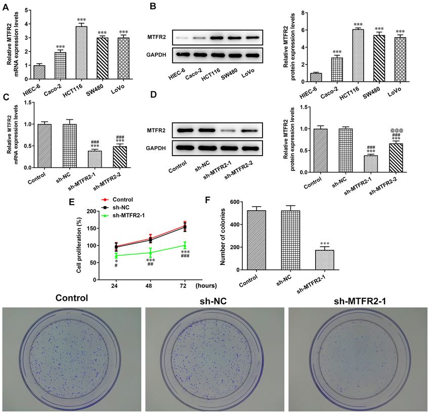
MOLECULAR MEDICINE REPORTS 24: 797, 2021                                                3

Table I. Histopathological characteristics of the patients.

           Age, 		                      Number of		                               Tumor              Differentiation        Distant
No.        years  Sex                     LNM     Grading                        location                degree            metastasis

 1 63 Female 1 Ⅱ                                                            Sigmoid colon       Middle‑low                    M1
 2 16 Male   1 Ⅳ                                                            Middle segment      Low                           M1
					                                                                       of transverse
					                                                                       colon
 3 69 Female 0 Ⅱ                                                            Rectum              Middle                        M0
 4 39 Male   0 Ⅱ                                                            Transverse colon    Middle                        M0
 5 66 Male   1 Ⅲ                                                            Junction of         Middle                        M0
					                                                                       rectum and
					                                                                       sigmoid colon
 6 43 Female 2 Ⅲ                                                            Sigmoid colon       Middle                        M0
 7 60 Male   0 Ⅰ                                                            Rectum              Middle                        M0
 8 75 Female 0 Ⅱ                                                            Ascending colon     Middle                        M0
 9 67 Female 0 Ⅰ                                                            Rectum              Middle                        M0
10 63 Female 0 Ⅱ                                                            Transverse colon    Middle                        M0
					                                                                       (proximal to
					                                                                       hepatic curvature)
11 48 Female 0 Ⅰ                                                            Rectum              Middle                        M0
12 54 Male   2 Ⅲ                                                            Sigmoid colon       Middle                        M0
13 54 Female 1 Ⅲ                                                            Colon               Middle                        M0
14 51 Male   2 Ⅳ                                                            Sigmoid colon       Middle‑low                    M1
15 66 Female 0 Ⅳ                                                            Ileocecal junction  Middle‑low                    M1
16 55 Male   1 Ⅲ                                                            Rectum              Middle                        M0

LNM, lymph node metastasis (32); M0, no distant metastasis; M1, have distant metastasis.

cells were fixed with 10% formaldehyde for 10 min at room             24 h, cells in the upper side of the chamber were removed
temperature and stained with 0.5% crystal violet for 5 min at         and those in the lower side of the chamber were fixed with
room temperature. The number of cloned cells was quantified           methanol for 20 min at room temperature and then stained
by eye.                                                               with crystal violet for 10 min at room temperature. Cells were
                                                                      observed and imaged using an inverted light microscope
Wound healing assay. The migratory ability of HCT116                  (magnification, x100), and the cell invasion rate was calculated
cells was detected by the wound healing assay. Following              using ImageJ (version 1.52r; National Institutes of Health). The
transfection, HCT116 cells (5x10⁵ cells/well) were seeded into        number of invasive cells were counted from five randomly
6‑well plates and cultured until cells reached 100% conflu‑           selected fields.
ence. The non‑transfected cells were used as the control
group. Serum‑free medium replaced normal medium and a                 Bioinformatics analysis. Japan Automotive Software Platform
sterile 100‑µl pipette tip was used to scratch the cells (0 h).       and Architecture [JASPAR, 8th (2020); http://jaspar.genereg.
Unattached cells were removed and the remaining cells were            net/] predicted that HOXC10 could bind to the promoter
cultured at 37˚C for 24 h. The wound gap was observed under           sequence of MTFR2.
an inverted light microscope (magnification, x100) and the
cell migration rate was calculated using ImageJ (version 1.52r;       Dual‑luciferase reporter assay. The interaction between
National Institutes of Health).                                       HOXC10 and the MTFR2 promoter in HCT116 cells was
                                                                      determined via the dual‑luciferase reporter assay. The
Transwell assay. The invasive ability of HCT116 cells was             wild‑type (WT) and mutant (MUT) sequences of MTFR2
detected via the Transwell assay. Following transfection,             untranslated region were amplified by Shanghai GenePharma
HCT116 cells (1x10⁵ cells/well) in 200 µl DMEM were placedin          Co., Ltd., cloned into a pGL3 luciferase vector (Promega
the upper Transwell chamber with the 8‑µm pore inserted               Corporation) and respectively named MTFR2‑WT and
membranes pre‑coated with Matrigel (BD Biosciences)                   MTFR2‑MUT. HCT116 cells at 80% confluence were
overnight at 37˚C. The non‑transfected cells were used as the         co‑transfected with MTFR2‑WT (50 nM) or MTFR2‑MUT
control group. In the lower chamber, 600 µl DMEM containing           (50 nM) and Oe‑HOXC10 (50 nM) or Oe‑NC (50 nM) using
10% FBS was added. After cells were cultured at 37˚C for              Lipofectamine 2000. Following incubation for 48 h at 37˚C,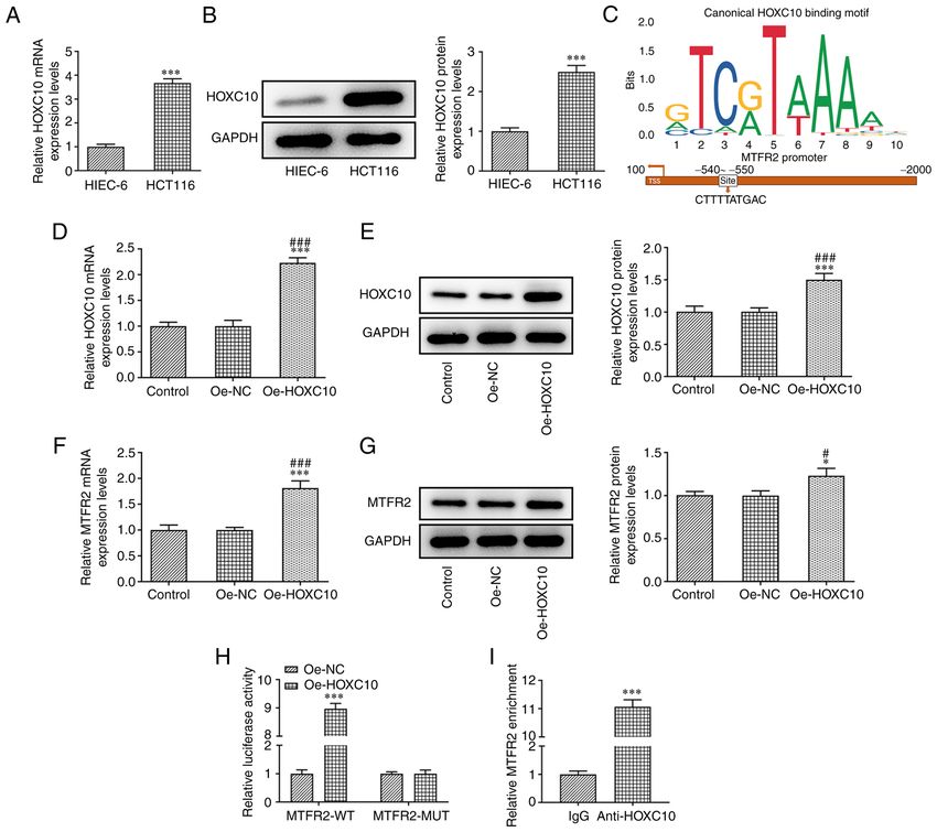
4                                         XIE et al: ROLE OF HOXC10/MTFR2 IN COLORECTAL CANCER

Figure 1. mRNA expression levels of HOXC10 and MTFR2 are upregulated in CRC tissues. mRNA expression levels of (A) MTFR2 and (B) HOXC10 in CRC
tissues and matched normal adjacent tissues were detected by reverse transcription‑quantitative PCR. ***P
MOLECULAR MEDICINE REPORTS 24: 797, 2021                                                               5

Figure 2. MTFR2 knockdown inhibits CRC cell proliferation. (A) mRNA and (B) protein expression levels of MTFR2 in CRC and the HIEC‑6 cell lines
were analyzed by RT‑qPCR and western blotting, respectively. ***P
6                                        XIE et al: ROLE OF HOXC10/MTFR2 IN COLORECTAL CANCER

Figure 3. MTFR2 knockdown inhibits colorectal cancer cell invasion and migration. (A) Migration and (B) invasion of HCT116 cells transfected with
sh‑MTFR2‑1 were analyzed by wound healing and Transwell assays, respectively (magnification, x100). Relative migration and invasion rates were expressed
normalized to the control group. (C) Expression of invasion‑ and migration‑related proteins in HCT116 cells transfected with sh‑MTFR2‑1 were detected via
western blotting. ***P
MOLECULAR MEDICINE REPORTS 24: 797, 2021                                                             7

Figure 4. HOXC10 is upregulated in colorectal cancer cells, and binds to the MTFR2 promoter to activate MTFR2 expression. (A) mRNA and (B) protein
expression levels of HOXC10 in HCT116 and HIEC‑6 cell lines were analyzed by RT‑qPCR and western blotting, respectively. ***P
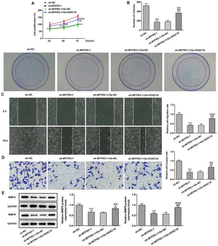
8                                         XIE et al: ROLE OF HOXC10/MTFR2 IN COLORECTAL CANCER

Figure 5. Overexpression of HOXC10 partially reverses the inhibitory effect of MTFR2 knockdown on colorectal cancer cell proliferation and migration.
(A) Proliferation, (B) clone formation, (C) migration and (D) invasion of HCT116 cells co‑transfected with sh‑MTFR2‑1 and Oe‑HOXC10 were in turn detected
by the Cell Counting Kit‑8, clone formation, wound healing and Transwell assays (magnification, x100). Relative migration and invasion rates were expressed
normalized to the control group. (E) Protein expression levels of invasion‑ and migration‑related proteins in HCT116 cells co‑transfected with sh‑MTFR2‑1
and Oe‑HOXC10 were detected by western blotting. **P
MOLECULAR MEDICINE REPORTS 24: 797, 2021                                                      9

    In conclusion, the present study demonstrated that                    6. Wang J, Xie Y, Bai X, Wang N, Yu H, Deng Z, Lian M,
                                                                             Yu S, Liu H, Xie W and Wang M: Targeting dual specificity
HOXC10 overexpression activated the expression of MTFR2                      protein kinase TTK attenuates tumorigenesis of glioblastoma.
to enhance the proliferation, clone formation, invasion and                  Oncotarget 9: 3081‑3088, 2018.
migration of CRC cells. HOXC10 may therefore be considered                7. Wang W, Xiong M, Jiang L, Chen Z and Shao Y: MTFR2
                                                                             promotes the proliferation, migration, and invasion of oral
an ideal therapeutic target for CRC. However, the study did not              squamous carcinoma by switching OXPHOS to glycolysis. Front
perform an animal model, which was a limitation of this study.               Oncol 10: 858, 2020.
                                                                          8. Lu W, Zang R, Du Y, Li X, Li H, Liu C, Song Y, Li Y and
                                                                             Wang Y: Overexpression of MTFR2 predicts poor prognosis of
Acknowledgements                                                             breast cancer. Cancer Manag Res 12: 11095‑11102, 2020.
                                                                          9. Lu G, Lai Y, Wang T, Lin W, Lu J, Ma Y, Chen Y, Ma H, Liu R
Not applicable.                                                              and Li J: Mitochondrial fission regulator 2 (MTFR2) promotes
                                                                             growth, migration, invasion and tumour progression in breast
                                                                             cancer cells. Aging (Albany NY) 11: 10203‑10219, 2019.
Funding                                                                  10. Enteghami M, Ghorbani M, Zamani M and Galehdari H:
                                                                             HOXC10 is significantly overexpressed in colorectal cancer.
                                                                             Biomed Rep 13: 18, 2020.
No funding was received.                                                 11. Peng Y, Li Y, Li Y, Wu A, Fan L, Huang W, Fu C, Deng Z,
                                                                             Wang K, Zhang Y, et al: HOXC10 promotes tumour metastasis
Availability of data and materials                                           by regulating the EMT‑related gene Slug in ovarian cancer.
                                                                             Aging (Albany NY) 12: 19375‑19398, 2020.
                                                                         12. Li J, Tong G, Huang C, Luo Y, Wang S, Zhang Y, Cheng B, Zhang Z,
The datasets used and/or analyzed during the current study                   Wu X, Liu Q, et al: HOXC10 promotes cell migration, invasion,
are available from the corresponding author on reasonable                    and tumor growth in gastric carcinoma cells through upregulating
                                                                             proinflammatory cytokines. J Cell Physiol 235: 3579‑3591, 2020.
request.                                                                 13. Guan Y, He Y, Lv S, Hou X, Li L and Song J: Overexpression of
                                                                             HOXC10 promotes glioblastoma cell progression to a poor prog‑
Authors' contributions                                                       nosis via the PI3K/AKT signalling pathway. J Drug Target 27:
                                                                             60‑66, 2019.
                                                                         14. Livak KJ and Schmittgen TD: Analysis of relative gene expres‑
YX, QW and GL conceived and designed the study. RC, LY                       sion data using real‑time quantitative PCR and the 2(‑Delta Delta
and ZJ were responsible for the acquisition, analysis and                    C(T)) method. Methods 25: 402‑408, 2001.
                                                                         15. Liang PS and Shaukat A: Assessing the impact of lowering the
interpretation of data. YX, RC and GL was responsible for                    colorectal cancer screening age to 45 years. Lancet Gastroenterol
manuscript preparation, writing and critical revisions. YX and               Hepatol 5: 523‑524, 2020.
RC confirm the authenticity of all the raw data. All authors             16. Song WY, Zhang X, Zhang Q, Zhang PJ and Zhang R: Clinical
                                                                             value evaluation of serum markers for early diagnosis of
have read and approved the manuscript.                                       colorectal cancer. World J Gastrointest Oncol 12: 219‑227, 2020.
                                                                         17. Zhang K, Xu H and Li HT: Safety and efficacy of propofol alone
Ethics approval and consent to participate                                   or in combination with other agents for sedation of patients
                                                                             undergoing colonoscopy: An updated meta‑analysis. Eur Rev
                                                                             Med Pharmacol Sci 24: 4506‑4518, 2020.
The present study was approved by the Clinical Research                  18. Park SW, Shin SP and Hong JT: Efficacy and tolerability of
Ethics Committee of Nanjing Medical University (Nanjing,                     prucalopride in bowel preparation for colonoscopy: A systematic
                                                                             review and Meta‑analysis. Adv Ther 37: 2507‑2519, 2020.
China; approval no. 2021‑448) and informed consent was                   19. Lima AR, Santos L, Correia M, Soares P, Sobrinho‑Simões M,
obtained from each patient.                                                  Melo M and Máximo V: Dynamin‑related protein 1 at the cross‑
                                                                             roads of cancer. Genes (Basel) 9: 115, 2018.
                                                                         20. Sheridan C and Martin SJ: Mitochondrial fission/fusion dynamics
Patient consent for publication                                              and apoptosis. Mitochondrion 10: 640‑648, 2010.
                                                                         21. Liu D, Duan W, Guo H, Xu X and Bai Y: Meta‑analysis of
Not applicable.                                                              associations between polymorphisms in the promoter regions of
                                                                             matrix metalloproteinases and the risk of colorectal cancer. Int
                                                                             J Colorectal Dis 26: 1099‑1105, 2011.
Competing interests                                                      22. Dong W, Li H, Zhang Y, Yang H, Guo M, Li L and Liu T:
                                                                             Matrix metalloproteinase 2 promotes cell growth and invasion
                                                                             in colorectal cancer. Acta Biochim Biophys Sin (Shanghai) 43:
The authors declare that they have no competing interests.                   840‑848, 2011.
                                                                         23. Morán A, Iniesta P, García‑Aranda C, De Juan C, Díaz‑López A,
References                                                                   Sánchez‑Pernaute A, Torres AJ, Díaz‑Rubio E, Balibrea JL and
                                                                             Benito M: Clinical relevance of MMP‑9, MMP‑2, TIMP‑1 and
                                                                             TIMP‑2 in colorectal cancer. Oncol Rep 13: 115‑120, 2005.
 1. Bray F, Ferlay J, Soerjomataram I, Siegel RL, Torre LA and           24. McGinnis W and Krumlauf R: Homeobox genes and axial
    Jemal A: Global cancer statistics 2018: GLOBOCAN estimates               patterning. Cell 68: 283‑302, 1992.
    of incidence and mortality worldwide for 36 cancers in 185 coun‑     25. Zhai Y, Kuick R, Nan B, Ota I, Weiss SJ, Trimble CL, Fearon ER
    tries. CA Cancer J Clin 68: 394‑424, 2018.                               and Cho KR: Gene expression analysis of preinvasive and inva‑
 2. Arnold M, Sierra MS, Laversanne M, Soerjomataram I, Jemal A              sive cervical squamous cell carcinomas identifies HOXC10 as a
    and Bray F: Global patterns and trends in colorectal cancer inci‑        key mediator of invasion. Cancer Res 67: 10163‑10172, 2007.
    dence and mortality. Gut 66: 683‑691, 2017.                          26. Feng X, Li T, Liu Z, Shi Y and Peng Y: HOXC10 up‑regulation
 3. Yin J, Bai Z, Zhang J, Zheng Z, Yao H, Ye P, Li J, Gao X and             contributes to human thyroid cancer and indicates poor survival
    Zhang Z: Burden of colorectal cancer in China, 1990‑2017:                outcome. Mol Biosyst 11: 2946‑2954, 2015.
    Findings from the Global Burden of disease study 2017. Chin          27. Tang XL, Ding BX, Hua Y, Chen H, Wu T, Chen ZQ and
    J Cancer Res 31: 489‑498, 2019.                                          Yuan CH: HOXC10 promotes the metastasis of human lung
 4. Clough E and Barrett T: The gene expression omnibus database.            adenocarcinoma and indicates poor survival outcome. Front
    Methods Mol Biol 1418: 93‑110, 2016.                                     Physiol 8: 557, 2017.
 5. Monticone M, Panfoli I, Ravera S, Puglisi R, Jiang MM, Morello R,    28. Li S, Zhang W, Wu C, Gao H, Yu J, Wang X, Li B, Jun Z, Zhang W,
    Candiani S, Tonachini L, Biticchi R, Fabiano A, et al: The nuclear       Zhou P, et al: HOXC10 promotes proliferation and invasion and
    genes Mtfr1 and Dufd1 regulate mitochondrial dynamic and                 induces immunosuppressive gene expression in glioma. FEBS
    cellular respiration. J Cell Physiol 225: 767‑776, 2010.                 J 285: 2278‑2291, 2018.
10 XIE et al: ROLE OF HOXC10/MTFR2 IN COLORECTAL CANCER
29. Zheng J, Ge P, Liu X, Wei J, Wu G and Li X: miR‑136 inhibits 32. Jin M and Frankel WL: Lymph node metastasis in colorectal
gastric cancer‑specific peritoneal metastasis by targeting cancer. Surg Oncol Clin N Am 27: 401‑412, 2018.
HOXC10. Tumour Biol 39: 1010428317706207, 2017.
30. Suo D, Wang Z, Li L, Chen Q, Zeng T, Liu R, Yun J, Guan XY
and Li Y: HOXC10 upregulation confers resistance to chemo‑ This work is licensed under a Creative Commons
radiotherapy in ESCC tumor cells and predicts poor prognosis. Attribution-NonCommercial-NoDerivatives 4.0
Oncogene 39: 5441‑5454, 2020. International (CC BY-NC-ND 4.0) License.
31. Yu J, Zhang X, Ma Y, Li Z, Tao R, Chen W, Xiong S and Han X:
miR‑129‑5p restrains apatinib resistance in human gastric
cancer cells via downregulating HOXC10. Cancer Biother
Radiopharm 36: 95‑105, 2021.You can also read