LINC00238 inhibits hepatic carcinoma progression by activating TMEM106C mediated apoptosis pathway
←
→
Page content transcription
If your browser does not render page correctly, please read the page content below
MOLECULAR MEDICINE REPORTS 24: 757, 2021

               LINC00238 inhibits hepatic carcinoma progression by
                activating TMEM106C‑mediated apoptosis pathway
                             CAIHUA JIANG1*, FENG LI2*, MENG YANG3, JIANPING DUAN2,
                                 JIANMING LAI4, SHULUN SUN5 and SHAOHUA FAN6

        Departments of 1Outpatients, 2Infectious Disease and 3Physical Therapy, Qingdao No. 6 People's Hospital, Qingdao,
                     Shandong 266033; 4Qingdao University Medical College, Qingdao, Shandong 266071;
                                 Departments of 5Liver Disease and 6Blood Purification Center,
                           Qingdao No. 6 People's Hospital, Qingdao, Shandong 266033, P.R. China

                                       Received February 6, 2021; Accepted June 22, 2021

                                                 DOI: 10.3892/mmr.2021.12397

Abstract. The present study aimed to explore the regulatory        In conclusion, LINC00238 inhibited HCC progression
mechanism of long intergenic non‑protein coding (LINC)00238        by inhibiting TMEM106 expression and activating the
in hepatocellular carcinoma (HCC). LINC00238 expression            TMEM106C‑mediated apoptosis pathway.
in HCC tissues and cell lines was measured using reverse
transcription‑quantitative PCR. LncTar was used to predict         Introduction
the binding sites between LINC00238 and transmembrane
protein 106C (TMEM106C). Survival analysis of LINC00238,           Hepatocellular carcinoma (HCC) is the fourth major cause of
TMEM106C and activating transcription factor 3 (ATF3) in           cancer mortality worldwide and the global morbidity of the
patients with HCC was performed based on TCGA data. The            disease is increasing (1). Surgical resection is the most effec‑
proliferation, apoptosis, migration, and invasion of HCC cells     tive treatment method of all therapy methods; however, most
were measured by 3‑(4,5‑dimethylthiazol‑2‑yl)‑5‑(3‑carboxy         patients are initially diagnosed as advanced and untreatable by
methoxyphenyl)‑2‑(4‑sulfophenyl)‑2H‑tetrazolium assay, flow         surgery (2). High rates of postoperative recurrence and metas‑
cytometer, wound healing and Transwell assays, respectively.       tasis are also reasons for the high mortality rate of HCC (3).
LINC00238 promoted apoptosis and inhibited proliferation,          Although great progress in the therapy methods has been
migration and invasion of HCC cells. LINC00238 was down‑           made in the last few decades, the survival rate of patients with
regulated in HCC. TMEM106C was a target of LINC00238               HCC remains poor (4). Hence, the search for new effective
and TMEM106C expression was negatively regulated                   therapeutic targets is of great significance for HCC treatment.
by LINC00238. TMEM106C suppressed the apoptosis                        In recent years, an increasing numbers of studies have
pathway and decreased the expression of caspase‑7, tissue          focused on the effect of lncRNAs on the occurrence and
inhibitor of metalloproteinase 2, programmed cell death 4          development of HCC (5‑7). Research has shown that aberrant
and ATF3. Notably, ATF3 was the upstream promoter of               expression of long non‑coding RNAs (lncRNAs) decelerates
LINC00238 and positively regulated LINC00238 expression.           or accelerates the progression of HCC. For instance, a study by
                                                                   Liu et al (8) demonstrates that lncRNA Ftx can inhibit prolif‑
                                                                   eration and metastasis of HCC and that higher lncRNA Ftx
                                                                   expression is associated with longer survival. Furthermore,
                                                                   long intergenic non‑protein coding (LINC)RNA‑p21 over‑
Correspondence to: Dr Shulun Sun, Department of Liver Disease,     expression reduces the levels of epithelial‑mesenchymal
Qingdao No. 6 People's Hospital, 9 Fushun Road, Shibei, Qingdao,
                                                                   transition (EMT)‑related proteins and decrease tumor metas‑
Shandong 266033, P.R. China
                                                                   tasis in HCC (7). The findings of Wang et al (9) indicate that
E‑mail: Sunshulun123@126.com
                                                                   high lncRNA ATB expression promotes proliferation of HCC
Dr Shaohua Fan, Department of Blood Purification Center,           and is positively correlated with poorer survival of patients
Qingdao No. 6 People's Hospital, 9 Fushun Road, Shibei, Qingdao,   with HCC. Gong et al (6) found that LINC00238 is expressed
Shandong 266033, P.R. China
                                                                   at low levels in HCC samples. However, the regulatory mecha‑
E‑mail: ShaohuaFan1123@126.com
                                                                   nism of LINC00238 in HCC remains to be elucidated.
*
    Contributed equally                                                The transmembrane protein (TMEM) family is a class
                                                                   of proteins containing one or more putative transmembrane
Key words: hepatocellular carcinoma, long intergenic non‑protein   segments that span partially or completely through biological
coding RNA 00238, transmembrane protein 106C, activating           membranes (10). As a member of the TMEM family,
transcription factor 3                                             TMEM106 is involved in several diseases, including cancer.
                                                                   In a study on non‑small cell lung carcinoma, Liu and Zhu (11)
                                                                   found that TMEM106A is an anti‑oncogene that inhibits the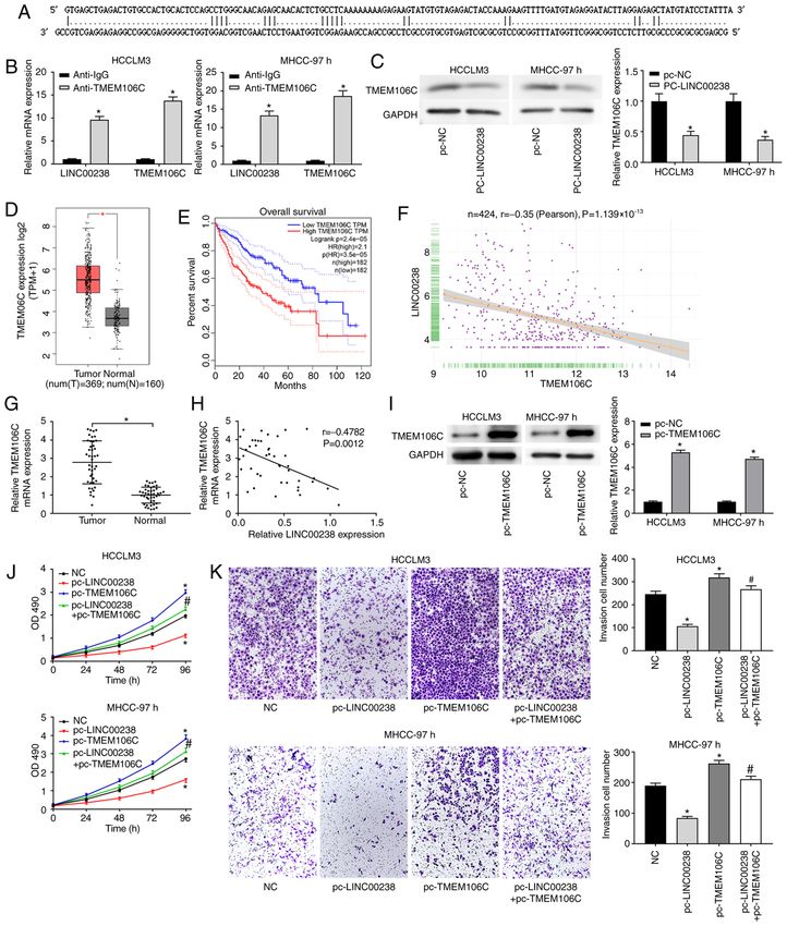
2                                 JIANG et al: LINC00238 INHIBITS HCC BY TARGETING TMEM106C

proliferation and invasion of lung cancer cells. Lang et al (12)   Cell transfection. LINC00238‑overexpressing plasmid
note that abnormal expression of TMEM106B is associated            pcDNA3.1 (pc‑LINC00238), negative control plasmid (pc‑NC),
with progression of frontotemporal lobar degeneration. In          short interfering (si)RNAs targeting LINC00238 (30 nM,
addition, Assassi et al (13) found that TMEM106C is aber‑          si‑LINC00238#1, 5'‑GAGGATACTTAGGAGAGCTAT‑3';
rantly expressed in ankylosing spondylitis. Luo et al (14) have    si‑LINC00238#2, 5'‑GGAGAGCTATGTATCCTATTT‑3'; and
indicate that TMEM106C is involved in HCC development.             si‑LINC00238#3, 5'‑GGCTGGAAGTGTTCAGTTAGT‑3')
However, remains to be elucidated whether lncRNAs are              and negative control siRNA (30 nM, si‑NC, 5'‑GAGGTATG
involved in the regulation of TMEM106C in HCC progres‑             GTATTCGGTCTGA‑3'), TMEM106C‑overexpressing plasmid
sion.                                                              pcDNA3.1 (pc‑TMEM106C) negative control plasmid (pc‑NC),
    The present study explored the regulatory role of              siRNAs targeting ATF3 (30 nM, si‑ATF3#1, 5'‑GAGCAGTTCG
LINC00238 in HCC progression. The findings suggested that          GTGCATATGGT‑3'; si‑ATF3#2, 5'‑GGGTTACTGGCAGGT
LINC00238 expression was decreased in HCC tissues and cell         TGAACT‑3'; and si‑ATF3#3, 5'‑GGGAAACAGTTGAGAGGT
lines and the low LINC00238 expression was associated with         TAT‑3') and negative control siRNA (30 nM, si‑NC, 5'‑GCT
the poor prognosis of patients with HCC. LINC00238 inhib‑          TGGAAGGTCGGTCATGTA‑3'), ATF3‑overexpressing
ited HCC progression by regulating the TMEM106C‑mediated           plasmid pcDNA3.1 (pc‑ATF3) negative control plasmid
apoptosis signaling pathway. Furthermore, ATF3, the gene           (pc‑NC) were purchased from Shanghai GenePharma Co., Ltd.
enriched in the apoptosis pathway, could regulate LINC00238        The transfection was performed by Lipofectamine® 3000
expression, consequently preventing HCC development.               (Invitrogen; Thermo Fisher Scientific, Inc.) according to the
                                                                   manufacturer's instructions. The cells were collected after
Materials and methods                                              transfection for 48 h.

Bioinformatics analysis. The online platform Gene Expression       Reverse transcription‑quantitative (RT‑q) PCR. Total RNA was
Profiling Interactive Analysis (GEPIA) (15) was used to            extracted from tissues and cells (1x10⁶) using TRIzol reagent
analyze the differentially expressed lncRNAs in HCC. A total       (Thermo Fisher Scientific, Inc.). The PrimeScript RT reagent kit
of 369 HCC cases and 160 normal were downloaded from               (Takara Bio, Inc.) was used to synthesize cDNA. qPCR was
The Cancer Genome Atlas (TCGA) database (https://portal.           carried out using SYBR Premix Ex Taq II kit (Takara Bio, Inc.)
gdc.cancer.gov/). The expression of LINC00238, TMEM106C            according to the manufacturer's protocol. The reaction conditions
and ATF3 in HCC tumor tissues and normal tissues were              were as follows: 95˚C for 5 min, followed by 40 cycles of 95˚C for
analyzed based on the downloaded data. The overall survival        15 sec and 60˚C for 30 sec. GAPDH was used as the internal
information in this study was downloaded from TCGA data‑           control for mRNA expression. Relative gene expression levels
base. Survival analysis was performed with the R package           were calculated using the 2‑ΔΔCq method (20). All experiments
‘survival’ (16,17) by using the Kaplan‑Meier curve method and      were performed in triplicate. The primers used in the study were:
TANRIC platform. The binding sites between LINC00238               LINC00238: 5'‑TTTGCAGAGTGGGTGCTAGG‑3' (forward)
and TMEM106C were predicted by LncTar (www.cuilab.                 and 5'‑TGCTTCATCTGGCAATGACCT‑3' (reverse);
cn/lnctar). Gene Set Enrichment Analysis (GSEA) (18) was           Transmembrane Protein 106C (TMEM106C): 5'‑GCCTGT
performed using three gene set datasets [GO‑BP (http://www.        CCAGCCAGATTCAG‑3' (forward) and 5'‑TTCCTACAGCC
geneontology.org), KEGG (http://www.genome.jp/kegg/) and           CCCTACTCT‑3' (reverse); activating transcription factor 3
Hallmark gene sets (19)] to analyze the impact of TMEM106C         (ATF3): 5'‑AGGTTTGCCATCCAGAACAA‑3' (forward) and
on signaling pathways. The JASPAR (http:///jaspar.genereg.         5'‑AGGCACTCCGTCTTCTCCTT‑3' (reverse); GAPDH:
net/) and PROMO (alggen.lsi.upc.es/cgi‑bin/promo_                  5'‑CTGGGCTACACTGAGCACC‑3' (forward) 5'‑AGTGGT
v3/promo/promoinit.cgi?dirDB=TF_8.3) databases were used           CGTTGAGGGCAATG‑3' (reverse).
to analyze the upstream transcription factor of LINC00238.
                                                                   Cell proliferation assay. HCC cells were trypsinized and
Patients and samples. A total of 43 human HCC tissues and          seeded into 96‑well culture plates (4,000 cells/well). The
corresponding normal tissues were collected from 22 male           cells were harvested at 24, 48, 72 and 96 h to detect prolif‑
(age range, 32‑64 years) and 21 female (age range, 34‑70 years)    eration using the 3‑(4,5‑dimethylthiazol‑2‑yl)‑5‑(3‑carboxym
patients with HCC between January 2018 and September 2019.         ethoxyphenyl)‑2‑(4‑sulfophenyl)‑2H‑tetrazolium (MTS) Cell
None of the patients received any treatment before surgical        Proliferation Colorimetric Assay kit (AmyJet Scientific, Inc.)
resection, including surgery, radiotherapy and chemotherapy.       and the absorption was detected at a wavelength of 490 nm.
The study was approved by the ethics committee of Qingdao
No. 6 People's Hospital [approval no. (2018) 26] and written       Cell apoptosis assay. The apoptosis of HCC cells was evalu‑
informed consent was obtained from all patients.                   ated using Annexin V/PI apoptosis‑detection kit (Invitrogen;
                                                                   Thermo Fisher Scientific, Inc.). HCC cells were washed with
Cell lines and culture. The human normal hepatic cell line         Annexin V‑binding buffer (Sigma‑Aldrich; Merck KGaA),
MIHA and HCC cell lines Huh7, HLF, SNU‑449, MHCC‑97h               followed by incubation in binding buffer containing
and HCCLM3 were purchased from Mlbio and cultured in               Annexin V‑FITC (25 µg/ml) and PI (25 µg/ml) in the dark
DMEM (Gibco; Thermo Fisher Scientific, Inc.) containing            for 10 min at room temperature. Apoptotic cells (early + late
antibiotics (Gibco; Thermo Fisher Scientific, Inc.) and            apoptotic cells) were analyzed using a BD Accuri C6 Plus
10% FBS (Gibco; Thermo Fisher Scientific, Inc.), in a humidi‑      flow cytometer and FACSDiva software (version 6.13;
fied atmosphere containing 5% CO2 at 37˚C.                         BD Biosciences).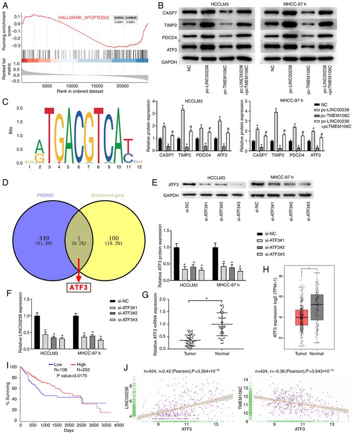
MOLECULAR MEDICINE REPORTS 24: 757, 2021                                               3

Wound healing assay. HCC cells were seeded into 6‑well               Statistical analysis. Data are shown as means ± SD and
plates and incubated to achieve ≥90% confluence. The mono‑           were analyzed using SPSS 23.0 (IBM Corp.) and GraphPad
layer cells were scratched with a 10‑µl sterile micropipette tip     Prism 7.0 (GraphPad Software, Inc.). Comparisons between
and cellular debris was removed by washing with PBS. Then,           two groups were analyzed using unpaired Student's t‑test and
cells were incubated in serum‑free medium with proliferation         the comparisons among multiple groups were analyzed by
inhibitor mitomycin C (10 µM; MedChemExpress) at 37˚C.               one‑way ANOVA followed by Tukey's post hoc test. Pearson's
Cell migration was observed at 0 and 48 h under a light              correlation analysis was used to assess the correlation among
microscope (magnification, x100, Olympus Corporation) and            LINC00238, TMEM106C and ATF3 expression. All experi‑
analyzed with ImageJ version 1.49 software (NIH). All experi‑        ments were performed in triplicate.
ments were performed in triplicate.
                                                                     Results
Transwell assay. The invasion of HCC cells was detected by
Transwell assay. The upper chambers (8 µm pore size) were            LINC00238 is downregulated in HCC. The online platform
coated with Matrigel at 37˚C for 30 min. Cells (1x10⁴ cells/well)    GEPIA was used to analyze the differentially expressed
were seeded into the upper chamber with serum‑free DMEM.             lncRNAs in HCC and the results suggested that LINC00238 was
DMEM containing 10% FBS was added to the lower chambers.             downregulated in HCC. The online analysis based on TCGA
The chambers were incubated for 48 h at 37˚C. Subsequently,          suggested that LINC00238 levels in HCC tissues were markedly
the noninvasive cells were removed with a cotton swab and            lower compared with those in normal tissues (Fig. 1A) and that
the invading cells were fixed with methanol. The cells were          LINC00238 levels was positively correlated with the overall
then stained with crystal violet at room temperature for 15          survival of patients with HCC (Fig. 1B). LINC00238 expres‑
min. The number of invasive cells was observed under a light         sion was measured in HCC tissues and corresponding normal
microscope (magnification, x200; Olympus Corporation).               tissues using RT‑qPCR. The data showed that LINC00238 was
                                                                     significantly downregulated in HCC tumor tissues (Fig. 1C).
Western blotting. HCC cells (1x10⁶) were lysed with RIPA buffer      High levels of LINC00238 expression were associated with
(Beyotime, Shanghai, China) to obtain total protein, centrifuged     local TNM stage (I/II; Fig. 1D) and non‑metastasis (Fig. 1E).
for 10 min at 14,000 x g and the supernatant was collected. The      LINC00238 expression in human normal hepatic cell line
protein concentration was determined using a BCA kit (Beijing        MIHA and HCC cell lines (Huh7, HLF, SNU‑449, MHCC‑97h
Solarbio Science & Technology Co., Ltd.). Subsequently, the          and HCCLM3) was measured using RT‑qPCR. The results
proteins (20 µg) were separated by 10% SDS‑PAGE and trans‑           indicated that LINC00238 expression in HCC cell lines was
ferred onto PVDF membranes. The membranes were incubated             markedly decreased compared with that in the normal hepatic
with 5% non‑fat dry milk for 1 h at room temperature and then        cell line (Fig. 1F). HCCLM3 and MHCC‑97h cell lines were
incubated with primary antibodies against TMEM106C (1:500;           selected for further study because of the low LINC00238
cat. no. PA5‑67816; Thermo Fisher Scientific, Inc.), ATF3            expression in these cells.
(1:1,000; cat. no. 18665S; Cell Signaling Technology, Inc.),
caspase‑7 (CASP7) (1:1,000; cat. no. 12827S; Cell Signaling          LINC00238 overexpression inhibits cell proliferation,
Technology, Inc.), tissue inhibitor of metalloproteinase 2           migration and invasion of HCC cells. The LINC00238
(TIMP2) (1:1,000; cat. no. 5738S; Cell Signaling Technology,         overexpressing plasmid pc‑LINC00238 was transfected into
Inc.), programmed cell death 4 (PDCD4) (1:1,000; cat. no. 9535S;     HCCLM3 and MHCC‑97h cells to detect the influence of
Cell Signaling Technology, Inc.) and GAPDH (1:1,000; cat.            LINC00238 on the biological function of HCC cells. As shown
no. 5174S; Cell Signaling Technology, Inc.) overnight at 4˚C,        in Fig. 2A, LINC00238 expression was significantly increased
followed by incubation with the Goat anti‑Rabbit IgG Secondary       in the pc‑LINC00238 group compared with that in pc‑NC
Antibody (1:5,000; cat. no. 31466; Thermo Fisher Scientific, Inc.)   group. The proliferation of HCC cells was measured using the
at room temperature. GAPDH served as the internal reference.         MTS assay. The data suggested that LINC00238 overexpres‑
The bands were visualized using enhanced chemiluminescence           sion suppressed the proliferation ability of HCCLM3 and
(ECL) detection reagents (Amersham Biosciences; Cytiva).             MHCC‑97h cells (Fig. 2B). Flow cytometry results revealed
Blots were semi‑quantitatively analyzed using ImageJ software        that the apoptotic rates of HCCLM3 and MHCC‑97h cells
(version 2.0; National Institutes of Health).                        were significantly increased after LINC00238 overexpres‑
                                                                     sion (Fig. 2C). The migration and invasion capacity of HCC
RNA immunoprecipitation (RIP) assay. To explore the binding          cells was detected by wound healing and Transwell assays,
relationship between LINC00238 and TMEM106C, the RIP                 respectively. The results in Fig. 2D showed that LINC00238
assay was performed using Magna RIP kit (EMD Millipore)              overexpression markedly decreased the migration capacity of
according the manufacturer's protocols. HCC cells were lysed         HCC cells. Additionally, the number of invasive cells in the
with RIPA lysis buffer (EMD Millipore) and then incubated            pc‑LINC00238 group was significantly reduced compared
with anti‑TMEM106C‑coated Dynabeads Protein G (cat.                  with that in the pc‑NC group (Fig. 2E).
no. PA5‑61466; Thermo Fisher Scientific, Inc.) beads at 4˚C.
IgG (cat. no. ab182931; Abcam) was used as the control. The          LINC00238 negatively regulates the expression of TMEM106C
beads were washed with PBS and incubated with TRIzol                 in HCC. A recent study showed that TMEM106C is highly
(Thermo Fisher Scientific, Inc.) for 30 min at 55˚C. The             expressed in HCC and serves an important role in HCC progres‑
levels of LINC00238 and TMEM106C were measured using                 sion (14). LncTar was performed to determine whether there is a
RT‑qPCR.                                                             targeted relationship between LINC00238 and TMEM106C (21)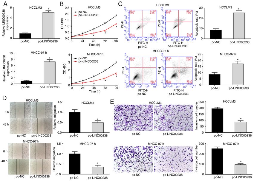
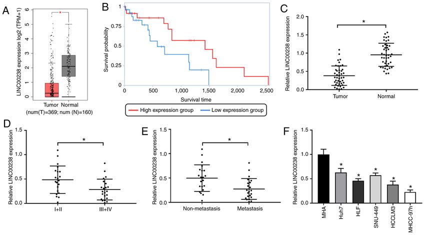
4                                     JIANG et al: LINC00238 INHIBITS HCC BY TARGETING TMEM106C

Figure 1. LINC00238 is downregulated in HCC. (A) Scatter plots comparing LINC00238 expression in HCC samples (n=396) and normal tissue samples
(n=160) showed that LINC00238 exhibited low expression in HCC tumor samples. (B) Kaplan‑Meier analysis indicated a correlation between low LINC00238
and poor overall survival in patients with HCC. The graph was conducted by TANRIC platform. (C) RT‑qPCR data showed that LINC00238 was down‑
regulated in HCC tumor tissues compared with normal tissues. (D) RT‑qPCR data showed that low expression level of LINC00238 in patients with HCC
was associated with early stage (I/II) disease of HCC. (E) RT‑qPCR data showed that the low expression levels of LINC00238 in patients with HCC was
associated with non‑metastasis. (F) RT‑qPCR data showed that LINC00238 was downregulated in HCC cell lines (Huh7, HLF, SNU‑449, MHCC‑97h and
HCCLM3) compared with human normal hepatic cell line MIHA. *P
MOLECULAR MEDICINE REPORTS 24: 757, 2021                                                            5

Figure 2. LINC00238 overexpression inhibits cell proliferation, migration and invasion of HCC. (A) RT‑qPCR data showed that LINC00238 expression in
HCCLM3 and MHCC‑97h cells was significantly increased by LINC00238 overexpression. (B) Data from MTS assay showed that the proliferation ability of
HCCLM3 and MHCC‑97h cells was significantly decreased by LINC00238 overexpression. (C) The flow cytometry data showed that the apoptotic rates of
HCCLM3 and MHCC‑97h cells were increased by LINC00238 overexpression. (D) Wound healing assay showed that the migration capacity of HCCLM3 and
MHCC‑97h cells was suppressed by LLINC00238 overexpression (magnification, x100). (E) The data of Transwell assay showed that the invasion capacity of
HCCLM3 and MHCC‑97h cells was suppressed by LLINC00238 overexpression. *P
6                                       JIANG et al: LINC00238 INHIBITS HCC BY TARGETING TMEM106C

Figure 3. LINC00238 negatively regulates the expression of TMEM106C in HCC. (A) The predicted result of LncTar showed that MEM106C was a target
gene of LINC00238. A dot in the graph indicates there is a paired of bases in the two sequences. A line in the graph indicates there is a paired of comple‑
mentary bases in the two sequences. (B) The data of RNA immunoprecipitation assay showed that TMEM106C was a target of LINC00238 in HCCLM3 and
MHCC‑97h cells. (C) The western blot results showed that protein expression of TMEM106C in HCC cells was decreased by LINC00238 overexpression.
(D) Scatter plots comparing TMEM106C expression in HCC samples (n=396) and normal tissue samples (n=160) showed that TMEM106C was upregulated
in HCC tumor samples compared with normal samples. (E) Kaplan‑Meier analysis indicated a correlation between high TMEM106 and poor overall survival
rates in patients with HCC. (F) Correlation analysis between LINC00238 and TMEM106C expression showed that the LINC00238 expression was negatively
correlated with TMEM106C expression. (G) RT‑qPCR data showed that TMEM106C expression was upregulated in HCC tumor tissues compared with
corresponding normal tissues. *P
MOLECULAR MEDICINE REPORTS 24: 757, 2021                                                                  7

Figure 4. TMEM106C regulates apoptosis pathway and LINC00238 is transcriptionally regulated by ATF3 in HCC. (A) The Enriched KEGG pathway
analysis showed that TMEM106C suppressed the apoptosis signaling pathway. (B) The results of western blotting showed that protein expression of ASP7,
TIMP2, PDCD4 and ATF3 in HCCLM3 and MHCC‑97h cells was increased by LINC00238 overexpression while decreased by TMEM1106C overexpression.
*
 P
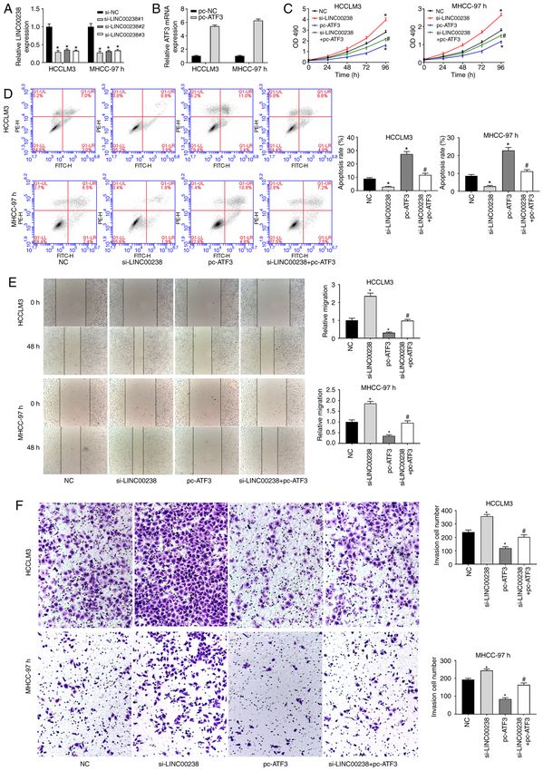
8                                     JIANG et al: LINC00238 INHIBITS HCC BY TARGETING TMEM106C

Figure 5. ATF3 overexpression reverses the promoting effect of LINC00238 silencing on the procession of HCC. (A) RT‑qPCR showed that LINC00238
expression was significantly decreased by LINC00238 silencing. (B) RT‑qPCR showed that ATF3 expression was significantly decreased by ATF3 overexpres‑
sion. (C) MTS assay showed that ATF3 overexpression reduced the proliferation ability of HCCLM3 and MHCC‑97h cells and eliminated the promoted effect
of LINC00238 silence on HCC proliferation. (D) Flow cytometry showed that ATF3 overexpression increased the apoptosis of HCCLM3 and MHCC‑97h
cells and eliminated the inhibitory effect of LINC00238 silence on HCC apoptosis. (E) Wound healing assay showed that ATF3 overexpression inhibited the
migration capacity of HCCLM3 and MHCC‑97h cells and eliminated the promoted effect of LINC00238 silence on HCC migration (magnification, x100).
(F) Transwell assay showed that ATF3 overexpression inhibited the invasion capacity of HCCLM3 and MHCC‑97h cells and eliminated the promoted effect of
LINC00238 silence on HCC invasion (magnification, x200). *P
MOLECULAR MEDICINE REPORTS 24: 757, 2021                                                 9

The present study found that LINC00238 was downregulated             reduced by ATF3 silencing. Overexpression of ATF3 reversed
in HCC. Overexpression of LINC00238 suppressed prolif‑               the promoting effect of LINC00238 silencing on proliferation,
eration, migration and invasion of HCC cells by activating the       migration and invasion of HCC cells. These results indicated
TMEM106C‑mediated apoptosis signaling pathway. Furthermore,          that ATF3 could suppress HCC progression and strengthen the
it was found that ATF3, a gene enriched in the apoptosis signaling   inhibitory effect of LINC00238 on HCC.
pathway, was the upstream promoter of LINC00238.                         Avoidance of apoptosis a major cause of cancer develop‑
     Previous studies have suggested that lncRNAs serve a vital      ment and progression (30). CASP7 is a member of the caspases
role in the initiation and progression of HCC. Mo et al (22)         family that participates in cervical cancer progression (31).
revealed that LINC01287 expression is increased in HCC cells         Palmerini et al (32) indicated that CASP7 is downregulated in
and knockdown of LINC01287 decreases the proliferation               colon cancer. Proteins encoded by TIMPs are natural inhibitors
and invasion ability of HCC cells. The lncRNA CTC‑297N7.9            of matrix metalloproteinases. Peeney et al (33) demonstrated
acts as a tumor suppressor in HCC and low expression of              that TIMP2 inhibits the proliferation and EMT progression
lncRNA CTC‑297N7.9 is associated with poor prognosis in              of triple‑negative breast cancer. PDCD4 is a tumor suppressor
patients with HCC (23). In the present study, the expression of      gene for various cancers and can inhibit cell proliferation,
LINC00238 was significantly decreased in both HCC tissues            migration and invasion, as well as promote tumor cell apop‑
and cell lines. High levels of LINC00238 were associated with        tosis (34). Hwang et al (35) indicated that overexpression of
a good prognosis. Overexpression of LINC00238 inhibited the          PDCD4 induces anti‑proliferation and apoptosis‑induced
proliferation, migration and reduced invasion capacity of HCC        effects on human lung cancer. In the present study, western blot‑
cells. These data indicated that LINC00238 acted as a tumor          ting data showed that LINC00238 overexpression increased the
suppressor in HCC and LINC00238 overexpression inhibited             protein expression of CASP7, TIMP2 and PDCD4. However,
HCC progression.                                                     the promoting effect of LINC00238 on CASP7, TIMP2 and
     TMEM106C is a member of the TMEM106 family and                  PDCD4 was eliminated by TMEM106C overexpression. These
is expressed at low levels in patients with ankylosing spondy‑       results suggested that LINC00238 inhibited the proliferation,
litis (13). Duan et al (24) found that TMEM106C expression is        migration and invasion of HCC cells by upregulating these
positively related to CENPM and negatively related to DLC‑1          genes that were enriched in the apoptosis pathway.
in HCC tumor tissues. Luo et al (14) showed that FOXO1 and               In conclusion, LINC00238 was downregulated in HCC
FOXO3 may be the key target genes of TMEM106C in HCC. In             tissues and cell lines and served a vital role in restraining
the present study, LINC00238 expression was higher in HCC            HCC progression. Overexpression of LINC00238 decreased
tissues compared with that in normal tissues. The predicted          TMEM106C expression and increased the expression of
result of LncTar suggested that TMEM106C was the target of           CASP7, TIMP2, PDCD4 and ATF3, which are apoptotic
LINC00238. LINC00238 overexpression reduced the protein              pathway genes and thus inhibited the proliferation, migration
expression of TMRM106C. TCGA analysis showed that high               and invasion of HCC cells. Notably, ATF3 positively regulated
TMEM106C expression was associated with poor prognosis of            the expression of LINC00238, thereby participating in the
HCC and negatively correlated with LINC00238 expression.             inhibitory regulation of HCC by LINC00238.
In addition, TMEM106C overexpression reversed the inhibi‑
tory effect of LINC00238 overexpression on the proliferation,        Acknowledgements
migration and invasion of HCC cells. These data suggested
that LINC00238 inhibited HCC progression by suppressing              Not applicable.
TMEM106C expression.
     The expression of the ATF3, a member of the ATF family          Funding
of transcription factors, can be induced by a variety of stress
signals (25). Previous studies have demonstrated that ATF3           No funding was received.
acts as a tumor inhibitor in cancer. ATF3 dysfunction allows
normal cells to be easily transformed by oncogenes (26). For         Availability of data and materials
instance, Li et al (27) found that overexpression of ATF3
inhibits the proliferation and migration in esophageal squa‑         The datasets used and/or analyzed during the current study are
mous cell carcinoma. The findings of Hackl et al (28) indicated      available from the corresponding author on reasonable request.
that downregulation of ATF3 promotes the migration capacity
of colon cancer cells in vitro and facilitates tumor growth          Authors' contributions
in vivo. Lv et al (29) observed that ATF3 is significantly
downregulated in human intrahepatic cholangiocarcinoma               CJ and FL designed the experiments, MY, JD and SF collected
with tumor metastasis. The results of the present study were         data. JL and SS analyzed the data and wrote the manuscript.
consistent with those of previous studies. The expression of         FL and CJ confirm the authenticity of all the raw data. All
ATF3 was significantly lower in HCC tumor tissues than that          authors reviewed and approved the final manuscript.
in normal tissues. Low ATF3 expression was associated with
poor prognosis in patients with HCC. ATF3 overexpression             Ethics approval and consent to participate
inhibited proliferation, migration and invasion of HCC cells.
In addition, it was found that ATF3 was an upstream transcrip‑       The study was approved by the ethics committee of Qingdao
tion factor of LINC00238. ATF3 expression was positively             No. 6 People's Hospital [Approval no. (2018) 26] and written
correlated to LINC00238 and LINC00238 expression could be            informed consent was obtained from all patients.
10 JIANG et al: LINC00238 INHIBITS HCC BY TARGETING TMEM106C
Patient consent for publication 17. R Core Team: A Language And Environment For Statistical
Computing. R Foundation for Statistical Computing, Vienna,
2012.
Not applicable. 18. Subramanian A, Tamayo P, Mootha VK, Mukherjee S,
Ebert BL, Gillette MA, Paulovich A, Pomeroy SL, Golub TR,
Lander ES, et al: Gene set enrichment analysis: A knowledge‑based
Competing interests approach for interpreting genome‑wide expression profiles. Proc
Natl Acad Sci USA 102: 15545‑15550, 2005.
The authors declare that they have no competing interests. 19. Liberzon A, Birger C, Thorvaldsdóttir H, Ghandi M, Mesirov JP
and Tamayo P: The Molecular Signatures Database (MSigDB)
hallmark gene set collection. Cell Syst 1: 417‑425, 2015.
References 20. Livak KJ and Schmittgen TD: Analysis of relative gene expression
data using real‑time quantitative PCR and the 2(‑ΔΔC(T)) method.
1. Bray F, Ferlay J, Soerjomataram I, Siegel RL, Torre LA and Methods 25: 402‑408, 2001.
Jemal A: Global cancer statistics 2018: GLOBOCAN estimates 21. Li J, Ma W, Zeng P, Wang J, Geng B, Yang J and Cui Q: LncTar:
of incidence and mortality worldwide for 36 cancers in 185 A tool for predicting the RNA targets of long noncoding RNAs.
countries. CA Cancer J Clin 68: 394‑424, 2018. Brief Bioinform 16: 806‑812, 2015.
2. Bellissimo F, Pinzone MR, Cacopardo B and Nunnari G: 22. Mo Y, He L, Lai Z, Wan Z, Chen Q, Pan S, Li L, Li D, Huang J,
Diagnostic and therapeutic management of hepatocellular Xue F, et al: LINC01287/miR‑298/STAT3 feedback loop
carcinoma. World J Gastroenterol 21: 12003‑12021, 2015. regulates growth and the epithelial‑to‑mesenchymal transition
3. Cheng D, Deng J, Zhang B, He X, Meng Z, Li G, Ye H, Zheng S, phenotype in hepatocellular carcinoma cells. J Exp Clin Cancer
Wei L, Deng X, et al: LncRNA HOTAIR epigenetically Res 37: 149‑149, 2018.
suppresses miR‑122 expression in hepatocellular carcinoma via 23. Zhu S, Huang X, Zhang K, Tan W, Lin Z, He Q, Chen Y and
DNA methylation. EBioMedicine 36: 159‑170, 2018. Shang C: Low expression of long noncoding RNA CTC‑297N7.9
4. Lou W, Liu J, Gao Y, Zhong G, Ding B, Xu L and Fan W: predicts poor prognosis in patients with hepatocellular carcinoma.
MicroRNA regulation of liver cancer stem cells. Am J Cancer Cancer Med 8: 7679‑7692, 2019.
Res 8: 1126‑1141, 2018. 24. Duan J, Qian Y, Fu X, Chen M, Liu K, Liu H, Yang J, Liu C
5. Huang Z, Zhou JK, Peng Y, He W and Huang C: The role of long and Chang Y: TMEM106C contributes to the malignant charac‑
noncoding RNAs in hepatocellular carcinoma. Mol Cancer19: teristics and poor prognosis of hepatocellular carcinoma. Aging
77, 2020. (Albany NY) 13: 5585‑5606, 2021.
6. Gong D, Feng PC, Ke X‑F, Kuang HL, Pan LL, Ye Q and Wu JB: 25. Hai T, Wolford CC and Chang YS: ATF3, a hub of the cellular
Silencing long non‑coding RNA LINC01224 inhibits hepato‑ adaptive‑response network, in the pathogenesis of diseases:
cellular carcinoma progression via MicroRNA‑330‑5p‑induced Is modulation of inflammation a unifying component? Gene
inhibition of CHEK1. Mol Ther Nucleic Acids 19: 482‑497, 2020. Expr 15: 1‑11, 2010.
7. Jia M, Jiang L, Wang YD, Huang JZ, Yu M and Xue HZ: 26. Yan C and Boyd DD: ATF3 regulates the stability of p53: A link
LincRNA‑p21 inhibits invasion and metastasis of hepatocellular to cancer. Cell Cycle 5: 926‑929, 2006.
carcinoma through Notch signaling induced epithelial‑mesen‑ 27. Li J, Yang Z, Chen Z, Bao Y, Zhang H, Fang X and Yang W:
chymal transition. Hepatol Res 46: 1137-1144, 2016. ATF3 suppresses ESCC via downregulation of ID1. Oncol
8. Liu F, Yuan JH, Huang JF, Yang F, Wang TT, Ma JZ, Zhang L, Lett 12: 1642‑1648, 2016.
Zhou CC, Wang F, Yu J, et al: Long noncoding RNA FTX 28. Hackl C, Lang SA, Moser C, Mori A, Fichtner-Feigl S, Hellerbrand C,
inhibits hepatocellular carcinoma proliferation and metastasis by Dietmeier W, Schlitt HJ, Geissler EK and Stoeltzing O: Activating
binding MCM2 and miR‑374a. Oncogene 35: 5422‑5434, 2016. transcription factor‑3 (ATF3) functions as a tumor suppressor in
9. Wang CZ, Yan GX, Dong DS, Xin H and Liu ZY: LncRNA‑ATB colon cancer and is up‑regulated upon heat‑shock protein 90
promotes autophagy by activating Yes‑associated protein and (Hsp90) inhibition. BMC Cancer 10: 668, 2010.
inducing autophagy‑related protein 5 expression in hepato‑ 29. Lv L, Wei M, Lin P, Chen Z, Gong P, Quan Z and Tang Z:
cellular carcinoma. World J Gastroenterol 25: 5310‑5322, 2019. Integrated mRNA and lncRNA expression profiling for exploring
10. Marx S, Dal Maso T, Chen JW, Bury M, Wouters J, Michiels C and metastatic biomarkers of human intrahepatic cholangiocar‑
Le Calvé B: Transmembrane (TMEM) protein family members: cinoma. Am J Cancer Res 7: 688‑699, 2017.
Poorly characterized even if essential for the metastatic process. 30. Reed JC: Mechanisms of apoptosis avoidance in cancer. Curr
Semin Cancer Biol 60: 96‑106, 2020. Opin Oncol 11: 68‑75, 1999.
11. Liu J and Zhu H: TMEM106A inhibits cell proliferation, migration, 31. Shi TY, He J, Wang MY, Zhu ML, Yu KD, Shao ZM, Sun MH,
and induces apoptosis of lung cancer cells. J Cell Biochem: Nov 19, Wu X, Cheng X and Wei Q: CASP7 variants modify suscepti‑
2018 (Epub ahead of print). doi: 10.1002/jcb.28057. bility to cervical cancer in Chinese women. Sci Rep 5: 9225‑9225,
12. Lang CM, Fellerer K, Schwenk BM, Kuhn PH, Kremmer E, 2015.
Edbauer D, Capell A and Haass C: Membrane orientation 32. Palmerini F, Devilard E, Jarry A, Birg F and Xerri L: Caspase 7
and subcellular localization of transmembrane protein 106B downregulation as an immunohistochemical marker of colonic
(TMEM106B), a major risk factor for frontotemporal lobar carcinoma. Hum Pathol 32: 461‑467, 2001.
degeneration. J Biol Chem 287: 19355‑19365, 2012. 33. Peeney D, Jensen SM, Castro NP, Kumar S, Noonan S, Handler C,
13. Assassi S, Reveille JD, Arnett FC, Weisman MH, Ward MM, Kuznetsov A, Shih J, Tran AD, Salomon DS, et al: TIMP‑2
Agarwal SK, Gourh P, Bhula J, Sharif R, Sampat K, et al: suppresses tumor growth and metastasis in murine model of
Whole‑blood gene expression profiling in ankylosing spondylitis triple‑negative breast cancer. Carcinogenesis 41: 313‑325, 2020.
shows upregulation of toll‑like receptor 4 and 5. J Rheumatol 38: 34. Afonja O, Juste D, Das S, Matsuhashi S and Samuels HH:
87‑98, 2011. Induction of PDCD4 tumor suppressor gene expression by RAR
14. Luo X, Han G, Lu R, Guan S, Wang Y, Ju L, Chen L, Shao J and agonists, antiestrogen and HER‑2/neu antagonist in breast cancer
Bian Z: Transmembrane protein 106C promotes the development cells. Evidence for a role in apoptosis. Oncogene 23: 8135‑8145,
of hepatocellular carcinoma. J Cell Biochem 121: 4484-4495, 2004.
2020. 35. Hwang SK, Jeong YJ and Chang Y‑C: PDCD4 inhibits lung
15. Tang Z, Li C, Kang B, Gao G, Li C and Zhang Z: GEPIA: A tumorigenesis by the suppressing p62‑Nrf2 signaling pathway and
web server for cancer and normal gene expression profiling and upregulating Keap1 expression. Am J Cancer Res 10: 424‑439,
interactive analyses. Nucleic Acids Res 45: W98‑W102, 2017. 2020.
16. Therneau TM: Survival Analysis [R package survival version This work is licensed under a Creative Commons
2.39‑5]. 46: 111‑112, 2015.
Attribution-NonCommercial-NoDerivatives 4.0
International (CC BY-NC-ND 4.0) License.You can also read


