Treatment Strategy for Multiple Myeloma to Improve Immunological Environment and Maintain MRD Negativity - MDPI
←
→
Page content transcription
If your browser does not render page correctly, please read the page content below
cancers
Review
Treatment Strategy for Multiple Myeloma to Improve
Immunological Environment and Maintain MRD Negativity
Kazuhito Suzuki 1,2, * , Kaichi Nishiwaki 1,2 and Shingo Yano 2

                                          1   Department of Internal Medicine, Division of Clinical Oncology and Hematology, The Jikei University
                                              Kashiwa Hospital, Tokyo 277-8567, Japan; nishiwaki@jikei.ac.jp
                                          2   Department of Internal Medicine, Division of Clinical Oncology and Hematology, The Jikei University School
                                              of Medicine, Tokyo 105-8461, Japan; yano@jikei.ac.jp
                                          *   Correspondence: kaz-suzuki@jikei.ac.jp

                                          Simple Summary: Improving the immunological environment and eradicating minimal residual
                                          disease (MRD) are the two main treatment goals for long-term survival in patients with multiple
                                          myeloma (MM). An improved immunological environment may be useful for maintaining MRD
                                          negativity. Whether the ongoing treatment should be continued or changed if the MRD status
                                          remains positive is controversial. In this case, genetic, immunophenotypic, and clinical analysis of
                                          residual myeloma cells may be necessary to select the effective treatment for the residual myeloma
                                          cells. The purpose of this review is to discuss the MM treatment strategy to “cure MM” based on
                                          currently available therapies and expected immunotherapies via improvement of the immunological
                                          environment and maintenance of MRD negativity.

                                Abstract: Improving the immunological environment and eradicating minimal residual disease
                                   (MRD) are the two main treatment goals for long-term survival in patients with multiple myeloma
Citation: Suzuki, K.; Nishiwaki, K.;      (MM). Immunomodulatory drugs (IMiDs), monoclonal antibody drugs (MoAbs), and autologous
Yano, S. Treatment Strategy for           grafts for autologous stem cell transplantation (ASCT) can improve the immunological microenvi-
Multiple Myeloma to Improve               ronment. ASCT, MoAbs, and proteasome inhibitors (PIs) may be important for the achievement of
Immunological Environment and             MRD negativity. An improved immunological environment may be useful for maintaining MRD
Maintain MRD Negativity. Cancers          negativity, although the specific treatment for persistent MRD negativity is unknown. However,
2021, 13, 4867. https://doi.org/
                                          whether the ongoing treatment should be continued or changed if the MRD status remains positive
10.3390/cancers13194867
                                          is controversial. In this case, genetic, immunophenotypic, and clinical analysis of residual myeloma
                                          cells may be necessary to select the effective treatment for the residual myeloma cells. The purpose
Academic Editor: Jo Caers
                                          of this review is to discuss the MM treatment strategy to “cure MM” based on currently available
Received: 11 August 2021
                                          therapies, including IMiDs, PIs, MoAbs, and ASCT, and expected immunotherapies, such as chimeric
Accepted: 24 September 2021               antigen receptor T cell (CAR-T) therapy, via improvement of the immunological environment and
Published: 28 September 2021              maintenance of MRD negativity.

Publisher’s Note: MDPI stays neutral      Keywords: multiple myeloma; immune environment; minimal residual disease; proteasome inhibitor;
with regard to jurisdictional claims in   immunomodulatory drug; monoclonal antibody; autologous stem cell transplantation
published maps and institutional affil-
iations.

                                          1. Introduction
                                               Multiple myeloma (MM) is a hematopoietic malignancy of the plasma cells, and
Copyright: © 2021 by the authors.         although the survival of patients with MM has been prolonged by the development of new
Licensee MDPI, Basel, Switzerland.        agents in the last few decades, it is still an incurable disease [1,2].
This article is an open access article         To cure MM, it is important to improve the immune environment and ensure persistent
distributed under the terms and           minimal residual disease (MRD) negativity [3–5]. Notably, the immune environment
conditions of the Creative Commons        of myeloma patients is characterized by an attenuated immune effect on tumor cells,
Attribution (CC BY) license (https://
                                          creating an environment suitable for the survival of myeloma cells [3,4]. However, an
creativecommons.org/licenses/by/
                                          improved immune environment leads to the long-term survival of patients with myeloma
4.0/).

Cancers 2021, 13, 4867. https://doi.org/10.3390/cancers13194867                                               https://www.mdpi.com/journal/cancers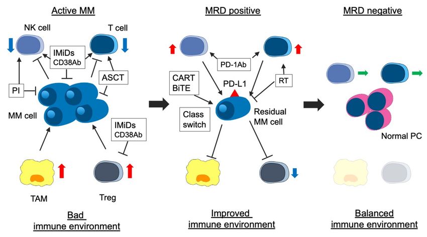
Cancers 2021, 13, 4867                                                                                           2 of 25

                         due to enhanced immunological potency against myeloma cells [6]. Recently, various
                         immunotherapeutic agents, including immunomodulatory drugs (IMiDs) and monoclonal
                         antibody drugs (MoAbs) against CD38 and signaling lymphocytic activation molecule
                         family 7 (SLAMF7), have been developed [7–11]. In addition, the clinical development
                         of an immune checkpoint inhibitor for myeloma, which has played an important role
                         in the treatment of solid malignant tumors, is under way [12]. Autologous grafts used
                         in autologous stem cell transplantation (ASCT), which is still the standard treatment for
                         patients with MM [13,14], have been reported to improve the immune environment [15].
                               MRD-negativity, which is analyzed using next-generation sequencing (NGS) and
                         next-generation flow cytometry (NGF), prolongs the progression-free survival (PFS) and
                         overall survival (OS) of patients [5]. Persistent MRD negativity in multiple assessments is
                         important for long-term survival [5]. However, the prognosis of MRD-positive patients is
                         not good, even if complete response (CR) is achieved. Therefore, eradicating all myeloma
                         cells should be the primary treatment goal for MRD-positive patients, although sustained
                         MRD-positivity is not always an unfavorable outcome [16]. Genetic and immunophe-
                         notypic characterization of residual myeloma cells, including the clinical course, can be
                         essential for defining and selecting a suitable treatment strategy.
                               The purpose of this review is to describe the importance of improving the immune
                         environment in MM patients and its therapeutic strategies, the clinical significance of MRD
                         status for long-term survival, and therapeutic strategies for persistent MRD negativity. We
                         also describe the treatment of residual myeloma cells in MRD-positive patients and the
                         future MRD status-adapted treatment strategies.

                         2. Immunological Environment in MM
                               The immune system plays an important role in the genesis of myeloma. The functions
                         of immune cells are suppressed by cytokines and the interaction between myeloma cells
                         and the bone marrow (BM) microenvironment [17,18]. A potential positive relationship
                         between the cellular components of the immune system, such as T cells, natural killer (NK)
                         cells, regulatory T cells (Treg), and B cells, and myeloma progression was suggested in
                         previous studies [17–19]. According to an earlier report, disease status, advanced stage in
                         the International Staging System (ISS), and high-risk cytogenetic abnormalities (HRCA)
                         were related to worse immune profiles [18].
                               T cells are categorized into cytotoxic CD8+ T cells and helper CD4+ T cells. Cytotoxic T
                         cells (CTL) are effector cells for adoptive immune responses and are activated by interleukin
                         (IL)-2 and exert their anti-tumor effect by releasing interferon-gamma following antigen
                         presentation [19]. CD4+ T cells mainly enhance the adaptive immune response [20]. T-
                         cells are quantitatively and functionally altered in MM and, consequently, have a role in
                         the immunodeficiency associated with myeloma pathogenesis [21]. The frequencies of
                         effector memory and effector CD8+ T cells in MM patients are higher than those in healthy
                         individuals, while the frequency of CD4+ T cells is similar between MM patients and
                         healthy individuals [22]. Low CD4+ T cell counts and low CD4/CD8 ratios in peripheral
                         blood (PB) are predictors of poor clinical outcomes [23].
                               T cells, especially in tumor sites, are exhausted in patients with MM compared to
                         those with monoclonal gammopathy of undetermined significance (MGUS) [22,24,25]. T
                         cell exhaustion is induced by inadequate IFN-gamma and upregulation of inhibitory recep-
                         tors on T cells, such as programmed cell death protein1 (PD-1), cytotoxic T-lymphocyte-
                         associated antigen 4 (CTLA-4), Tim-3, and lymphocyte activation gene-3 (LAG-3), in MM
                         patients [24,26]. Galectin-9 and a proliferation-inducing ligand (APRIL) derived from
                         osteoclasts, which constitute the microenvironment of myeloma cells, induce Tim-3 and
                         programmed death-ligand 1 (PD-L1) on MM cells, respectively, and contribute to immune
                         escape [27]. Soluble PD-L1, derived from MM cells, suppresses the immune system by
                         binding to PD-1 on CTL [28]. T cell anergy is a tolerance mechanism due to the inactivation
                         of lymphocytes, and anergic T cells remain alive for an extended period in a hypore-
                         sponsive state [29]. Anergic T cells are induced by co-stimulation of the T cell receptor
Cancers 2021, 13, 4867 3 of 25
(TCR) and low expression of CD28 and high expression of CTLA-4 on MM cells [24,30].
The positivity of PD-1 and CTLA-4 on both CD4+ and CD8+ T cells in MM patients was
higher than that in healthy individuals in BM [24]. PD-1+ lymphocytes contribute to the
proliferation of functionally impaired tumor-specific lymphocytes [31]. The high frequency
of PD-1- or CTLA-4-expressing CD8+ T cells was not significantly different in MM patients
before and after IMiDs treatment [22]. In addition, PD-1+ CD38+ lymphocytes suppress
anti-cancer activity and have been identified in patients with malignancies [32], especially
after administration of anti-PD-1 MoAbs [33]. Thus, PD-1 and PD-L1 are therapeutic targets
in MM.
NK cells are effector lymphocytes for the innate immune response, control several
types of tumors and infections, and regulate the activities of T cells, macrophages, and
dendritic cells [34]. Elevated NK cell counts in PB and BM are noted in patients with
early-phase myeloma, but the number of NK cells in PB decreases as myeloma progresses.
Moreover, NK cell activity is reduced in patients with MM [35]. In antibody-dependent
cellular cytotoxicity (ADCC) activity induced by the binding of Fcγ receptors to the Fc
tail of the MoAbs, NK cells release toxic proteins, including granzymes and perforins,
which kill myeloma cells [36]. However, myeloma cells have reduced ADCC activity due to
downregulated expression of NK cell receptors, such as natural killer group 2D (NKG2D),
NKp30, CD244, and DNAX accessory molecule 1 (DNAM-1) [37,38]. In addition, the
expression of PD-1 on NK cells prevents immune recognition of tumor cells in myeloma
patients [39]. ADCC activity can be induced by several MoAbs, including PD-1 blockade,
and is essential for MM treatment.
Treg cells comprise 5–7% of CD4+ T cells and develop from CD4+ T cells under
conditions of high levels of transforming growth factor-β (TGF-β) [40]. They suppress
the immune response in the functional homeostasis of the immune system [35]. Tregs
induce immune tolerance by modulating antigen presentation by expressing soluble anti-
inflammatory mediators, such as IL-10 and TGFβ, the consumption of IL-2, and the
expression of negative regulatory cell surface receptors, including CTLA-4 [41,42]. Patients
with MM have elevated Treg level, which is a marker of poor prognosis [43–45]. MM
cells secrete an inducible T-cell co-stimulator ligand (ICOS-L) and transform non-Tregs
into Tregs [46]. Thus, decreasing the Treg count can enhance the immune activity against
myeloma cells.
Data on the kinetics of B cells are limited compared with those of T cells. Decreasing
levels of polyclonal immunoglobulin reflect suppression of CD19+ B cells, which is inversely
correlated with disease progression and affects normal B-cell differentiation [21,47]. TGF-β
contributes to B-cell dysfunction in myeloma [21,48]. Regulatory B cells (Breg), a small
B-cell subset, regulate immune responses via stimulation of IL-10, an anti-inflammatory
cytokine, and modulation of CD4+ T cell activation and differentiation [49]. Breg induces
an immunosuppressive BM microenvironment, which may, in turn, affect therapeutic
response and disease outcome in patients with MM [50]. Therefore, Breg inhibition is a
potential therapeutic target.
Macrophages are blood cells derived from monocytes and show various activities
depending on the body site. Macrophages contribute to antibody-dependent cellular
phagocytosis (ADCP) activity, which is the phagocytosis of antibody-opsonized tumor
cells via binding to Fcγ receptors present on macrophages or monocytes [51]. In contrast,
some macrophages suppress immune activity in myeloma. Tumor-associated macrophages
(TAMs) are categorized as M2 and have a pro-tumoral function. They infiltrate the tumors
and are associated with the growth, angiogenesis, and metastasis of various cancers, includ-
ing MM [52,53]. TAMs in MM have little cytotoxicity and suppress T cell activity [54]. In
addition, TAMs regulate fibroblast function in BM [52] and induce resistance to chemother-
apy via inhibition of Bcl-XL-dependent caspase activation [55]. Thus, TAM inhibition is a
potential therapeutic target as well. A summary of function of immune cells for healthy
individuals and MM patients is shown in Table 1.Cancers 2021, 13, 4867 4 of 25
Table 1. Summary of function of immune cells and anti-myeloma agents for immune system. IMiDs, immunomodulatory drugs; MoAb, monoclonal antibody; PD-1, programed death-1;
HD-MEL, high dose-melphalan; ASCT, autologous stem cell transplantation; PIs, proteasome inhibitor; CTL, cytotoxic T-cell; NK cell, natural killer cell; Treg, regulatory T-cell; Breg,
regulatory B cell; TAM, tumor associated macrophage; MM, multiple myeloma; IL-2, interleukine-2; IFN-gamma, interferon gamma; ICOS-L, inducible T-cell co-stimulator ligand; ADCC,
antibody-dependent cellular cytotoxicity; ADCP, antibody-dependent cellular phagocytosis; HMGB-1, high-mobility group box-1; FOXP3, forkhead box P3; and ADO, adenosine.
Characteristics CTL NK Cell Treg Breg Macrophage TAM
Suppression for immune
response via immune
Adoptive immune Innate immune response, tolerance by modulating
Regulation of immune
Function in healthy responses activated by IL-2 and regulate the activities of antigen presentation, the Various activities
responses via stimulation of -
individuals and anti-tumor effect by T cells, macrophages, and consumption of IL-2, and depending on the body site
IL-10 [49]
releasing IFN-gamma [19] dendritic cells [34] the expression of negative
regulatory cell surface
receptors [35,41,42]
Elevated Treg level predicts
Exhaustion in tumor site via poor prognosis [43–45]; Induction of an Suppress T-cell activity [54].
NK cell activity is reduced
Function in MM patients upregulation of inhibitory transformation from immunosuppressive BM Relation with ADPC [51] Induction of resistance to
in patients with MM [35]
receptors [22,24,26] non-Tregs into Tregs by micro-environment [50] chemotherapy [55]
secretion of ICOS-L [46]
Drugs - - - - - -
Activate via INF-gamma
Activate ADCC by Decrease TAM via
and IL-2 from T-cell from Inhibit via down regulation
IMiDs INF-gamma and IL-2 from - - conversion from TAM into
CD4+ T-cell and PD-1 of FOXP3 [60]
CD4+ T-cell [56,58,59] M1 macrophage [61]
blockade [56,57]
Decrease CD38+ NK cell but
Induce clonal increasing Inhibit activity via Inhibit activity via
Anti-CD38 MoAb activate ADCC by Activate ADCP [69] -
CTL [62] suppression ADO [66–68] suppression ADO [66–68]
CD38−/low NK cell [63–65]
Activate via PD-1 Activate via PD-1
Anti-PD-1 MoAb - - - -
blockade [31] blockade [31]
Belantamab Mafodotin Activate ADCC [70] - - Activate ADCP [70] -
Decrease T-cell but activate
Decrease Treg by
HD-MEL + ASCT via activated dendritic cell - - - -
HD-MEL [72,73]
by releasing HMGB1 [71,72]
Activate ADCC via HLA
PIs - - - - -
class1 blockade [74,75]Cancers 2021, 13, 4867 5 of 25
3. Importance of Immunological Environment for Long-Term Survival in MM
Immune reconstitution, which is indicated by lymphocyte count in PB and im-
munoglobulin levels, predicts good prognosis in patients with MM. Changes in the BM
immune microenvironment have recently been analyzed using cytometry by time of flight
(CyTOF) and NGF.
Early immune reconstitution, which is defined as recovery to the normal count of
both lymphocytes and monocytes one month after treatment, predicts long OS. The OS in
patients with early immune reconstitution was reported to be similar to that in patients
without immune deregulation at diagnosis. The frequency of early immune reconstitution
is high in patients treated with IMiDs and low in patients with HRCA [76]. The recovery of
absolute lymphocyte count (ALC), defined as ≥1400 cells/µL at day 0, day 15, and day
90 after ASCT, predict long OS [15]. Decreased CD4+ T cell count and the CD4/8 ratio
are associated with poor prognosis [23,77]. The number of clonal CD8+ T cells, which are
identified as effector memory T cells with a restricted T-cell receptor (TCR) Vβ expression,
was associated with persistent stimulation by myeloma-associated antigens [78]. The count
of clonal CD8+ T cells in PB was higher in myeloma patients who survived for more than
10 years than in those who died in less than 10 years [79]. Among patients with long-term
CR after ASCT, the distribution of CD4+ and CD8+ memory T cells and naïve B cells in PB
was higher than that in age-matched healthy individuals [80]. In contrast, the presence
of naïve and terminally differentiated T cells in the BM predicted a short survival time
in myeloma patients who received ASCT using CyTOF [81]. These results suggested that
naïve T cells could not activate antigen engagement, and terminally differentiated T cells
could not mediate effective clearance of myeloma cells because of T cell exhaustion. In the
transplant-ineligible patients enrolled in the PETHEMA/GEM2010MAS65 study, PFS and
OS were longer in the groups rich in naïve and memory B cells in BM using NGF instead
of the ISS and those with cytogenetic abnormality and MRD status [82]. Thus, improving
the immune microenvironment is associated with long-term survival and is a surrogate
marker for good outcomes in myeloma patients.
Immunoparesis is associated with poor outcomes in patients treated with novel agents
and cytotoxic agents [83–85]. In a multivariate analysis including age, ISS stage, and genetic
risk of MM patients treated with novel agents, a low immunoglobulin (Ig)M level was a
significant predictor of short PFS and OS compared with IgA and IgG levels [83].
The oligoclonal band is often identified in patients with CR and is an immunoglobulin
derived from myeloma cells and other polyclonal B cells [86,87]. In a previous study, the
oligoclonal band predicted longer survival and disappeared before relapse [86]. However,
in MM patients with extramedullary disease (EMD) or light chain escape, the oligoclonal
band may remain. Thus, the oligoclonal band is also considered a form of immune
reconstitution [88]. Oligoclonal bands, identified as clonal isotype switches, are often
detected after ASCT or chemotherapy [89,90] and predict long-term survival in patients
receiving ASCT [89,91]. In patients treated with PI or IMiDs induction therapy followed by
ASCT, the frequency of oligoclonal bands in the patients treated with IMiDs was higher than
in those without IMiDs. The CD8+ T cell count was significantly lower, and the CD4/CD8
T cell ratio was significantly higher in patients with oligoclonal bands than in those without
oligoclonal bands [89]. Thus, improvement of immune status concerning T cells and B
cells predicts good outcomes in MM patients. In addition, immune reconstitution of T cells
could be correlated with that of B cells.
Immune status in BM was similar among healthy individuals, MGUS patients, and
MM patients treated with ASCT using CyTOF [92]. Thus, the immune environment in
active MM patients is suitable for the proliferation and survival of myeloma cells, while
the immune environment in MM patients with remission is unfit for myeloma cells and
normal immune cells increases, leading to immune reconstitution.Cancers 2021, 13, 4867 6 of 25
4. Treatment to Improve the Immunological Environment
Currently, IMiDs, PIs, and MoAbs play an important role in MM treatment. Many
studies have demonstrated that IMiDs and MoAbs improve the immunological environ-
ment via activation of T and NK cells and suppression of Tregs. ASCT improves the
immunological environment via the supply of autografts and high-dose melphalan (HD-
MEL). In addition, PIs activate ADCC via the downregulation of human leukocyte antigen
(HLA) on myeloma cells. A summary of anti-myeloma agents for immune system is shown
in Table 1.
4.1. IMiDs
IMiDs induce immune modulation and exhibit anti-myeloma activity. They also
enhance both the adaptive and innate immune systems via co-stimulation of T cells and
enhancement of NK in vitro [56]. These drugs enhance the tumor-specific Th1 type immune
response via the generation of IFN-gamma and IL-2 derived from CD4+ and CD8+ T
cells [56]. Examples of IMiDs include lenalidomide (LEN) and pomalidomide (POM),
and thalidomide (THAL). LEN and POM, excluding THAL, augment ADCC and innate
cytotoxicity of NK cells and promote the proliferation of NK cells dependent on IL-2 [58].
LEN and POM upregulate the expression of FasL and Granzyme B on NK cells [59]. In
addition, IMiDs suppress Treg function by downregulating Foxp3 gene expression in
Tregs [60]. The effect of IMiDs depends on cereblon (CRBN) burden concerning both
direct antitumor effects and immune activity [93]. IMiDs also act on immune checkpoint
molecules to enhance immune responses. They suppress PD-1 expression on T and NK
cells [57], and LEN suppresses PD-L1 expression in MM cells [43]. Finally, LEN coverts M2
macrophages, identified as TAM, into M1 macrophages via the degradation of the IKAROS
family zinc finger 1 (IKZF1) of macrophages [61]. Iberdomide (IBER), a next-generation
cereblon-targeting agent, showed direct anti-myeloma activity for LEN- and/or POM-
resistant human myeloma cell lines via faster degradation of IKZF1 due to high cereblon-
binding affinity and enhanced immunomodulatory effect in a co-culture with peripheral
blood mononuclear cells via elevated IL-2 secretion and granzyme-B degranulation [94,95].
In addition, IBER combined with daratumumab (DARA) enhanced complement-dependent
cytotoxicity (CDC) compared with either drug alone [95], demonstrating the clinical efficacy
of IMiDs and/or anti-CD38 MoAb in refractory MM patients [96].
4.2. MoAbs
Daratumumab (DARA) is a monoclonal antibody against CD38 antigen with complement-
dependent cytotoxicity (CDC) and enhanced ADCC activities [63]. CD38 is expressed on
the surface of myeloma cells as well as T and NK cells [62]. Notably, DARA reduces NK cell
count [64]. However, CD38−/low NK cells play a major role in ADCC activity, an immune
response that is expected even after DARA administration [65]. DARA also contributes to a
clonal increase in CTL and suppression of Treg and Breg [62]. DARA-containing treatment
is an option for post-transplant treatment as it might reduce the number of CD38+ Treg,
which increases early after ASCT and suppresses the immune effect [62]. In the POLLUX
trial, the clonal expansion of CD8+ T cell and reduction of CD38+ Treg were observed more
frequently in the deep responders treated with DRd (DARA, LEN, plus dexamethasone
(DEX)) than in those treated with Rd (LEN plus DEX) [97]. Anti-CD38 MoAbs also activate
T cell function via suppression of adenosine (ADO) production because CD38 functions
as an ectoenzyme and promotes ADO production from NAD+ via regulation of calcium
signaling, which suppresses T cell activity [66–68]. Isatuximab (ISA), a new anti-CD38
MoAb, has been approved for relapsed/refractory MM (RRMM). Similar to DARA, ISA
can also modulate the immune system. ISA has a higher direct killing activity of myeloma
cells [98] but lower CDC activity than DARA [99]. A combination of ISA and POM showed
higher anti-myeloma activity than a combination of ISA and LEN [98].
Anti-PD-1 MoAb is expected to be active against several solid malignancies because
PD-1-expressing lymphocytes expand tumor-specific CTLs and suppress the anti-myelomaCancers 2021, 13, 4867 7 of 25
activity [31]. In the KEYNOTE-185 trial, PFS of patients in the pembrolizumab, an anti-PD-1
MoAb, plus Rd group was similar to that of patients in the Rd group, while severe adverse
events and treatment-related mortality of patients in the pembrolizumab plus Rd group
were higher than those of patients in the Rd group [100].
CD38/PD-1 double-positive lymphocytes have been identified in patients with malig-
nancies [32]. CD38 expression on T cells might induce escape from PD-1/PD-L1 blockade
in tumor cells [33]. However, in a previous study, durvalumab, an anti-PD-L1 MoAb,
and daratumumab were ineffective in myeloma patients with daratumumab refractori-
ness [101]. A clinical trial on combined anti CD38 and PD-1 monoclonal antibodies for
myeloma patients is ongoing.
Belantamab mafodotin is the first anti-B-cell mature antigen (BCMA) antibody drug
conjugate (ADC) with mono methyl auristatin F (MMAF) and has been approved for
RRMM in the United States [102]. Belantamab mafodotin not only delivers MMAF into
the BCMA-expressed myeloma cells and induces apoptosis but also enhances ADCC and
ADCP [70].
4.3. ASCT
The therapeutic effect of ASCT depends on HD-MEL therapy for MM. However,
autografts play an important role in the therapeutic effect of ASCT. Granulocytes, platelets,
and red blood cells recover within a few weeks, while the recovery of other blood cells, such
as lymphocytes and monocytes, takes a longer duration after HSC transplantation [103].
Immune reconstitution, which has been shown to increase absolute lymphocyte count,
CD4/CD8 ratio, and oligoclonal band, is associated with good clinical outcomes in patients
receiving ASCT [15,77,79–81,89].
HD-MEL is the mainstay of pre-transplantation conditioning chemotherapy. HD-MEL
induces lymphodepletion, which could affect immunological activity, including that of
T cells, in myeloma cells [71]. However, T cell count from autografts is upregulated by
IL-7 and IL-15, whose serum levels increase after HD-MEL followed by ASCT, although
T cell count decreases in the absence of these cytokines [71]. Melphalan has been shown
to activate CD8+ T cells via dendritic cell activation due to immunogenic cell death and
antigen presentation, including the release of high-mobility group box 1 (HMGB-1), in
myeloma-bearing mice [72]. In addition, the combination of melphalan and CD4+ T cell
adoptive cell therapy is more effective than either treatment alone in mice. HD-MEL can
enhance immune activity by reducing the levels of Treg and myeloid-derived suppressor
cells, which inhibit the anti-myeloma T cell-mediated immune response in BM [72,73].
4.4. Proteasome Inhibitors
PIs enhance ADCC activity by suppressing HLA class 1 expression on MM cells [74].
Recently, bortezomib, a proteasome inhibitor, enhanced the ADCC of DARA in mice when
human myeloma cell lines and ex vivo NK cells were co-cultured [75]. Additionally, PIs ac-
tivate dendritic cells by increasing exposure to tumor antigens, thus inducing immunogenic
cell death [104,105].
5. Clinical Significance of MRD Negativity in MM
MRD can be analyzed in patients who have achieved CR using NGF or NGS. Im-
munophenotypic and molecular CR are defined as achievement of MRD negativity using
NGF and NGS, respectively, according to the International Myeloma Working Group recom-
mendation [106]. Several reports indicate that achievement of negative MRD is associated
with prolonged PFS and OS, but the curability of MM is still debatable [107,108]. According
to a recent meta-analysis, MRD negativity predicted long PFS and OS independent of
transplant eligibility, disease status, cytogenetic risk, MRD sensitivity threshold, and MRD
detection methods [5]. A summary of phase 3 clinical trials investigating MRD status was
shown in Table 2.Cancers 2021, 13, 4867 8 of 25
Table 2. Summary of phase 3 clinical trials investigating MRD status. MRD, minimal residual disease; TE-NDMM, transplantation eligible newly diagnosed multiple myeloma;
NTE-NDMM, not transplantation eligible newly diagnosed multiple myeloma; RRMM, relapsed or refractory multiple myeloma; NGS, next generation sequencing; MCF, multi-color
flowcytometry; DARA, daratumumab; LEN, lenalidomide; K, carfilzomib (CFZ); V, bortezomib (BOR); T, thalidomide (THAL); M, melphalan (MEL); C, cyclophosphamide (CPA); d,
dexamethasone (DEX); ASCT, autologous stem cell transplantation; CVd, CPA, BOR, plus DEX; VMP, BOR, MEL, plus prednisone; VRd, BOR, LEN, plus DEX; VTd, BOR, THAL, plus DEX;
D-VTd, DARA, plus VTd; KRd, CFZ, LEN, plus DEX; KCd, CFZ, CPA, plus DEX; Rd, LEN, plus DEX; DRd, DARA, plus Rd; VMP, BOR, MEL, plus prednisone; D-VMP; DARA, plus VMP;
Vd, BOR, plus DEX; DVd, DARA, plus Vd; Kd, CFZ, plus DEX; DKd, DARA, plus Kd; CONS, consolidation therapy; MT, maintenance therapy; OBS, observation; PFS, progression free
survival; OS, overall survival; NR, not reached; and mo, months.
Cutoff of MRD Negativity
Trial Disease Status Treatment MRD Negative Rate Outcome (MRD− vs. MRD+)
(Method)
VRd followed by ASCT followed by
ASCT arm 30%,
IFM2009 [108] TE-NDMM 10−6 (NGS) VRd vs. VRd alone followed by Median PFS: NR vs. 29 mo
VRd alone arm 20%
LEN-MT
CVD followed by VMP vs. ASCT
EMN02/HO95 [109] TE-NDMM 10−5 (MCF) followed by VRd as CONS vs. OBS Post CONS: 76% 5yr PFS: 79% vs. 48%
followed by LEN-MT
After 1year post first ASCT MRD,
Tandem ASCT vs. single ASCT vs.
RPIMeR [110] TE-NDMM 10−5 (MCF) tandem ASCT 92%, single ASCT 78%, PFS 76% vs. 44%, OS 96% vs. 66%
ASCT + 4cycles VRd
ASCT + VRd 85%
Arm A: 42%, Arm B: 58%,Arm C: 54%
KRd followed by ASCT followed by
using NGS (10−5 )
FORTE [111] TE-NDMM 10−5 (NGS) KRd (A) vs. KRd 12 (B) vs. KCd -
Arm A: 42%, Arm B: 58%, Arm C:
followed by ASCT followed by KCd (C)
54% using NGS (10−6 )
Post IND, D-VTd 35%, VTd 23%; Post
D-VTd followed by ASCT followed by
10−5 (MCF) CONS, D-VTd 64% vs. VTd 44%
CASSIOPEIA [112] TE-NDMM D-VTd vs. VTd followed by ASCT -
10−6 (NGS) using MCF (10−5 ); D-VTd 39% vs.
followed by VTd
VTd 23% using NGS (10−6 )
D-MT 6.61%, OBS 55.2% using MCF
10−5 (MCF)
CASSIOPEIA2 [113] TE-NDMM D-MT vs. OBS (10−5 ); D-MT 49.5%, OBS 36.7% using -
10−6 (NGS)
NGS (10−6 )
MAIA [7] NTE-NDMM 10−5 (NGS) DRd vs. Rd DRd 24.1%, Rd 7.3% -
D-VMP followed by D-MT vs.
ALCYONE [114] NTE-NDMM 10−5 (NGS) D-VMP 28%, VMP 7% -
VMP alone
DRd arm, NR vs. 27.5 mo;
POLLUX [115] RRMM 10−5 (NGS) DRd vs. Rd DRd 32.5%, Rd 6.7%
Rd arm, 55.3 mo vs. 15.7 mo
DVd arm, NR vs. 12.4 mo; Vd
CASTOR [115] RRMM 10−5 (NGS) DVd vs. Vd DVd 15.1%, Vd 1.6%
arm, 37.6 mo vs. 6.8 mo
CANDOR [116] RRMM 10−5 (NGS) DKd vs. Kd DKd 18%, Kd 4%Cancers 2021, 13, 4867 9 of 25
In a phase 2 trial on LEN maintenance therapy after ASCT, no relapse was noted in
patients with sustained MRD negativity for two years after a median follow-up time of
40.7 months, while the PFS in patients with loss of MRD negativity was shorter than in
those with persistent MRD positivity [16]. MRD status might affect decisions regarding
treatment discontinuation or escalation/de-escalation of treatment intensity [117], and
several response-adapted clinical trials on MRD are ongoing [118]. Thus, MRD nega-
tivity is an important prognostic factor for long-term survival and a biomarker of the
treatment strategy.
MRD status is assessed using BM samples of myeloma patients, but the invasiveness
of BM tests has necessitated the use of PB samples [119–121]. Although MRD assessment
using PB samples is less invasive, it is less accurate than using BM samples. Nevertheless,
MDS-negativity using PB samples can predict survival in myeloma patients [119,120].
Meanwhile, MRD status using PB might reflect not only residual myeloma cells in the BM
but also EMD due to the detection of circulating residual myeloma cells [121]. Therefore,
we considered that the use of PB samples is suitable for checking MRD-negativity, while
the use of BM samples is necessary for evaluating precise MRD-negativity. Sustained
MRD-negativity is important for long-term survival among myeloma patients, but it
is controversial whether BM and PB samples are suitable for assessing sustained MRD-
negativity. There is no consensus about the optimal timing of the next treatment for patients
whose MRD status has changed from negative to positive. We consider that PB samples
may be more suitable than BM samples for analysis of sustained MRD-negativity because
MRD status using PB reflects residual myeloma cells in not only testing site of BM but also
the other sites including EMD.
6. Current Treatment to Achieve Persistent MRD-Negativity
High-dose chemotherapy followed by ASCT was developed in the 1990s and is still a
standard treatment for patients with MM. In addition, various treatment agents, including
IMiDs, PIs, and MoAbs, have been developed in the last two decades, thus increasing
CR ratios [1,2]. Many clinical trials have demonstrated that the administration of several
treatment agents with different modes of action helps achieve MRD negativity in addition
to CR [5,7,8,111,112,114,115,122–124]. For MM patients who achieve MRD negativity after
induction therapy and ASCT, consolidation and/or maintenance therapy is needed to
enhance and maintain the therapeutic effect.
Combination chemotherapies with PIs and IMiDs might induce MRD negativity more
frequently than PIs or IMiDs alone for newly diagnosed multiple myeloma (NDMM)
and RRMM, although there was no direct comparison between them [122,123]. ASCT
and DARA play an important role in achieving MRD negativity after administration
of PIs and IMiDs combination chemotherapies [7,14,111,112,114,115,124]. However, no
clinical trials have compared up-front ASCT and DARA-containing treatments for NDMM.
Considering the improvement of the immune environment after ASCT [125], IMiDs should
be one of the best treatment choices. According to a meta-analysis, LEN maintenance
therapy prolonged PFS and OS [126,127]. In addition, LEN maintenance therapy achieved
and maintained MRD negativity compared with no maintenance therapy [128]. In the
TOURMALINE-MM3 trial, post-transplantation ixazomib maintenance therapy prolonged
PFS and prevented conversion from MRD negative to positive compared with placebo
in MRD-negative patients before maintenance therapy [129]. Moreover, use of MoAbs
for consolidation and/or maintenance therapies has been studied [13,112,130]. However,
in the CASSIOPEIA trial, the clinical significance of DARA maintenance therapy was
relatively low in patients treated with D-VTD (DARA, BOR, THAL, and DEX) therapy as
induction therapy compared with those treated with VTD therapy [131]. Thus, further
studies are needed on DARA maintenance therapy regarding the duration and timing
of discontinuation.Cancers 2021, 13, 4867 10 of 25
7. Characteristics of Residual MM Cells in MRD Positive Patients
Combination treatment of several agents with different modes of action, including
ASCT, contributes to the achievement of MRD negativity. Some clinical trials concerning
MRD status-adapted treatment strategies are currently ongoing (Table 3). The MASTER
trial investigated the efficacy of four cycles of D-KRd (DARA, CFZ, LEN, plus DEX) fol-
lowed by ASCT and eight cycles of D-KRd consolidation treatment in achieving MRD
negativity for NDMM [132]. If MRD negativity was achieved, chemotherapy was discon-
tinued. Approximately 62–78% of patients achieved MRD negativity after the treatment,
including eight cycles of consolidation therapy, and treatment could be discontinued.
However, some patients do not achieve MRD negativity due to the presence of residual
myeloma cells that are resistant to the administered treatment. Such patients might require
a change of treatment strategy; however, to our best knowledge, no MRD status-adapted
treatment strategy has been established. In addition, the clinical significance of pre-emptive
therapy for RRMM with conversion from MRD negativity to positivity has not been ana-
lyzed to date. The PREDATOR trial is investigating the efficacy of pre-emptive therapy
for patients with RRMM who have achieved MRD negativity after the last chemotherapy
and may reveal the improvement of survival time of patients with RRMM whose MRD
status converted to positivity [NCT03697655]. Therefore, it is important to understand the
characteristics of residual myeloma cells and select suitable treatment strategies. Previous
reports have demonstrated that residual myeloma cells are immature, have low CD38
expression, and are rich in integrin-related antigens [133,134]. Thus, it has been suggested
that residual myeloma cells may be resistant to ongoing chemotherapy through adhesion
to bone marrow stromal cells (BMSCs) and have some advantages, such as growth, prolif-
eration, survival, and resistance to chemotherapy [134,135]. In addition, a previous study
reported that when the residual myeloma cells among patients who received VMP (BOR,
melphalan, and prednisone) and alternate treatment with VMP and Rd were analyzed
by flow cytometry, the incidence of surface antigen changes from the time of diagnosis to
MRD assessment in the VMP alone group was higher than in the alternative treatment
group [134]. These data suggest that the use of several therapeutic agents might reduce
acquired chemotherapy resistance.Cancers 2021, 13, 4867 11 of 25
Table 3. Clinical trials concerning MRD status adapted treatment strategies. NDMM, newly diagnosed multiple myeloma; RRMM, relapsed or refractory multiple myeloma; MRD,
minimal residual disease; PD, progressive disease; Tx, treatment; DARA, daratumumab; LEN, lenalidomide; K, carfilzomib (CFZ); V, bortezomib (BOR); d, dexamethasone (DEX); ASCT,
autologous stem cell transplantation; DR, DARA, plus LEN; DRd, DARA, LEN, plus DEX; DKd, DARA, CFZ, plus DEX; D-KRd, DARA, CFZ, LEN, plus DEX; D-VRd, DARA, BOR, LEN,
plus DEX; and IMWG, international myeloma working group.
Phase Disease Status Study Design Primary Endpoint
DR versus LEN alone maintenance after ASCT (randomization1). If
DRAMMATIC Overall survival between DR and
3 NDMM MRD+, maintenance Tx continues. If MRD−, maintenance Tx
(NCT04071457) LEN alone
continue versus stop (randomization 2).
REMMANT Salvage Tx, DKd until PD for the patients with loss MRD− (Arm A)
3 RRMM Progression-free survival
(NCT04513639) versus PD according to IMWG criteria.
D-KRd 4 cycles followed by ASCT; consolidation Tx, D-KRd
MASTER 8 cycles; maintenance Tx, LEN alone until PD. If MRD− achieved MRD− rate at the completion of
2 NDMM
(NCT03224507) after ASCT, 4 or 8 cycles of D-KRd as consolidation Tx, treatment consolidation Tx
free observation.
DARA monotherapy every week in 1–8 weeks and every 2 weeks in
DART4MM
2 NDMM 9–24 weeks. If MRD+, DARA every 4 weeks for 80 weeks; if MRD−, Overall response rate
(NCT03992170)
DARA stop.
Induction Tx, DRd 1–24 weeks; consolidation Tx, D-VRD for only
MRD− rate after induction and
(NCT04140162) 2 NDMM MRD+ 25–36weeks; maintenance Tx, DR 37–88 weeks followed by
consolidation Tx
LEN alone until PD.
PREDATOR Pre-emptive DARA until PD versus observation for MRD+ RRMM
2 RRMM Event-free survival
(NCT03697655) after MRD− by last line chemotherapy.Cancers 2021, 13, 4867 12 of 25
8. Drug Resistance concerning Gene Mutation
To date, few reports have examined the genetic characteristics of residual myeloma
cells [136]. Possibly, the residual myeloma cells in MRD-positive patients on LEN mainte-
nance therapy are resistant to LEN due to decreased CRBN burden, CRBN gene mutation,
and c-MYC overexpression [136–139]. IMiDs may not be effective against residual myeloma
cells with reduced CRBN burden and CBRN gene mutations. In these cases, chemotherapy
with a different mechanism of action, such as PI or MoAb, should be selected. PI is a
therapeutic option for residual myeloma cells with refractoriness for LEN via c-MYC over-
expression because PI has been reported to have a therapeutic effect in patients with c-MYC
overexpression [140–142]. MRD positivity during PI maintenance therapy means that the
residual myeloma cells may be resistant to PIs. One of the causes of refractoriness to PIs
is proteasome 20S subunit beta 5 (PSMB5) gene mutations [136,143]. Notably, a previous
study found that the frequencies of gene mutations concerning refractoriness to LEN or
PIs were low, suggesting that these mutations were detected in subclonal myeloma cells,
and the clinical significance of these mutations, including gene mutation-guided treatment
strategy, should be analyzed in future studies [144].
The causes of resistance to anti-CD38 MoAbs are categorized into decreased or loss of
CD38 expression, neutralization of CD38, and decreased immunological effects, including
ADCC, CDC, and ADCP, via reduced numbers of NK cells, complement inhibitory proteins,
and CD47 expression on myeloma cells, respectively [145]. CD38 expression decreased
in myeloma cells just after DARA was administered and recovered approximately six
months after DARA was discontinued [146]. In a phase 2 trial of ISA monotherapy in the
RRMM with refractoriness to DARA, a high CD38 expression was associated with a long
interval from the last DARA administration, and the disease control rate was higher in
the six months or longer DARA-free interval group than in the three months or shorter
DARA-free interval group [147]. In the ICARIA-MM trial, the PFS of DARA as the first
subsequent therapy in the ISA and POM plus DEX (Pd) group was shorter than that in the
Pd group [148]. Thus, the efficacy of anti-CD38 MoAb could be related to the anti-CD38
free interval, and the immunophenotypic characteristics of residual myeloma cells could
predict resistance to anti-CD38 MoAbs.
9. Immunological Treatment to Eradicate Residual MM Cells
Residual myeloma cells acquire drug resistance via gene mutation and immune escape.
Thus, an immunological approach can be essential for eradicating residual myeloma cells.
The commonly used immunotherapy is allogeneic hematopoietic stem cell transplantation
(HSCT) up to a few years ago. Treatment of young high-risk myeloma patients with allo-
geneic HSCT using reduced-intensity conditioning treatment after up-front ASCT has been
investigated for several decades, but there is no consensus on whether these treatment
strategies can prolong survival [149]. Two clinical trials have revealed long survival of
patients treated with ASCT followed by allogeneic HSCT compared to those treated with
tandem ASCT [150,151]. In contrast, a clinical trial on allogeneic HSCT upfront ASCT
reported that OS in patients who received allogeneic HSCT was similar to that in patients
who received ASCT alone because some patients experienced recurrence and transplant-
related mortality after allogeneic transplantation [152]. Therefore, other immunotherapies
are required. Since PD-L1 is highly expressed in residual myeloma cells in MRD-positive
patients [153], the inhibition of immune checkpoints via blocking PD1/PDL1 is expected.
Although several reports of post-ASCT treatment with pembrolizumab have shown tolera-
bility, no therapeutic effect or survival benefit has been demonstrated [154,155].
Chimeric antigen receptor T cell (CAR-T) is a new treatment option as immunotherapy
and is currently approved by the US FDA (Food and Drug Administration) for patients
with RRMM. Several clinical trials are investigating the efficacy and tolerability of CAR-T
as an earlier line of treatment such as consolidation therapy [156]. The most popular target
antigen for CAR-T is the BCMA, which is specifically expressed in myeloma cells [156–159].
The clinical outcome of CAR-T cells is associated with the quality of harvested autologousCancers 2021, 13, 4867 13 of 25
T-cells [160,161]. In a clinical trial for RRMM, anti-BCMA CAR-T expansion and response
were related to the preserved CD4/8 ratio and high levels of naïve and stem cell memory
T cells during mobilization and CAR-T manufacturing [145,146]. The T-cells suitable
for CAR-T were identified more frequently in early-phase MM patients than in heavily
treated MM patients [162]. Thus, post-ASCT is one of the best timings for immune therapy,
including CAR-T.
Bispecific T-cell engager (BiTE) is an important immunotherapy option. Generally,
BiTE is more tolerable than CAR-T, considering the incidence of cytokine release syndrome
and neurological toxicity, and less effective than CAR-T according to the results of clinical
trials, although no clinical trial has directly compared the two therapies [163]. BiTE might
be suitable for MRD-positive patients after ASCT because the immune environment is
improved by the autograft. The excellent therapeutic effect of anti-BCMA CAR-T is clear,
as evidenced by the high MRD negativity rate, but the PFS is not as long as expected [158].
The resistance mechanisms to anti-BCMA CAR-T include immune escape through reduced
expression or disappearance of BCMA on the surface of myeloma cells [164,165]. To
maintain MRD negativity, continuous stimulation of CAR-T cells could be important.
Considering this suggestion, clinical trials on the addition of IMiDs after CAR-T are under
way [166]. It has also been reported that the use of BiTE after CAR-T treatment stimulates
CAR-T cells and reactivates the immune response. In addition, anti-CD19 and BCMA dual
CAR-T was developed to overcome the resistance due to exhaustion of CAR-T cells [167].
In the future, it may be necessary to develop a treatment strategy for maintenance of
immune activation with IMiDs, MoAb, or BiTE after CART for the purpose of maintaining
MRD negativity.
10. Correlation between MRD Status and Immune Environment
Both the immunological environment and MRD-negativity are essential for long-term
survival in patients with MM. The immunological environment may be associated with
the MRD status [168]. Previous studies reported that the number of TAM, erythroblasts,
Tregs, memory B cells, and CD4+ T cells (especially CD27+ ) in BM of MRD-positive patients
were significantly higher than those in BM of MRD-negative patients [18,82]. Notably, the
number of immune cells in PB does not reflect that in BM [18]. Effector Treg, a form of
Treg that strongly suppresses immune activity in myeloma cells, exists in the BM tumor
site, although there is no significant difference in Treg counts in PB and BM [168,169]. In
patients who received ASCT followed by LEN maintenance therapy, NK cells decreased,
and exhausted T cells increased in PB of MRD-positive patients compared with that of
MRD-negative patients [170]. In addition, the expression of killer cell immunoglobulin
like receptor, 2 Ig domains and short cytoplasmic tail 4 (KIR2DS4), which activates im-
munity, decreased, and that of NKG2A, which suppresses immunity, increased in the
MRD-negative patients compared with the MRD-positive patients [170]. Thus, improving
the immune environment can contribute to the achievement of MRD, and eradicating resid-
ual myeloma cells can balance the immune environment. The correlation between disease
status, including MRD status, and immunological environment is shown in Figure 1.Cancers 2021, 13, 4867 14 of 25
Figure 1. Association between disease status and immune environment in myeloma. The immune environment contributes
to immune escape, proliferation, and survival of myeloma cells via several cytokines and activated immunosuppressed
cellular components (i.e., TAM and Treg) in patients with active myeloma. IMiDs, PIs, anti-CD38 MoABs, and ASCT
improve the immune environment. ASCT contributes to immune reconstitution. In patients with MRD positivity, the
immune environment is improving, but immunosuppressive cells are still active. In addition, residual myeloma cells
express immune checkpoint molecules (i.e., PD-L1) and escape from immune attack. Two treatment strategies for residual
myeloma cells are considered: the first is the activation of immune response using agents with another mode of action (i.e.,
anti-PD-1 MoAb). The second is a change into a treatment approach suitable for residual myeloma cells (class switch) or a
new mode of action agents (i.e., BCMA-targeting CAR-T). Finally, in patients with MRD-negativity, immune-suppressive
cells decrease, the activity of immune cells (CTL and NK cells) is equivalent, and immune reconstitution occurs (balanced
immune environment). MM, multiple myeloma; MRD, minimal residual disease; IMiDs, immunomodulatory drugs; PIs,
proteasome inhibitors; MoAbs, monoclonal antibodies; ASCT, autologous stem cell transplantation; NK cell, natural killer
cell; TAM, tumor associated macrophage; Treg, regulatory T cell; PD-1, progress-death 1; PD-L1 progress–death ligand 1;
BCMA, B-cell mature antigen; and CAR-T, chimeric antigen receptor T cell.
11. Future Directions
IMiDs, PIs, MoAbs, and ASCT are important for improving the immune environment
and maintaining MRD negativity. We previously reported that the total therapy approach
combining these four treatment approaches could be essential, considering the BM mi-
croenvironment, such as adhesion to BMSCs, vascular niche, and endosteal niche [171]. If
MRD-negativity is achieved, current treatment should continue, although the timing of
treatment discontinuation is still controversial. However, immune-activating agents, such
as IMiDs, anti-CD38 MoAb, or anti-PD-1 MoAb, might be suitable because the immune
environment is equivalent in patients with MRD negativity. If MRD status is positive, the
characteristics of residual myeloma cells, such as genetics and immunophenotypes, should
be analyzed to optimize treatment. In particular, we considered that assessing MRD status
using PB samples might be more suitable for optimizing treatment than using BM samples
because MRD status using PB samples can reflect both residual myeloma cells in testing
site of BM and the other sites, including EMD [121]. Besides, the use of PB sample eases
analysis of residual myeloma cells, and the immunological microenvironment might be
activated compared with those in patients with active disease or MRD negativity. TreatmentCancers 2021, 13, 4867 15 of 25
algorithm concerning MRD and immunological status for NDMM when anti-CD38 MoAb,
IMiDs, and PIs are available was shown in Figure 2.
Figure 2. Treatment algorithm concerning MRD and immunological status. Combination therapy with anti-CD38 MoAb,
IMiDs, PIs, and ASCT may be suitable for MM patients considering the efficacy against myeloma cells and improved
immune system. If the MRD status is negative, the current treatment should continue. However, if the MRD status is
positive, the genetic mutation of residual myeloma cells should be analyzed to optimize treatment. If mutation related
with resistance for IMiDs or PIs is detected, treatment should be changed. If mutation related with resistance for IMiDs or
PIs is not detected, agents for activation of immune system might be added. MRD assessment should be repeated in the
patients with MRD negativity. If MRD status convert into positivity in PB sample, imaging technique should be evaluated to
detect EMD. Thereafter, the current Tx could continue with close monitoring MRD status. MRD, minimal residual disease;
IMiDs, immunomodulatory drugs; PIs, proteasome inhibitors; MoAbs, monoclonal antibodies; ASCT, autologous stem cell
transplantation; Tx, treatment; PB, peripheral blood; and EMD, extramedullary disease.
If anti-BCMA CAR-T is available, these agents may be reasonable considering their
different modes of action. Meanwhile, immune-activating agents, except anti-PD-1 MoAb,
may not be effective because the immune environment is still activated in patients with
MRD positivity. Blockade of PD-1/PD-L1 may be necessary as residual myeloma cells
express PD-L1. The treatment strategies considering the MRD status are shown in Figure 3.
However, analysis and characterization of residual myeloma cells are currently difficult
in most hospitals, underscoring the need to consider the resistance to the current anti-
myeloma agents. For example, if MRD status is positive during LEN maintenance therapy,
the treatment can be changed, including class switching, considering decreased CBRN
burden, CBRN mutation, and/or c-MYC upregulation. However, it has also been argued
when the treatment should be changed during persistent MRD-positivity considering
the possibility of a late responder to current treatment. Data from clinical trials on the
current MRD-driven treatment will provide more insights into the effective treatment
approaches [172].Cancers 2021, 13, 4867 16 of 25
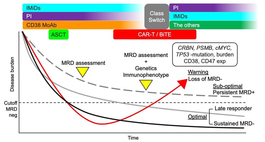
Figure 3. Treatment strategy considering MRD status. A total therapy approach combining IMiDs, PIs, anti-CD38 MoAb,
and ASCT may be suitable for MM patients considering the efficacy against myeloma cells and improved microenvironment.
MRD status after the total therapy approach can be useful in further treatment decisions. If the MRD status is negative, the
current treatment should continue (optimal). However, if the MRD status is positive, the genetic and immunophenotypic
characteristics of residual myeloma cells should be analyzed to optimize treatment. Loss of MRD negativity can lead to
aggressive recurrence (warning). The clinical outcome of persistent MRD positivity is better than that of loss of MRD-
negativity (sub-optimal). Repeated MRD assessment may be necessary for patients with persistent MRD positivity to
identify late responders and detect early-phase recurrence. MM, multiple myeloma; MRD, minimal residual disease;
IMiDs, immunomodulatory drugs; PIs, proteasome inhibitors; MoAbs, monoclonal antibodies; ASCT, autologous stem
cell transplantation; CAR-T, chimeric antigen receptor T cell; CRBN, cereblon; PSMB5, proteasome 20S subunit beta 5; and
exp, expression.
Examining the improvement of the immune environment is difficult in current prac-
tice, but it can be predicted using recently reported indicators, such as the lymphocyte
to monocyte ratio (LMR) [173–175]. A high LMR status reflects a good immunological
environment and is associated with a long survival time among MM patients. Recently,
we demonstrated that PFS in patients with both MRD-positivity and low LMR status
was significantly shorter than in those with MRD-negativity and/or high LMR status,
despite the achievement of CR [176]. Thus, a treatment change might be considered in
patients with both MRD positivity and low LMR status. However, there is no current
evidence showing the clinical significance of changing treatment approaches to enhance
the treatment response and improve the immune environment.
12. Conclusions
We consider that improvement of the immune environment and maintenance of
MRD negativity are key factors for the long-term survival of MM patients. Considering
the microenvironment around myeloma cells, initial treatment encompassing IMiDs, PIs,
anti-CD38 MoAb, and ASCT is important. This total therapy approach can improve the
immune environment and help achieve MRD negativity. This review suggests that an
MRD-driven treatment strategy may be promising, but genetic and immunophenotypic
analyses of residual myeloma cells should be repeated to select a suitable treatment for
residual myeloma cells. Before these analyses are available in clinical practice, treatmentCancers 2021, 13, 4867 17 of 25
can be selected based on “class switch.” In the future, there is a need to develop a treatment
strategy that not only treats the myeloma cells but also improves the immune environment
and targets the residual myeloma cells.
Author Contributions: Conceptualization, K.S.; writing—original draft preparation, K.S.; writing—
review and editing, K.S.; supervision, K.N. and S.Y. All authors have read and agreed to the published
version of the manuscript.
Funding: This research received no external funding.
Institutional Review Board Statement: Not applicable.
Informed Consent Statement: Not applicable.
Data Availability Statement: No new data were created or analyzed in this study. Data sharing is
not applicable to this article.
Conflicts of Interest: The authors declare no conflict of interest.
References
1. Palumbo, A.; Anderson, K. Multiple myeloma. N. Engl. J. Med. 2011, 364, 1046–1060. [CrossRef]
2. Van de Donk, N.W.C.J.; Pawlyn, C.; Yong, K.L. Multiple myeloma. Lancet 2021, 397, 410–427. [CrossRef]
3. B˛ebnowska, D.; Hrynkiewicz, R.; Grywalska, E.; Pasiarski, M.; Sosnowska-Pasiarska, B.; Smarz-Widelska, I.; Góźdź, S.; Roliński, J.;
Niedźwiedzka-Rystwej, P. Immunological Prognostic Factors in Multiple Myeloma. Int. J. Mol. Sci. 2021, 22, 3587. [CrossRef]
[PubMed]
4. Leone, P.; Solimando, A.G.; Malerba, E.; Fasano, R.; Buonavoglia, A.; Pappagallo, F.; De Re, V.; Argentiero, A.; Silvestris, N.;
Vacca, A.; et al. Actors on the Scene: Immune Cells in the Myeloma Niche. Front. Oncol. 2020, 10, 599098. [CrossRef] [PubMed]
5. Munshi, N.C.; Avet-Loiseau, H.; Anderson, K.C.; Neri, P.; Paiva, B.; Samur, M.; Dimopoulos, M.; Kulakova, M.; Lam, A.;
Hashim, M.; et al. A large meta-analysis establishes the role of MRD negativity in long-term survival outcomes in patients with
multiple myeloma. Blood Adv. 2020, 4, 5988–5999. [CrossRef]
6. Pessoa de Magalhães, R.J.; Vidriales, M.B.; Paiva, B.; Fernandez-Gimenez, C.; García-Sanz, R.; Mateos, M.V.; Gutierrez, N.C.;
Lecrevisse, Q.; Blanco, J.F.; Hernández, J.; et al. Analysis of the immune system of multiple myeloma patients achieving long-term
disease control by multidimensional flow cytometry. Haematologica 2013, 98, 79–86. [CrossRef]
7. Facon, T.; Kumar, S.; Plesner, T.; Orlowski, R.Z.; Moreau, P.; Bahlis, N.; Basu, S.; Nahi, H.; Hulin, C.; Quach, H.; et al. Daratumumab
plus Lenalidomide and Dexamethasone for Untreated Myeloma. N. Engl. J. Med. 2019, 380, 2104–2115. [CrossRef] [PubMed]
8. Dimopoulos, M.A.; Oriol, A.; Nahi, H.; San-Miguel, J.; Bahlis, N.J.; Usmani, S.Z.; Rabin, N.; Orlowski, R.Z.; Komarnicki, M.;
Suzuki, K.; et al. Daratumumab, Lenalidomide, and Dexamethasone for Multiple Myeloma. N. Engl. J. Med. 2016, 375, 1319–1331.
[CrossRef]
9. Lonial, S.; Dimopoulos, M.A.; Palumbo, A.; White, D.; Grosicki, S.; Spicka, I.; Walter-Croneck, A.; Moreau, P.; Mateos, M.V.;
Magen, H.; et al. Elotuzumab Therapy for Relapsed or Refractory Multiple Myeloma. N. Engl. J. Med. 2015, 373, 621–631.
[CrossRef] [PubMed]
10. Dimopoulos, M.A.; Dytfeld, D.; Grosicki, S.; Moreau, P.; Takezako, N.; Hori, M.; Leleu, X.; LeBlanc, R.; Suzuki, K.; Raab, M.S.; et al.
Elotuzumab plus Pomalidomide and Dexamethasone for Multiple Myeloma. N. Engl. J. Med. 2018, 379, 1811–1822. [CrossRef]
11. Attal, M.; Richardson, P.G.; Rajkumar, S.V.; San-Miguel, J.; Beksac, M.; Spicka, I.; Leleu, X.; Schjesvold, F.; Moreau, P.;
Dimopoulos, M.A.; et al. Isatuximab plus pomalidomide and low-dose dexamethasone versus pomalidomide and low-dose
dexamethasone in patients with relapsed and refractory multiple myeloma (ICARIA-MM): A randomised, multicentre, open-label,
phase 3 study. Lancet 2019, 394, 2096–2107. [CrossRef]
12. Jelinek, T.; Paiva, B.; Hajek, R. Update on PD-1/PD-L1 Inhibitors in Multiple Myeloma. Front. Immunol. 2018, 9, 2431. [CrossRef]
13. Voorhees, P.M.; Kaufman, J.L.; Laubach, J.; Sborov, D.W.; Reeves, B.; Rodriguez, C.; Chari, A.; Silbermann, R.; Costa, L.J.;
Anderson, L.D., Jr.; et al. Daratumumab, lenalidomide, bortezomib, and dexamethasone for transplant-eligible newly diagnosed
multiple myeloma: The GRIFFIN trial. Blood 2020, 136, 936–945. [CrossRef]
14. Attal, M.; Lauwers-Cances, V.; Hulin, C.; Leleu, X.; Caillot, D.; Escoffre, M.; Arnulf, B.; Macro, M.; Belhadj, K.; Garderet, L.; et al.
Lenalidomide, Bortezomib, and Dexamethasone with Transplantation for Myeloma. N. Engl. J. Med. 2017, 376, 1311–1320.
[CrossRef]
15. Jimenez-Zepeda, V.H.; Reece, D.E.; Trudel, S.; Chen, C.; Franke, N.; Winter, A.; Tiedemann, R.; Kukreti, V. Absolute lymphocyte
count as predictor of overall survival for patients with multiple myeloma treated with single autologous stem cell transplant.
Leuk. Lymphoma 2015, 56, 2668–2673. [CrossRef]
16. Diamond, B.; Korde, N.; Lesokhin, A.M.; Smith, E.L.; Shah, U.; Mailankody, S.; Hultcrantz, M.; Hassoun, H.; Lu, S.X.; Tan, C.; et al.
Dynamics of minimal residual disease in patients with multiple myeloma on continuous lenalidomide maintenance: A single-arm,
single-centre, phase 2 trial. Lancet Haematol. 2021, 8, e422–e432. [CrossRef]You can also read