Unique expression pattern of the EMT markers Snail, Twist and E-cadherin in benign and malignant parathyroid neoplasia
←
→
Page content transcription
If your browser does not render page correctly, please read the page content below

European Journal of Endocrinology (2009) 160 695–703                                                                              ISSN 0804-4643

CLINICAL STUDY

Unique expression pattern of the EMT markers Snail, Twist and
E-cadherin in benign and malignant parathyroid neoplasia
Volker Fendrich1,*, Jens Waldmann1,*, Georg Feldmann4,5, Katja Schlosser1, Alexander König2,
Annette Ramaswamy3, Detlef K Bartsch1 and Elias Karakas1
Departments of 1Surgery, 2Gastroenterology and Endocrinology and 3Pathology, Philipps-University Marburg, Baldingerstraße, D-35043 Marburg,
Germany, 4Department of Pathology and 5The Sol Goldman Pancreatic Cancer Research Center, Johns Hopkins University School of Medicine, Baltimore,
Maryland, USA
(Correspondence should be addressed to V Fendrich; Email: fendrich@med.uni-marburg.de)
*(V Fendrich and J Waldmann contributed equally to this work)

                            Abstract
                            Background: Epithelial and mesenchymal transitions (EMT) are essential for embryonic development
                            and progression of non-invasive tumor cells into malignant, metastatic carcinomas. During
                            embryogenesis, the parathyroid glands develop from pharyngeal pouches and migrate to their final
                            destinations, densely enclosed by mesenchymal neural crest cells. In this study, we examined the
                            expression of the EMT markers Snail, Twist and E-cadherin in normal parathyroid glands and benign
                            and malignant parathyroid diseases.
                            Methods: Using immunohistochemistry, we compared expression of E-cadherin, Snail and Twist in 25
                            patients with parathyroid adenoma, 25 patients with parathyroid hyperplasia, and nine patients with
                            parathyroid cancer with normal parathyroid glands.
                            Results: Normal parathyroid glands, parathyroid adenomas, and parathyroid hyperplasias showed a
                            typical membranous E-cadherin staining pattern. Expression of Snail was found in 22/25 parathyroid
                            adenomas and in all parathyroid hyperplasias. Twist was expressed in 22/25 of parathyroid adenomas
                            and in 20/25 parathyroid hyperplasias. Snail and Twist positive cells were homogeneously distributed
                            throughout the gland. However, in all nine parathyroid carcinomas, membranous E-cadherin staining
                            was lost. In addition, the expression pattern of Snail and Twist was changed and mostly limited to the
                            invasive front of cancer tissue samples.
                            Conclusion: Expression of Snail and Twist at the invasive front and consecutive loss of E-cadherin in
                            parathyroid carcinomas suggests a key role of EMT in the tumorigenesis of this cancer. The unique
                            expression pattern could help to distinguish between an adenoma and a non-metastatic carcinoma.
                            Loss of E-cadherin and change of the expression pattern of Snail and Twist together should result in an
                            en bloc resection or a close follow-up.

                            European Journal of Endocrinology 160 695–703

Introduction                                                              a highly unstable protein, is rapidly phosphorylated by
                                                                          glycogen synthase kinase-3b (GSK-3b) and sub-
In epithelial–mesenchymal transition (EMT), epithelial                    sequently degraded. Conversely, inhibition of GSK-3b
cells acquire fibroblast-like properties and show reduced                 function results in upregulation of Snail by an NF-kB-
intercellular adhesion and increased motility (1).                        dependent pathway, loss of E-cadherin expression, and
During progression to metastatic competence, carci-                       EMT. Additional protein modification further stabilizes
noma cells acquire mesenchymal gene-expression                            Snail protein and promotes EMT and tumor invasion
patterns and properties. This results in changed                          (4). Expression of Snail in epithelial tumors increases
adhesive properties and the activation of proteolysis                     their aggressiveness, as seen in experimentally induced
and motility, which allows tumor cells to invade into                     breast tumors, where high Snail expression correlates
surrounding stroma and finally to metastasize and                         with an increased risk of tumor relapse and poor
establish secondary tumors at distant sites (2). During                   survival rates in human breast cancer (5). Twist is a
EMT, the E-cadherin promoter is frequently repressed by                   highly conserved basic helix-loop-helix transcription
specific transcriptional repressors. E-cadherin levels                    factor that has important regulatory functions during
become limiting, which results in the loss of                             embryogenesis. In Drosophila, Twist protein is crucial
E-cadherin-dependent intercellular epithelial (3). Snail,                 for proper gastrulation and mesoderm formation (6).

q 2009 European Society of Endocrinology                                                                               DOI: 10.1530/EJE-08-0662
                                                                                                             Online version via www.eje-online.org

                                                                                                           Downloaded from Bioscientifica.com at 03/05/2022 02:07:17AM
                                                                                                                                                         via free access
696     V Fendrich, J Waldmann and others                                   EUROPEAN JOURNAL OF ENDOCRINOLOGY (2009) 160

While Twist proteins are only expressed in a subset of        and heart arrest. Cure is dependent on precise diagnosis
mesodermally and ectodermally derived tissues, Twist is       with consecutive surgical en bloc resection (12, 13). The
overexpressed in various human solid tumors including         only definite criteria for diagnosing parathyroid carci-
numerous types of carcinomas as well as sarcomas,             noma are local recurrence or metastases, making it
gliomas, neuroblastomas, and melanomas. The role of           sometimes difficult. Most patients with parathyroid
Twist in tumor progression has been convincingly              cancer have clinical manifestations that are virtually
associated with the metastatic process (7). Exogenous         indistinguishable from those in patients with a para-
overexpression of Twist increases the invasive and            thyroid adenoma, although severe hypercalcaemia may
metastatic abilities of cancer cells by promoting the         be regarded as a risk factor for malignancy (13).
downregulation of E-cadherin and the induction of an            The molecular mechanism for most of these para-
EMT (7).                                                      thyroid disorders is unknown and poorly understood.
   The increased motility and invasiveness of cancer          Some groups tried to characterize the global gene
cells in the first phase of metastasis are reminiscent of     expression profiles in a series of sporadic parathyroid
EMT during embryonic development. Following neural            adenomas in an attempt to obtain an improved picture
tube closure, multipotent neural crest cells undergo          of the genetic etiology behind parathyroid tumor
EMT, delaminate from the dorsal aspect of the neural          development (14, 15).
tube and migrate extensively throughout the embryo              Recently, our group described that Snail is over-
before giving rise to a diverse set of derivatives, such as   expressed in a large subset of neuroendocrine tumors of
for the development of the mesoderm in amniotes, or           the ileum, presenting the first evidence of Snail
the neural crest in all vertebrates (8). It has been          expression in endocrine tumors (16). In the present
demonstrated that as development proceeds, the neural         study, we analyzed the expression pattern of E-cadherin,
crest mesenchyme contributes connective tissue                Snail and Twist in normal parathyroid glands and
elements to organs developing in the pharyngeal region,       parathyroid disorders. For the first time, we show that
including thymus and parathyroid (9). EMT also                Snail and Twist are expressed in parathyroid carci-
explains why epithelial cells from one region can             nomas. Furthermore, we demonstrate that E-cadherin,
dissociate and migrate to a new location. One classic         Snail and Twist are expressed simultaneously in normal
example for such a cell movement during embryogen-            parathyroid gland and benign parathyroid disorders,
esis is the descent of the parathyroid glands. The inferior   based on the embryonic background of epithelial and
parathyroid glands that originate from the third              mesenchymal cells of the glands.
pharyngeal pouch migrate caudally with the thymus,
normally only as far as the inferior poles of the thyroid
gland, but may descend with the thymus gland into the         Patients and methods
thorax. The position of the larger pair of superior
parathyroid glands, which develop from the fourth             Patients and tissue collection
pharyngeal pouch is more constant, with 99% located
behind the upper poles of the thyroid lobes.                  Tissue from 25 patients with pHPT due to parathyroid
   Three parathyroid disorders can lead to an enlarge-        adenoma, 25 patients with sHPT due to renal failure,
ment of one or more glands. Primary hyperparathy-             nine patients with parathyroid cancer, and two patients
roidism (pHPT) is classically thought of as the somatic       with normal parathyroid glands were obtained from the
manifestation of hypercalcemia in which patients suffer       tissue bank of the Department of Pathology at the
from a variety of complaints including abdominal pain,        University Hospital of Marburg, Germany. All patients
nephrolithiasis, osteopenia, and mental status changes.       underwent parathyroid surgery in the Department of
Ninety percent of cases of pHPT are caused by a single        Surgery from the University Hospital of Marburg,
enlarged parathyroid adenoma. Multiglandular invol-           Germany. Patients with a hereditary background of
vement is less common and may be associated with              the parathyroid disease, e.g., MEN I, were excluded from
the multiple endocrine neoplasia syndromes MEN I              the study. Furthermore, patients with pHPT due to
and II (10).                                                  double adenomas or with persistent hypercalcaemia
   Secondary hyperparathyroidism (sHPT) usually               after previous surgery were excluded. Also patients with
develops in patients with chronic renal failure, where        sHPT with an unidentified gland during operation were
decreased levels of calcitriol with consecutive               excluded. The study was approved by the local Ethics
                                                              Committee.
hypocalcemia and a reduced phosphate clearance
lead to an increase of parathyroid hormone (PTH)
                                                              Diagnosis
synthesis and secretion (11).
   Parathyroid carcinoma is a rare cause of pHPT,             Histological diagnosis was confirmed by an experienced
affecting 0.2 to 1% of patients undergoing surgery (12).      pathologist (A R). The sporadic parathyroid adenomas
The major cause of death in patients with parathyroid         (pHPT) all showed a single enlarged hypercellular
cancer is severe hypercalcemia with its metabolic             parathyroid gland with or without a rim of normal
complications, such as malnutrition, acute pancreatitis,      parathyroid tissue and a biopsy of at least one other

www.eje-online.org

                                                                                      Downloaded from Bioscientifica.com at 03/05/2022 02:07:17AM
                                                                                                                                    via free access
EUROPEAN JOURNAL OF ENDOCRINOLOGY (2009) 160                                                          Snail and Twist in parathyroid neoplasia         697

parathyroid gland with findings consistent with normal                        Biochemical data
parathyroid tissue. The renal-induced hyperplasias
(sHPT) all showed hypercellular parathyroid tissue                            Preoperative levels of serum calcium and plasma PTH
                                                                              were obtained from clinical records. The values
involving three or more glands. A parathyroid tumor
                                                                              corresponding to the first hospital visit before any
was defined as carcinoma only when it showed invasion
                                                                              medical treatment, and of drugs with possible influence
of the tumor capsule or of surrounding structures. The
                                                                              on calcium metabolism were registered. The normal
presence of lymph node and/or distant metastasis was
                                                                              range for intact PTH was 11–65 pg/ml. The normal
also considered diagnostic of malignancy.
                                                                              range for calcium was 2.2–2.7 mmol/l.

Surgery                                                                       Immunostaining
Standard surgical treatment of sporadic pHPT consisted                        For immunolabeling, formalin-fixed and paraffin
of a bilateral exploration and identification of all four                     embedded archived tumor samples and corresponding
parathyroid glands with consecutive removal of the                            normal tissues were stained as previously described
enlarged gland. Standard surgical treatment of sHPT                           (17). Concentrations and sources of primary antibodies
consisted of a total parathyroidectomy with or without                        were used as follows: a-E-cadherin 1:200 (Zymed, San
autotransplantation and with bilateral thymectomy.                            Francisco, CA, USA), a-Twist and a-SNAIL 1:100
Standard surgical treatment of parathyroid carcinomas                         (Santa Cruz, Santa Cruz, CA, USA). Briefly, slides from
consisted of an en bloc resection. This procedure includes                    archived normal parathyroid glands, parathyroid ade-
the resection of the ipsilateral thyroid lobe together with                   nomas, parathyroid hyperplasias, and parathyroid
the isthmus, as well as a lymphadenectomy of the                              carcinomas were heated to 60 8C for 1 h, deparaffinized
central compartment of the neck. All areas suspicious                         using xylene, and hydrated by a graded series of ethanol
for local invasion must be resected, even if important                        washes. Antigen retrieval was accomplished by micro-
structures (e.g., recurrent laryngeal nerve, esophagus                        wave heating in 10 mM sodium citrate buffer of pH 6.0
or great vessels) are affected. Avoiding rupture of the                       for 10 min. For immunohistochemistry, endogenous
tumor capsule is of utmost importance and a complete                          peroxidase activity was quenched by 10 min incubation
resection of all tumor bearing tissue is inevitable to                        in 3% H2O2. Non-specific binding was blocked with 10%
avoid local recurrence.                                                       serum. Sections were then probed with primary

Table 1 Clinical characteristics and results of E-cadherin, Snail and Twist immunohistochemistry in 25 patients with PHPT.

Patient           Age                                   Ca2C               PTH          Gland         E-cadherin           Snail                Twist
number          (years)    Sex      Diagnosis           (mmol/l)           (pg/l)      weight (g)     expression        expression            expression

1                 41       M        pHPT                2.7                 128            1.1       CC                CC                     C
2                 72       F        pHPT                2.8                 151            1.8       CC                C                      C
3                 67       M        pHPT                3                   230            2.3       CC                C                      C
4                 66       M        pHPT                2.8                  93            0.2       CC                CC                     CC
5                 66       F        pHPT                2.8                 149            0.8       CC                C                      CC
6                 81       M        pHPT                2.5                  99            0.3       CC                CC                     C
7                 67       M        pHPT                2.4                  81            0.4       CC                Negative               Negative
8                 64       M        pHPT                3.3                 243            1.4       CC                C                      C
9                 53       F        pHPT                2.8                  88            1.8       CC                C                      C
10                56       M        pHPT                3                   134            2.2       CC                C                      C
11                48       M        pHPT                3.4                1418           16.0       C                 CC                     CC
12                69       M        pHPT                2.9                 182            1.0       CC                Negative               Negative
13                68       F        pHPT                2.9                 147            1.1       CC                CC                     CC
14                54       F        pHPT                3.3                 191            3.1       C                 CC                     C
15                63       F        pHPT                2.6                  84            0.3       CC                CC                     CC
16                26       F        pHPT                2.6                 157            2.0       CC                CC                     CC
17                38       M        pHPT                2.7                 359            0.7       CC                C                      C
18                60       M        pHPT                2.8                 150            1.4       CC                C                      C
19                57       F        pHPT                2.8                 110            2.1       CC                C                      CC
20                62       F        pHPT                2.8                 183            0.7       CC                CC                     C
21                53       M        pHPT                2.8                 140            4.1       C                 C                      C
22                43       M        pHPT                2.6                 190            0.5       CC                CC                     CC
23                43       M        pHPT                2.6                 106            1.3       CC                C                      C
24                46       F        pHPT                2.8                 129            0.8       CC                Negative               Negative
25                47       M        pHPT                2.6                 108            0.4       CC                C                      C

Ca2C, serum calcium level at diagnosis; PTH, serum parathyroid hormone level at diagnosis; pHPT, primary hyperparathyroidism due to parathyroid adenoma.

                                                                                                                                        www.eje-online.org

                                                                                                                 Downloaded from Bioscientifica.com at 03/05/2022 02:07:17AM
                                                                                                                                                               via free access
698     V Fendrich, J Waldmann and others                                                      EUROPEAN JOURNAL OF ENDOCRINOLOGY (2009) 160

antibodies overnight at 4 8C. For immunohistochem-                           65 years) at the time of surgery were also included.
istry, bound antibodies were detected using the avidin–                      Clinical and biochemical characteristics are listed in
biotin-complex (ABC) peroxidase method (ABC Elite Kit,                       Table 3.
Vector Labs, Burlingame, CA, USA). Final staining was
developed with the Sigma FAST DAB peroxidase
substrate kit (Sigma). To avoid misleading results, we                       E-cadherin, Snail and Twist are simul-
used the exact same amount of time for all sections to be                    taneously expressed in normal
developed.                                                                   parathyroid glands
   The immunohistochemistry results for E-cadherin,                          First, we analyzed the expression of E-cadherin, Snail
Snail and Twist were scored as described previously                          and Twist in normal parathyroid tissue from two
(16): negativeZless than 5% cells positive; CZ!30%                           patients with thyroid diseases and concurrently resected
cells positive; CCZO30% cells positive.                                      normal parathyroid glands. Immunohistochemical
                                                                             staining revealed expression of E-cadherin (Fig. 1A),
                                                                             Snail (Fig. 2A) and, Twist (Fig. 3A) in both glands. The
Results                                                                      pattern of E-cadherin expression showed a typical
                                                                             membranous staining. Cells with cytoplasmic Snail
Patients                                                                     and Twist expression were distributed throughout
Altogether, 59 patients with parathyroid disorders were                      larger areas of the glands.
included in the study. For evaluating E-cadherin, Snail
and Twist in pHPT, fifteen males and ten females with a                      E-cadherin, Snail and Twist are simul-
median age of 56 years (range 26 to 81 years) at the                         taneously expressed in benign
time of surgery were included. Clinical and biochemical                      parathyroid disorders
characteristics are listed in Table 1. To study expression
of EMT-markers in sHPT, thirteen females and twelve                          All parathyroid glands obtained from patients with
males with a median age of 53 years (range 29 to 78                          pHPT or sHPT showed a strong membranous staining of
years) at the time of surgery were enclosed. Clinical and                    E-cadherin (Fig. 1B and C, Tables 1 and 2).
biochemical characteristics are listed in Table 2. Five                         Immunohistochemical staining revealed expression
males and four females presenting with parathyroid                           of Snail in 22/25 (88%) of parathyroid adenomas out of
carcinoma with a median age of 46 years (range 21 to                         patients with pHPT (Fig. 2B, Table 1) and in all 25

Table 2 Clinical characteristics and results of E-cadherin, Snail and Twist immunohistochemistry in 25 patients with sHPT.

Patient           Age                                   Ca2C            PTH          Gland          E-cadherin            Snail                Twist
number          (years)    Sex       Diagnosis         (mmol/l)         (pg/l)      weight (g)      expression         expression            expression

1                    31    F         sHPT                2.6            1090            1.3       CC                 CC                      C
2                    58    F         sHPT                2.8             793            0.9       C                  CC                      C
3                    56    F         sHPT                2.4            1667            0.1       CC                 CC                      C
4                    36    M         sHPT                2.7            1208            1.6       CC                 CC                      CC
5                    58    M         sHPT                2.4            1536            0.1       CC                 CC                      Negative
6                    63    F         sHPT                2.6            1274            0.3       CC                 C                       C
7                    61    F         sHPT                2.6            2271            1.1       CC                 CC                      C
8                    48    F         sHPT                2.8             870            0.9       CC                 CC                      CC
9                    52    M         sHPT                2.8            1198            1.5       CC                 C                       Negative
10                   61    F         sHPT                2.8             322            0.1       CC                 CC                      C
11                   46    M         sHPT                2.4            1129            0.5       CC                 CC                      CC
12                   51    M         sHPT                2.7             518            0.5       CC                 C                       C
13                   45    F         sHPT                2.9             588            1.1       C                  C                       Negative
14                   65    M         sHPT                2.7            3011            2.3       CC                 CC                      CC
15                   68    F         sHPT                2.7            1739            0.6       C                  C                       C
16                   44    M         sHPT                2.6             794            0.9       CC                 CC                      C
17                   46    F         sHPT                1.9             554            0.3       CC                 CC                      CC
18                   62    M         sHPT                2.7             557            1.4       CC                 C                       Negative
19                   63    F         sHPT                2.7            2123            1.4       CC                 C                       C
20                   35    M         sHPT                2.8            1090            1.0       C                  C                       Negative
21                   29    F         sHPT                2.4            1116            0.3       CC                 CC                      CC
22                   59    F         sHPT                2.3             708            0.6       CC                 C                       C
23                   78    M         sHPT                2.7            1324            1.4       CC                 C                       C
24                   74    M         sHPT                2.9            1318            1.8       CC                 C                       C
25                   27    M         sHPT                2.6            2800            0.8       CC                 CC                      CC

Ca2C, serum calcium level at diagnosis; PTH, serum parathyroid hormone level at diagnosis; sHPT, secondary hyperparathyroidism due to renal failure.

www.eje-online.org

                                                                                                            Downloaded from Bioscientifica.com at 03/05/2022 02:07:17AM
                                                                                                                                                          via free access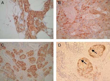
EUROPEAN JOURNAL OF ENDOCRINOLOGY (2009) 160                                                         Snail and Twist in parathyroid neoplasia        699

Table 3 Clinical characteristics and results of E-cadherin, Snail and Twist immunohistochemistry in 9 patients with parathyroid carcinoma.

Patient         Age                                 Ca2C             PTH           Gland          E-cadherin               Snail              Twist
number        (years)     Sex    Diagnosis         (mmol/l)          (pg/l)       weight (g)      expression            expression          expression

1                59       M      Pc                   4.0             489              4.0       Negative          CC                       CC
2                34       F      Pc                   3.3            1365              6.0       Negative          C                        CC
3                21       F      Pc                   5.8             880              1.2       Negative          CC                       CC
4                64       M      Pc                   4.9            1120             11.5       Negative          CC                       Negative
5                44       F      Pc                   4.0             714              1.4       Negative          C                        C
6                65       M      Pc                   4.2            1480             12.0       Negative          CC                       CC
7                58       F      Pc                   3.4             358              7.3       Negative          CC                       C
8                31       M      Pc                   3.8            2319             10.5       Negative          C                        C
9                42       M      Pc                   3.8             480              1.0       Negative          CC                       C

Ca2C, serum calcium level at diagnosis; PTH, serum parathyroid hormone level at diagnosis; Pc, parathyroid carcinoma.

parathyroid tissues from patients with sHPT (Fig. 2C,                         The expression patterns of Snail and Twist are
Table 2). Furthermore, immunohistochemical staining                           changed in parathyroid cancer
revealed expression of Twist in 22/25 (88%) patients
                                                                              Consistent with loss of E-cadherin expression in
with pHPT (Fig. 3B, Table 1) and in 20/25 (80%)
                                                                              parathyroid carcinomas, the expression pattern of
patients with sHPT (Fig. 3C, Table 2).
                                                                              Snail (Fig. 2D, Table 3) and Twist (Fig. 3D, Table 3)
   Snail and Twist positive cells were homogeneously                          changed in malignant tumors. Snail was no longer
distributed throughout the whole gland, comparable                            expressed homogenously throughout the whole tumor,
with the pattern seen in normal parathyroid glands.                           but was mostly limited to the invasive front (Fig. 2D,
                                                                              arrows). In addition, Twist was now stronger and
Expression of E-cadherin is lost in                                           expressed along the front of the tumor.
parathyroid carcinoma
In all nine parathyroid carcinomas analyzed, membra-                          Discussion
nous E-cadherin staining was lost (Fig. 1D, Table 3).
Immunohistochemical staining now revealed a cyto-                             EMT occurs during embryonic morphogenesis in multi-
plasmic expression of E-cadherin protein, which is a                          cellular organisms, in which embryonic mesenchymal
hallmark of EMT.                                                              cells are formed and become motile following the loss
                                                                              of epithelial cell polarity. In recent years, EMT has
                                                                              also been recognized as a potential mechanism for
                                                                              cancer progression (18). A central event in EMT is

Figure 1 IHC staining for E-cadherin in normal parathyroid gland
and parathyroid disorders. (A) Representative example of
E-cadherin expression in a normal parathyroid gland, showing a                Figure 2 IHC staining for Snail in normal parathyroid gland and
typical membranous staining pattern for the cell-adhesion marker.             parathyroid disorders. (A–C) Snail expression was found in normal
(B) Strong membranous staining of E-cadherin in pHPT and                      parathyroid gland (A) and in benign parathyroid disorders (B, C).
(C) sHPT. (D) By contrast, in parathyroid carcinoma, the                      Snail positive cells were distributed throughout larger areas of the
membranous pattern of E-cadherin expression was lost, showing                 glands. (D) By contrast, in parathyroid cancer, Snail was no longer
now a cytoplasmic expression of E-cadherin protein, which is a                expressed homogenously throughout the whole tumor, but was
hallmark of EMT. Full colour version of this figure available via             mostly limited to the invasive front (arrows). Full colour version of
http://dx.doi.org/10.1530/EJE-08-0662.                                        this figure available via http://dx.doi.org/10.1530/EJE-08-0662.

                                                                                                                                      www.eje-online.org

                                                                                                               Downloaded from Bioscientifica.com at 03/05/2022 02:07:17AM
                                                                                                                                                             via free access
700     V Fendrich, J Waldmann and others                                             EUROPEAN JOURNAL OF ENDOCRINOLOGY (2009) 160

                                                                       that neural crest cells undergo EMT, delaminate from the
                                                                       neural epithelium, and migrate throughout the embryo,
                                                                       differentiating at their destination sites into a wide array
                                                                       of cell types. Subsequent to the specification of neural
                                                                       crest progenitors at the neural plate border, a group of
                                                                       genes that primarily encode transcription factors (25),
                                                                       including the Snail family genes Snail and Slug, are
                                                                       induced in neural crest progenitor cells (26). EMT can be
                                                                       triggered by different signaling molecules, such as bone
                                                                       morphogenetic proteins (BMPs) (8). Thériault et al.
                                                                       recently demonstrated an upregulation of Snail mRNA
                                                                       and protein in response to exogenous BMP4 in ovarian
                                                                       cancer cells (27). Interestingly, BMP4 also plays a critical
                                                                       role in thymus and parathyroid organogenesis (28) and
                                                                       induces Snail during neural crest development (29).
                                                                       Very recently, Franci et al. showed that at day E9.5 in the
Figure 3 IHC staining for Twist in normal parathyroid gland and        mouse, Snail activity can be detected in the pharyngeal
parathyroid disorders. Expression of Twist was found in normal         arches (30).
parathyroid gland (A) and in patients with pHPT (B) and sHPT (C).
The expression pattern was comparable with Snail (Fig. 2), with a
                                                                          In the present study, in normal parathyroid glands
distribution throughout the gland. (D) In parathyroid carcinoma, the   and tissue obtained out of patients with benign
staining pattern changed again. Twist was more strongly                parathyroid disorders, Snail positive cells were distrib-
expressed along the front of the tumor (arrows). Full colour version   uted throughout large areas of the glands. Furthermore,
of this figure available via http://dx.doi.org/10.1530/EJE-08-0662.    we found the same expression pattern for the transcrip-
                                                                       tion factor Twist. It has been shown that Twist is
downregulation of membranous E-cadherin expression                     required for the maintenance of cell viability and
(19), which leads to the loss of cell–cell contact and the             proliferation in pharyngeal arch tissues (31). In mouse
consecutive progression of the cells towards a malignant               embryos, Twist-positive cells were found at E9.5 along
phenotype. The transcription factor Snail is one major                 the migratory paths of the hindbrain neural crest and in
suppressor of E-cadherin and a strong inducer of EMT.                  branchial arches (32). Twist is also required for the
Snail downregulates E-cadherin in different types of                   activation of Snail, which is crucial for proper gastrula-
tumors e.g., hepatocellular carcinomas (20), carcinomas                tion and for maintenance of Twist expression (33). We
from the esophagus, cardia, stomach (21), and colorectal               also found a typical membranous staining of E-cadherin
carcinomas (22). Recently, our group was the first to                  in normal parathyroids and benign parathyroid
describe activation of Snail in endocrine tumors (16).                 disorders, which has been reported before (34). By
   The presented study is now the first to show that Snail,            contrast, in parathyroid carcinoma, the membranous
Twist and E-cadherin are expressed simultaneously in                   pattern of E-cadherin expression is lost, showing now
normal parathyroid glands and benign parathyroid                       a cytoplasmic expression of E-cadherin protein.
disorders; at first glance a surprising result, having                 Cytoplasmic expression of E-cadherin protein and/or
these opposed characters of EMT expressed at the same                  transcriptional repression of its mRNA are hallmarks of
time. But, most likely, the explanation could be found in              EMT, both in embryonic development and in cancer
organogenesis of the parathyroid glands. The pharyn-                   progression (4). Our results are in line with the results
geal glandular organs in mammals have complex                          reported by Haven et al. They undertook an expression
developmental origins. The parathyroid, thymus, and                    profiling of 53 hereditary and sporadic parathyroid
ultimobranchial primordial develop from the pharyn-                    tumors and found an upregulation of E-cadherin mRNA
geal pouches and migrate to their final destinations.                  in parathyroid carcinomas, with aberrant staining
During their descent to the neck, these pharyngeal                     noted, indicating loss of function in cell adhesion (35).
organs are surrounded by mesenchyme derived from the                      Another striking result of our study was the change
cranial neural crest (9). The cranial neural crest arising             of staining pattern of Snail and Twist in parathyroid
from the embryonic midbrain and hindbrain plays a                      cancer tissue. Snail and Twist were no longer expressed
critical role in the development of the pharyngeal arches              homogenously throughout the whole tumor, but were
and pouches, initially by providing the mesenchymal                    mostly limited to the invasive front, a hallmark of EMT
cells which populate this region. As development                       (2, 4). The invasive front of a tumor is formed by cells
proceeds, the neural crest mesenchyme contributes                      that migrate into and invade the surrounding tissue
directly to the formation of some structures in the                    either as single cells (Figs 6 and 7) or in collective
pharyngeal region, including thymus and parathyroid,                   clusters (Figs 4 and 5) (4). In order to acquire motility
and forms the calcitonin producing cells of thyroid gland              and invasiveness, malignant cells must lose some of
(9, 23, 24). The molecular basis for control of these                  their epithelial characters and undergo EMT. While
events is largely unknown. Recently, it has been shown                 these steps are crucial for embryonic development, they

www.eje-online.org

                                                                                                Downloaded from Bioscientifica.com at 03/05/2022 02:07:17AM
                                                                                                                                              via free access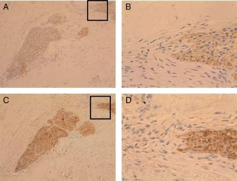
EUROPEAN JOURNAL OF ENDOCRINOLOGY (2009) 160                                                       Snail and Twist in parathyroid neoplasia         701

Figure 4 IHC staining for Snail (4AB) and Twist (4CD) in parathyroid         Figure 6 IHC staining for Snail (6AB) and Twist (6CD) in multiple
carcinomas. B and D show selected areas from A and C marked by               samples of parathyroid carcinomas. B and D show selected areas
the box. Snail and Twist expression is found at the invasive front of        from A and C marked by the box. Snail and Twist expression is
the tumor where tumor cells invade the surrounding tissue either as          found at the invasive front of the tumor where tumor cells invade the
single cells (Figs 6 and 7) or in collective clusters (Figs 4 and 5). Full   surrounding tissue either as single cells (Figs 6 and 7) or in
colour version of this figure available via http://dx.doi.org/10.1530/       collective clusters (Figs 4 and 5). Full colour version of this figure
EJE-08-0662.                                                                 available via http://dx.doi.org/10.1530/EJE-08-0662.

become fatal in pathological situations in the adult. The                    switch-on EMT, leading to the loss of E-cadherin and the
strong similarity between the process of tumor invasion                      typical staining pattern of Snail and Twist at the edges
and cell migration observed during organ development                         of the parathyroid carcinoma (Figs 6 and 7).
suggest that carcinoma cells can change their own                               Parathyroid adenoma shares some histological
morphology, motility, and ability to invade surrounding                      features with parathyroid cancer and at the time of
structures. In parathyroid tissue, in which Snail, Twist                     initial surgery differentiation from parathyroid cancer
and E-cadherin are expressed simultaneously in normal                        can be difficult. Hence, some lesions have been reported
and benign states, undefined oncogenic factors must                          as parathyroid cancer, but their clinical behavior has

Figure 5 IHC staining for Snail (5AB) and Twist (5CD) in multiple            Figure 7 IHC staining for Snail (7AB) and Twist (7CD) in multiple
samples of parathyroid carcinomas. B and D show selected areas               samples of parathyroid carcinomas. B and D show selected areas
from A and C marked by the box. Snail and Twist expression is                from A and C marked by the box. Snail and Twist expression is
found at the invasive front of the tumor where tumor cells invade the        found at the invasive front of the tumor where tumor cells invade the
surrounding tissue either as single cells (Figs 6 and 7) or in               surrounding tissue either as single cells (Figs 6 and 7) or in
collective clusters (Figs 4 and 5). Full colour version of this figure       collective clusters (Figs 4 and 5). Full colour version of this figure
available via http://dx.doi.org/10.1530/EJE-08-0662.                         available via http://dx.doi.org/10.1530/EJE-08-0662.

                                                                                                                                     www.eje-online.org

                                                                                                              Downloaded from Bioscientifica.com at 03/05/2022 02:07:17AM
                                                                                                                                                            via free access
702 V Fendrich, J Waldmann and others EUROPEAN JOURNAL OF ENDOCRINOLOGY (2009) 160
not always been consistent with this diagnosis. Indeed, 7 Yang J, Mani SA, Donaher JL, Ramaswamy S, Itzykson RA,
the presence of local recurrence or metastatic disease is Come C, Savagner P, Gitelman I, Richardson A & Weinberg RA.
Twist, a master regulator of morphogenesis, plays an essential role
the only reliable feature that differentiates benign from in tumor metastasis. Cell 2004 117 927–939.
malignant parathyroid disease. At the initial operation, 8 Barrallo-Gimeno A & Nieto MA. The Snail genes as inducers of cell
features of presumed malignancy, such as firm texture, movement and survival: implications in development and cancer.
grey color and gross adherence to adjacent tissue, are Development 2005 132 3151–3161.
not proven differentiators of APA from parathyroid 9 LeLievre CS & LeDouarin NM. Mesenchymal derivatives of the
neural crest: analysis of chimeric quail and chick embryos. Journal of
cancer. Thus, the risk for the surgeon is overtreatment Embryology and Experimental Morphology 1975 34 125–154.
of an APA or undertreatment of a true parathyroid 10 Suliburk JW & Perrier ND. Primary hyperparathyroidism.
cancer (13). The presented findings could help to Oncologist 2007 12 644–653.
distinguish between an adenoma and a non-metastatic 11 Schlosser K, Zielke A & Rothmund M. Medical and surgical
carcinoma. Loss of E-cadherin and change of the treatment for secondary and tertiary hyperparathyroidism.
Scandinavian Journal of Surgery 2004 93 288–297.
expression pattern of Snail and Twist together with a 12 Rawat N, Khetan N, Williams ND & Baxter JN. Parathyroid
clinical suspicious lesion should result in an en bloc carcinoma. British Journal of Surgery 2005 92 1345–1353.
resection as described above. If this is not possible, these 13 Ippolito G, Palazzo FF, Sebag F, De Micco C & Henry JF.
patients should undergo a very close follow up, because Intraoperative diagnosis and treatment of parathyroid cancer
they might be at a higher risk of developing distant and atypical parathyroid adenoma. British Journal of Surgery 2007
94 566–570.
metastases. 14 Forsberg L, Björck E, Hashemi J, Zedenius J, Höög A, Farnebo LO,
In conclusion, we show for the first time, that EMT Reimers M & Larsson C. Distinction in gene expression profiles
plays a role in the tumorigenesis of parathyroid demonstrated in parathyroid adenomas by high-density oligoarray
carcinomas. Loss of membranous E-cadherin and technology. European Journal of Endocrinology 2005 152 459–470.
aberrant Snail and Twist expression patterns along 15 Morrison C, Farrar W, Kneile J, Williams N, Liu-Stratton Y,
Bakaletz A, Aldred MA & Eng C. Molecular classification of
the invasive front in parathyroid cancer tissue samples
parathyroid neoplasia by gene expression profiling. American
as compared with benign tumors and histologically Journal of Pathology 2004 165 565–576.
normal parathyroid tissues is in line with changes 16 Fendrich V, Waldmann J, Esni F, Ramaswamy A, Mullendore M,
usually observed in EMT. Buchholz M, Maitra A & Feldmann G. Snail and Sonic Hedgehog
activation in neuroendocrine tumors of the ileum. Endocrine
Related Cancer 2007 14 865–874.
17 Esni F, Stoffers DA, Takeuchi T & Leach SD. Origin of exocrine
Declaration of interest pancreatic cells from nestin-positive precursors in developing
We declare that there is no conflict of interest that could be perceived mouse pancreas. Mechanisms of Development 2004 121 15–25.
as prejudicing the impartiality of the research reported. 18 Thiery JP. Epithelial–mesenchymal transitions in tumour pro-
gression. Nature Reviews. Cancer 2002 2 442–454.
19 Perl AK, Wilgenbus P, Dahl U, Semb H & Christofori G. A causal
role for E-cadherin in the transition from adenoma to carcinoma.
Funding Nature 1998 392 190–193.
Grant support: V F was supported by a Research Grant of the 20 Jiao W, Miyazaki K & Kitajima Y. Inverse correlation between
University Medical Center Giessen and Marburg. G F was supported by E-cadherin and Snail expression in hepatocellular carcinoma cell
a fellowship grant within the postdoc-program of the German lines in vitro and in vivo. British Journal of Cancer 2002 86 98–101.
Academic Exchange Service (DAAD). 21 Rosivatz E, Becker KF, Kremmer E, Schott C, Blechschmidt K,
Hofler H & Sarbia M. Expression and nuclear localization of Snail,
an E-cadherin repressor, in adenocarcinomas of the upper
gastrointestinal tract. Virchows Archiv 2006 448 277–287.
22 Roy HK, Smyrk TC, Koetsier J, Victor TA & Wali RK. The
References transcriptional repressor SNAIL is overexpressed in human colon
cancer. Digestive Diseases and Sciences 2005 50 42–46.
1 Zhou BP & Hung MC. Wnt, hedgehog and snail: sister pathways 23 Manley NR & Capecchi MR. The role of Hoxa-3 in mouse thymus
that control by GSK-3beta and beta-Trcp in the regulation of and thyroid development. Development 1995 121 1989–2003.
metastasis. Cell Cycle 2005 4 772–776. 24 Manley NR & Capecchi MR. Hox Group 3 paralogs regulate the
2 Thiery JP & Sleeman JP. Complex networks orchestrate epithelial– development and migration of the thymus, thyroid, and
mesenchymal transitions. Nature Reviews. Molecular Cell Biology parathyroid glands. Developmental Biology 1998 195 1–15.
2006 7 131–142. 25 Clouthier DE, Williams SC, Yanagisawa H, Wieduwilt M,
3 Cano A, Pérez-Moreno MA, Rodrigo I, Locascio A, Blanco MJ, del Richardson JA & Yanagisawa M. Signaling pathways crucial for
Barrio MG, Portillo F & Nieto MA. The transcription factor Snail craniofacial development revealed by endothelin-A receptor-
controls epithelial–mesenchymal transitions by repressing deficient mice. Developmental Biology 2000 217 10–24.
E-cadherin expression. Nature Cell Biology 2000 2 76–83. 26 Murray SA & Gridley T. Snail family genes are required for left-
4 Christofori G. New signals from the invasive front. Nature 2006 44 right asymmetry determination, but not neural crest formation, in
444–450. mice. PNAS 2006 103 10300–10304.
5 Moody SE, Perez D, Pan TC, Sarkisian CJ, Portocarrero CP, 27 Thériault BL, Shepherd TG, Mujoomdar ML & Nachtigal MW.
Sterner CJ, Notorfrancesco KL, Cardiff RD & Chodosh LA. The BMP4 induces EMT and Rho GTPase activation in human ovarian
transcriptional repressor Snail promotes mammary tumor recur- cancer cells. Carcinogenesis 2007 28 1153–1162.
rence. Cancer Cell 2005 8 197–209. 28 Patel SR, Gordon J, Mahbub F, Blackburn CC & Manley NR. Bmp4
6 O’Rourke MP & Tam PP. Twist functions in mouse development. and Noggin expression during early thymus and parathyroid
International Journal of Developmental Biology 2002 46 401–413. organogenesis. Gene Expression Patterns 2006 6 794–799.
www.eje-online.org
Downloaded from Bioscientifica.com at 03/05/2022 02:07:17AM
via free accessEUROPEAN JOURNAL OF ENDOCRINOLOGY (2009) 160 Snail and Twist in parathyroid neoplasia 703
29 Liem KF, Tremml G, Roelink H & Jessell T. Dorsal differentiation of 34 Zeromski J, Lawniczak M, Galbas K, Jenek R & Golusiński P.
neural plate cells induced by BMP-mediated signals from Expression of CD56/N-CAM antigen and some other adhesion
epidermal ectoderm. Cell 1995 82 969–979. molecules in various human endocrine glands. Folia Histochem-
30 Francı́ C, Takkunen M, Dave N, Alameda F, Gómez S, Rodrı́guez R, istry and Cytobiology 1998 36 119–125.
Escrivà M, Montserrat-Sentı́s B, Baró T, Garrido M, Bonilla F, 35 Haven CJ, Howell VM, Eilers PH, Dunne R, Takahashi M, van
Virtanen I & Garcı́a de Herreros A. Expression of Snail protein in Puijenbroek M, Furge K, Kievit J, Tan MH, Fleuren GJ,
tumor–stroma interface. Oncogene 2006 25 5134–5144. Robinson BG, Delbridge LW, Philips J, Nelson AE, Krause U,
31 Ota MS, Loebel DA, O’Rourke MP, Wong N, Tsoi B & Tam PP. Twist Dralle H, Hoang-Vu C, Gimm O, Morreau H, Marsh DJ & Teh BT.
is required for patterning the cranial nerves and maintaining the Gene expression of parathyroid tumors: molecular subclassifica-
viability of mesodermal cells. Developmental Dynamics 2004 230 tion and identification of the potential malignant phenotype.
216–228. Cancer Research 2004 64 7405–7411.
32 Gitelman I. Twist protein in mouse embryogenesis. Developmental
Biology 1997 189 205–214.
33 Castanon I & Baylies MK. Twist in fate: evolutionary comparison of Received 30 December 2008
Twist structure and function. Gene 2002 287 11–22. Accepted 22 January 2009
www.eje-online.org
Downloaded from Bioscientifica.com at 03/05/2022 02:07:17AM
via free accessYou can also read