A Review of the Primary Nutritional and Environmental Factors Associated with Parkinson's Disease
←
→
Page content transcription
If your browser does not render page correctly, please read the page content below
Volume 10 Issue 2 (2021)

A Review of the Primary Nutritional and Environmental
Factors Associated with Parkinson’s Disease
Eleanor Considine 1* , Lucy Yin 2* and Mitra Hartmann 3

1
  Skyline High School, Salt Lake City, UT, USA
2
  Aragon High School, San Mateo, CA, USA
3
  Northwestern University, Evanston, IL, USA
DOI: https://doi.org/10.47611/jsrhs.v10i2.1482

ABSTRACT

Parkinson’s disease is a progressive, degenerative nervous system disorder that produces both motor and nonmotor
symptoms. This literature review begins by examining evidence for two possible origins for the disease: does it begin
in the brain and progress to the gut, or vice versa, or does it begin in both places concurrently? Next, we examine
several environmental factors that have been shown to either increase or decrease risk of Parkinson’s disease. These
are primarily nutritional factors, specifically caffeine, nicotine, and dairy products. Studies in both animals and hu-
mans provide weak evidence that increased consumption of low fat dairy is associated with an increased risk of Par-
kinson’s disease development. Additionally, there is strong evidence that nicotine has a neuroprotective effect which
also lowers the risk. Finally, there is similarly strong evidence that caffeine exerts neuroprotective effects which lower
the overall risk of developing Parkinson’s disease.

Introduction: The gut-brain axis: Does PD start in the gut or in the brain or
both?

Traditionally, Parkinson’s disease (PD) has been thought to originate in the brain: it is associated with a gradual
aggregation of the alpha synuclein protein, which damages nerve cells, in the substantia nigra (Stefanis, 2012).
          However, in 2003, neuroanatomist Heiko Braak suggested a possible route by which misfolded alpha-synu-
clein aggregates travel: the vagus nerve. Cases of idiopathic PD, or a spontaneous form where the origin is uncertain,
which comprise the vast majority of PD cases, showed involvement of both the enteric nervous system and the vagus
nerve, leading Braak to hypothesize that the aggregates may originate outside of the central nervous system (Braak,
Rub, Gai, & Del Tredici, 2003).
          Building off of Braak’s observations, researchers injected misfolded alpha synuclein into the intestines of
healthy mice. Over the course of 10 months, they observed a buildup of these proteins both in the vagus nerve and the
brain. They hypothesized that this neurological disorder may begin in the gut for some patients, indicating that the
damage begins in the enteric nervous system, and then travels up to the brain (S. Kim et al., 2019).
          Consistent with the possibility that there may be two variants of the disease— one that begins in the brain,
and the other in the gut— the symptoms of Parkinson's disease are unique to each patient, some developing intestinal
discomfort—such as constipation— first, while others develop neurological symptoms first (Borghammer & Van Den
Berge, 2019). In addition, the progression of symptoms also varies from person to person. The discovery of a gut-
related origin is promising, as it could potentially be easier to prevent Parkinson’s disease development if it starts in
the gut, knowing that it can be stopped before it enters the brain.
          By what possible mechanism(s) could these two putative variants induce Parkinson’s disease? As shown in
Figure 1 the brain is connected to the gut via the vagus nerve, which offers transportation for proteins. The brain and
the gut communicate through a multitude of pathways including the bloodstream, the neuroendocrine system and

ISSN: 2167-1907                                      www.JSR.org                                                        1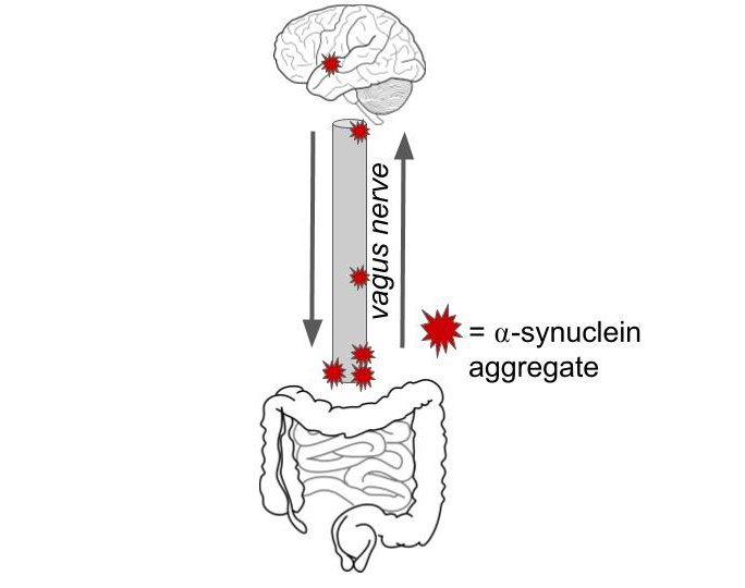
Volume 10 Issue 2 (2021)

immune response (cytokines) but the vagus nerve offers the most direct form of communication. The vagus nerve is
bidirectional which adds to the confusion surrounding the origin of Parkinson’s pathology. There have been many
studies on the vagus nerve attempting to determine its role in the aggregation of alpha-synuclein in the brain.
          A study that used genetic modification to deliberately overproduce alpha synuclein in two groups of mice
found that the mice raised in germ free cages had fewer motor deficit symptoms and fewer Lewy Bodies (formed from
aggregates of alpha synuclein) in their brain compared to mice raised in a non-sterile environment. In addition, mice
raised in the nonsterile environment displayed a decrease in PD symptoms when given an antibiotic, indicating that
their microbiome was associated with PD development. Finally, to further support their results, the research team
injected gut bacteria from human Parkinson’s patients into germ-free mice, and healthy human gut bacteria into others.
As expected, the mice injected with the Parkinson’s gut microbiota began to display degenerative motor symptoms
(Sampson et al., 2016).

Figure 1. Diagram of the function of the vagus nerve as it is hypothesized in the gut-brain axis argument on Parkin-
son’s pathology. Image of the gut was obtained from https://pixabay.com/vectors/intestines-bowel-guts-intestinal-
293929/ and image of the brain from https://pixy.org/153514/. Both images are CCBY

In addition to rodent studies, evidence from human studies supports the theory of a variant form of PD that originates
in the gut. One study found that individuals who underwent truncal vagotomy (a procedure in which both trunks of
the vagus nerve are cut) had decreases in both PD symptoms and risks (Svensson et al., 2015).
         Other studies of the vagus nerve in humans also provide evidence for a variant that starts in the gut. In one
study, researchers studied a cohort of more than 9,000 vagotomized patients. They found that those who underwent a
truncal vagotomy were 15% less likely to develop Parkinson’s disease 5 years after their procedure compared to the
general population. The truncal vagotomy’s association with a decreased rate of developing PD indicates the vagus
nerve’s possible role as a pathway in which alpha synuclein aggregates travel (Liu, Chan, & Stoessl, 2017).
         If, indeed, at least one type of PD originates in the gut, then this potentially opens new avenues for treatment.
Nutrition is one of the most researched aspects of Parkinson’s prevention (Boulos, Yaghi, El Hayeck, Heraoui, &
Fakhoury-Sayegh, 2019). To write this review, we performed a broad survey of the literature to reveal five primary

ISSN: 2167-1907                                      www.JSR.org                                                        2
Volume 10 Issue 2 (2021)
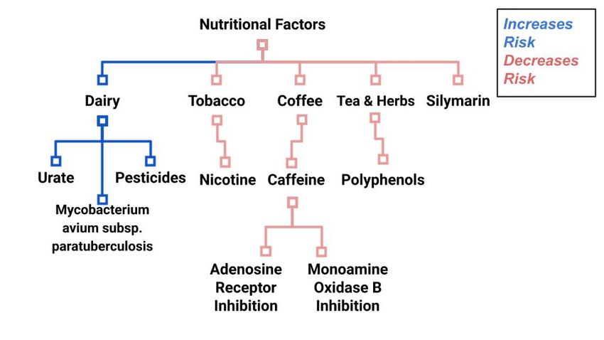
nutritional and environmental factors associated with risk of developing PD: consumption of dairy, coffee, tea, nico-
tine, and silymarin. These factors are diagrammed in Figure 2. Although we anticipate that the present review will
mostly be relevant to the second mechanism in which it originates in the gut, it is also possible that some factors (e.g.,
nicotine) are also neuroprotective. When we began this review, we were unaware of the work of Seidl et al. (2014),
which reviews research up through 2014 (Seidl, Santiago, Bilyk, & Potashkin, 2014). The present work represents an
independent review of some of the same studies, and also extends the work to include research through 2020.
Figure 2. Organizational chart outlining the content of entire review.
1.Dairy
Studies in both humans and mice indicate that dairy consumption is associated with an increased risk of the develop-
ment of Parkinson’s disease (Ascherio & Schwarzschild, 2016; Godos et al., 2020; Hughes et al., 2017; Jiang, Ju,
Jiang, & Zhang, 2014). However, the association is complicated by sex differences as well as the type of dairy product
consumed.
One of the largest studies (H. L. Chen et al., 2007) prospectively investigated the association between dairy
intake and Parkinson’s risk among over 55,000 men and over 70,000 women. These participants were recruited in
1982, and an initial questionnaire was conducted in 1992 on “demographic, medical, environmental, and lifestyle
factors and dietary habits.” Then, dairy intake was assessed in 1997, 1999, and 2001 through follow-up surveys asking
about consumption habits and any new diagnoses, including Parkinson’s disease. From 1992 to 2001, a total of 250
men and 138 women were diagnosed with PD. The authors concluded that dairy consumption was positively associ-
ated with increased PD risk for both men and women (H. L. Chen et al., 2007).
Other studies have supported the increased risks of dairy for both men and women, but found that men were
at higher risk. One study examined 1,083 PD cases among 304,193 subjects. Overall, the combined risk of PD was
found to be ~1.4 times higher in men than women (Jiang et al., 2014). In terms of types of dairy, the PD risk was
ISSN: 2167-1907 www.JSR.org 3Volume 10 Issue 2 (2021)
highest for milk and cheese, and lowest for yogurt and butter. Analysis indicated that PD risk increased by 17 % for
every 200 g/day increment in milk intake, pointing towards a positive correlation.
Yet other studies have found a positive correlation between dairy intake and PD risk for men, but not for
women (H. L. Chen, Zhang, Hernan, Willett, & Ascherio, 2002). This study asked participants how much they con-
sumed of a certain dairy product on a range from “never” to “six or more times a day.” These data were then processed
into a Food Composition Database at Harvard University, which showed positive associations with Parkinson’s dis-
ease risk for dairy calcium, dairy vitamin, dairy protein, lactose, and dairy fat intake. However, calcium, vitamin D,
and protein from other dietary or supplemental sources were not related to PD risk, leaving lactose and fat. One limi-
tation of this study was that the survey asked subjects to remember their dietary intake; thus, the inconsistency of
recalling memories could affect the accuracy of the results (H. L. Chen et al., 2002).
In summary, several epidemiological studies consisting of large samples have consistently found an associ-
ation between dairy intake— particularly low-fat dairy consumption— and a higher risk for Parkinson's disease
(PD). Although the reason for this association is not known, the literature provides three possible reasons: (1) in-
creased consumption of non-fat and low-fat dairy reduces the bioavailability of urate. (2) increased dairy consumption
increases exposure to Mycobacterium avium subspecies paratuberculosis (MAP, a bacterium which causes Johne's
Disease, an inflammatory bowel disease); (3) increased dairy consumption could be linked to increase in pesticide
exposure (Kistner & Krack, 2014).
1.1 Dairy -- Potential mechanism: urate levels
It is known that consumption of low fat and nonfat milk (but not full-fat milk) is associated with a lower concentration
of serum urate, an endogenous antioxidant(Jakse, Jakse, Pajek, & Pajek, 2019). Recent studies suggest that higher
concentration of serum urate is associated with a lower risk of PD due to its neuroprotective effect (H. L. Chen &
Marder, 2016; Gao et al., 2008; Kistner & Krack, 2014).
One study consisted of 4,695 participants that were 55 years and older, who were followed for an average of
9.4 years. Higher serum levels of uric acid were associated with a large decreased risk of PD, which supports the
hypothesis that oxidative stress contributes to the risk of PD (de Lau, Koudstaal, Hofman, & Breteler, 2005). Further-
more, the natural antioxidant and free radical scavenger uric acid seems to have a potential neuroprotective effect
(Schlesinger & Schlesinger, 2008).
Another group examined whether a diet that increases plasma urate level is associated with a reduced risk of
Parkinson's disease (Gao et al., 2008). They found that a higher dietary urate index was associated with a lower risk
of PD, supporting the suggestion that urate could be a protective factor. A limitation that the authors emphasize is that
potential benefits of increased plasma urate levels must be weighed with adverse effects like risk of gout (a form of
arthritis caused by too much uric acid in the body).
1.2 Dairy -- Potential mechanism: Mycobacterium avium s. paratuberculosis (MAP)
MAP is a bacterium that is known to cause Johne's and Crohn's disease (Dow & Sechi, 2019). Constant and broad
exposure to MAP through dairy products and the environment explains why MAP has been found within granulomas
in humans. One group of researchers hypothesizes that PD could originate from MAP exposure. Specifically, the
authors hypothesize that, beginning as an enteric infection, MAP initiates a pathologic process that leads to neuroin-
vasion of the central nervous system via the vagus nerve (similar to a suggested pathway of alpha synuclein from the
gut to the brain). They propose that a MAP infection and the resultant PD pathology are similar in that protein quality
control systems play a significant role in both (Dow, 2014).
Furthermore, mutations of the LKK2 gene are one of the most common genetic causes of Parkinson’s disease
(Cabezudo, Baekelandt, & Lobbestael, 2020). Studies have also indicated that mutations on this gene increase the
vulnerability of developing an infection from MAP bacteria (Weindel et al., 2020). While LKK2 gene mutations
ISSN: 2167-1907 www.JSR.org 4Volume 10 Issue 2 (2021)
contribute to the development of neurodegenerative diseases, they are also linked to an increased immune response.
One study found a link between “neurodegeneration proteins and their first shell interactors in human B lymphocytes”
(Nataf, Guillen, & Pays, 2019). Thus, researchers propose a link between MAP and Parkinson’s development, and
continue to study associated proteins and genes.
1.3 Dairy -- Potential mechanism: increased dairy consumption has been linked to increased pes-
ticide exposure
The data are unequivocal that increased pesticide exposure increases risk for Parkinson’s (Chade, Kasten, & Tanner,
2006; Kistner & Krack, 2014). Two pesticides in particular have been linked: rotenone and paraquat (H. L. Chen &
Marder, 2016).
Evidence for the role of pesticides is found in several studies (Abbott et al., 2016; H. L. Chen & Marder,
2016). For example, researchers collected milk intake data from 1965 to 1968 in 449 middle aged men. In their post-
mortem examinations from 1992 to 2004, neuron density was measured in the substantia nigra. Brain residues of
heptachlor epoxide, a pesticide found at extreme levels in milk, was also measured. When data were collected, neuron
density was lowest in nonsmokers who consumed more than 16oz of milk. Furthermore, residues of the pesticide
heptachlor epoxide were found in 9 of 10 brains of milk consuming patients compared to about 6 of 10 in non-dairy
consumers. They concluded that milk intake is associated with neuron loss in the substantia nigra and confirmed that
pesticides are linked to increased dairy consumption (Abbott et al., 2016).
Another pesticide, dieldrin, has been found in postmortem brain tissue of PD patients, indicating its role in nigral
cell death. Although dieldrin has been banned, it continues to contaminate dairy products and meat due to its previous
accumulation in the environment (Kanthasamy, Kitazawa, Kanthasamy, & Anantharam, 2005).
2.Nicotine
Because tobacco is notorious for being packed with pesticides (McDaniel, Solomon, & Malone, 2005), one might
think that it would be associated with an increased risk of Parkinson’s disease. However, studies consistently show
that nicotine seems to offer protection for dopaminergic cells (Ascherio & Schwarzschild, 2016; Ma et al., 2020).
Nicotine has been shown to provide promising therapeutic potential in relation to Parkinson’s disease and other neu-
rodegenerative disorders, displaying an inverse relationship between tobacco use and PD development. Nicotine offers
neuroprotective effects, specifically in the nigrostriatal pathway (Ascherio & Schwarzschild, 2016). In non-human
primates, doses of nicotine were shown to enhance the effects of levodopa, an amino acid that is used to manage
Parkinson’s symptoms by increasing dopamine transmission. Nicotine’s enhancement of levodopa and its neuropro-
tective abilities in the CNS make it a strong candidate for treating Parkinson’s disease (Quik, O'Leary, & Tanner,
2008).
Additionally, nicotine has been shown to degrade SIRT6 which is a gene that increases inflammation and
promotes the development of PD (Nicholatos et al., 2018). Brain tissue samples and neuronal culture data collected
from patients with Parkinson’s showed an overall abundance of SIRT6 in the brain, while tobacco users showed
decreased levels. Nicotine contributes to a decrease in production of the SIRT6 gene, which could explain its negative
correlation with the development of Parkinson's (Nicholatos et al., 2018).
Furthermore, a certain species of Solanaceae, a flowering plant family, are edible and contain nicotine
(Nielsen, Franklin, Longstreth, Swanson, & Checkoway, 2013). Examples include bell peppers, tomatoes, and chilis.
In the presence of nicotine, researchers found that dopaminergic neurons were more resistant to the toxic effects of
the misfolded proteins, as it was found to reduce the level of mutated and misfolded proteins (Nielsen et al., 2013).
ISSN: 2167-1907 www.JSR.org 5Volume 10 Issue 2 (2021)
Surprisingly, a study on Parkinson’s patients indicates that nicotine from nicotine patches does not have an effect on
the development of Parkinson’s disease. For a year, half of the participants received high doses from real nicotine
patches, while the other half received placebo patches. At the conclusion of the treatment, the patients were tested on
motor, memory, and thinking abilities, as well as behavior and mood through the Unified Parkinson’s Disease Rating
Scale (UPDRS). UPDRS scores were worse in those treated with the nicotine patch, than those who received a placebo
patch. However, the results were not statistically significant. Thus – despite the well-document effect that smoking
reduces PD risk – the researchers concluded that nicotine patches at 28mg per day did not affect the progression of
PD (Oertel et al., 2018).
3.Coffee and Tea
Repeated consumption of substances with antioxidant properties seems to have neuroprotective effects (Faraone et
al., 2019; Kim, Jung, Jeong, & Chung, 2019). The pathways these substances interact with besides those that attack
reactive oxygen species are also involved with protecting against protein aggregation which causes Lewy bodies to
form, and the regulation of dopamine in the substantia nigra. All these interactions are potential reasons for why we
see a strong correlation between caffeine intake, and simply drinking coffee and/or tea, and a decrease in PD.
Studies are uniform in the finding that caffeine is strongly correlated with a decreased risk of Parkinson’s
disease (Belvisi et al., 2020; Munoz & Fujioka, 2018). There are many possible reasons why this correlation is so
frequently observed. Here we look at adenosine receptor inhibition, MAO B inhibition, and polyphenols specifically
Epigallocatechin gallate (EGCG).
3.1 Coffee and Tea - Adenosine receptor antagonism
Caffeine is an adenosine receptor antagonist (Ribeiro & Sebastiao, 2010). Adenosine is involved in many harmful
neuro-inflammatory pathways; thus, its inhibition via caffeine and other antagonists can have general neuroprotective
effects (Bobermin, Roppa, & Quincozes-Santos, 2019). Studies have shown that caffeine’s neuroprotective effects at
least partially emerge from regulation of adenosine receptors.
In a study on C. elegans caffeine once again was shown to protect dopaminergic neurons from neurodegen-
eration in a dopamine rich environment through the regulation of adenosine receptors (Manalo & Medina, 2018).
Similar results were found in rotenone-induced PD mice where caffeine components through antioxidant properties
prevented neurodegeneration (Miyazaki et al., 2018). In MPTP induced rodents and non-human primates, caffeine
was found to protect dopaminergic neurons in the substantia nigra through regulation of striatum specific adenosine
receptor A(2A)R (Munoz & Fujioka, 2018). In species from worms to monkeys, adenosine receptor antagonists have
been shown to reduce inflammation and increase the number of surviving dopaminergic neurons in the brain.
Because the beneficial effect of caffeine through its interaction with adenosine receptors is so clear, adenosine
antagonists have been found to be an effective therapy in PD patients. It has been suggested that adenosine receptor
antagonists would be most helpful in treating non-motor symptoms, and decreasing negative side effects of levodopa
- the leading Parkinson’s medication (Domenici et al., 2019). Adenosine receptor antagonists have been approved by
the FDA and used successfully in patients with PD in Japan when administered in conjunction with levodopa (J. F.
Chen & Cunha, 2020).
One group created a model outlining the structures involved in the interaction between adenosine antagonists
and dopamine in striatopallidal neurons (A2AR-D2R heterotetramer-AC5 complex), which supported potential treat-
ments using adenosine receptor antagonists (Ferre et al., 2018).
ISSN: 2167-1907 www.JSR.org 6Volume 10 Issue 2 (2021)
3.2 Coffee and Tea - Monoamine oxidase inhibition
Another possible process by which caffeine protects against Parkinson’s is through monoamine oxidase Type B
(MAO). MAO is an enzyme that breaks down neurotransmitters such as dopamine and acetylcholine (Dhiman, Malik,
& Khatkar, 2018). Introducing MAO inhibitors causes increases in the amount of dopamine in the brain, which im-
proves the motor symptoms associated with PD (Cesura, 2007). Coffee is a weak MAO inhibitor on its own (Herraiz
& Chaparro, 2006) but some studies have tested caffeine’s effect in combination with other MAO inhibitors.
There have been experiments in which a MAO inhibitors in combination with existing Parkinsonian combat-
ant substances (including caffeine) yields “increased extracellular dopamine levels in the striatum” in rats (Faro, Justo,
Alfonso, & Duran, 2020). Further rat studies showed similar results as far as a combination of MAO inhibition and
caffeine resulting in neuroprotective effects (Kasabova-Angelova et al., 2020). In a similar study, one caffeine deriv-
ative compound (out of eight tested) was found to have neuroprotective effects in terms of enzyme activity and via-
bility (Kondeva-Burdina, Georgieva, Kasabova-Angelova, Tzankova, & Zlatkov, 2019).
Finally, we have looked at the combination of adenosine receptor antagonists and MAOB inhibitors. The use
of dual-acting ligands targeting both sites induced neuroprotective effects (Kuder et al., 2020). Some studies looked
at adenosine receptor antagonists’ effect on motor symptoms. One group found it helped ease gait freezing in human
subjects (Iijima et al., 2019). Another group found a decrease in tremors and catalepsy using a light-activated antago-
nist in mice (Taura et al., 2018).
3.3 Coffee and Tea – Polyphenols
Polyphenols, especially those found in tea, have been associated with a decreased risk of PD as well. Green tea cate-
chins, a type of polyphenol, have been found to inhibit microglial activation and thus reduce inflammation
(Farkhondeh et al., 2020). Other groups have identified many neuroprotective targets that catechins interact with, and
results have been positive in some in vitro and animal models but have not yet been proven effective for prevention
in humans (Farzaei, Bahramsoltani, Abbasabadi, Braidy, & Nabavi, 2019). Polyphenol catechins protect against glu-
tamate-induced oxidative stress via cell apoptosis proteins and other related enzymes (He, Xu, Yang, & Sun, 2019).
Along with a general decrease in inflammatory activity, one study investigated polyphenol’s effect on misfolded pro-
tein aggregation, another known risk factor of PD (Yan et al., 2018).
Epigallocatechin gallate, or EGCG, the green tea polyphenol, is a major component of this discussion. This
type of catechin with antioxidant properties works to prevent the buildup of protein fibers. This means that when used
preventatively, EGCG could combat alpha synuclein aggregation (Pervin et al., 2018). In addition to its neuroprotec-
tive effects, EGCG has been shown to decrease motor symptoms in rotenone-induced PD mice (Tseng et al., 2020).
The effect of green tea catechins has been statistically investigated with the conclusion that there is a significant
correlation between consumption of non-black tea and decreased risk of PD (Zhen et al., 2019). Additionally, EGCG
has been seen to interact with gut microbiota on some level. In one study, administration of EGCG resulted in major
compositional changes of gut microbiota. Furthermore, the beneficial effects that the EGCG treatment brought about
were stopped with the introduction of antibiotics and blocking genes related to the microbiota (Xu et al., 2020).
In the context of treatment, EGCG has also proven helpful. Through its interaction with the immune response,
EGCG protected substantia nigra pars compacta cells from toxicity and restored some motor function in MPTP-
induced PD mice (Zhou, Zhu et al. 2018). Similar results were found in 6-OHDA induced PD rats, in that EGCG
treatment decreased a-synuclein expression and apoptosis, and improved motor function (Zhou, Chen, Hu, Cao, &
Yang, 2019). EGCG and other tea polyphenols have illuminated many possible drug targets.
Overall, polyphenols and specifically catechins, interact with antioxidant pathways, gut microbiota, pathways
related to motor symptoms of PD and result in neuroprotective effects.
ISSN: 2167-1907 www.JSR.org 7Volume 10 Issue 2 (2021)
4.Silymarin
Silymarin is an extract of the milk thistle seeds normally used to treat liver disorders, but has been discovered to be a
neuroprotective agent against neurologic diseases like Parkinson's disease (Baluchnejadmojarad, Roghani, &
Mafakheri, 2010).
One group investigated the antioxidant effects of silymarin as well as its involvement with Na+/K+-ATPase
and monoamine oxidase activity in a rat brain. Silymarin was chosen because it displayed a total antioxidant capacity.
In vitro data from the study suggests that silymarin’s antioxidant and modulatory effects on MAO and Na+/K+-
ATPase activity offer neuroprotective effects (de Oliveira et al., 2015).
Neuroprotective effects including various “antioxidant mechanisms, kinases involved in cell signaling path-
ways, inhibition of the inflammatory response generated during neurodegeneration, neurotrophic effects, regulation
of neurotransmitters and inhibition of apoptosis” are in effect when silymarin is present. However, the bioavailability
of silymarin is low, and the neuroprotective efficacy still needs to be tested further in humans (Devi, Malar, Braidy,
Nabavi, & Nabavi, 2017).
Conclusion
Currently, we have come across substantial evidence that supports that nicotine, caffeine, and silymarin have a neu-
roprotective effect that decreases risk of developing Parkinson’s disease. In contrast, we found weaker conclusions
that indicate that low-fat dairy is associated with an increased risk.
Overall, our review has left us with more questions than answers, and we list some potential directions for
future research below.
There is clearly a difference between the sexes when it comes to prevention and development of Parkinson’s
in humans, but we see no mention of it in rodent studies. Is it significant only in human patients? If not, further
experimentation on rodents with a focus on sex dependency may reveal helpful information for treatment methods.
We also see very few studies on drug-free patients. Is there an interaction between levodopa or other common
PD drugs and gut microbiota? Also, how do these mechanisms for neuroprotection change for patients who are taking
medication and those who are not?
Studies have found that nicotine patches do not provide the same neuroprotective effect as when nicotine is
consumed through tobacco. Why doesn’t transdermal nicotine have a neuroprotective effect?
We lack definitive answers of why low-fat dairy is associated with an increased risk of developing PD. In-
formation about the chemical breakdown of dairy or its relationship with the gut microbiota would be helpful. We
also wonder why low-fat dairy is associated with an increased risk, but high-fat dairy is not.
Acknowledgments
We would like to thank our families for support, encouragement, and reading a draft of the manuscript. This work was
partially sponsored by award NSF-IOS 1558068 to MH.
References
Abbott, R. D., Ross, G. W., Petrovitch, H., Masaki, K. H., Launer, L. J., Nelson, J. S., . . . Tanner, C. M. (2016).
Midlife milk consumption and substantia nigra neuron density at death. Neurology, 86(6), 512-519.
doi:10.1212/wnl.0000000000002254
ISSN: 2167-1907 www.JSR.org 8Volume 10 Issue 2 (2021)
Ascherio, A., & Schwarzschild, M. A. (2016). The epidemiology of Parkinson's disease: risk factors and prevention.
Lancet Neurology, 15(12), 1255-1270. Retrieved from ://WOS:000386315700021
Baluchnejadmojarad, T., Roghani, M., & Mafakheri, M. (2010). Neuroprotective effect of silymarin in 6-
hydroxydopamine hemi-parkinsonian rat: Involvement of estrogen receptors and oxidative stress.
Neuroscience Letters, 480(3), 206-210. doi:10.1016/j.neulet.2010.06.038
Belvisi, D., Pellicciari, R., Fabbrini, G., Tinazzi, M., Berardelli, A., & Defazio, G. (2020). Modifiable risk and
protective factors in disease development, progression and clinical subtypes of Parkinson's disease: What
do prospective studies suggest? Neurobiology of Disease, 134. doi:10.1016/j.nbd.2019.104671
Bobermin, L. D., Roppa, R. H. A., & Quincozes-Santos, A. (2019). Adenosine receptors as a new target for
resveratrol-mediated glioprotection. Biochimica Et Biophysica Acta-Molecular Basis of Disease, 1865(3),
634-647. doi:10.1016/j.bbadis.2019.01.004
Borghammer, P., & Van Den Berge, N. (2019). Brain-First versus Gut-First Parkinson's Disease: A Hypothesis.
Journal of Parkinsons Disease, 9, S281-S295. doi:10.3233/jpd-191721
Boulos, C., Yaghi, N., El Hayeck, R., Heraoui, G., & Fakhoury-Sayegh, N. (2019). Nutritional Risk Factors,
Microbiota and Parkinson's Disease: What Is the Current Evidence? Nutrients, 11(8).
doi:10.3390/nu11081896
Braak, H., Rub, U., Gai, W. P., & Del Tredici, K. (2003). Idiopathic Parkinson's disease: possible routes by which
vulnerable neuronal types may be subject to neuroinvasion by an unknown pathogen. Journal of Neural
Transmission, 110(5), 517-536. doi:10.1007/s00702-002-0808-2
Cabezudo, D., Baekelandt, V., & Lobbestael, E. (2020). Multiple-Hit Hypothesis in Parkinson's Disease: LRRK2
and Inflammation. Frontiers in Neuroscience, 14, 8. doi:10.3389/fnins.2020.00376
Chade, A. R., Kasten, M., & Tanner, C. M. (2006). Nongenetic causes of Parkinson's disease. Journal of Neural
Transmission-Supplement(70), 147-151. Retrieved from ://WOS:000240329000024
Chen, H. L., & Marder, K. (2016). Milk consumption and the risk of nigral degeneration. Neurology, 86(6), 496-
497. doi:10.1212/wnl.0000000000002268
Chen, H. L., O'Reilly, E., McCullough, M. L., Rodriguez, C., Schwarzschild, M. A., Calle, E. E., . . . Ascherio, A.
(2007). Consumption of dairy products and risk of Parkinson's disease. American Journal of Epidemiology,
165(9), 998-1006. doi:10.1093/aje/kwk089
Chen, H. L., Zhang, S. M. M., Hernan, M. A., Willett, W. C., & Ascherio, A. (2002). Diet and Parkinson's disease:
A potential role of dairy products in men. Annals of Neurology, 52(6), 793-801. doi:10.1002/ana.10381
Chen, J. F., & Cunha, R. A. (2020). The belated US FDA approval of the adenosine A(2A) receptor antagonist
istradefylline for treatment of Parkinson's disease. Purinergic Signalling, 16(2), 167-174.
doi:10.1007/s11302-020-09694-2
ISSN: 2167-1907 www.JSR.org 9Volume 10 Issue 2 (2021)
de Lau, L. M. L., Koudstaal, P. J., Hofman, A., & Breteler, M. M. B. (2005). Serum uric acid levels and the risk of
Parkinson disease. Annals of Neurology, 58(5), 797-800. doi:10.1002/ana.20663
de Oliveira, D. R., Schaffer, L. F., Busanello, A., Barbosa, C. P., Peroza, L. R., de Freitas, C. M., . . . Fachinetto, R.
(2015). Silymarin has antioxidant potential and changes the activity of Na+/K+-ATPase and monoamine
oxidase in vitro. Industrial Crops and Products, 70, 347-355. doi:10.1016/j.indcrop.2015.03.060
Devi, K. P., Malar, D. S., Braidy, N., Nabavi, S. M., & Nabavi, S. F. (2017). A Mini Review on the Chemistry and
Neuroprotective Effects of Silymarin. Current Drug Targets, 18(13), 1529-1536.
doi:10.2174/1389450117666161227125121
Dhiman, P., Malik, N., & Khatkar, A. (2018). 3D-QSAR and in-silico Studies of Natural Products and Related
Derivatives as Monoamine Oxidase Inhibitors. Current Neuropharmacology, 16(6), 881-900.
doi:10.2174/1570159x15666171128143650
Domenici, M. R., Ferrante, A., Martire, A., Chiodi, V., Pepponi, R., Tebano, M. T., & Popoli, P. (2019). Adenosine
A(2A) receptor as potential therapeutic target in neuropsychiatric disorders. Pharmacological Research,
147. doi:10.1016/j.phrs.2019.104338
Dow, C. T. (2014). M. paratuberculosis and Parkinson's disease - Is this a trigger. Medical Hypotheses, 83(6), 709-
712. doi:10.1016/j.mehy.2014.09.025
Dow, C. T., & Sechi, L. A. (2019). Cows Get Crohn's Disease and They're Giving Us Diabetes. Microorganisms,
7(10). doi:10.3390/microorganisms7100466
Faraone, I., Rai, D. K., Russo, D., Chiummiento, L., Fernandez, E., Choudhary, A., & Milella, L. (2019).
Antioxidant, Antidiabetic, and Anticholinesterase Activities and Phytochemical Profile of Azorella glabra
Wedd. Plants-Basel, 8(8). doi:10.3390/plants8080265
Farkhondeh, T., Pourbagher-Shahri, A. M., Ashrafizadeh, M., Folgado, S. L., Rajabpour-Sanati, A., Khazdair, M.
R., & Samarghandian, S. (2020). Green tea catechins inhibit microglial activation which prevents the
development of neurological disorders. Neural Regeneration Research, 15(10), 1792-1798.
doi:10.4103/1673-5374.280300
Faro, L. R. F., Justo, L. A., Alfonso, M., & Duran, R. (2020). Possible synergies between isatin, an endogenous
MAO inhibitor, and antiparkinsonian agents on the dopamine release from striatum of freely moving rats.
Neuropharmacology, 171. doi:10.1016/j.neuropharm.2020.108083
Farzaei, M. H., Bahramsoltani, R., Abbasabadi, Z., Braidy, N., & Nabavi, S. M. (2019). Role of green tea catechins
in prevention of age-related cognitive decline: Pharmacological targets and clinical perspective. Journal of
Cellular Physiology, 234(3), 2447-2459. doi:10.1002/jcp.27289
Ferre, S., Bonaventura, J., Zhu, W., Hatcher-Solis, C., Taura, J., Quiroz, C., . . . Zwilling, D. (2018). Essential
Control of the Function of the Striatopallidal Neuron by Pre-coupled Complexes of Adenosine A(2A)-
Dopamine D-2 Receptor Heterotetramers and Adenylyl Cyclase. Frontiers in Pharmacology, 9.
doi:10.3389/fphar.2018.00243
ISSN: 2167-1907 www.JSR.org 10Volume 10 Issue 2 (2021)
Gao, X., Chen, H. L., Choi, H. K., Curhan, G., Schwarzschild, M. A., & Ascherio, A. (2008). Diet, urate, and
Parkinson's disease risk in men. American Journal of Epidemiology, 167(7), 831-838.
doi:10.1093/aje/kwm385
Godos, J., Tieri, M., Ghelfi, F., Titta, L., Marventano, S., Lafranconi, A., . . . Grosso, G. (2020). Dairy foods and
health: an umbrella review of observational studies. International Journal of Food Sciences and Nutrition,
71(2), 138-151. doi:10.1080/09637486.2019.1625035
He, J. T., Xu, L., Yang, L., & Sun, C. X. (2019). Anti-oxidative effects of catechins and theaflavins on glutamate-
induced HT22 cell damage. Rsc Advances, 9(37), 21418-21428. doi:10.1039/c9ra02721a
Herraiz, T., & Chaparro, C. (2006). Human monoamine oxidase enzyme inhibition by coffee and beta-carbolines
norharman and harman isolated from coffee. Life Sciences, 78(8), 795-802. doi:10.1016/j.lfs.2005.05.074
Hughes, K. C., Gao, X., Kim, I. Y., Wang, M. L., Weisskopf, M. G., Schwarzschild, M. A., & Ascherio, A. (2017).
Intake of dairy foods and risk of Parkinson disease. Neurology, 89(1), 46-52.
doi:10.1212/wnl.0000000000004057
Iijima, M., Orimo, S., Terashi, H., Suzuki, M., Hayashi, A., Shimura, H., . . . Okuma, Y. (2019). Efficacy of
istradefylline for gait disorders with freezing of gait in Parkinson's disease: A single-arm, open-label,
prospective, multicenter study. Expert Opinion on Pharmacotherapy, 20(11), 1405-1411.
doi:10.1080/14656566.2019.1614167
Jakse, B., Jakse, B., Pajek, M., & Pajek, J. (2019). Uric Acid and Plant-Based Nutrition. Nutrients, 11(8), 15.
doi:10.3390/nu11081736
Jiang, W. J., Ju, C. X., Jiang, H., & Zhang, D. F. (2014). Dairy foods intake and risk of Parkinson's disease: a dose-
response meta-analysis of prospective cohort studies. European Journal of Epidemiology, 29(9), 613-619.
doi:10.1007/s10654-014-9921-4
Kanthasamy, A. G., Kitazawa, M., Kanthasamy, A., & Anantharam, V. (2005). Dieldrin-induced neurotoxicity:
Relevance to Parkinson's disease pathogenesis. Neurotoxicology, 26(4), 701-719.
doi:10.1016/j.neuro.2004.07.010
Kasabova-Angelova, A., Kondeva-Burdina, M., Mitkov, J., Georgieva, M., Tzankova, V., & Zlatkov, A. (2020).
Neuroprotective and MAOB inhibitory effects of a series of caffeine-8-thioglycolic acid amides. Brazilian
Journal of Pharmaceutical Sciences, 56. doi:10.1590/s2175-97902019000318255
Kim, M., Jung, J., Jeong, N. Y., & Chung, H. J. (2019). The natural plant flavonoid apigenin is a strong antioxidant
that effectively delays peripheral neurodegenerative processes. Anatomical Science International, 94(4),
285-294. doi:10.1007/s12565-019-00486-2
Kim, S., Kwon, S. H., Kam, T. I., Panicker, N., Karuppagounder, S. S., Lee, S., . . . Ko, H. S. (2019). Transneuronal
Propagation of Pathologic alpha-Synuclein from the Gut to the Brain Models Parkinson's Disease. Neuron,
103(4), 627-+. doi:10.1016/j.neuron.2019.05.035
Kistner, A., & Krack, P. (2014). Parkinson's disease: no milk today? Frontiers in Neurology, 5.
doi:10.3389/fneur.2014.00172
ISSN: 2167-1907 www.JSR.org 11Volume 10 Issue 2 (2021)
Kondeva-Burdina, M., Georgieva, M., Kasabova-Angelova, A., Tzankova, V., & Zlatkov, A. (2019). Preliminary in
vitro evaluation of neuroprotective and monoamine oxidase type B inhibitory effects of newly synthesized
8-aminocaffeines. Neural Regeneration Research, 14(6), 971-972. doi:10.4103/1673-5374.250573
Kuder, K. J., Zaluski, M., Schabikowski, J., Latacz, G., Olejarz-Maciej, A., Jasko, P., . . . Kiec-Kononowicz, K.
(2020). Novel, Dual Target-Directed Annelated Xanthine Derivatives Acting on Adenosine Receptors and
Monoamine Oxidase B. Chemmedchem, 15(9), 772-786. doi:10.1002/cmdc.201900717
Liu, S. Y., Chan, P., & Stoessl, A. J. (2017). The underlying mechanism of prodromal PD: insights from the
parasympathetic nervous system and the olfactory system. Translational Neurodegeneration, 6, 9.
doi:10.1186/s40035-017-0074-8
Ma, C. R., Molsberry, S., Li, Y. P., Schwarzschild, M., Ascherio, A., & Gao, X. (2020). Dietary nicotine intake and
risk of Parkinson disease: a prospective study. American Journal of Clinical Nutrition, 112(4), 1080-1087.
doi:10.1093/ajcn/nqaa186
Manalo, R. V. M., & Medina, P. M. B. (2018). Caffeine Protects Dopaminergic Neurons From Dopamine-Induced
Neurodegeneration via Synergistic Adenosine-Dopamine D2-Like Receptor Interactions in Transgenic
Caenorhabditis elegans. Frontiers in Neuroscience, 12. doi:10.3389/fnins.2018.00137
McDaniel, P. A., Solomon, G., & Malone, R. E. (2005). The tobacco industry and pesticide regulations: Case studies
from tobacco industry archives. Environmental Health Perspectives, 113(12), 1659-1665.
doi:10.1289/ehp.7452
Miyazaki, I., Isooka, N., Kikuoka, R., Wada, K., Kitamura, Y., & Asanuma, M. (2018). Neuroprotective effects of
coffee ingredients against rotenone-induced neurodegeneration in parkinsonian model. Movement
Disorders, 33, S85-S85. Retrieved from ://WOS:000446176700184
Munoz, D. G., & Fujioka, S. (2018). Caffeine and Parkinson disease: A possible diagnostic and pathogenic
breakthrough. Neurology, 90(5), 205-206. doi:10.1212/wnl.0000000000004898
Nataf, S., Guillen, M., & Pays, L. (2019). Common Neurodegeneration-Associated Proteins Are Physiologically
Expressed by Human B Lymphocytes and Are Interconnected via the Inflammation/Autophagy-Related
Proteins TRAF6 and SQSTM1. Frontiers in Immunology, 10, 16. doi:10.3389/fimmu.2019.02704
Nicholatos, J. W., Francisco, A. B., Bender, C. A., Yeh, T., Lugay, F. J., Salazar, J. E., . . . Libert, S. (2018).
Nicotine promotes neuron survival and partially protects from Parkinson's disease by suppressing SIRT6. Acta
Neuropathologica Communications, 6, 18. doi:10.1186/s40478-018-0625-y
Nielsen, S. S., Franklin, G. M., Longstreth, W. T., Swanson, P. D., & Checkoway, H. (2013). Nicotine from Edible
Solanaceae and Risk of Parkinson Disease. Annals of Neurology, 74(3), 472-477. doi:10.1002/ana.23884
Oertel, W., Muller, H., Schade-Brittinger, C., Kamp, C., Balthasar, K., Articus, K., . . . Boyd, J. (2018). The NIC-
PD-study - A randomized, placebo-controlled, double-blind, multi-centre trial to assess the disease-
ISSN: 2167-1907 www.JSR.org 12Volume 10 Issue 2 (2021)
modifying potential of transdermal nicotine in early Parkinson's disease in Germany and N. America.
Movement Disorders, 33, S159-S159. Retrieved from ://WOS:000446176700345
Pervin, M., Unno, K., Ohishi, T., Tanabe, H., Miyoshi, N., & Nakamura, Y. (2018). Beneficial Effects of Green Tea
Catechins on Neurodegenerative Diseases. Molecules, 23(6). doi:10.3390/molecules23061297
Quik, M., O'Leary, K., & Tanner, C. M. (2008). Nicotine and Parkinson's disease: Implications for therapy.
Movement Disorders, 23(12), 1641-1652. doi:10.1002/mds.21900
Ribeiro, J. A., & Sebastiao, A. M. (2010). Caffeine and Adenosine. Journal of Alzheimers Disease, 20, S3-S15.
doi:10.3233/jad-2010-1379
Sampson, T. R., Debelius, J. W., Thron, T., Janssen, S., Shastri, G. G., Ilhan, Z. E., . . . Mazmanian, S. K. (2016).
Gut Microbiota Regulate Motor Deficits and Neuroinflammation in a Model of Parkinson's Disease. Cell,
167(6), 1469-+. doi:10.1016/j.cell.2016.11.018
Schlesinger, I., & Schlesinger, N. (2008). Uric acid in Parkinson's disease. Movement Disorders, 23(12), 1653-1657.
doi:10.1002/mds.22139
Seidl, S., Santiago, J., Bilyk, H., & Potashkin, J. (2014). The emerging role of nutrition in Parkinson's disease.
Frontiers in Aging Neuroscience, 6(36). doi:10.3389/fnagi.2014.00036
Stefanis, L. (2012). alpha-Synuclein in Parkinson's Disease. Cold Spring Harbor Perspectives in Medicine, 2(2), 23.
doi:10.1101/cshperspect.a009399
Svensson, E., Horvath-Puho, E., Thomsen, R. W., Djurhuus, J. C., Pedersen, L., Borghammer, P., & Sorensen, H. T.
(2015). Vagotomy and Subsequent Risk of Parkinson's Disease. Annals of Neurology, 78(4), 522-529.
doi:10.1002/ana.24448
Taura, J., Nolen, E. G., Cabre, G., Hernando, J., Squarcialupi, L., Lopez-Cano, M., . . . Ciruela, F. (2018). Remote
control of movement disorders using a photoactive adenosine A(2A) receptor antagonist. Journal of
Controlled Release, 283, 135-142. doi:10.1016/j.jconrel.2018.05.033
Tseng, H. C., Wang, M. H., Chang, K. C., Soung, H. S., Fang, C. H., Lin, Y. W., . . . Tsai, C. C. (2020). Protective
Effect of (-)Epigallocatechin-3-gallate on Rotenone-Induced Parkinsonism-like Symptoms in Rats.
Neurotoxicity Research, 37(3), 669-682. doi:10.1007/s12640-019-00143-6
Weindel, C. G., Bell, S. L., Vail, K. J., West, K. O., Patrick, K. L., & Watson, R. O. (2020). LRRK2 maintains
mitochondrial homeostasis and regulates innate immune responses to Mycobacterium tuberculosis. Elife, 9,
31. doi:10.7554/eLife.51071
Xu, Y., Xie, M. M., Xue, J. S., Xiang, L., Li, Y. L., Xiao, J., . . . Wang, H. L. (2020). EGCG ameliorates neuronal
and behavioral defects by remodeling gut microbiota and TotM expression in Drosophila models of
Parkinson's disease. Faseb Journal, 34(4), 5931-5950. doi:10.1096/fj.201903125RR
Yan, R., Zhang, J., Park, H. J., Park, E. S., Oh, S., Zheng, H. Y., . . . Mouradian, M. M. (2018). Synergistic
neuroprotection by coffee components eicosanoyl-5-hydroxytryptamide and caffeine in models of
ISSN: 2167-1907 www.JSR.org 13Volume 10 Issue 2 (2021)
Parkinson's disease and DLB. Proceedings of the National Academy of Sciences of the United States of
America, 115(51), E12053-E12062. doi:10.1073/pnas.1813365115
Zhen, C., Li, D. M., Wang, H. Y., Wang, P., Zhang, W. J., Yu, J. T., . . . Wang, X. (2019). Tea consumption and risk
of Parkinson's disease: A meta-analysis. Neurology Asia, 24(1), 31-40. Retrieved from ://WOS:000463141900006
Zhou, W., Chen, L., Hu, X. Q., Cao, S. S., & Yang, J. X. (2019). Effects and mechanism of epigallocatechin-3-
gallate on apoptosis and mTOR/AKT/GSK-3 beta pathway in substantia nigra neurons in Parkinson rats.
Neuroreport, 30(2), 60-65. doi:10.1097/wnr.0000000000001149
ISSN: 2167-1907 www.JSR.org 14You can also read