A Systematic Review of Natural Language Processing Applied to Radiology Reports
←
→
Page content transcription
If your browser does not render page correctly, please read the page content below
A Systematic Review of Natural Language Processing
                                                       Applied to Radiology Reports
                                         Arlene Casey1,* , Emma Davidson2 , Michael Poon2 , Hang Dong3,4 , Daniel Duma1 ,
                                           Andreas Grivas5 , Claire Grover5 , Vı́ctor Suárez-Paniagua3,4 , Richard Tobin5 ,
                                                    William Whiteley2,6 , Honghan Wu4,7 , and Beatrice Alex1,8
                                         1
                                           School of Literatures, Languages and Cultures (LLC), University of Edinburgh
arXiv:2102.09553v1 [cs.CL] 18 Feb 2021

                                                       2
                                                         Centre for Clinical Brain Sciences, University of Edinburgh
                                          3
                                            Centre for Medical Informatics, Usher Institute of Population Health Sciences
                                                                  and Informatics, University of Edinburgh
                                                                        4
                                                                          Health Data Research, UK
                                            5
                                              Institute for Language, Cognition and Computation, School of Informatics,
                                                                           University of Edinburgh
                                                   6
                                                     Nuffield Department of Population Health, University of Oxford
                                                     7
                                                       Institute of Health Informatics, University College of London
                                                          8
                                                            Edinburgh Futures Institute, University of Edinburgh
                                                              *
                                                                Corresponding author:arlene.casey AT ed.ac.uk

                                                                                      Abstract
                                                  Background Natural language processing (NLP) has a significant role in ad-
                                              vancing healthcare and has been found to be key in extracting structured information
                                              from radiology reports. Understanding recent developments in NLP application to
                                              radiology is of significance but recent reviews on this are limited. This study sys-
                                              tematically assesses and quantifies recent literature in NLP applied to radiology
                                              reports.
                                                  Methods We conduct an automated literature search yielding 4,799 results us-
                                              ing automated filtering, metadata enriching steps and citation search combined with
                                              manual review. Our analysis is based on 21 variables including radiology characteris-
                                              tics, NLP methodology, performance, study, and clinical application characteristics.
                                                  Results We present a comprehensive analysis of the 164 publications retrieved
                                              with publications in 2019 almost triple those in 2015. Each publication is categorised
                                              into one of 6 clinical application categories. Deep learning use increases in the period
                                              but conventional machine learning approaches are still prevalent. Deep learning
                                              remains challenged when data is scarce and there is little evidence of adoption into
                                              clinical practice. Despite 17% of studies reporting greater than 0.85 F1 scores, it
                                              is hard to comparatively evaluate these approaches given that most of them use
                                              different datasets. Only 14 studies made their data and 15 their code available with
                                              10 externally validating results.
                                                  Conclusions Automated understanding of clinical narratives of the radiology
                                              reports has the potential to enhance the healthcare process and we show that re-
                                              search in this field continues to grow. Reproducibility and explainability of models

                                                                                         1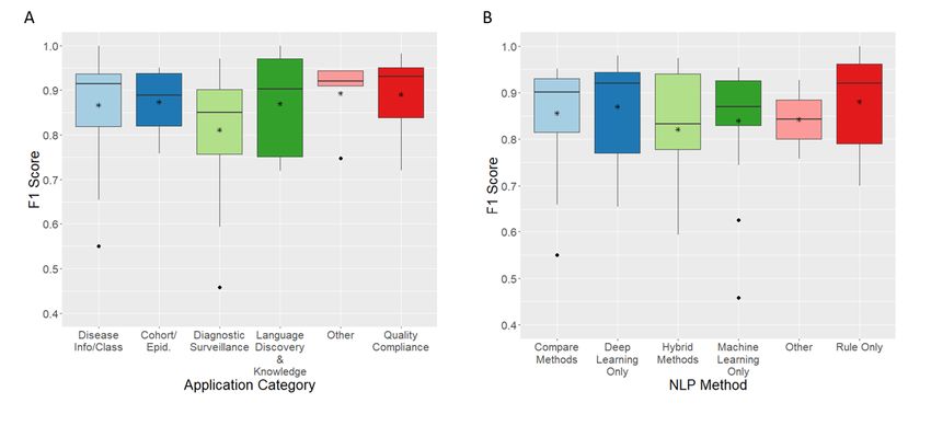
are important if the domain is to move applications into clinical use. More could
      be done to share code enabling validation of methods on different institutional data
      and to reduce heterogeneity in reporting of study properties allowing inter-study
      comparisons. Our results have significance for researchers in the field providing a
      systematic synthesis of existing work to build on, identify gaps, opportunities for
      collaboration and avoid duplication.

Background
Medical imaging examinations interpreted by radiologists in the form of narrative reports
are used to support and confirm diagnosis in clinical practice. Being able to accurately
and quickly identify the information stored in radiologists’ narratives has the potential
to reduce workloads, support clinicians in their decision processes, triage patients to get
urgent care or identify patients for research purposes. However, whilst these reports are
generally considered more restricted in vocabulary than other electronic health records
(EHR), e.g. clinical notes, it is still difficult to access this efficiently at scale [Bates et al.,
2016]. This is due to the unstructured nature of these reports and Natural Language
Processing (NLP) is key to obtaining structured information from radiology reports
[Pons et al., 2016].
    NLP applied to radiology reports is shown to be a growing field in earlier reviews
[Pons et al., 2016, Cai et al., 2016]. In recent years there has been an even more extensive
growth in NLP research in general and in particular deep learning methods which is not
seen in the earlier reviews. A more recent review of NLP applied to radiology-related
research can be found but this focuses on one NLP technique only, deep learning models
[Sorin et al., 2020]. Our paper provides a more comprehensive review comparing and
contrasting all NLP methodologies as they are applied to radiology.
    It is of significance to understand and synthesise recent developments specific to
NLP in the radiology research field as this will assist researchers to gain a broader
understanding of the field, provide insight into methods and techniques supporting and
promoting new developments in the field. Therefore, we carry out a systematic review
of research output on NLP applications in radiology from 2015 onward, thus, allowing
for a more up to date analysis of the area. An additional listing of our synthesis of
publications detailing their clinical and technical categories along with anatomical scan
regions can be made available on request. Also different to the existing work, we look
at both the clinical application areas NLP is being applied in and consider the trends in
NLP methods. We describe and discuss study properties, e.g. data size, performance,
annotation details, quantifying these in relation to both the clinical application areas
and NLP methods. Having a more detailed understanding of these properties allows
us to make recommendations for future NLP research applied to radiology datasets,
supporting improvements and progress in this domain.

                                                 2
Related Work
Amongst pre-existing reviews in this area, [Pons et al., 2016] was the first that was both
specific to NLP on radiology reports and systematic in methodology. Their literature
search identified 67 studies published in the period up to October 2014. They examined
the NLP methods used, summarised their performance and extracted the studies’ clinical
applications, which they assigned to five broad categories delineating their purpose.
Since Pons et al.’s paper, several reviews have emerged with the broader remit of NLP
applied to electronic health data, which includes radiology reports. [Kreimeyer et al.,
2017] conducted a systematic review of NLP systems with a specific focus on coding free
text into clinical terminologies and structured data capture. The systematic review by
[Spasic and Nenadic, 2020] specifically examined machine learning approaches to NLP
(2015-2019) in more general clinical text data, and a further methodical review was
carried out by [Wu et al., 2020] to synthesise literature on deep learning in clinical NLP
(up to April 2019) although the did not follow the PRISMA guideline completely. With
radiology reports as their particular focus, [Cai et al., 2016] published, the same year
as Pons et al.’s review, an instructive narrative review outlining the fundamentals of
NLP techniques applied in radiology. More recently, [Sorin et al., 2020] published a
systematic review focused on deep learning radiology-related research. They identified
10 relevant papers in their search (up to September 2019) and examined their deep
learning models, comparing these with traditional NLP models and also considered their
clinical applications but did not employ a specific categorisation. We build on this
corpus of related work, and most specifically Pons et al.’s work. In our initial synthesis
of clinical applications we adopt their application categories and further expand upon
these to reflect the nature of subsequent literature captured in our work. Additionally,
we quantify and compare properties of the studies reviewed and provide a series of
recommendations for future NLP research applied to radiology datasets in order to
promote improvements and progress in this domain.
Methods
Our methodology followed the Preferred Reporting Items for Systematic Reviews and
Meta-Analysis (PRISMA) [Moher et al., 2015], and the protocol is registered on proto-
cols.io.
Eligibility for Literature Inclusion and Search Strategy
We included studies using NLP on radiology reports of any imaging modality and
anatomical region for NLP technical development, clinical support, or epidemiological
research. Exclusion criteria included: (1) case reports; (2) published before 2015; (3) in
language other than English; (4) processing of radiology images; (5) reviews, conference
abstracts, comments, patents, or editorials; (6) not reporting outcomes of interest; (7)
not radiology reports; (8) not using NLP methods; (9) not available in full text; (10)
duplicates.
3We used Publish or Perish [Harzing A. W., 2007], a citation retrieval and analysis
software program, to search Google Scholar. Google Scholar has a similar coverage to
other databases [Gehanno et al., 2013] and is easier to integrate into search pipelines.
We conducted an initial pilot search following the process described here, but the search
terms were too specific and restricted the number of publications. However, we did
include papers found in the pilot search in full-text screening. We use the following
search query restricted to research articles published in English between January 2015
and October 2019. (”radiology” OR ”radiologist”) AND (”natural language” OR ”text
mining” OR ”information extraction” OR ”document classification” OR ”word2vec”)
NOT patent. We automated the addition of publication metadata and applied filtering
to remove irrelevant publications. These automated steps are described in Table 1 &
Table 2.
Table 1: Metadata enriching steps undertaken for each publication
Metadata Enriching Steps
1. Match the paper with its DOI via the Crossref API
2. If DOI matched, check Semantic Scholar for metadata/abstract
3. If no DOI match and no abstract, search PubMed for abstract
4. Search arXiv(for a pre-print)
5. If no PDF link, search Unpaywall for available open access versions
6. If PDF but no separate abstract via Semantics Scholar/PubMed, extract abstract from the PDF
Table 2: Automated filtering steps to remove irrelevant publications
Automated Filtering Steps
1. Document language is English
2. Word ’patent’ in title or URL
3. Year of publication out of range (cussed disagreements. Fleiss’ kappa [Fleiss, 1971] agreement between reviewers was 0.70,
indicating substantial agreement [Landis and Koch, 1977]. After this screening process,
each full-text article was reviewed by a team of eight (six NLP researchers and two epi-
demiologists) and double reviewed by a NLP researcher. We resolved any discrepancies
by discussion in regular meetings.
Data Extraction for Analysis
We extracted data on: primary clinical application and technical objective, data source(s),
study period, radiology report language, anatomical region, imaging modality, disease
area, dataset size, annotated set size, training/validation/test set size, external valida-
tion performed, domain expert used, number of annotators, inter-annotator agreement,
NLP technique(s) used, best-reported results (recall, precision and F1 score), availability
of dataset, and availability of code.
Results
The literature search yielded 4,799 possibly relevant publications from which our auto-
mated exclusion process removed 4,402, and during both our screening processes, 233
were removed, leaving 164 publications. See Figure 1 for details of exclusions at each
step.
General Characteristics
2015 and 2016 saw similar numbers of publications retrieved (22 and 21 respectively)
with the volume increasing almost three-fold in 2019 (55), noting 2019 only covers 10
months (Figure 2). Imaging modality (Table 3) varied considerably and 38 studies
used reports from multiple modalities. Of studies focusing on a single modality, the
most featured were CT scans (36) followed by MRI (16), X-Ray (18), Mammogram (5)
and Ultrasound (4). Forty-seven studies did not specifying scan modality. For the study
samples (Table 4), 33 papers specified that they used consecutive patient images, 38 used
non-consecutive image sampling and 93 did not clearly specify their sampling strategy.
The anatomical regions for scans varied (Table 5) with mixed being the highest followed
by Thorax and Head/neck. Disease categories are presented in Table 6 with the largest
disease category being Oncology. The majority of reports were in English (142) and a
small number in other languages e.g., Chinese (5), Spanish (4), German (3) (Table 7).
Clinical Application Categories
In synthesis of the literature each publication was classified by the primary clinical pur-
pose. Pons’ work in 2016 categorised publications into 5 broad categories: Diagnostic
Surveillance, Cohort Building for Epidemiological Studies, Query-based Case Retrieval,
Quality Assessment of Radiological Practice and Clinical Support Services. We found
5Figure 1: PRISMA diagram for search publication retrieval
Table 3: Scan modality Table 4: Image sampling method
Scan Modality No. Studies Sampling Method No. Studies
Multiple Modalities 38 Consecutive Images 33
MRI 16 Non-Consecutive Images 38
CT 36 Not specified 93
X-Ray 18 TOTAL 164
Mammogram 5
Ultrasound 4
Not specified 47
TOTAL 164
6Table 5: Anatomical region scanned Table 6: Disease category
Anatomical Region No. Studies Disease Category No. Studies
Mixed 45 Not specific disease related 40
Thorax 31 Oncology 39
Head/Neck 25 Various 20
Abdomen 15 Musculoskeletal 10
Breast 15 Cerebrovascular 13
Extremities 8 Other 13
Spine 5 Respiratory 10
Other 1 Trauma 7
Unspecified 19 Cardiovascular 6
TOTAL 164 Gastrointestinal 3
Hepatobiliary 2
Genitourinary 1
TOTAL 164
Table 7: Radiology report language
Report Language No. Studies
English 142
Chinese 5
Spanish 4
German 3
Italian 2
French 2
Hebrew 1
Polish 1
Brazilian Portuguese 1
Unspecified 3
TOTAL 164
some changes in this categorisation schema and our categorisation consisted of six cat-
egories: Diagnostic Surveillance, Disease information and classification, Quality Com-
pliance, Cohort/Epidemiology, Language Discovery and Knowledge Structure, Technical
NLP. The main difference is we found no evidence for a category of Clinical Support Ser-
vices which described applications that had been integrated into the workflow to assist.
Despite the increase in the number of publications, very few were in clinical use with
more focus on the category of Disease Information and Classification. We describe each
clinical application area in more detail below and where applicable how our categories
differ from the earlier findings. A listing of all publications and their corresponding clin-
ical application category can be made available on request. Table 8 shows the clinical
application category by the technical classification and Figure 2 shows the breakdown
7Table 8: Clinical Application Category by Technical Objective
Application Information Report/ Lexicon/ Clustering
Category Extraction Sentence Ontol- (n=1)
(n=81) Classifi- ogy
cation Discov-
(n=73) ery
(n=9)
Disease Information & Classification 14 31 - -
Diagnostic Surveillance 28 17 - -
Quality Compliance 7 14 - -
Cohort-Epid. 6 10 - -
Language Discovery & Knowledge 13 4 9 1
Technical NLP 6 4 - -
of clinical application category by publication year. There were more publications in
2019 compared with 2015 for all categories except Language Discovery & Knowledge
Structure, which fell by ≈ 25% (Figure 2).
Figure 2: Clinical application of publication by year
8Diagnostic Surveillance
A large proportion of studies in this category focused on extracting disease information
for patient or disease surveillance e.g. investigating tumour characteristics [Peng et al.,
2019, Bozkurt et al., 2019]; changes over time [Hassanpour et al., 2017] and worsen-
ing/progression or improvement/response to treatment [Kehl et al., 2019, Chen et al.,
2018]; identifying correct anatomical labels [Cotik et al., 2018]; organ measurements and
temporality [Sevenster et al., 2015a]. Studies also investigated pairing measurements
between reports [Sevenster et al., 2015b] and linking reports to monitoring changes
through providing an integrated view of consecutive examinations [Oberkampf et al.,
2016]. Studies focused specifically on breast imaging findings investigating aspects, such
as BI-RADS MRI descriptors (shape, size, margin) and final assessment categories (be-
nign, malignant etc.) e.g., [Liu et al., 2019, Gupta et al., 2018, Castro et al., 2017, Short
et al., 2019, Lacson et al., 2017, 2015]. Studies focused on tumour information e.g., for
liver [Yim et al., 2016b] and hepatocellular carcinoma (HPC) [Yim et al., 2017, 2016a]
and one study on extracting information relevant for structuring subdural haematoma
characteristics in reports [Pruitt et al., 2019].
Studies in this category also investigated incidental findings including on lung imag-
ing [Farjah et al., 2016, Karunakaran et al., 2017, Tan et al., 2018], with [Farjah et al.,
2016] additionally extracting the nodule size; for trauma patients [Trivedi et al., 2019];
and looking for silent brain infarction and white matter disease [Fu et al., 2019]. Other
studies focused on prioritising/triaging reports, detecting follow-up recommendations,
and linking a follow-up exam to the initial recommendation report, or bio-surveillance
of infectious conditions, such as invasive mould disease.
Disease Information and Classification
Disease Information and Classification publications use reports to identify information
that may be aggregated according to classification systems. These publications focused
solely on classifying a disease occurrence or extracting information about a disease with
no focus on the overall clinical application. This category was not found in Pons’ work.
Methods considered a range of conditions including intracranial haemorrhage [Jnawali
et al., 2019, Banerjee et al., 2017], aneurysms [Klos et al., 2018], brain metastases [Desh-
mukh et al., 2019], ischaemic stroke [Kim et al., 2019, Garg et al., 2019], and several
classified on types and severity of conditions e.g., [Deshmukh et al., 2019, Shin et al.,
2017, Wheater et al., 2019, Gorinski et al., 2019, Alex et al., 2019]. Studies focused
on breast imaging considered aspects such as predicting lesion malignancy from BI-
RADS descriptors [Bozkurt et al., 2016], breast cancer subtypes [Patel et al., 2017],
and extracting or inferring BI-RADS categories, such as [Banerjee et al., 2019a, Miao
et al., 2018]. Two studies focused on abdominal images and hepatocellular carcinoma
(HCC) staging and CLIP scoring. Chest imaging reports were used to detect pulmonary
embolism e.g., [Dunne et al., 2015, Banerjee et al., 2019b, Chen et al., 2017], bacterial
pneumonia [Meystre et al., 2017], and Lungs-RADS categories [Beyer et al., 2017]. Func-
tional imaging was also included, such as echocardiograms, extracting measurements to
9evaluate heart failure, including left ventricular ejection fractions (LVEF). Other studies
investigated classification of fractures and abnormalities and the prediction of ICD codes
from imaging reports.
Language Discovery and Knowledge Structure
Language Discovery and Knowledge Structure publications investigate the structure of
language in reports and how this might be optimised to facilitate decision support and
communication. Pons et al. reported on applications of Query-based retrieval which has
similarities to Language Discovery and Knowledge Structure but it is not the same. Their
category contains studies that retrieve cases and conditions that are not predefined and
in some instances could be used for research purposes or are motivated for educational
purposes. Our category is broader and encompasses papers that investigated different
aspects of language including variability, complexity simplification and normalising to
support extraction and classification tasks.
Studies focus on exploring lexicon coverage and methods to support language simpli-
fication for patients looking at sources, such as the consumer health vocabulary [Qenam
et al., 2017] and the French lexical network (JDM) [Lafourcade and Ramadier, 2017].
Other works studied the variability and complexity of report language comparing free-
text and structured reports and radiologists. Also investigated was how ontologies and
lexicons could be combined with other NLP methods to represent knowledge that can
support clinicians. This work included improving report reading efficiency [Hong and
Zhang, 2015]; finding similar reports [Comelli et al., 2015]; normalising phrases to sup-
port classification and extraction tasks, such as entity recognition in Spanish reports
[Cotik et al., 2015]; imputing semantic classes for labelling [Johnson et al., 2015], sup-
porting search [Mujjiga et al., 2019] or to discover semantic relations [Lafourcade and
Ramadier, 2016].
Quality and Compliance
Quality and Compliance publications use reports to assess the quality and safety of
practice and reports similar to Pons’ category. Works considered how patient indica-
tions for scans adhered to guidance e.g., [Shelmerdine et al., 2019, Mabotuwana et al.,
2018b, Dalal et al., 2020, Bobbin et al., 2017, Kwan et al., 2019, Mabotuwana et al.,
2018a] or protocol selection [Brown and Marotta, 2017, Trivedi et al., 2018, Zhang et al.,
2018, Brown and Marotta, 2018, Yan et al., 2016] or the impact of guideline changes
on practice, such as [Kang et al., 2019]. Also investigated was diagnostic utilisation
and yield, based on clinicians or on patients, which can be useful for hospital planning
and for clinicians to study their work patterns e.g.[Brown and Kachura, 2019]. Other
studies in this category looked at specific aspects of quality, such as, classification for
long bone fractures to support quality improvement in paediatric medicine [Grundmeier
et al., 2016], automatic identification of reports that have critical findings for auditing
purposes [Heilbrun et al., 2019], deriving a query-based quality measure to compare
structured and free-text report variability [Maros et al., 2018], and [Minn et al., 2015]
10who describe a method to fix errors in gender or laterality in a report.
Cohort and Epidemiology
This category is similar to Pons’ earlier review but we treated the studies in this category
slightly attempting to differentiate which papers described methods for creating cohorts
for research purposes, and those which also reported the outcomes of an epidemiologi-
cal analysis. Ten studies use NLP to create specific cohorts for research purposes and
six reported the performance of their tools. Out of these papers, the majority (n=8)
created cohorts for specific medical conditions including fatty liver disease [Goldshtein
et al., 2020, Redman et al., 2017] hepatocellular cancer [Sada et al., 2016], ureteric
stones [Li and Elliot, 2019], vertebral facture [Tan and Heagerty, 2019], traumatic brain
injury [Yadav et al., 2016, Mahan et al., 2019], and leptomeningeal disease secondary to
metastatic breast cancer [Brizzi et al., 2019]. Five papers identified cohorts focused on
particular radiology findings including ground glass opacities (GGO) [Van Haren et al.,
2019], cerebral microbleeds (CMB) [Noorbakhsh-Sabet et al., 2018], pulmonary nodules
[Gould et al., 2015], [Huhdanpaa et al., 2018], changes in the spine correlated to back
pain [Bates et al., 2016] and identifying radiological evidence of people having suffered
a fall. One paper focused on identifying abnormalities of specific anatomical regions of
the ear within an audiology imaging database [Masino et al., 2016] and another paper
aimed to create a cohort of people with any rare disease (within existing ontologies - Or-
phanet Rare Disease Ontology ORDO and Radiology Gamuts Ontology RGO). Lastly,
one paper took a different approach of screening reports to create a cohort of people with
contraindications for MRI, seeking to prevent iatrogenic events. Amongst the epidemiol-
ogy studies there were various analytical aims, but they primarily focused on estimating
the prevalence or incidence of conditions or imaging findings and looking for associations
of these conditions/findings with specific population demographics, associated factors or
comorbidities. The focus of one study differed in that it applied NLP to healthcare
evaluation, investigating the association of palliative care consultations and measures of
high-quality end-of-life (EOL) care [Brizzi et al., 2019].
Technical NLP
This category is for publications that have a primary technical aim that is not focused
on radiology report outcome, e.g. detecting negation in reports, spelling correction [Zech
et al., 2019], fact checking [Zhang et al., 2019, Steinkamp et al., 2019] methods for sample
selection, crowd source annotation [Cocos et al., 2017]. This category did not occur in
Pons’ earlier review.
NLP Methods in Use
NLP methods capture the different techniques an author applied broken down into rules,
machine learning methods, deep learning, ontologies, lexicons and word embeddings.
11We discriminate machine learning from deep learning, using the former to represent
traditional machine learning methods.
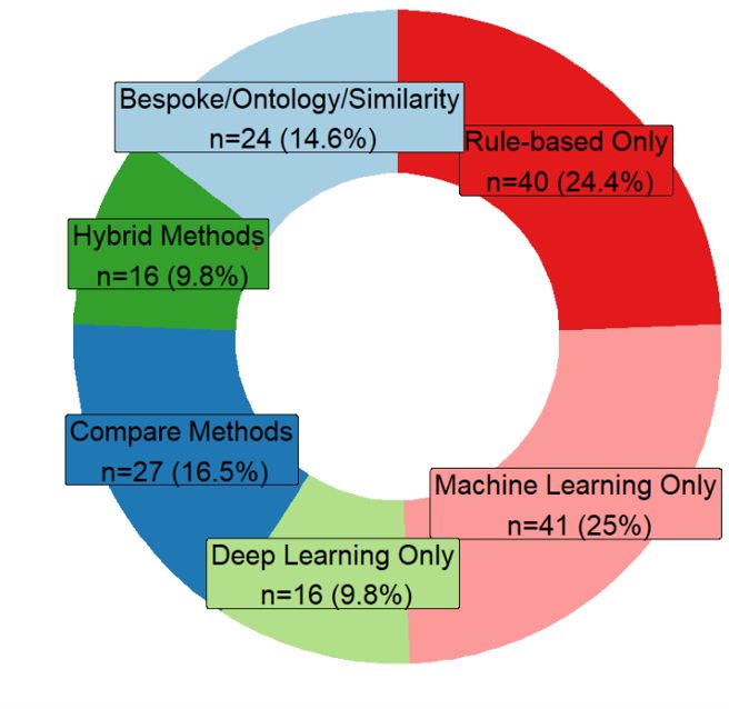
Over half of the studies only applied one type of NLP method and just over a quarter
of the studies compared or combined methods in hybrid approaches. The remaining
studies either used a bespoke proprietary system or focus on building ontologies or
similarity measures (Figure 3). Rule-based method use remains almost constant across
the period, whereas use of machine learning decreases and deep learning methods rises,
from five publications in 2017 to twenty-four publications in 2019 (Figure 4).
Figure 3: NLP method breakdown
Table 9: Breakdown of NLP Method
ML (n=74) No studies Deep Learning (n=36) No studies
SVM 34 RNN variants 14
Logistic Regression 23 CNN 10
Random Forest 18 Other 5
Naı̈ve Bayes 17 Compare CNN, RNN 4
Maximum Entropy 7 Combine CNN+RNN 3
Decision Trees 4
A variety of machine classifier algorithms were used, with SVM and Logistic Regres-
sion being the most common (Table 9). Recurrent Neural Networks (RNN) variants were
the most common type of deep learning architectures. RNN methods were split between
long short-term memory (LSTM) and bidirectional-LSTM (Bi-LSTM), bi-directional
12Figure 4: NLP method by year
gated recurrent unit (Bi-GRU), and standard RNN approaches. Four of these studies
additionally added a Conditional Random Field (CRF) for the final label generation
step. Convolutional Neural Networks (CNN) were the second most common architec-
ture explored. Eight studies additionally used an attention mechanism as part of their
deep learning architecture. Other neural approaches included feed-forward neural net-
works, fully connected neural networks and a proprietary neural system IBM Watson
[Trivedi et al., 2018] and Snorkel [Ratner et al., 2018]. Several studies proposed combined
architectures, such as [Zhu et al., 2019, Short et al., 2019].
NLP Method Features
Most rule-based and machine classifying approaches used features based on bag-of-words,
part-of-speech, term frequency, and phrases with only two studies alternatively using
word embeddings. Three studies use feature engineering with deep learning rather than
word embeddings. Thirty-three studies use domain-knowledge to support building fea-
tures for their methods, such as developing lexicons or selecting terms and phrases.
Comparison of embedding methods is difficult as many studies did not describe their
embedding method. Of those that did, Word2Vec [Mikolov et al., 2013] was the most
popular (n=19), followed by GLOVE embeddings [Pennington et al., 2014] (n=6), Fast-
Text [Mikolov et al., 2018] (n=3), ELMo [Peters et al., 2018] (n=1) and BERT [Devlin
et al., 2018] (n=1). Ontologies or lexicon look-ups are used in 100 studies; however, even
though publications increase over the period in real terms, 20% fewer studies employ the
13use of ontologies or lexicons in 2019 compared to 2015. The most widely used resources
were UMLS [National Library of Medicine, 2021b] (n=15), Radlex [RSNA, 2021] (n=20),
SNOMED-CT [National Library of Medicine, 2021a] (n=14). Most studies used these as
features for normalising words and phrases for classification, but this was mainly those
using rule-based or machine learning classifiers with only six studies using ontologies
as input to their deep learning architecture. Three of those investigated how existing
ontologies can be combined with word embeddings to create domain-specific mappings,
with authors pointing to this avoiding the need for large amounts of annotated data.
Other approaches looked to extend existing medical resources using a frequent phrases
approach, e.g. [Bulu et al., 2018]. Works also used the derived concepts and relations
visualising these to support activities, such as report reading and report querying (e.g.
[Hassanpour and Langlotz, 2016, Zhao et al., 2018])
Annotation and Inter-Annotator Agreement
Eighty-nine studies used at least two annotators, 75 did not specify any annotation
details, and only one study used a single annotator. Whilst 69 studies use a domain ex-
pert for annotation (a clinician or radiologist) only 56 studies report the inter-annotator
agreement. Some studies mention annotation but do not report on agreement or annota-
tors. Inter-annotator agreement values for Kappa range from 0.43 to perfect agreement
at 1. Whilst most studies reported agreement by Cohen’s Kappa [Cohen, 1960] some
reported precision, and percent agreement. Studies reported annotation data sizes differ-
ently, e.g., on the sentence or patient level. Studies also considered ground truth labels
from coding schemes such as ICD or BI-RADS categories as annotated data. Of studies
which detailed human annotation at the radiology report level, only 45 specified inter-
annotator agreement and/or the number of annotators. Annotated report numbers for
these studies varies with 15 papers having annotated less than 500, 12 having annotated
between 500 and less than 1,000, 15 between 1,000 and less than 3,000, and 3 between
4,000 and 8,288 reports.
Data Sources and Availability
Only 14 studies reported that their data is available, and 15 studies reported that their
code is available. Most studies sourced their data from medical institutions, a number
of studies did not specify where their data was from, and some studies used publicly
available datasets: MIMIC-III (n=5), MIMIC-II (n=1), MIMIC-CXR (n=1); Radcore
(n=5) or STRIDE (n=2). Four studies used combined electronic health records such as
clinical notes or pathology reports.
Reporting on data size and splits differed across studies with some not giving exact
data sizes and others reporting numbers of sentences, patients, or mixed data sources
rather than radiology reports. Data sizes for those reporting at the radiology report
level is n=135 or 82.32% of the studies (Table 10). The biggest variation of data size
by NLP Method is in studies that apply other methods or are rule-based. Machine
learning also varies in size; however, the median value is lower compared to rule-based
14Table 10: NLP Method by data size properties, minimum data size, maximum data size and median value, studies reporting in numbers of radiology reports NLP Method Min Size Max Size Median Compare Methods 513 2,167,445 2,845 Hybrid Methods 40 34,926 918 Deep Learning (Only) 120 1,567,581 5,000 Machine Learning (Only) 101 2,977,739 2,531 Rules (Only) 31 10,000,000 8,000 Other 25 12,377,743 10,000 Table 11: Grouped data size and number of studies in each group, only for studies reporting in numbers of radiology reports Data Size Group No. Studies (%)
testing on these comparisons. Issues of heterogeneity make it difficult and unrealistic to
compare performance between methods applied, hence, we use summary measures as a
broad overview (Figure 5). Performance reported varies, but both the mean and median
values for the F1 score appear higher for methods using rule-based only or deep learning
only methods. Whilst differences are less discernible between F1 scores for application
areas, Diagnostic Surveillance looks on average lower than other categories.

Figure 5: Application Category and NLP Method, Mean and Median Summaries. Mean
value is indicated by a vertical bar, the box shows error bars and the asterisk is the
median value.

Discussion and Future Directions
Our work shows there has been a considerable increase in the number of publications
using NLP on radiology reports over the recent time period. Compared to 67 publications
retrieved in the earlier review of [Pons et al., 2016], we retrieved 164 publications. In
this section we discuss and offer some insight into the observations and trends of how
NLP is being applied to radiology and make some recommendations that may benefit
the field going forward.

Clinical Applications and NLP Methods in Radiology
The clinical applications of the publications is similar to the earlier review of Pons et al.
but whilst we observe an increase in research output we also highlight that there appears
to be even less focus on clinical application compared to their review. Like many other
fields applying NLP the use of deep learning has increased, with RNN architectures
being the most popular. This is also observed in a review of NLP in clinical text[Wu

                                             16
et al., 2020]. However, although deep learning use increases, rules and traditional ma-
chine classifiers are still prevalent and often used as baselines to compare deep learning
architectures against. One reason for traditional methods remaining popular is their in-
terpretability compared to deep learning models. Understanding the features that drive
a model prediction can support decision-making in the clinical domain but the complex
layers of non-linear data transformations deep learning is composed of does not easily
support transparency [Shickel et al., 2018]. This may also help explain why in synthesis
of the literature we observed less focus on discussing clinical application and more em-
phasis on disease classification or information task only. Advances in interpretability of
deep learning models are critical to its adoption in clinical practice.
Other challenges exist for deep learning such as only having access to small or im-
balanced datasets. Chen et al. [Chen et al., 2019] review deep learning methods within
healthcare and point to these challenges resulting in poor performance but that these
same datasets can perform well with traditional machine learning methods. We found
several studies highlight this and when data is scarce or datasets imbalanced, they intro-
duced hybrid approaches of rules and deep learning to improve performance, particularly
in the Diagnostic Surveillance category. Yang et al. [Yang et al., 2018] observed rules
performing better for some entity types, such as time and size, which are proportionally
lower than some of the other entities in their train and test sets; hence they combine a
bidirectional-LSTM and CRF with rules for entity recognition. Peng et al. [Peng et al.,
2019] comment that combining rules and the neural architecture complement each other,
with deep learning being more balanced between precision and recall, but the rule-based
method having higher precision and lower recall. The authors reason that this provides
better performance as rules can capture rare disease cases, particularly when multi-class
labelling is needed, whilst deep learning architectures perform worse in instances with
fewer data points.
In addition to its need for large-scale data, deep learning can be computationally
costly. The use of pre-trained models and embeddings may alleviate some of this burden.
Pre-trained models often only require fine-tuning, which can reduce computation cost.
Language comprehension pre-learned from other tasks can then be inherited from the
parent models, meaning fewer domain-specific labelled examples may be needed [Wood
et al., 2020]. This use of pre-trained information also supports generalisability, e.g.,
[Banerjee et al., 2019b] show that their model trained on one dataset can generalise to
other institutional datasets.
Embedding use has increased which is expected with the application of deep learn-
ing approaches but many rule-based and machine classifiers continue to use traditional
count-based features, e.g., bag-of-words and n-grams. Recent evidence [Ong et al.,
2020] suggests that the trend to continue to use feature engineering with traditional
machine learning methods does produce better performance in radiology reports than
using domain-specific word embeddings.
Banerjee et al. [Banerjee et al., 2017] found that there was not much difference be-
tween a uni-gram approach and a Word2vec embedding, hypothesising this was due to
their narrow domain, intracranial haemorrhage. However, the NLP research field has
17seen a move towards bi-directional encoder representations from transformers (BERT)
based embedding models not reflected in our analysis, with only one study using BERT
generated embeddings [Deshmukh et al., 2019]. Embeddings from BERT are thought to
be superior as they can deliver better contextual representations and result in improved
task performance. Whilst more publications since our review period have used BERT
based embeddings with radiology reports e.g. [Wood et al., 2020, Smit et al., 2020a]
not all outperform traditional methods [Grivas et al., 2020]. Recent evidence shows that
embeddings generated by BERT fail to show a generalisable understanding of negation
[Ettinger, 2020], an essential factor in interpreting radiology reports effectively. Spe-
cialised BERT models have been introduced such as ClinicalBERT [Alsentzer et al.,
2019] or BlueBERT [Smit et al., 2020a]. BlueBERT has been shown to outperform Clin-
icalBERT when considering chest radiology [Smit et al., 2020b] but more exploration
of the performance gains versus the benefits of generalisability are needed for radiology
text.
All NLP models have in common that they need large amounts of labelled data for
model training [Yasaka and Abe, 2018]. Several studies [Percha et al., 2018, Tahmasebi
et al., 2019, Banerjee et al., 2018] explored combining word embeddings and ontologies
to create domain-specific mappings, and they suggest this can avoid a need for large
amounts of annotated data. Additionally, [Percha et al., 2018, Tahmasebi et al., 2019]
highlight that such combinations could boost coverage and performance compared to
more conventional techniques for concept normalisation.
The number of publications using medical lexical knowledge resources is still rel-
atively low, even though a recent trend in the general NLP field is to enhance deep
learning with external knowledge [Young et al., 2018]. This was also observed by [Wu
et al., 2020], where only 18% of the deep learning studies in their review utilised knowl-
edge resources. Although pre-training supports learning previously known facts it could
introduce unwanted bias, hindering performance. The inclusion of domain expertise
through resources such as medical lexical knowledge may help reduce this unwanted bias
[Wu et al., 2020]. Exploration of how this domain expertise can be incorporated with
deep learning architectures in future could improve the performance when having access
to less labelled data.
Task Knowledge
Knowledge about the disease area of interest and how aspects of this disease are lin-
guistically expressed is useful and could promote better performing solutions. Whilst
[Donnelly et al., 2019] find high variability between radiologists, with metric values
(e.g. number of syntactic, clinical terms based on ontology mapping) being significantly
greater on free-text than structured reports, [Xie et al., 2019] who look specifically
at anatomical areas find less evidence for variability. Zech et al. [Zech et al., 2018]
suggest that the highly specialised nature of each imaging modality creates different
sub-languages and the ability to discover these labels (i.e. disease mentions) reflects the
consistency with which labels are referred to. For example, edema is referred to very con-
sistently whereas other labels are not, such as infarction/ischaemic. Understanding the
18language and the context of entity mentions could help promote novel ideas on how to
solve problems more effectively. For example, [Yim et al., 2017] discuss how the accuracy
of predicting malignancy is affected by cues being outside their window of consideration
and [Yim et al., 2018] observe problems of co-reference resolution within a report due
to long-range dependencies. Both these studies use traditional NLP approaches, but
we observed novel neural architectures being proposed to improve performance in sim-
ilar tasks specifically capturing long-range context and dependency learning, e.g., [Zhu
et al., 2019, Short et al., 2019]. This understanding requires close cooperation of health-
care professionals and data scientists, which is different to some other fields where more
disconnection is present [Chen et al., 2019].
Study Heterogeneity, a Need for Reporting Standards
Most studies reviewed could be described as a proof-of-concept and not trialled in a clin-
ical setting. Pons et al. [Pons et al., 2016] hypothesised that a lack of clinical application
may stem from uncertainty around minimal performance requirements hampering imple-
mentations, evidence-based practice requiring justification and transparency of decisions,
and the inability to be able to compare to human performance as the human agreement
is often an unknown. These hypotheses are still valid, and we see little evidence that
these problems are solved.
Human annotation is generally considered the gold standard at measuring human
performance, and whilst many studies reported that they used annotated data, overall,
reporting was inconsistent. Steps were undertaken to measure inter-annotator agree-
ment (IAA), but in many studies, this was not directly comparable to the evaluation
undertaken of the NLP methods. The size of the data being used to draw experimental
conclusions from is important and accurate reporting of these measures is essential to
ensure reproducibility and comparison in further studies. Reporting on the training,
test and validation splits was varied with some studies not giving details and not using
held-out validation sets.
Most studies use retrospective data from single institutions but this can lead to a
model over-fitting and, thus, not generalising well when applied in a new setting. Over-
coming the problem of data availability is challenging due to privacy and ethics concerns,
but essential to ensure that performance of models can be investigated across institu-
tions, modalities, and methods. Availability of data would allow for agreed benchmarks
to be developed within the field that algorithm improvements can be measured upon.
External validation of applied methods was extremely low, although, this is likely due to
the availability of external datasets. Making code available would enable researchers to
report how external systems perform on their data. However, only 15 studies reported
that their code is available. To be able to compare systems there is a need for common
datasets to be available to benchmark and compare systems against.
Whilst reported figures in precision and recall generally look high more evidence is
needed for accurate comparison to human performance. A wide variety of performance
measures were used, with some studies only reporting one measure, e.g., accuracy or
F1 scores, with these likely representing the best performance obtained. Individual
19studies are often not directly comparable for such measures, but none-the-less clarity
and consistency in reporting is desirable. Many studies making model comparisons did
not carry out any significance testing for these comparisons.
The make the following recommendations to help move the field forward, enable more
inter-study comparisons, and increase study reproducibility:
1. Clarity in reporting study properties is required: (a) Data characteristics including
size and the type of dataset should be detailed, e.g., the number of reports, sen-
tences, patients, and if patients how many reports per patient. The training, test
and validation data split should be evident, as should the source of the data. (b)
Annotation characteristics including the methodology to develop the annotation
should be reported, e.g., annotation set size, annotator details, how many, exper-
tise. (c) Performance metrics should include a range of metrics: precision, recall,
F1, accuracy and not just one overall value.
2. Significance testing should be carried out when a comparison between methods is
made.
3. Data and code availability are encouraged. While making data available will often
be challenging due to privacy concerns, researchers should make code available to
enable inter-study comparisons and external validation of methods.
4. Common datasets should be used to benchmark and compare systems.
Limitations of Study
Publication search is subject to bias in search methods and it is likely that our search
strategy did inevitably miss some publications. Whilst trying to be precise and objec-
tive during our review process some of the data collected and categorising publications
into categories was difficult to agree on and was subjective. For example, many of the
publications could have belonged to more than one category. One of the reasons for this
was how diverse in structure the content was which was in some ways reflected by the
different domains papers were published in. It is also possible that certain keywords
were missed in recording data elements due to the reviewers own biases and research
experience.
Conclusions
This paper presents an systematic review of publications using NLP on radiology reports
during the period 2015 to October 2019. We show there has been substantial growth
in the field particularly in researchers using deep learning methods. Whilst deep learn-
ing use has increased, as seen in NLP research in general, it faces challenges of lower
performance when data is scarce or when labelled data is unavailable, and is not widely
used in clinical practice perhaps due to the difficulties in interpretability of such models.
Traditional machine learning and rule-based methods are, therefore, still widely in use.
20Exploration of domain expertise such as medial lexical knowledge must be explored fur-
ther to enhance performance when data is scarce. The clinical domain faces challenges
due to privacy and ethics in sharing data but overcoming this would enable development
of benchmarks to measure algorithm performance and test model robustness across in-
stitutions. Common agreed datasets to compare performance of tools against would help
support the community in inter-study comparisons and validation of systems. The work
we present here has the potential to inform researchers about applications of NLP to
radiology and to lead to more reliable and responsible research in the domain.
Acknowledgements
Not applicable
Funding
This research was supported by the Alan Turing Institute, MRC, HDR-UK and the Chief
Scientist Office. B.A.,A.C,D.D.,A.G. and C.G. have been supported by the Alan Turing
Institute via Turing Fellowships (B.A,C.G.) and Turing project funding (ESPRC grant
EP/N510129/1). A.G. was also funded by a MRC Mental Health Data Pathfinder Award
(MRC-MCPC17209). H.W. is MRC/Rutherford Fellow HRD UK (MR/S004149/1).
H.D. is supported by HDR UK National Phemomics Resource Project. V.S-P. is sup-
ported by the HDR UK National Text Analytics Implementation Project. W.W. is
supported by a Scottish Senior Clinical Fellowship (CAF/17/01).
Abbreviations
NLP - natural language processing
e.g. - example
ICD - international classification of diseases
BI-RADS - Breast Imaging-Reporting and Data System
IAA - inter-annotator agreement
No. - number
UMLS - unified medical language system
ELMo - embeddings from Language Models
BERT - bidirectional encoder representations form transformers
SVM - support vector machine
CNN - convolutional neural network
LSTM - long short-term memory
Bi-LSTM - bi-directional long short-term memory
Bi-GRU - bi-directional gated recurrent unit
CRF - conditional random field
21GLOVE - Global Vectors for Word Representation
Bibliography
Beatrice Alex, Claire Grover, Richard Tobin, Cathie Sudlow, Grant Mair, and William
Whiteley. Text mining brain imaging reports. Journal of Biomedical Semantics, 10
(1):23, November 2019. ISSN 2041-1480. doi: 10.1186/s13326-019-0211-7. URL
https://doi.org/10.1186/s13326-019-0211-7.
Emily Alsentzer, John Murphy, William Boag, Wei-Hung Weng, Di Jindi, Tristan Nau-
mann, and Matthew McDermott. Publicly Available Clinical BERT Embeddings. In
Proceedings of the 2nd Clinical Natural Language Processing Workshop, pages 72–78,
Minneapolis, Minnesota, USA, June 2019. Association for Computational Linguistics.
doi: 10.18653/v1/W19-1909. URL https://www.aclweb.org/anthology/W19-1909.
Imon Banerjee, Sriraman Madhavan, Roger Eric Goldman, and Daniel L. Rubin. Intel-
ligent Word Embeddings of Free-Text Radiology Reports. AMIA Annual Symposium
Proceedings, pages 411–420, 2017. ISSN 1942-597X. URL https://www.ncbi.nlm.
nih.gov/pmc/articles/PMC5977573/.
Imon Banerjee, Matthew C. Chen, Matthew P. Lungren, and Daniel L. Rubin. Radiology
report annotation using intelligent word embeddings: Applied to multi-institutional
chest CT cohort. Journal of Biomedical Informatics, 77:11–20, January 2018. ISSN
1532-0464. doi: 10.1016/j.jbi.2017.11.012. URL http://www.sciencedirect.com/
science/article/pii/S1532046417302575.
Imon Banerjee, Selen Bozkurt, Emel Alkim, Hersh Sagreiya, Allison W. Kurian, and
Daniel L. Rubin. Automatic inference of BI-RADS final assessment categories
from narrative mammography report findings. Journal of Biomedical Informatics,
92:103137, April 2019a. ISSN 1532-0464. doi: 10.1016/j.jbi.2019.103137. URL
http://www.sciencedirect.com/science/article/pii/S1532046419300553.
Imon Banerjee, Yuan Ling, Matthew C. Chen, Sadid A. Hasan, Curtis P. Langlotz,
Nathaniel Moradzadeh, Brian Chapman, Timothy Amrhein, David Mong, Daniel L.
Rubin, Oladimeji Farri, and Matthew P. Lungren. Comparative effectiveness of
convolutional neural network (CNN) and recurrent neural network (RNN) archi-
tectures for radiology text report classification. Artificial Intelligence in Medicine,
97:79–88, June 2019b. ISSN 0933-3657. doi: 10.1016/j.artmed.2018.11.004. URL
http://www.sciencedirect.com/science/article/pii/S0933365717306255.
Jonathan Bates, Samah J. Fodeh, Cynthia A. Brandt, and Julie A. Womack. Classifi-
cation of radiology reports for falls in an HIV study cohort. Journal of the Ameri-
can Medical Informatics Association, 23(e1):e113–e117, April 2016. ISSN 1067-5027.
doi: 10.1093/jamia/ocv155. URL https://academic.oup.com/jamia/article/23/
e1/e113/2379897.
22Sebastian E. Beyer, Brady J. McKee, Shawn M. Regis, Andrea B. McKee, Sebastian
Flacke, Gilan El Saadawi, and Christoph Wald. Automatic Lung-RADS™ classification
with a natural language processing system. Journal of Thoracic Disease, 9(9):3114–
3122, September 2017. ISSN 2072-1439. doi: 10.21037/jtd.2017.08.13. URL https:
//www.ncbi.nlm.nih.gov/pmc/articles/PMC5708435/.
Mark D. Bobbin, Ivan K. Ip, V. Anik Sahni, Atul B. Shinagare, and Ramin Kho-
rasani. Focal Cystic Pancreatic Lesion Follow-up Recommendations After Publica-
tion of ACR White Paper on Managing Incidental Findings. Journal of the Amer-
ican College of Radiology, 14(6):757–764, June 2017. ISSN 1546-1440. doi: 10.
1016/j.jacr.2017.01.044. URL http://www.sciencedirect.com/science/article/
pii/S1546144017301771.
Selen Bozkurt, Francisco Gimenez, Elizabeth S. Burnside, Kemal H. Gulkesen, and
Daniel L. Rubin. Using automatically extracted information from mammography
reports for decision-support. Journal of Biomedical Informatics, 62:224–231, Au-
gust 2016. ISSN 1532-0464. doi: 10.1016/j.jbi.2016.07.001. URL http://www.
sciencedirect.com/science/article/pii/S1532046416300557.
Selen Bozkurt, Emel Alkim, Imon Banerjee, and Daniel L. Rubin. Automated Detection
of Measurements and Their Descriptors in Radiology Reports Using a Hybrid Natural
Language Processing Algorithm. Journal of Digital Imaging, 32(4):544–553, August
2019. ISSN 1618-727X. doi: 10.1007/s10278-019-00237-9. URL https://doi.org/
10.1007/s10278-019-00237-9.
Simon Briscoe, Alison Bethel, and Morwenna Rogers. Conduct and reporting of citation
searching in Cochrane systematic reviews: A cross-sectional study. Research Synthesis
Methods, 11(2):169–180, 2020. ISSN 1759-2887. doi: 10.1002/jrsm.1355. URL https:
//onlinelibrary.wiley.com/doi/abs/10.1002/jrsm.1355.
Kate Brizzi, Sophia N. Zupanc, Brooks V. Udelsman, James A. Tulsky, Alexi A.
Wright, Hanneke Poort, and Charlotta Lindvall. Natural Language Processing to
Assess Palliative Care and End-of-Life Process Measures in Patients With Breast
Cancer With Leptomeningeal Disease. American Journal of Hospice and Palliative
Medicine, 37(5):371–376, 2019. doi: https://doi.org/10.1177/1049909119885585. URL
https://journals.sagepub.com/doi/abs/10.1177/1049909119885585.
A. D. Brown and J. R. Kachura. Natural Language Processing of Radiology Reports
in Patients With Hepatocellular Carcinoma to Predict Radiology Resource Utiliza-
tion. Journal of the American College of Radiology, 16(6):840–844, June 2019. ISSN
1546-1440. doi: 10.1016/j.jacr.2018.12.004. URL http://www.sciencedirect.com/
science/article/pii/S1546144018315539.
Andrew D. Brown and Thomas R. Marotta. A Natural Language Processing-
based Model to Automate MRI Brain Protocol Selection and Prioritization. Aca-
demic Radiology, 24(2):160–166, February 2017. ISSN 1076-6332. doi: 10.1016/
23j.acra.2016.09.013. URL http://www.sciencedirect.com/science/article/pii/
S1076633216303270.
Andrew D. Brown and Thomas R. Marotta. Using machine learning for sequence-
level automated MRI protocol selection in neuroradiology. Journal of the Amer-
ican Medical Informatics Association, 25(5):568–571, May 2018. ISSN 1067-5027.
doi: 10.1093/jamia/ocx125. URL https://academic.oup.com/jamia/article/25/
5/568/4569611.
Hakan Bulu, Dorothy A. Sippo, Janie M. Lee, Elizabeth S. Burnside, and Daniel L.
Rubin. Proposing New RadLex Terms by Analyzing Free-Text Mammography Re-
ports. Journal of Digital Imaging, 31(5):596–603, October 2018. ISSN 1618-727X. doi:
10.1007/s10278-018-0064-0. URL https://doi.org/10.1007/s10278-018-0064-0.
Tianrun Cai, Andreas A. Giannopoulos, Sheng Yu, Tatiana Kelil, Beth Ripley,
Kanako K. Kumamaru, Frank J. Rybicki, and Dimitrios Mitsouras. Natural Language
Processing Technologies in Radiology Research and Clinical Applications. Radio-
Graphics, 36(1):176–191, January 2016. ISSN 0271-5333. doi: 10.1148/rg.2016150080.
URL https://pubs.rsna.org/doi/full/10.1148/rg.2016150080.
Sergio M. Castro, Eugene Tseytlin, Olga Medvedeva, Kevin Mitchell, Shyam
Visweswaran, Tanja Bekhuis, and Rebecca S. Jacobson. Automated annotation
and classification of BI-RADS assessment from radiology reports. Journal of
Biomedical Informatics, 69:177–187, May 2017. ISSN 1532-0464. doi: 10.1016/
j.jbi.2017.04.011. URL http://www.sciencedirect.com/science/article/pii/
S1532046417300813.
David Chen, Sijia Liu, Paul Kingsbury, Sunghwan Sohn, Curtis B. Storlie, Elizabeth B.
Habermann, James M. Naessens, David W. Larson, and Hongfang Liu. Deep learning
and alternative learning strategies for retrospective real-world clinical data. npj Digital
Medicine, 2(1):1–5, May 2019. ISSN 2398-6352. doi: 10.1038/s41746-019-0122-0. URL
https://www.nature.com/articles/s41746-019-0122-0.
Matthew C. Chen, Robyn L. Ball, Lingyao Yang, Nathaniel Moradzadeh, Brian E. Chap-
man, David B. Larson, Curtis P. Langlotz, Timothy J. Amrhein, and Matthew P.
Lungren. Deep Learning to Classify Radiology Free-Text Reports. Radiology, 286(3):
845–852, November 2017. ISSN 0033-8419. doi: 10.1148/radiol.2017171115. URL
https://pubs.rsna.org/doi/full/10.1148/radiol.2017171115.
Po-Hao Chen, Hanna Zafar, Maya Galperin-Aizenberg, and Tessa Cook. Integrat-
ing Natural Language Processing and Machine Learning Algorithms to Categorize
Oncologic Response in Radiology Reports. Journal of Digital Imaging, 31(2):178–
184, April 2018. ISSN 1618-727X. doi: 10.1007/s10278-017-0027-x. URL https:
//doi.org/10.1007/s10278-017-0027-x.
24Anne Cocos, Ting Qian, Chris Callison-Burch, and Aaron J. Masino. Crowd con-
trol: Effectively utilizing unscreened crowd workers for biomedical data annota-
tion. Journal of Biomedical Informatics, 69:86–92, May 2017. ISSN 1532-0464.
doi: 10.1016/j.jbi.2017.04.003. URL http://www.sciencedirect.com/science/
article/pii/S1532046417300746.
Jacob Cohen. A Coefficient of Agreement for Nominal Scales. Educational and Psy-
chological Measurement, 20(1):37–46, April 1960. ISSN 0013-1644. doi: 10.1177/
001316446002000104. URL https://doi.org/10.1177/001316446002000104.
A. Comelli, L. Agnello, and S. Vitabile. An ontology-based retrieval system for mammo-
graphic reports. In 2015 IEEE Symposium on Computers and Communication (ISCC),
pages 1001–1006, Larnaca, July 2015. IEEE. doi: 10.1109/ISCC.2015.7405644.
Viviana Cotik, Dario Filippo, and Jose Castano. An Approach for Automatic Classifica-
tion of Radiology Reports in Spanish. Studies in Health Technology and Informatics,
216:634–638, jan 2015. ISSN 0926-9630, 1879-8365. URL https://europepmc.org/
article/med/26262128.
Viviana Cotik, Horacio Rodrı́guez, and Jorge Vivaldi. Spanish Named Entity Recogni-
tion in the Biomedical Domain. In Juan Antonio Lossio-Ventura, Denisse Muñante,
and Hugo Alatrista-Salas, editors, Information Management and Big Data, vol-
ume 898 of Communications in Computer and Information Science, pages 233–248,
Lima, Peru, 2018. Springer International Publishing. ISBN 978-3-030-11680-4. doi:
10.1007/978-3-030-11680-4-23.
Sandeep Dalal, Vadiraj Hombal, Wei-Hung Weng, Gabe Mankovich, Thusitha Mabo-
tuwana, Christopher S. Hall, Joseph Fuller, Bruce E. Lehnert, and Martin L.
Gunn. Determining Follow-Up Imaging Study Using Radiology Reports. Journal
of Digital Imaging, 33(1):121–130, February 2020. ISSN 1618-727X. doi: 10.1007/
s10278-019-00260-w. URL https://doi.org/10.1007/s10278-019-00260-w.
Neil Deshmukh, Selin Gumustop, Romane Gauriau, Varun Buch, Bradley Wright,
Christopher Bridge, Ram Naidu, Katherine Andriole, and Bernardo Bizzo. Semi-
Supervised Natural Language Approach for Fine-Grained Classification of Medical
Reports. arXiv:1910.13573 [cs.LG], November 2019. URL http://arxiv.org/abs/
1910.13573.
Jacob Devlin, Ming-Wei Chang, Kenton Lee, and Kristina Toutanova. Bert: Pre-
training of deep bidirectional transformers for language understanding. arXiv preprint
arXiv:1810.04805, 2018.
Lane F. Donnelly, Robert Grzeszczuk, Carolina V. Guimaraes, Wei Zhang, and George S.
Bisset III. Using a Natural Language Processing and Machine Learning Algorithm
Program to Analyze Inter-Radiologist Report Style Variation and Compare Variation
Between Radiologists When Using Highly Structured Versus More Free Text Report-
ing. Current Problems in Diagnostic Radiology, 48(6):524–530, November 2019. ISSN
25You can also read