AN ACRIDINE-BASED FLUORESCENT SENSOR FOR MONITORING CLO
←
→
Page content transcription
If your browser does not render page correctly, please read the page content below
sensors
Letter
An Acridine-Based Fluorescent Sensor for Monitoring
ClO− in Water Samples and Zebrafish
Su Chan Lee 1 , Soyoung Park 1 , Haeri So 1 , Gyudong Lee 2 , Ki-Tae Kim 2, * and Cheal Kim 1, *
 1    Department of Fine Chemistry, Seoul National University of Science and Technology, Seoul 136-741, Korea;
      rlthro456@naver.com (S.C.L.); pp113833@hanmail.net (S.P.); gofl0988@naver.com (H.S.)
 2    Department of Environmental Engineering, Seoul National University of Science and Technology,
      Seoul 136-741, Korea; rbehd8024@gmail.com
 *    Correspondence: ktkim@snut.ac.kr (K.-T.K.); chealkim@snut.ac.kr (C.K.); Tel.: +82-2-960-6683 (K.-T.K. & C.K.);
      Fax: +82-2-971-9139 (C.K.)

 Received: 30 July 2020; Accepted: 20 August 2020; Published: 23 August 2020

 Abstract: A novel acridine-based fluorescent chemosensor, BK ((E)-2-((acridine-9-ylimino)methyl)-
 N-benzhydrylhydrazine-1-carbothioamide), for monitoring ClO− was prepared. The sensor BK was
 synthesized by introducing a new synthetic route of making aldehyde group using formic hydrazide.
 Probe BK displayed notable fluorescence quenching in the presence of ClO− and showed a great
 selectivity over other guest analytes. The detection limit was calculated to be 7.65 µM. Additionally,
 BK was satisfactorily applied for sensing ClO− in water samples and zebrafish.

 Keywords: acridine; fluorescent chemosensor; hypochlorite; zebrafish; theoretical calculations

1. Introduction
     Interest in quantification of reactive oxygen species (ROS) has grown owing to the indispensable
role played by ROS in pathological and physiological processes [1–5]. For instance, recent research
revealed that cancer cells steadily produce high concentrations of intracellular ROS, owing to
carcinogenic deformation [6–8]. Hypochlorite (ClO− ) is one of the ROS, which was produced
from the oxidation reaction of Cl− and H2 O2 catalyzed by the heme protein myeloperoxidase [9–11].
Hypochlorite is well known as a bactericidal agent because of the capability to kill the deleterious
bacteria and pathogens [12–15]. However, the abnormal levels of ClO− in life systems are related to
various diseases like cystic fibrosis, neuron degeneration, kidney disease, arthritis, atherosclerosis,
and cancer [16–20]. Hence, there is urgent need to develop effective and dependable sensors for
detecting ClO− to understand the role of hypochlorite in organisms.
     Until now, various analytical methods for the sensing of ClO− have been established, such as
electrochemistry, colorimetry assays, spectrophotometry and fluorescent chemosensors [21–24].
Among the methods for detecting ClO− , fluorescence imaging techniques have various virtues like
specificity, superior sensitivity, manageability and fast response times [25–28]. Hitherto, several probes
having fluorophores have been developed for sensing ClO− like 1,8-diaminonaphthalene,
phenanthrene, BODIPY, anthracene, rhodamine, 1,8-naphthalimide, coumarin and fluorescein [29–36].
However, some of them have disadvantages like complicated synthetic routes, poor water solubility
and being unsuitable for biological application. Thus, there is still a need to further exploit new
fluorescent chemosensors for sensing ClO− in vitro as well as in biological systems [37–39].
     Acridine and its derivatives have been interesting subjects to researchers for a long time because of
their ability to bind DNA and act as a good fluorophore [40–44]. In addition, benzhydryl isothiocyanate
is water-soluble and used for a linker [45]. Therefore, we linked acridine moiety with benzhydryl

Sensors 2020, 20, 4764; doi:10.3390/s20174764                                         www.mdpi.com/journal/sensors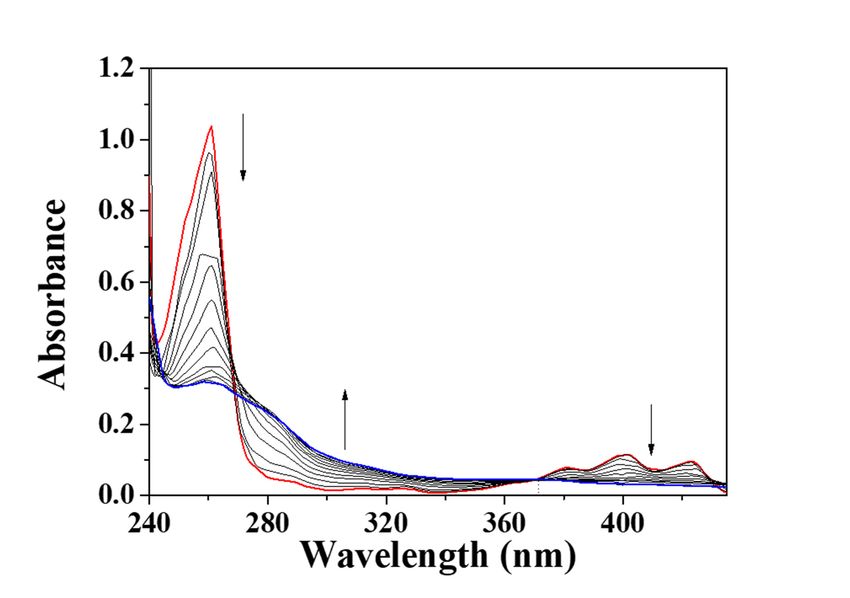
Sensors 2020, 20, 4764                                                                               2 of 13

isothiocyanate to develop a sensor having the unique photophysical, water-soluble and biocompatible
properties for the detection of ClO− .
     Herein, we represent a novel acridine-based fluorescent probe, BK, for detecting ClO− . The reaction
of BK with ClO− showed a fluorescent quenching in aqueous media. The sensing ability of the probe
was investigated by fluorescent and UV-visible titrations. The sensing mechanism of BK towards ClO−
was also demonstrated via ESI-mass spectrometry and theoretical calculations. In addition, the BK
probe was satisfactorily examined to monitor ClO− in environmental water samples and zebrafish.

2. Experiments

2.1. Materials and Equipment
     All reagents and solvents were purchased from Sigma-Aldrich. 13 C NMR (100 MHz) and 1 H
NMR (400 MHz) data were provided on a Varian spectrometer. UV-vis and fluorescence measurements
were performed on Perkin Elmer UV/Vis and fluorescence spectrometers. ESI-mass data were obtained
by a single-quadrupole liquid chromatography detector (ACQUITY QDa).

2.2. Synthesis of KT (N-benzhydryl-2-formylhydrazine-1-carbothioamide)
     An amount of 2 mmol of formic hydrazide and 2 mmol of benzhydryl isothiocyanate were
dissolved in 5 mL of EtOH. The mixture was stirred until a white-colored powder was gained.
The resultant powder was filtered and washed with ether. 1 H NMR (deuterated DMSO, 400 MHz) δ
(ppm): 11.4 (s, 2H), 10.7 (m, 2H), 8.35 (m, 4H), 8.25 (t, 2H), 8.15 (s, 2H), 8.05 (d, 2H), 7.95 (t,1H).

2.3. Synthesis of Sensor BK
((E)-N”-(acridine-9-yl)-N’-((benzhydrylamino)(oxo-l4-sulfanylidene)methyl)formimidohydrazide)
     An amount of 1 mmol of KT and 1 mmol of 9-aminoacridine (ACR) were dissolved in 5 mL
ethanol. After the solution was stirred at 23 ◦ C overnight, the yellowish powder was filtered and
washed with methanol and ether. 1 H NMR in DMSO-d6 , δ: 8.51 (s, 1H), 8.43 (s, 1H), 8.39 (s, 1H),
7.8 (d, 2H, J = 8.8 Hz), 7.65 (t, 2H), 7.5 (m, 10H), 7.3 (m, 5H), 7.2 (s, 1H). 13 C NMR: δ = 142.90, 140.87,
138.05, 132.82, 128.90, 128.26, 128.21, 128.02, 127.73, 124.05, 123.51, 122.71, 112.19, 61.93, 61.05 ppm.
ESI-Mass: m/z calcd for [C28 H23 N5 S – H+ + 2 Na+ + 2 Cl− ]− : 576.08 found, 575.92.

2.4. General Procedure for the Spectroscopic Studies
     Probe BK stock solution (1 mM) was prepared in DMF. ClO− stock solution was prepared by
diluting NaClO (500 µmol, 12%, dissolved in H2 O) in distilled water to make 100 mM. All anion and
ROS stock solutions (1.0 × 10⁻¹ M) were prepared in bis-tris buffer. The fluorescence and UV-vis data
were measured in near 100% aqueous solution (bis-tris, 1 × 10⁻² M, pH 7.0).

2.5. Calculation of Quantum Yield
    Quantum yield (Φ) was calculated by using quinine as a standard fluorophore (ΦF: 0.54 in 0.1 M
H2 SO4 ). Equation for quantum yield is:

                                ΦF(X) = ΦF(S) (AS FX /AX FS) (nX / nS)2

(ΦF(X) : fluorescent quantum yield, x: unknown, s: standard, A: absorbance, n: refractive index of the
solvent and F: the area of fluorescence emission curve).

2.6. Imaging Experiments in Zebrafish
     Zebrafish embryos were cultured under the former conditions [46]. The 6-day-old embryos were
incubated in E2 media replenished with 2 × 10⁻⁵ M of BK for 15 min and rinsed with E2 media to get
rid of the remnant BK. One was a control group and the other group further treated with 50 µM of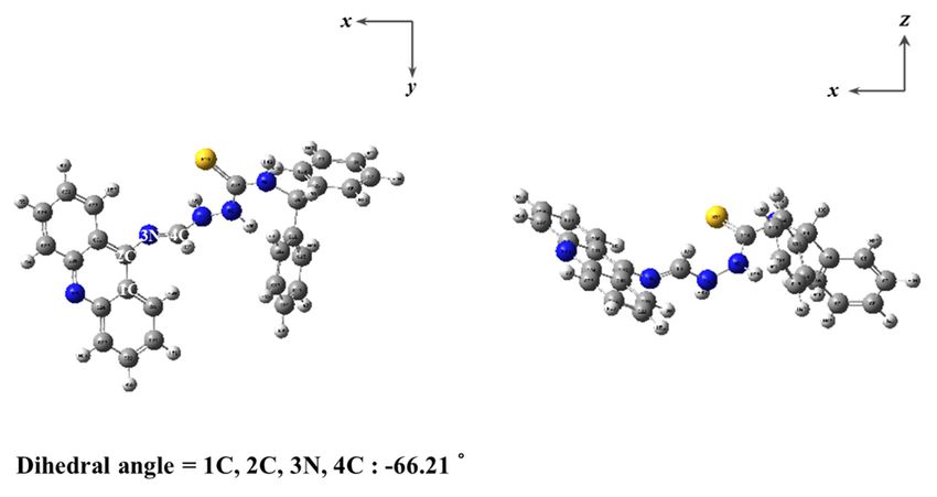
Sensors 2020, 20, 4764 3 of 13
ClO− for 15 min was prepared and washed with E2 media. Zebrafish were anesthetized by adding
ethyl-3-aminobenzoate. The image of zebrafish was achieved by a fluorescence microscope.
Sensors 2020, 20, x FOR PEER REVIEW 3 of 13
2.7. Cytotoxicity in Zebrafish
2.7. Cytotoxicity in Zebrafish
Zebrafish larvae at 6-day old were exposed to 0 and 20 µM of BK and 20 µM of 9-aminoacridine
Zebrafish larvae at 6-day old were exposed to 0 and 20 μM of BK and 20 μM of 9-aminoacridine
in E2 media at 0.05% of DMSO for 20 min. Later they were diverted into 10 µg/mL of AO reagent
in E2 media at 0.05% of DMSO for 20 min. Later they were diverted into 10 μg/mL of AO reagent
(Sigma-Aldrich,
(Sigma-Aldrich, St. Louis,MO,
St. Louis, MO,USA)
USA)ininE2
E2media
mediafor
for6060min.
min.After
After
thethe larvae
larvae were
were washed
washed with
with E2
E2 three times, the prepared larvae were mounted and photographed under a Leica
three times, the prepared larvae were mounted and photographed under a Leica fluorescence fluorescence
microscope
microscope (MZ10F,
(MZ10F,Singapore).
Singapore).Apoptosis
Apoptosiswas
wasidentified
identifiedas
asananobvious
obviousbright
brightspot.
spot.
2.8. Theoretical Calculations
2.8. Theoretical Calculations
DFT/TDDFT calculations on the basis of the hybrid exchange-correlation functional B3LYP were
DFT/TDDFT calculations on the basis of the hybrid exchange-correlation functional B3LYP were
accomplished with the Gaussian 09 W program [47,48]. The 6-31G basis set was applied for all elements
accomplished with the Gaussian 09 W program [47,48]. The 6-31G basis set was applied for all
(C, S, N, O and H) [49,50]. Frequency calculations of BK and ACR (9-aminoacridine) were achieved to
elements (C, S, N, O and H) [49,50]. Frequency calculations of BK and ACR (9-aminoacridine) were
prove that the optimized forms displayed local minima, and imaginary frequencies were not observed
achieved to prove that the optimized forms displayed local minima, and imaginary frequencies were
at all. Cossi and Barone’s CPCM was employed to consider solvent effect of water [51,52]. To study the
not observed at all. Cossi and Barone’s CPCM was employed to consider solvent effect of water
electronic properties of singlet excited states, TD-DFT was conducted for the ground state forms of
[51,52]. To study the electronic properties of singlet excited states, TD-DFT was conducted for the
BK and ACR. The twenty singlet-singlet excitations were quantitatively analyzed. GaussSum 2.2 was
ground state forms of BK and ACR. The twenty singlet-singlet excitations were quantitatively
used to analyze the contributions of MOs [53].
analyzed. GaussSum 2.2 was used to analyze the contributions of MOs [53].
3. Results and Discussion
3. Results and Discussion
The synthetic route for chemosensor BK is outlined in Scheme 1 and it was successfully
The synthetic
characterized route
by 1 H and for
13 C chemosensor
NMR BK
and ESI-MS. is outlined
Sensing in of
behavior Scheme 1 and
BK toward ClOit− was successfully
was investigated
characterized by 1H and 13C NMR and ESI-MS. Sensing behavior of BK toward ClO− was investigated
1
by using several analytical tools like UV-visible and fluorescent spectroscopy, H NMR titration,
by using
and several analytical tools like UV-visible and fluorescent spectroscopy, 1H NMR titration, and
calculations.
calculations.
Scheme 1. Synthesis of BK ((E)-2-((acridine-9-ylimino)methyl)-N-benzhydrylhydrazine-1-carbothioamide).
Scheme 1. Synthesis of BK ((E)-2-((acridine-9-ylimino)methyl)-N-benzhydrylhydrazine-1-
3.1. Spectroscopic Investigations of BK to ClO−
carbothioamide).
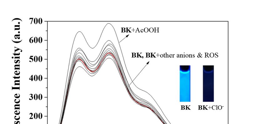
Selectivity was one of the essential indicators for measuring capacity of fluorescent probe.
Reactivity of BK Investigations
3.1. Spectroscopic of BK to(N
to diverse analytes ClO−−, Cl− , H O , CN− , S2− , I− , tBuOOH, SCN− , OAc− , Br− ,
3 2 2
AcOOH, H2 PO4 −was
Selectivity
− , F− , NO − and ClO− ) in bis-tris buffer (1 × 10⁻² M, pH 7.0, Figure 1) was tested
, BzOone of the2 essential indicators for measuring capacity of fluorescent probe.
to evaluateof
Reactivity the selectivity
BK to diverse ClO− . With
foranalytes (N3−, excitation
Cl−, H2O2, at
CN350
−, Snm,
2−, I−free sensor BK
, tBuOOH, SCNdisplayed
−, OAc−, Bra−markedly
, AcOOH,
strong
H2PO4 fluorescence
− , BzO , F , NO
− − emission
2 and ClO
− at 455
− ) innm (Φ =buffer
bis-tris 0.6659).
(1 ×Upon
10 M,
−2 addition
pH 7.0,ofFigure
each analyte into BK
1) was tested to solution,
evaluate
the selectivity for ClO−. With excitation at 350 nm, free sensor BK displayed a markedly strong
fluorescence emission at 455 nm (Ф = 0.6659). Upon addition of each analyte into BK solution, only
ClO− induced the prominent fluorescent decrease (Ф = 0.0047) whereas other analytes did not show
any noticeable changes. Therefore, the sensor BK could serve as a preeminent fluorescent sensor for ClO−.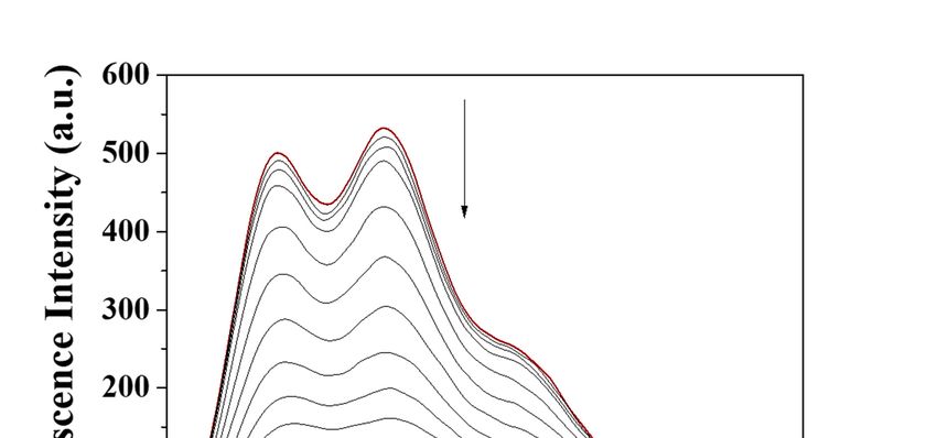
Sensors 2020, 20, 4764                                                                                   4 of 13

     ClO−2020,
onlySensors induced   thePEER
               20, x FOR      REVIEWfluorescent decrease (Φ = 0.0047) whereas other analytes did not
                          prominent                                                                4 of 13
show any noticeable changes. Therefore, the sensor BK could serve as a preeminent fluorescent sensor
for ClO− .

      Figure 1. Fluorescence spectra of BK (1 × 10⁻⁵ M) with varied analytes (220 equiv). ROS: Reactive
          Figure 1. Fluorescence spectra of BK (1 × 10⁻⁵ M) with varied analytes (220 equiv). ROS: Reactive
      oxygen species.
          oxygen species.
     To examine the response of BK to ClO− , the spectral titrations were carried out (Figure 2). On the
addition To  examine ClOthe response   ofto
                                          BK  to ClO
                            − amounts                −, the spectral titrations were carried out (Figure 2). On the
          of different                      BK,  the fluorescence    emission of 455 nm constantly decreased
    addition  of  different  ClO − amounts   to BK,  the  fluorescence
and was saturated at 220 equiv. The measured limit of detection          emission
                                                                           (CDL = of   455for
                                                                                    3σ/k)  nmClO
                                                                                               constantly  decreased
                                                                                                  - was 7.65 µM
    and   was saturated    at 220  equiv. The  measured     limit of detection  (C DL = 3σ/k) for ClO- was 7.65 μM
(Figure S1 in the Supplementary Materials). The UV-visible titration displayed that the absorbance of
    (Figure
280 nm       S1 in the
         constantly      Supplementary
                     increased             Materials).
                                and the bands    at 260 The   UV-visible
                                                         nm and   400 nm titration
                                                                           reduced displayed    that the
                                                                                     with an obvious     absorbance
                                                                                                       isosbestic
    ofat280
point    370nm
             nm constantly
                  (Figure 3). increased and the bands at 260 nm and 400 nm reduced with an obvious
    isosbestic
     To elucidate theat
                point     370 nm mechanism
                       detecting   (Figure 3). of ClO− , we conducted the ESI-mass experiment (Figure S2).
The peak of 322.08 (m/z) was indicative of [ACRO + Cl− + MeCN + 2H2 O]− [calcd, 322.10], indicating
the cleavage reaction product of BK by ClO− . 1 H NMR titration was carried out to observe the
formation of ACR (Figure 4). With gradual addition of ClO− to BK, the imine proton H5 disappeared,
and the amine proton HA of ACR and the aldehyde proton HB of KT appeared.
     On the other hand, as 9-aminoacridine was highly fluorescent in nature, we examined the
interaction of 9-aminoacridine with ClO− . As shown in Figure S3, ClO− quenched the fluorescence of
9-aminoacridine, most likely due to an oxidation reaction. These observations led us to propose that the
probe BK was cleaved by ClO− via the cleavage process of the C=N bond to produce 9-aminoacridine.
Then, the fluorescent 9-aminoacridine was subsequently quenched by ClO− (Scheme 2).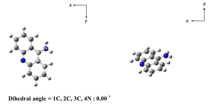
Sensors 2020, 20, x FOR PEER REVIEW                                                                            5 of 13

     Sensors 2020, 20, x FOR PEER REVIEW                                                                            5 of 13

Sensors 2020, 20, 4764                                                                                         5 of 13

                   Figure 2. Fluorescent changes of BK (1 × 10⁻⁵ M) with different concentrations of ClO−.

             Figure 2. Fluorescent changes of BK (1 × 10⁻⁵ M) with different concentrations of ClO− .
                  Figure 2. Fluorescent changes of BK (1 × 10⁻⁵ M) with different concentrations of ClO−.

           Figure 3. UV-vis variations of probe BK (1 × 10⁻⁵ M) with different concentrations of ClO− .
               Figure 3. UV-vis variations of probe BK (1 × 10⁻⁵ M) with different concentrations of ClO−.

                 Figure 3. UV-vis variations of probe BK (1 × 10⁻⁵ M) with different concentrations of ClO−.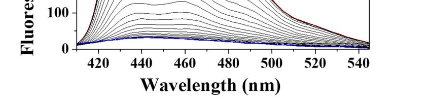
Sensors
and  the2020, 20, x proton
          amine     FOR PEER
                           HREVIEW
                             A of ACR and the aldehyde proton HB of KT appeared.
                                                                                                    6 of 13

      To elucidate the detecting mechanism of ClO−, we conducted the ESI-mass experiment (Figure S2).
The peak of 322.08 (m/z) was indicative of [ACRO + Cl− + MeCN + 2H2O]− [calcd, 322.10], indicating
the cleavage reaction product of BK by ClO−. 1H NMR titration was carried out to observe the
Sensors 2020, 20, 4764                                                                         6 of 13
formation of ACR (Figure 4). With gradual addition of ClO− to BK, the imine proton H5 disappeared,
and the amine proton HA of ACR and the aldehyde proton HB of KT appeared.

                               Figure 4. 1H NMR titration of BK with ClO−.

     On the other hand, as 9-aminoacridine was highly fluorescent in nature, we examined the
interaction of 9-aminoacridine with ClO−. As shown in Figure S3, ClO− quenched the fluorescence of
9-aminoacridine, most likely due to an oxidation reaction. These observations led us to propose that
the probe BK was cleaved by ClO− via the cleavage process of the C=N bond to produce
                               Figure     11H NMR titration of BK with ClO−−
                                Figure4.4.
9-aminoacridine. Then, the fluorescent      H NMR titration
                                        9-aminoacridine     of BK
                                                          was      with ClO . quenched by ClO− (Scheme 2).
                                                                subsequently

     On the other hand, as 9-aminoacridine was highly fluorescent in nature, we examined the
interaction of 9-aminoacridine with ClO−. As shown in Figure S3, ClO− quenched the fluorescence of
9-aminoacridine, most likely due to an oxidation reaction. These observations led us to propose that
the probe BK was cleaved by ClO− via the cleavage process of the C=N bond to produce
9-aminoacridine. Then, the fluorescent 9-aminoacridine was subsequently quenched by ClO− (Scheme 2).

                          Scheme 2. Cleavage sensing mechanism of BK by ClO− .
                          Scheme 2. Cleavage sensing mechanism of BK by ClO−.
     To further comprehend the cleavage sensing mechanism of BK by ClO− , computational calculations
were achieved. Energy-optimized forms of BK and ACR were analyzed with DFT/B3LYP/6-31G (d,p)
basis sets (Figure 5). Using the optimized forms of BK and ACR, TD-DFT calculations were performed
for analyzing transition energies and molecular orbitals (Figures S4–S6). The MOs of BK at the first
lowest excited state were identified as the HOMO → LUMO transitions (391.73 nm, Figure S5), which
                           Scheme 2. Cleavage
turned out to be π → π* transitions.  For ACR,sensing mechanism
                                                the MOs           of BK
                                                         at the first   by ClO
                                                                      lowest   −.
                                                                             excited state were identified
as the HOMO → LUMO (391.84 nm, Figure S6), which turned out to be π → π* transitions. In addition,
the decreased oscillator strength of ACR was corresponded with the decreased UV-vis absorbance.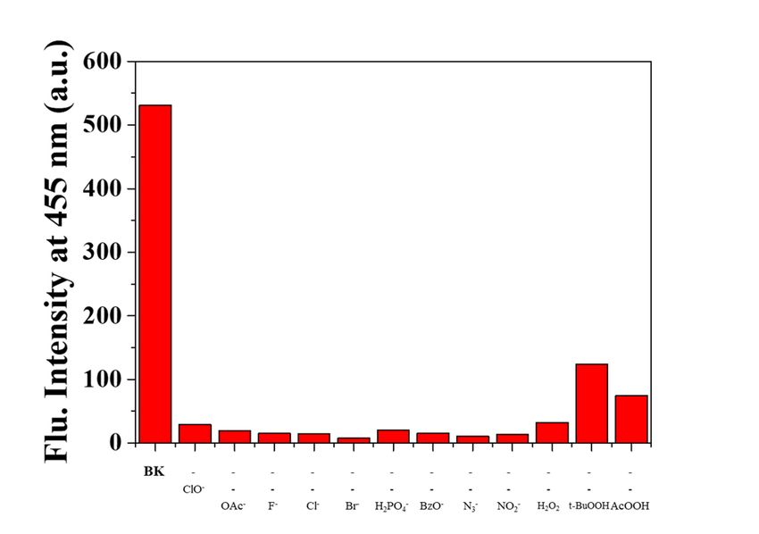
calculations were achieved. Energy-optimized forms of BK and ACR were analyzed with
DFT/B3LYP/6-31G (d,p) basis sets (Figure 5). Using the optimized forms of BK and ACR, TD-DFT
calculations were performed for analyzing transition energies and molecular orbitals (Figures S4–S6).
The MOs of BK at the first lowest excited state were identified as the HOMO → LUMO transitions
(391.732020,
Sensors  nm, 20,Figure
                 4764  S5), which turned out to be π → π* transitions. For ACR, the MOs at the 7first
                                                                                                  of 13
lowest excited state were identified as the HOMO → LUMO (391.84 nm, Figure S6), which turned
out to be π → π* transitions. In addition, the decreased oscillator strength of ACR was corresponded
These outcomes drove us to elucidate that fluorescent sensor BK was quenched due to the cleavage of
with the decreased UV-vis absorbance. These outcomes drove us to elucidate that fluorescent sensor
the C=N bond.
BK was quenched due to the cleavage of the C=N bond.

                                                   (a)

                                                   (b)
                Figure
                Figure 5.
                       5. Energy-minimized
                          Energy-minimized forms
                                            forms of
                                                  of (a)
                                                      (a) BK
                                                          BKand
                                                             and(b)
                                                                 (b)9-aminoacridine
                                                                     9-aminoacridine(ACR).
                                                                                     (ACR).

     Another vital indicator in the sensing process is the ability of the fluorescent probe not to be
disturbed by the competitive analytes. When BK was exposed with both ClO− and diverse analytes
(N3−, Cl−, H2O2, tBuOOH, OAc−, Br−, AcOOH, H2PO4−, BzO−, F− and NO2−), the guest analytes did not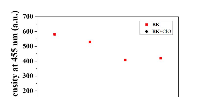
Sensors 2020, 20, 4764                                                                                           8 of 13

      Another
    Sensors       vital
            2020, 20,    indicator
                      x FOR        in the sensing process is the ability of the fluorescent probe not to be
                            PEER REVIEW                                                                     8 of 13
disturbed by the competitive analytes. When BK was exposed with both ClO− and diverse analytes
    − , Cl− , H O , tBuOOH, OAc
(N3hinder                              − , Br− , AcOOH, H PO − , BzO− , F− and NO − ), the guest     analytes
              the
                2 sensing
                    2         of ClO− (Figure      6). Only, the2  presence
                                                                    4         of tBuOOH 2and OAc− showed   slight
didinterference
     not hinder the       sensing of ClO   −− (Figure   6).  Only, the  presence  of tBuOOH and OAc − showed
                    in determining ClO . CN , S , I and SCN were excluded from the competition because
                                                   −   2−  −          −

slight  interference    in determining         − . CN− , S2− , I− and SCN− were excluded from the competition
    they  reacted directly     with ClO−ClO
                                          .
because they reacted directly with ClO− .

    Figure 6. Competitive selectivity of BK (1 × 10⁻⁵ M) to ClO− (220 equiv) with other analytes (220 equiv).
           Figure 6. Competitive selectivity of BK (1 × 10⁻⁵ M) to ClO− (220 equiv) with other analytes (220 equiv).
      The pH was a vital condition related with physiological processes and even cellular behaviors.
Thus, weThe     pH was
            checked   theaeffect
                           vital ofcondition  relatedproperty
                                     pH on sensing     with physiological
                                                                of BK for ClO    − at pHand
                                                                             processes          even from
                                                                                           ranging    cellular
                                                                                                           six behaviors.
                                                                                                               to nine
    Thus,
(Figure  7).weBKchecked   thesignificant
                  displayed   effect of pH    on sensingintensity
                                           fluorescence   propertyatofthe
                                                                        BKpHforrange
                                                                                 ClO− atof pH  ranging
                                                                                           six to        fromintensity
                                                                                                  nine. The    six to nine
    (Figure
of BK  treated7). BK   ClO− was
                  withdisplayed    significant
                                     completelyfluorescence
                                                  quenched intensity
                                                              at pH sixattothe  pH These
                                                                             nine.  range of   six to nine. The
                                                                                            observations         intensity
                                                                                                            indicated
thatofBK
       BKcould
             treated  with ClO as
                  be applicable  −  was  completely
                                      a probe         quenched
                                               for detecting      −
                                                             ClOat at
                                                                    pHpH sixsix
                                                                              totonine.
                                                                                   nine.These observations indicated
    that BK could
      To explore      be applicable
                   practical utility ofasBK,
                                          a probe  for detecting
                                              the application     ClOin
                                                               of BK  − at pHsamples
                                                                         real   six to nine.
                                                                                         was accomplished in both
tap and drinking water samples. The reliable R.S.D. values and recoveries demonstrated that sensor
BK had a valuable potential for being used as a dependable tool to monitor ClO− in real water samples
(Table 1).
Sensors 2020, 20, x FOR PEER REVIEW 9 of 13
Sensors 2020, 20, 4764 9 of 13
FigureFigure 7. Fluorescence
7. Fluorescence intensity
intensity (at 455(at
nm)455
ofnm) of BKClO
BK with with pH−values
− atClO at pH from
valuessix
from six to nine.
to nine.
Table 1. Determination of ClO− .a.
To explore practical utility of BK, the application of BK in real samples was accomplished in
both tap and drinking water samples. The reliable R.S.D. values and recoveriesR.S.D.
Recovery demonstrated
(n = 3) that
Sample. ClO− Added (µM) ClO− Found (µM)
sensor BK had a valuable potential for being used as a dependable tool to monitor ClO in real water
(%) (%)
−
samples (Table 1). water
Drinking 0.00 0.00
60.00 b 62.88 104.80 0.32
Table 1. Determination of ClO−.a.
Tap water 0.00 0.00
b Recovery R.S.D. (n = 3)
Sample. ClO− Added
60.00 (µM) 62.16(µM)
ClO− Found 103.60 0.41
(%) (%)
a Conditions: [BK] = 10 µM in bis-tris buffer. b 60.00 µM of ClO− was artificially added.
Drinking water 0.00 0.00
60.00 b 62.88 104.80 0.32
Tap water
3.2. In Vivo Imaging in Zebrafish 0.00 0.00
60.00 b 62.16 103.60 0.41
To test whether the probe BK is applicable under biological conditions, zebrafish were incubated
a Conditions: [BK] = 10 μM in bis-tris buffer. b 60.00 μM of ClO− was artificially added.
with BK (20 µM) and sequentially treated with two different concentrations of ClO− (0 and 50 µM)
forInimaging
3.2. (Figure
vivo Imaging 8). Zebrafish incubated with BK exhibited a green fluorescence image, but the
in Zebrafish
green fluorescence was eliminated in the presence of ClO− . Meanwhile, the cytotoxicity test of BK
wasTo test whether
examined the probe
by AO BK(Figure
staining is applicable under
S7). The AObiological conditions,
stained results zebrafish
showed were
that no incubated
apoptosis was
with BK (20 μM) and sequentially treated with two different concentrations of ClO
observed in control and the presence of BK and 9-aminoacridine. Zebrafish experiments proved that
− (0 and 50 μM)
for
BK imaging (Figure 8). Zebrafish
was organism-permeable andincubated withClO
can monitor BK− exhibited a green fluorescence
in living organisms. Importantly,image,
BK isbut
thethe
first
green fluorescence was eliminated in the presence − of ClO − . Meanwhile, the
fluorescent turn-off sensor capable of sensing ClO in zebrafish (Table S1) [1,38,54–59]. cytotoxicity test of BK
was examined by AO staining (Figure S7). The AO stained results showed that no apoptosis was
observed in control and the presence of BK and 9-aminoacridine. Zebrafish experiments proved that
BK was organism-permeable and can monitor ClO− in living organisms. Importantly, BK is the first
fluorescent turn-off sensor capable of sensing ClO− in zebrafish (Table S1) [1,38,54–59].Sensors 2020, 20, x FOR PEER REVIEW 10 of 13
Sensors 2020, 20, 4764 10 of 13
Figure 8. Fluorescence images of zebrafish treated with BK followed by addition of ClO− (λex = 502 nm
and λem = 526 nm). (a1 –a3 ): BK only; (b1 –b3 ): BK with 5 × 10⁻⁵ M ClO− . [BK] = 2 × 10⁻⁵ M. Scale bar:
0.89 mm.
Figure 8. Fluorescence images of zebrafish treated with BK followed by addition of ClO− (λex = 502 nm
and λem = 526 nm). (a1–a3): BK only; (b1–b3): BK with 5 × 10⁻⁵ M ClO-. [BK] = 2 × 10⁻⁵ M. Scale bar: 0.89 mm.
4. Conclusions
We have synthesized an acridine-based chemosensor for monitoring ClO− in a near-perfect
4. Conclusion
aqueous media. Probe BK selectively detected ClO− over other relevant species including ROS, and its
intenseWe have
blue synthesized
fluorescence wasan acridine-based
notably quenched chemosensor
with the additionfor monitoring
of ClO− . TheClO
− in a near-perfect
detection limit of BK
aqueous
for media.
ClO− was Probe BK
analyzed selectively
to be 7.65 µM. detected ClO over other
−
BK was employed relevant species
for quantitative including
measurement ofROS,
ClO−and
in
its intense blue fluorescence was notably quenched with the addition of ClO −. The detection limit of
real water samples and zebrafish. Significantly, BK is the first fluorescent turn-off sensor capable of
BK for ClO
sensing ClO−was
− analyzedtotodate.
in zebrafish be 7.65
TheμM. BK was outcomes
promising employedindicate
for quantitative
that BK measurement
can serve as a of ClO− in
potential
real water samples
chemosensor and zebrafish.
for monitoring ClO- inSignificantly, BK is the first
chemical, environmental andfluorescent
biologicalturn-off
systems.sensor capable
We believe of
that
sensing ClO − in zebrafish to date. The promising outcomes indicate that BK can serve as a potential
these results will be merited for further development of ClO− sensors.
chemosensor for monitoring ClO- in chemical, environmental and biological systems. We believe that
these results will
Supplementary be merited
Materials: for further
The following aredevelopment ofatClO
available online − sensors.
http://www.mdpi.com/1424-8220/20/17/4764/s1,
Table S1: Examples of fluorescent chemosensors for detecting ClO− in zebrafish, Figure S1: Determination of the
Supplementary
detection ClO− by BK
limit forMaterials: Table S1: Examples
(10 µM) based on the of fluorescent
fluorescence chemosensors
emission at 455 fornm;
detecting
FigureClO in zebrafish,
S2: −Negative-ion
electrospray ionization mass
Figure S1: Determination ofspectrum of BK
the detection (10 µM)
limit uponbyaddition
for ClO − BK (10 of μM)NaClO
based (200
on equiv); Figure S3: emission
the fluorescence Molecular at
orbital diagrams and excitation energies of (a) BK and (b) ACR; Figure S4: (a) The theoretical excitation energies
455 nm; Figure S2: Negative-ion electrospray ionization mass spectrum of BK (10 μM) upon addition of NaClO
and the experimental UV-vis spectrum of BK. (b) The major electronic transition energies and molecular orbital
(200 equiv); Figure
contributions for BKS3:(H
Molecular
= HOMO orbital
and Ldiagrams
= LUMO); and Figure
excitation
S5: energies
(a) The of (a) BK and
theoretical (b) ACR;energies
excitation Figure S4: (a)
and
the
Theexperimental UV-vis spectrum
theoretical excitation energiesofandACR.the(b)experimental
The major electronic transitionofenergies
UV-vis spectrum BK. (b)andThemolecular orbital
major electronic
contributions for ACR
transition energies and(H = HOMO
molecular orbital L = LUMO); Figure
and contributions for BK (HS6. =(a) The theoretical
HOMO excitation
and L = LUMO); energies
Figure S5: (a)and
The
the experimental UV-vis spectrum of ACR. (b) The major electronic transition energies and molecular orbital
theoretical excitation energies and the experimental UV-vis spectrum of ACR. (b) The
contributions for ACR (H = HOMO and L = LUMO); Figure S7. AO-stained zebrafish exposed to (a) 0 µM and major electronic transition
energies
(b) 20 µMand molecular
of BK orbital
and (c) 20 µM ofcontributions for ACR (H = HOMO and L = LUMO); Figure S6. (a) The theoretical
9-aminoacridine.
excitation energies and the experimental UV-vis
Author Contributions: S.C.L. and C.K. provided the spectrum of ACR.
initial idea (b)work;
for this The major
S.C.L.,electronic transition
S.P. and H.S. energies
contributed to
andcollection
the molecular andorbital contributions
analysis for ACR
of field test data; (H and
K.-T.K. = HOMO and L = LUMO);
G.L. contributed Figure of
to the analyses S7.results;
AO-stained
S.C.L. zebrafish
and C.K.
wrote the to
exposed paper.
(a) 0 All
μMauthors
and (b) have
20 μM read andand
of BK agreed
(c) 20toμM
the of
published version of the manuscript.
9-aminoacridine.
Funding: This research received
Author Contributions: no C.K.
S.C.L. and external funding.
provided the initial idea for this work; S.C.L., S.P. and H.S. contributed
Acknowledgments: KEITI (Korea
to the collection and analysis of fieldEnvironment Industry
test data; K.-T.K. and G.L.and Technology
contributed Institute)
to the analyses(2016001970001)
of results; S.C.L. are
and
kindly acknowledged.
C.K. wrote the paper. All authors have read and agreed to the published version of the manuscript.
Conflicts of Interest: The authors declare no conflict of interest.
Acknowledgments: KEITI (Korea Environment Industry and Technology Institute) (2016001970001) are kindly
acknowledged.
Conflicts of Interest: The authors declare no conflicts of interest.Sensors 2020, 20, 4764 11 of 13
References
1. He, X.; Xu, C.; Xiong, W.; Qian, Y.; Fan, J.; Ding, F.; Deng, H.; Chen, H.; Shen, J. The ICT-based fluorescence
and colorimetric dual sensing of endogenous hypochlorite in living cells, bacteria, and zebrafish. Analyst
2020, 145, 29–33. [CrossRef]
2. Kang, J.; Huo, F.; Zhang, Y.; Chao, J.; Strongin, R.M.; Yin, C. Detecting intracellular ClO− with ratiometric
fluorescent signal and its application in vivo. Sens. Actuators B Chem. 2018, 273, 1532–1538. [CrossRef]
3. Huo, B.; Du, M.; Shen, A.; Li, M.; Lai, Y.; Bai, X.; Gong, A.; Fang, L.; Yang, Y. A “light-up” fluorescent probe
based on TEMPO-oxidation for the detection of ClO− and application in real samples. Sens. Actuators B Chem.
2019, 284, 23–29. [CrossRef]
4. Kettle, A.J.; Albrett, A.M.; Chapman, A.L.; Dickerhof, N.; Forbes, L.V.; Khalilova, I.; Turner, R. Measuring
chlorine bleach in biology and medicine. Biochim. Biophys. Acta Gen. Subj. 2014, 1840, 781–793. [CrossRef]
5. Zhang, Y.R.; Liu, Y.; Feng, X.; Zhao, B.X. Recent progress in the development of fluorescent probes for the
detection of hypochlorous acid. Sens. Actuators B Chem. 2017, 240, 18–36. [CrossRef]
6. Reja, S.I.; Bhalla, V.; Sharma, A.; Kaur, G.; Kumar, M. A highly selective fluorescent probe for hypochlorite
and its endogenous imaging in living cells. Chem. Commun. 2014, 50, 11911–11914. [CrossRef] [PubMed]
7. Wang, L.; Hu, Y.; Qu, Y.; Xu, J.; Cao, J. Aggregated-induced emission phenothiazine probe for selective
ratiometric response of hypochlorite over other reactive oxygen species. Dye. Pigment. 2016, 128, 54–59.
[CrossRef]
8. Shim, M.S.; Xia, Y. A reactive oxygen species (ROS)-responsive polymer for safe, efficient, and targeted gene
delivery in cancer cells. Angew. Chem. Int. Ed. 2013, 52, 6926–6929. [CrossRef]
9. Yang, Y.; Yin, C.; Huo, F.; Chao, J.; Zhang, Y.; Jin, S. Simple 1,8-diaminonaphthalene-based fluorescence
chemosensor for hypochlorites and its practical application. Sens. Actuators B Chem. 2014, 199, 226–231.
[CrossRef]
10. Huang, Y.; Zhang, Y.; Huo, F.; Chao, J.; Yin, C. A near-infrared ratiometric fluorescent probe with large stokes
based on isophorone for rapid detection of ClO− and its bioimaging in cell and mice. Sens. Actuators B Chem.
2019, 287, 453–458. [CrossRef]
11. Jiang, Y.; Wu, S.; Jin, C.; Wang, B.; Shen, J. Novel diaminomaleonitrile-based fluorescent probe for ratiometric
detection and bioimaging of hypochlorite. Sens. Actuators B Chem. 2018, 265, 365–370. [CrossRef]
12. Huo, F.J.; Zhang, J.J.; Yang, Y.T.; Chao, J.B.; Yin, C.X.; Zhang, Y.B.; Chen, T.G. A fluorescein-based highly
specific colorimetric and fluorescent probe for hypochlorites in aqueous solution and its application in tap
water. Sens. Actuators B Chem. 2012, 166, 44–49. [CrossRef]
13. Lv, J.; Chen, Y.; Wang, F.; Wei, T.; Zhang, Z.; Qiang, J.; Chen, X. A mitochondria-targeted fluorescent probe
based on fluorescein derivative for detection of hypochlorite in living cells. Dye. Pigment. 2018, 148, 353–358.
[CrossRef]
14. Du, J.; Hu, M.; Fan, J.; Peng, X. Fluorescent chemodosimeters using “mild” chemical events for the detection
of small anions and cations in biological and environmental media. Chem. Soc. Rev. 2012, 41, 4511–4535.
[CrossRef] [PubMed]
15. Lee, S.C.; Kim, C. Naphthol-naphthalimide based ‘turn-on’ fluorescent sensor for ClO− in aqueous media
and test kit. Inorg. Chem. Commun. 2019, 108, 107545. [CrossRef]
16. Sam, C.H.; Lu, H.K. The role of hypochlorous acid as one of the reactive oxygen species in periodontal
disease. J. Dent. Sci. 2009, 4, 45–54. [CrossRef]
17. Steinbeck, M.J.; Nesti, L.J.; Sharkey, P.F.; Parvizi, J. Myeloperoxidase and chlorinated peptides in osteoarthritis:
Potential biomarkers of the disease. J. Orthop. Res. 2007, 25, 1128–1135. [CrossRef]
18. Pattison, D.I.; Davies, M.J. Evidence for rapid inter- and intramolecular chlorine transfer reactions of
histamine and carnosine chloramines: Implications for the prevention of hypochlorous-acid-mediated
damage. Biochemistry 2006, 45, 8152–8162. [CrossRef]
19. Tian, F.; Jia, Y.; Zhang, Y.; Song, W.; Zhao, G.; Qu, Z.; Li, C.; Chen, Y.; Li, P. A HClO-specific near-infrared
fluorescent probe for determination of Myeloperoxidase activity and imaging mitochondrial HClO in living
cells. Biosens. Bioelectron. 2016, 86, 68–74. [CrossRef]
20. Song, X.; Dong, B.; Kong, X.; Wang, C.; Zhang, N.; Lin, W. Construction of a ratiometric fluorescent probe
with an extremely large emission shift for imaging hypochlorite in living cells. Spectrochim. Acta Part A Mol.
Biomol. Spectrosc. 2018, 188, 394–399. [CrossRef]Sensors 2020, 20, 4764 12 of 13
21. Jin, L.; Xu, M.; Jiang, H.; Wang, W.; Wang, Q. A simple fluorescein derived colorimetric and fluorescent
“off-on” sensor for the detection of hypochlorite. Anal. Methods 2018, 10, 4562–4569. [CrossRef]
22. Zhang, P.; Wang, H.; Hong, Y.; Yu, M.; Zeng, R.; Long, Y.; Chen, J. Selective visualization of endogenous
hypochlorous acid in zebrafish during lipopolysaccharide-induced acute liver injury using a polymer
micelles-based ratiometric fluorescent probe. Biosens. Bioelectron. 2018, 99, 318–324. [CrossRef] [PubMed]
23. Adegoke, O.; Forbes, P.B.C. Challenges and advances in quantum dot fluorescent probes to detect reactive
oxygen and nitrogen species: A review. Anal. Chim. Acta 2015, 862, 1–13. [CrossRef] [PubMed]
24. Jiao, X.; Liu, C.; Wang, Q.; Huang, K.; He, S.; Zhao, L.; Zeng, X. Fluorescence probe for hypochlorous acid in
water and its applications for highly lysosome-targetable live cell imaging. Anal. Chim. Acta 2017, 969, 49–56.
[CrossRef] [PubMed]
25. Goswami, S.; Paul, S.; Manna, A. Carbazole based hemicyanine dye for both “naked eye” and ‘NIR’
fluorescence detection of CN− in aqueous solution: From molecules to low cost devices (TLC plate sticks).
Dalton Trans. 2013, 42, 10682–10686. [CrossRef]
26. Goswami, S.; Manna, A.; Paul, S.; Quah, C.K.; Fun, H.K. Rapid and ratiometric detection of hypochlorite
with real application in tap water: Molecules to low cost devices (TLC sticks). Chem. Commun. 2013, 49,
11656–11658. [CrossRef]
27. Kim, H.N.; Lee, M.H.; Kim, H.J.; Kim, J.S.; Yoon, J. A new trend in rhodamine-based chemosensors:
Application of spirolactam ring-opening to sensing ions. Chem. Soc. Rev. 2008, 37, 1465–1472. [CrossRef]
28. Chen, X.; Baek, K.H.; Kim, Y.; Kim, S.J.; Shin, I.; Yoon, J. A selenolactone-based fluorescent chemodosimeter
to monitor mecury/methylmercury species in vitro and in vivo. Tetrahedron 2010, 66, 4016–4021. [CrossRef]
29. Wang, X.; Song, F.; Peng, X. A versatile fluorescent probe for imaging viscosity and hypochlorite in living
cells. Dye. Pigment. 2016, 125, 89–94. [CrossRef]
30. Lou, X.; Zhang, Y.; Li, Q.; Qin, J.; Li, Z. A highly specific rhodamine-based colorimetric probe for hypochlorites:
A new sensing strategy and real application in tap water. Chem. Commun. 2011, 47, 3189–3191. [CrossRef]
31. Long, L.; Zhang, D.; Li, X.; Zhang, J.; Zhang, C.; Zhou, L. A fluorescence ratiometric sensor for hypochlorite
based on a novel dual-fluorophore response approach. Anal. Chim. Acta 2013, 775, 100–105. [CrossRef]
[PubMed]
32. Maity, D.; Karthigeyan, D.; Kundu, T.K.; Govindaraju, T. FRET-based rational strategy for ratiometric
detection of Cu2+ and live cell imaging. Sens. Actuators B Chem. 2013, 176, 831–837. [CrossRef]
33. Hwang, S.M.; Chae, J.B.; Kim, C. A Phenanthroimidazole-based Fluorescent Turn-Off Chemosensor for the
Selective Detection of Cu2+ in Aqueous Media. Bull. Korean Chem. Soc. 2018, 39, 925–930. [CrossRef]
34. Koide, Y.; Urano, Y.; Hanaoka, K.; Terai, T.; Nagano, T. Development of an Si-rhodamine-based far-red to
near-infrared fluorescence probe selective for hypochlorous acid and its applications for biological imaging.
J. Am. Chem. Soc. 2011, 133, 5680–5682. [CrossRef]
35. Shiraishi, Y.; Yamada, C.; Hirai, T. A coumarin-dihydroperimidine dye as a fluorescent chemosensor for
hypochlorite in 99% water. RSC Adv. 2019, 9, 28636–28641. [CrossRef]
36. Grabchev, I.; Petkov, C.; Bojinov, V. 1,8-Naphthalimides as blue emitting fluorophores for polymer materials.
Macromol. Mater. Eng. 2002, 287, 904–908. [CrossRef]
37. Liu, Y.; Zhao, C.; Zhao, X.; Liu, H.; Wang, Y.; Du, Y.; Wei, D. A selective N,N-dithenoyl-rhodamine based
fluorescent probe for Fe3+ detection in aqueous and living cells. J. Environ. Sci. 2020, 90, 180–188. [CrossRef]
38. He, X.; Chen, H.; Xu, C.; Fan, J.; Xu, W.; Li, Y.; Deng, H.; Shen, J. Ratiometric and colorimetric fluorescent
probe for hypochlorite monitor and application for bioimaging in living cells, bacteria and zebrafish.
J. Hazard. Mater. 2020, 388, 122029. [CrossRef]
39. Wang, Y.; He, J.; Zheng, M.; Qin, M.; Wei, W. Dual-emission of Eu based metal-organic frameworks hybrids
with carbon dots for ratiometric fluorescent detection of Cr (VI). Talanta 2019, 191, 519–525. [CrossRef]
40. Wang, C.; Fu, J.; Yao, K.; Xue, K.; Xu, K.; Pang, X. Acridine-based fluorescence chemosensors for selective
sensing of Fe3+ and Ni2+ ions. Spectrochim. Acta Part A Mol. Biomol. Spectrosc. 2018, 199, 403–411. [CrossRef]
41. Nunes, M.C.; dos Santos Carlos, F.; Fuganti, O.; Galindo, D.D.M.; De Boni, L.; Abate, G.; Nunes, F.S.
Turn-on fluorescence study of a highly selective acridine-based chemosensor for Zn2+ in aqueous solutions.
Inorg. Chim. Acta 2020, 499, 119191. [CrossRef]
42. Wang, C.; Wang, P.; Liu, X.; Fu, J.; Xue, K.; Xu, K. Novel enantioselective fluorescent sensors for tartrate anion
based on acridinezswsxa. Luminescence 2017, 32, 1313–1318. [CrossRef] [PubMed]Sensors 2020, 20, 4764 13 of 13
43. Janovec, L.; Kožurková, M.; Sabolová, D.; Ungvarský, J.; Paulíková, H.; Plšíková, J.; Vantová, Z.; Imrich, J.
Cytotoxic 3,6-bis((imidazolidinone)imino)acridines: Synthesis, DNA binding and molecular modeling.
Bioorganic Med. Chem. 2011, 19, 1790–1801. [CrossRef] [PubMed]
44. Dai, Y.; Xu, K.; Li, Q.; Wang, C.; Liu, X.; Wang, P. Acridine-based complex as amino acid anion fluorescent
sensor in aqueous solution. Spectrochim. Acta Part A Mol. Biomol. Spectrosc. 2016, 157, 1–5. [CrossRef]
45. Kim, A.; Kim, C. A hydrazono-quinoline-based chemosensor sensing In3+ and Zn2+ via fluorescence turn-on
and ClO− via color change in aqueous solution. New J. Chem. 2019, 43, 7320–7328. [CrossRef]
46. Jung, J.M.; Lee, S.Y.; Nam, E.; Lim, M.H.; Kim, C. A highly selective turn-on chemosensor for Zn2+ in aqueous
media and living cells. Sens. Actuators B Chem. 2017, 244, 1045–1053. [CrossRef]
47. Becke, A.D. Density-functional thermochemistry. III. The role of exact exchange. J. Chem. Phys. 1993, 98,
5648–5652. [CrossRef]
48. Lee, C.; Yang, W.; Parr, R.G. Development of the Colle-Salvetti correlation-energy formula into a functional
of the electron density. Phys. Rev. B 1988, 37, 785. [CrossRef]
49. Hariharan, P.C.; Pople, J.A. The influence of polarization functions on molecular orbital hydrogenation
energies. Chim. Acta 1973, 28, 213–222. [CrossRef]
50. Gordon, M.S.; Binkley, J.S.; Pople, J.A.; Pietro, W.J.; Hehre, W.J. Self-Consistent Molecular-Orbital Methods.
22. Small Split-Valence Basis Sets for Second-Row Elements. J. Am. Chem. Soc. 1982, 104, 2797–2803.
[CrossRef]
51. Barone, V.; Cossi, M. Quantum calculation of molecular energies and energy gradients in solution by a
conductor solvent model. J. Phys. Chem. A 1998, 102, 1995–2001. [CrossRef]
52. Cossi, M.; Barone, V. Time-dependent density functional theory for molecules in liquid solutions. J. Chem. Phys.
2001, 115, 4708. [CrossRef]
53. O’boyle, N.M.; Tenderholt, A.L.; Langner, K.M. Cclib: A library for package-independent computational
chemistry algorithms. J. Comput. Chem. 2008, 29, 839–845. [CrossRef] [PubMed]
54. Chen, H.; Sun, T.; Qiao, X.G.; Tang, Q.O.; Zhao, S.C.; Zhou, Z. Red-emitting fluorescent probe for detecting
hypochlorite acid in vitro and in vivo. Spectrochim. Acta Part A Mol. Biomol. Spectrosc. 2018, 204, 196–202.
[CrossRef] [PubMed]
55. Lan, J.; Guo, J.; Jiang, X.; Chen, Y.; Hu, Z.; Que, Y.; Li, H.; Gu, J.; Ho, R.J.Y.; Zeng, R.; et al. A new
dicyanoisophorone-based ratiometric and colorimetric near-infrared fluorescent probe for specifically
detecting hypochlorite and its bioimaging on a model of acute inflammation. Anal. Chim. Acta 2020, 1094,
70–79. [CrossRef] [PubMed]
56. Duan, C.; Won, M.; Verwilst, P.; Xu, J.; Kim, H.S.; Zeng, L.; Kim, J.S. In vivo imaging of endogenously
produced HClO in zebrafish and mice using a bright, photostable ratiometric fluorescent probe. Anal. Chem.
2019, 91, 4172–4178. [CrossRef]
57. Wang, N.; Xu, W.; Song, D.; Ma, P. A fluorescein-carbazole-based fluorescent probe for imaging of endogenous
hypochlorite in living cells and zebrafish. Spectrochim. Acta Part A Mol. Biomol. Spectrosc. 2020, 227, 117692.
[CrossRef]
58. So, H.; Cho, H.; Lee, H.; Tran, M.C.; Kim, K.T.; Kim, C. Detection of zinc (II) and hypochlorite by a
thiourea-based chemosensor via two emission channels and its application in vivo. Microchem. J. 2020, 155,
104788. [CrossRef]
59. Liu, C.; Li, Z.; Yu, C.; Chen, Y.; Liu, D.; Zhuang, Z.; Jia, P.; Zhu, H.; Zhang, X.; Yu, Y.; et al. Development of a
Concise Rhodamine-Formylhydrazine Type Fluorescent Probe for Highly Specific and Ultrasensitive Tracing
of Basal HOCl in Live Cells and Zebrafish. ACS Sens. 2019, 4, 2156–2163. [CrossRef]
© 2020 by the authors. Licensee MDPI, Basel, Switzerland. This article is an open access
article distributed under the terms and conditions of the Creative Commons Attribution
(CC BY) license (http://creativecommons.org/licenses/by/4.0/).You can also read