Cambridge Institute for Medical Research - University of Cambridge School of Clinical Medicine - Cambridge Institute for Medical ...
←
→
Page content transcription
If your browser does not render page correctly, please read the page content below
University of Cambridge
School of Clinical Medicine

Cambridge Institute for Medical Research

                              Research Report 2008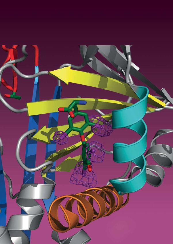
Front cover illustration:
Part of the structure of thyroxine-binding globulin showing the binding site for
thyroxine (green), a hormone that controls cellular development as well as the rate
of body metabolism. Verification of the binding site was achieved using the program
Phaser developed in CIMR. (See also page 26, Randy Read).

Cambridge Institute for Medical Research
University of Cambridge
Wellcome Trust/MRC Building
Hills Road
Cambridge CB2 0XY

www.cimr.cam.ac.uk

Photography by Stephen Bond www.stephenbond.com and
University of Cambridge Medical Photography Department

Printed by Cambridge University Press, www.cambridge.org/printing
School of Clinical Medicine

Cambridge Institute for Medical Research
                                  Research Report 2008
Contents

5 Foreword by Paul Luzio                   42 MRC Dunn Human Nutrition Unit

7 CIMR Principal Investigators             43 CIMR Affiliated Principal Investigators
                                              43 Jennie Blackwell
9 Science in the Institute                    43 Sadaf Farooqi
   9 Folma Buss                               43 Stephen O’Rahilly
  10 David Clayton
  11 Bertie Göttgens                       44 Principal Investigators who will be moving on
  12 Allison Green                            from CIMR shortly
  13 Tony Green                               44 Roger Pedersen
  14 Fiona Gribble                            44 John Yates
  15 Gillian Griffiths
  16 James Huntington                      46 Principal Investigators who have moved on
  17 Brian Huntly                             from CIMR since Research Report 2006
  18 Fiona Karet                              46 Krish Chatterjee
  19 Paul Lehner                              46 Hisao Kondo
  20 David Lomas                              46 Kerstin Meyer
  21 Paul Luzio                               46 Gillian Murphy
  22 Katrin Ottersbach                        46 Bruce Ponder/Doug Winton
  23 David Owen                               46 Karin Römisch
  24 Andrew Peden
  25 Lucy Raymond                          47 Postgraduate Opportunities in the Institute
  26 Randy Read
  27 Evan Reid                             48 CIMR Research Retreat 2007
  28 Margaret Robinson
  29 David Rubinsztein                     49 Administration, IT and Technical Support
  30 Christopher Rudd
  31 Peter St George-Hyslop                51 Funding of CIMR
  32 Richard Sandford
  33 Matthew Seaman                        52 Other Information
  34 Symeon Siniossoglou
  35 Ken Smith                             56 Publications for Scientists in CIMR
  36 John Todd                                since 2006
  37 The Juvenile Diabetes Research
      Foundation/Wellcome Trust Diabetes   78 Regulations for CIMR
      and Inflammation Laboratory
  38 John Trowsdale
  39 Linda Wicker
  40 Geoff Woods

                                                                                            3
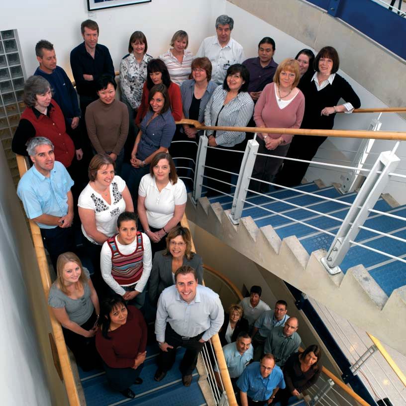
The Wellcome Trust/MRC Building which houses the Cambridge Institute for Medical Research and the MRC Dunn Human Nutrition Unit.

4

Foreword: Cambridge Institute for Medical Research

The late summer of 2008 will mark the tenth          and Chris Rudd who has brought part of his
anniversary of the first research groups moving      group from the Department of Pathology and
into the Cambridge Institute for Medical             will in the future operate at both locations.
Research (CIMR). Throughout the ten years we         Added to our existing cohort of PRFs (David
have tried to develop and maintain an institute      Clayton, Randy Read, Margaret Robinson and
that provides a unique interface between basic       Linda Wicker), this now takes CIMR’s total to
and clinical science and has as its major goal,      seven, 19% of the national total and certainly
the determination and understanding of the           the most at any single location. Amongst our
molecular mechanisms of disease. Over 40%            PIs we also have six Wellcome Trust or MRC
of our Principal Investigators (PIs) are medically   Senior Clinical Research Fellows and four
qualified and clinically active, a percentage that   non-clinical Wellcome Trust or MRC Senior            Paul Luzio
has remained constant since CIMR opened.             Research Fellows.                                    CIMR Director
CIMR is a cross-departmental institute, within
the University of Cambridge Clinical School,
but the departments represented have altered         A very important milestone was reached in
somewhat over the years. The Departments of          the recent period when CIMR was given a five
Medicine, Medical Genetics, Clinical Biochem-        year Strategic Award by the Wellcome Trust to
istry, Haematology and Pathology have always         support our core scientific facilities, to provide
had a presence, but in recent years members          PhD studentships and allow us funds to attract
of the Departments of Surgery and Clinical           young clinicians back into research. In our
Neurosciences have joined the Institute. With        application to the Wellcome Trust we pointed
the establishment and opening of the CRUK            out that within CIMR there are major ongoing
Cambridge Research Institute headed by               strengths in medical genetics, immunology,
Sir Bruce Ponder (a former CIMR PI), all             structural biology applied to medicine, molecular
remaining members of the Department of               cell biology and developmental/stem cell
Oncology have now left our building. Similarly,      biology, which are brought to bear on a number
PIs in the field of metabolic medicine from the      of diseases. There are also major research
Departments of Medicine and Clinical Bio-            themes that transcend individual research
chemistry have moved out into the new Institute      groups, namely misfolded proteins and disease,
of Metabolic Science headed by Stephen               intracellular membrane traffic, autoimmune
O’Rahilly, who previously led a group in CIMR in     disease and haematopoietic stem cell biology.
addition to his group housed in the Department       Within these themes the Institute’s present
of Clinical Biochemistry. During 2008 we also        scientific goals include: (i) determination of the
expect Roger Pedersen to move into refur-            molecular mechanisms of intracellular protein
bished laboratories for stem cell medicine on        aggregate formation and breakdown in health
the Forvie site. We hope to maintain close links     and disease, including the identification of novel
with former PIs in Cambridge through such            therapeutic targets for protein conformational
mechanisms as establishing some as affiliated
PIs (who now include Stephen O’Rahilly, Sadaf
Farooqi and Jennie Blackwell) and co-opting
others as external members of our Manage-
ment Committee.

Despite all the moves out, both within
Cambridge and to other locations, we have
continued to strengthen our scientific com-
munity. Amongst new PIs that we have recently
welcomed are three Wellcome Trust Principal
Research Fellows (PRFs): Gillian Griffiths from
Oxford, Peter St George-Hyslop from Toronto          The 7 Wellcome Trust Principal Research Fellows in CIMR

                                                                                                                          5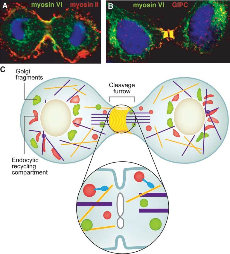
diseases; (ii) identifying   Trust/MRC Building, for access to the Dunn’s
                                                                 and characterising the       excellent proteomic facilities. In the foreword
                                                                 molecular machinery of       to the last Research Report in 2006 I thanked
                                                                 intracellular membrane       Sir Keith Peters and Martin Bobrow for their
                                                                 traffic and determining      great contribution to the establishment of CIMR.
                                                                 how traffic pathways         This time I want to thank Jennie Blackwell.
                                                                 are coordinated,             Jennie has recently left Cambridge to take up a
                                                                 regulated and modified       Chair in Genetics and Health at the University
                                                                 in health and disease;       of Western Australia in Perth. She was the
                                                                 (iii) the identification     first Director of CIMR, but her contribution to
                                                                 of genes, proteins and       establishing our Institute started much earlier.
                                                                 pathways increas-            Jennie came to Cambridge in 1991 as the
                                                                 ing susceptibility to,       Glaxo Professor of Molecular Parasitology first
                                                                 or protection from,          being based in the Department of Pathology
                                                                 autoimmune diseases;         and later the Department of Medicine. Almost
                                                                 (iv) determining the         immediately, Keith Peters identified Jennie as
    CIMR Seminar Posters                                         transcriptional regula-      the person to articulate the rationale and objec-
                                                                 tion of haematopoietic       tives for the new Institute in an application to
                                     stem cells. A major objective for CIMR over              the Wellcome Trust. Jennie later described that
                                     the next five years is to understand protein             period in her introduction to our first Research
                                     localisation, function and metabolism in a range         Report in 2000 where she wrote “Dreams of a
                                     of diseases in which genetic studies have                modern research facility in Cambridge, where
                                     identified the causative genes. We argued to             clinical and basic science could converge in
                                     the Wellcome Trust that underpinning our core            the study of molecular mechanisms of disease,
                                     facilities is essential to achieve this objective        turned to reality in 1993 with a major capital
                                     and will provide added value to the considerable         award from the Wellcome Trust to the School
                                     investment that the Wellcome Trust is already            of Clinical Medicine. At the same time the
                                     making to our scientific activities (currently           Medical Research Council was looking to
                                     about 60% of annual grant spend). In giving us           re-house its Dunn Nutrition Unit in Cambridge.
                                     a Strategic Award we believe that the Wellcome           A joint project was conceived and, two years
                                     Trust has helped to ensure that CIMR is a                of planning and two years of building later, the
                                     flagship in the UK for interdisciplinary research        new Wellcome Trust/MRC Building emerged
                                     at the interface between clinical and basic              on the Addenbrooke’s Hospital Site”. Jennie
                                     research.                                                was the University’s main representative on
                                                                                              the building project team for four years, led
                                                         There are many people                many applications for equipment grants and
                                                         whom I want to thank for             was one of a small group of senior scientists
                                                         their support of CIMR. These         who selected our initial PIs. She established
                                                         include the members of               the management structure and appointed our
                                                         the International Scientific         first management and technical support teams.
                                                         Advisory Board, chaired by           When we finally got into the building it was
                                                         Professor Nick Hastie, who           Jennie as our first Director who established a
                                                         always give us valuable              CIMR style and ethos underlying the way that
                                                         advice and suggestions at            the Institute functions. All of us at CIMR wish
                                                         their biennial visits and the        Jennie success in her new venture in Australia
                                                         Institute’s Strategy Commit-         and we are very pleased that we will still see
                                                         tee, made up of Heads of             her from time to time because she will maintain
                                                         Departments with staff in            some research activity here and is now an affili-
                                                         CIMR, chaired by the Regius          ated PI in CIMR as well as an Honorary Senior
                                                         Professor, Patrick Sissons.          Visiting Fellow to the Department of Medicine.
                                                         We remain very grateful to
                                                         Professor Sir John Walker,
                                                         the Director of the MRC
                                                         Dunn Human Nutrition Unit            Paul Luzio
                                                         housed in the Wellcome               January 2008
    Jennie Blackwell, First Director of CIMR

6
CIMR Principal Investigators

Principal Investigator   Investigator Status                                                 Home Department

Folma Buss               Wellcome Trust Senior Research Fellow in Basic Biomedical Science   Clinical Biochemistry
David Clayton            JDRF/Wellcome Trust Principal Research Fellow and Personal Chair    Medical Genetics
Bertie Göttgens          University Reader                                                   Haematology
Allison Green            Wellcome Trust Senior Research Fellow in Basic Biomedical Science   Pathology
Tony Green               University Chair                                                    Haematology
Fiona Gribble            Wellcome Trust Senior Research Fellow in Clinical Sciences          Clinical Biochemistry
Gillian Griffiths        Wellcome Trust Principal Research Fellow and Personal Chair         Medicine
James Huntington         MRC Senior Research Fellow and University Reader                    Haematology
Brian Huntly             MRC Senior Clinical Research Fellow                                 Haematology
Fiona Karet              Wellcome Trust Senior Research Fellow in Clinical Sciences and      Medical Genetics
                         Personal Chair
Paul Lehner              Wellcome Trust Senior Research Fellow in Clinical Sciences and      Medicine
                         Personal Chair
David Lomas              University Chair and Deputy Director CIMR                           Medicine
Paul Luzio               Personal Chair and Director CIMR                                    Clinical Biochemistry
Katrin Ottersbach        Kay Kendall Leukaemia Fund Intermediate Fellow                      Haematology
David Owen               Wellcome Trust Senior Research Fellow in Basic Biomedical Science   Clinical Biochemistry
                         and University Reader
Andrew Peden             MRC Career Development Fellow                                       Clinical Biochemistry
Roger Pedersen           University Chair                                                    Surgery
Lucy Raymond             University Senior Lecturer                                          Medical Genetics
Randy Read               Wellcome Trust Principal Research Fellow and Personal Chair         Haematology
Evan Reid                Wellcome Trust Senior Research Fellow in Clinical Sciences          Medical Genetics
Margaret Robinson        Wellcome Trust Principal Research Fellow and Personal Chair         Clinical Biochemistry
David Rubinsztein        Wellcome Trust Senior Research Fellow in Clinical Sciences and      Medical Genetics
                         Personal Chair
Christopher Rudd         Wellcome Trust Principal Research Fellow and Personal Chair         Pathology
Peter St George-Hyslop   Wellcome Trust Principal Research Fellow and Personal Chair         Clinical Neurosciences
Richard Sandford         University Reader                                                   Medical Genetics
Matthew Seaman           Senior Research Associate (formerly MRC Senior Research Fellow)     Clinical Biochemistry
Symeon Siniossoglou      Senior Research Associate (formerly Wellcome Trust Career           Clinical Biochemistry
                         Development Fellow)
Ken Smith                University Chair                                                    Medicine
John Todd                University Chair and Director DIL                                   Medical Genetics
John Trowsdale           University Chair                                                    Pathology
Linda Wicker             JDRF/Wellcome Trust Principal Research Fellow, Personal Chair       Medical Genetics
                         and Deputy Director DIL
Geoff Woods              University Reader                                                   Medical Genetics
John Yates               University Chair                                                    Medical Genetics

                                                                                                                      7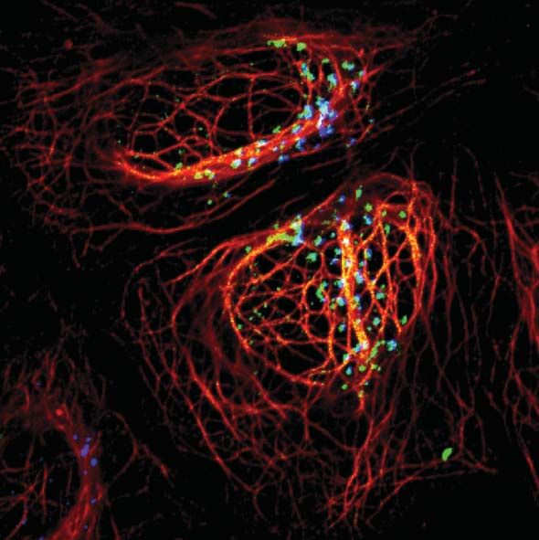
Current Membership of the CIMR Strategy                               Current Membership of the International
    Committee:                                                            Scientific Advisory Board:

    Patrick Sissons (Chairman, Regius Professor of Physic),               Nick Hastie, Chairman (University of Edinburgh), Dennis
    Andrew Bradley (Surgery), Alastair Compston (Clinical                 Ausiello (Harvard University), John Dick (University of
    Neurosciences), Tim Cox (Medicine), John Danesh                       Toronto), Chris Haslett (University of Edinburgh), Louise
    (IPH, Faculty Board), Tony Green (Haematology),                       Johnson (University of Oxford), Carl Nathan (Cornell
    David Lomas (Deputy Director, CIMR), Paul Luzio                       University), Graham Warren (Max Perutz Laboratories, Austria)
    (Director, CIMR), Stephen O’Rahilly (Clinical Biochemistry),
    Bruce Ponder (CRUK CRI, Faculty Board), John Todd
    (Diabetes and Inflammation Laboratory), Andrew Wyllie
    (Pathology), John Yates (Medical Genetics)

    Screening apparatus funded through a Wellcome Trust equipment grant

8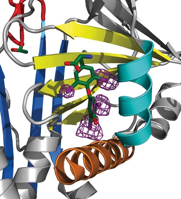
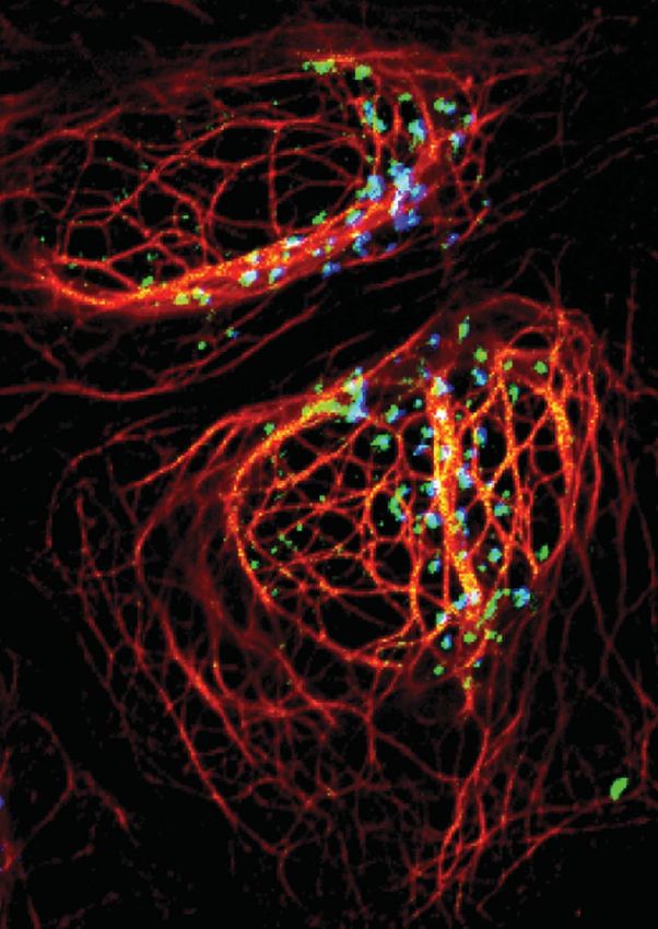
Myosin Motor Proteins in Health and Disease                                                                  Folma Buss

We are studying the role(s) of myosin motor         mid-body during cytokinesis and have shown
proteins in intracellular transport of organelles   that it is involved in a wide variety of intracellular
and vesicles, and in cell migration and cytoki-     processes such as endocytosis, secretion, main-
nesis. In humans at least 40 different myosins      tenance of Golgi morphology, cell movement
operate to control and drive these complex          and cell division. The multiple roles of myosin
range of motile and transport processes. Each       VI in these membrane transport pathways are
myosin is composed of a motor domain that uses      mediated by interaction of its specific C-terminal       Sue Arden
the energy released from ATP hydrolysis to drive    tail domain with a host of distinct binding              Margarita Chibalina
along actin filament tracks around the cell and     partners which are currently one of our main
                                                                                                             Claudia Puri
a tail domain that binds cargo and targets it to    areas of research. Mutations or the absence
specific cellular locations. We have focused on     of myosin VI have been linked to such diverse
myosin VI, which unlike all the other myosins so    pathological processes as deafness, cardiomy-            Funding:
far characterised moves in the reverse direction    opathy, neurodegeneration and cancer. Our long           The Wellcome Trust
                                                                                                             Cancer Research UK
along actin filaments and therefore has unique      term aim is therefore to establish the precise
molecular properties and intracellular func-        intracellular functions of myosin VI so as to allow
tions. We have localized myosin VI in membrane      us to develop therapeutic strategies to combat           Sahlender, D. A., Roberts,
                                                                                                             R. C., Arden, S. D., Spudich,
ruffles, at the Golgi complex, in clathrin coated   or alleviate these pathological disorders.
                                                                                                             G., Taylor, M. J., Luzio, J. P.,
pits and vesicles, at the centrosome and in the                                                              Kendrick-Jones, J. & Buss, F.
                                                                                                             (2005). Optineurin links myosin
                                                                                                             VI to the Golgi complex and is
                                                                                                             involved in Golgi organization
                                                                                                             and exocytosis. J Cell Biol 169,
                                                                                                             285–295.

                                                                                                             Au, J. S., Puri, C., Ihrke, G.,
                                                                                                             Kendrick-Jones, J. & Buss, F.
                                                                                                             (2007). Myosin VI is required
                                                                                                             for sorting of AP-1B-dependent
                                                                                                             cargo to the basolateral domain
                                                                                                             in polarized MDCK cells. J Cell
                                                                                                             Biol 177, 103–114.

                                                                                                             Arden, S. D., Puri, C., Au, J. S.,
                                                                                                             Kendrick-Jones, J. & Buss, F.
                                                                                                             (2007). Myosin VI is required
                                                                                                             for targeted membrane
                                                                                                             transport during cytokinesis.
                                                                                                             Mol Biol Cell 18, 4750–4761.

Myosin VI is recruited to
cleavage furrow in early (A) and
late (B) cytokinesis. (C) Cartoon
illustrating the possible function
of myosin VI in transporting
vesicles into the cleavage
furrow for the final abscission
process.

                                                                                                                                     9
David Clayton Biostatistical Methods in Genetics
Our aim is to develop and apply statistical the management of the huge quantities of data
methods in genetic epidemiology and, to a lesser generated by this study. The main results of the
extent, in other aspects of biostatistics. The main study were published in early 2007.
focus of this work is provided by the Diabetes
and Inflammation Laboratory (DIL), although I The WTCCC has been remarkably successful in
have long-standing interests in studies into the identifying new disease susceptibility loci, particu-
Jeff Barrett genetic epidemiology of other complex diseases, larly for type 1 diabetes, and has generated much
Jason Cooper including hypertension, and senile macular follow-on work. Fine mapping of causal variants
Geoffrey Dolman degeneration. Implementation and dissemination remains a considerable challenge and refine-
of analytical methods as an important part of our ment of methods for the design and analysis
Vin Everett
work. of fine mapping studies remains a high priority
Joanna Howson for my group. In addition the Type 1 Diabetes
Hin-Tak Leung A major commitment over the last two years Consortium has funded a further genome-wide
Vincent Plagnol has been the Wellcome Trust Case-Control association study of 4,000 new cases and 2,500
Neil Walker Consortium (WTCCC). This is a genome-wide new controls (all drawn from the existing DIL
association (GWA) study in which 2,000 cases collections) and my group will be responsible for
Chris Wallace
of each of 7 different diseases will be compared the analysis of the new data and for a combined
with two shared control groups, together compris- analysis of the new data in conjunction with the
Funding: ing 3,000 subjects. Each subject was typed for existing WTCCC T1D data (2,000 cases and
The Wellcome Trust ~500,000 SNPs. I co-chaired the analysis group 3,000 controls). I also hope to provide advice and
Juvenile Diabetes Research
and we have developed analysis software, which software for several proposed GWA association
Foundation
we have made freely available on the Internet. studies.
Medical Research Council
My group has also made a major contribution to
Todd, J. A., Walker, N. M.,
Cooper, J. D., Smyth, D. J.,
Downes, K., Plagnol, V.,
Bailey, R., Nejentsev, S.,
Field, S. F., Payne, F.,
Lowe, C. E., Szeszko, J. S.,
Hafler, J. P., Zeitels, L.,
Yang, J. H., Vella, A.,
Nutland, S., Stevens, H. E.,
Schuilenburg, H., Coleman, G.,
Maisuria, M., Meadows, W.,
Smink, L. J., Healy, B.,
Burren, O. S., Lam, A. A.,
Ovington, N. R., Allen, J.,
Adlem, E., Leung, H. T.,
Wallace, C., Howson, J. M.,
Guja, C., Ionescu-Tirgoviste, C.,
Genetics of Type 1 Diabetes
in Finland, Simmons, M. J.,
Heward, J. M., Gough, S. C.,
Dunger, D. B., The Wellcome
Trust Case Control
Consortium, Wicker, L. S.
& Clayton, D. G. (2007).
Robust associations of four
new chromosome regions from
genome-wide analyses of type
1 diabetes. Nat Genet 39,
857–864.
The Wellcome Trust Case
Control Consortium (2007).
Genome-wide association
study of 14,000 cases of seven
common diseases and 3,000
shared controls. Nature 447,
661–678.
10Transcriptional Control of Normal and Leukaemic Bertie Göttgens
Blood Stem / Progenitor Cells
Haematopoiesis has served as a model process predicted in vivo activity. In parallel, transgenic
for studying stem cell biology, and a close and molecular studies are performed to dissect
developmental link between the formation the transcriptional regulation of LMO2, Lyl1,
of embryonic blood and endothelial cells has SCL and endoglin, four key regulators of early
long been recognised. However, the transcrip- blood development. The latter bottom-up studies
tional networks that determine these early cell continue to inform the design of bioinformatic
fate decisions are still poorly understood. The search strategies to allow refinement of top- Nicolas Bonadies
Göttgens laboratory is part of a consortium of down computational approaches. Yi-Han Cheng
Cambridge groups, largely in the CIMR, focusing
Dean Griffiths
on complementary aspects of normal and leu- Identification of transcriptional hierarchies in nor-
kaemic stem cell biology. The specific focus of mal cells will illuminate the molecular hierarchy of Sarah Kinston
the Göttgens group is the combination of com- transcriptional programmes responsible for blood Diego Miranda
putational approaches with mouse experimental stem cell development. The importance of tran- Helen Oram
model systems for the analysis of transcriptional scriptional control in both normal and leukaemic Aileen Smith
networks during the formation of embryonic cells is underlined by the large number of tran-
Nicola Wilson
blood and endothelial cells. scription factor genes that are disrupted as part
of the pathogenesis of haematological malignan- Andrew Wood
Recent work has employed both top-down and cies. Future work will address how transcriptional
bottom-up approaches to identify and charac- programmes are perturbed in specific subtypes of Funding:
terise key regulatory elements of blood stem leukaemia and may thus open up new avenues Leukaemia Research Fund
cell transcriptional networks. A new suite of for the development of targeted therapies. Cancer Research UK
bioinformatics tools has been developed for Newton Trust
genome-wide top-down computational iden- Further details can be found at The Wellcome Trust
tification of gene regulatory elements with http://hscl.cimr.cam.ac.uk IBM
Kay Kendall Leukaemia Fund
Leukemia and Lymphoma
Society
Pimanda, J. E., Chan, W. Y.,
Donaldson, I. J., et al. (2006).
Endoglin expression in the
endothelium is regulated by
Fli-1, Erg, and Elf-1 acting on
the promoter and a -8-kb enhancer.
Blood 107, 4737–4745.
Pimanda, J. E., Donaldson, I. J.,
de Bruijn, M. F. et al. (2007).
The SCL transcriptional network
and BMP signaling pathway
interact to regulate RUNX1 activity.
Proc Natl Acad Sci USA 104,
840–845.
Chan, W. Y., Follows, G. A.,
Lacaud, G. et al. (2007). The
paralogous hematopoietic
regulators Lyl1 and Scl are
coregulated by Ets and GATA
factors, but Lyl1 cannot rescue
the early Scl-/- phenotype. Blood
A nascent HSC transcriptional network composed 109, 1908–1916.
of 14 genes and 35 connections all of which have
been verified by chromatin-immunoprecipitation and
transgenic assays.
11Allison Green Deciphering Immunological Mechanisms to
Control, or Promote, Autoimmune Diseases
The goal of our laboratory is to understand the the molecular requirement for CD40-CD154 in
mechanisms by which inflammation promotes CD4+Foxp3+ Treg biology is ongoing.
autoimmune disease, particularly type 1 diabetes.
Using a series of murine models either transgenic We also investigate how the suppressor cytokine
or knockout for key cytokines/co-stimulatory TGFß impedes diabetes development. Using a
molecules, we exam the impact such events have unique transgenic model where islet specific
Eunice Amofah on the ability of the immune system to generate expression of TGFß can be controlled by an
Philip Spence CD4+Foxp3+ Treg cells, or autoaggressive CD8+ on/off switch, we have defined a phase in the
T cells. disease where a short pulse of TGFß significantly
Jennifer Varian
delays diabetes progression. The mechanisms by
Maja Wallberg We have established that CD154-CD40 inter- which TGFß controls the autoimmune response
action is critical for the thymic development of is subject to continued investigation.
Funding: CD4+Foxp3+ Treg cells. Deficiency in either
The Wellcome Trust molecule reduces CD4+Foxp3+ Treg numbers by We have shown that, surprisingly, B cells are
Diabetes UK 50%. Bone marrow chimeric mice showed that central for promoting acceleration to diabetes in
CD40 signals derived from thymic epithelial cells the presence of an inflammatory response. Impact-
McGregor, C. M., (mTECs) or thymic dendritic cells (tDCs) promiscu- ing on end stage disease, B cells promote CD8+
Schoenberger, S. P. & ously promote CD4+Foxp3+ Treg cell development. T cells differentiate to CTL and survival within the
Green, E. A. (2004). CD154 Further, CD154 requirement is restricted to islets. The mechanisms by which B cells manipu-
is a negative regulator of
developing CD4+Foxp3+ Treg cell and signals to late CD8+ T cell responses are under investigation.
autoaggressive CD8+ T cells in
type 1 diabetes. Proc Natl Acad induce its differentiation/expansion. In contrast to Finally, we are using a series of congenic mice to
Sci USA 101, 9345–9350. developing CD4+Foxp3+ Treg cells, CD40-CD154 establish if key genes that control diabetes are
Chamberlain, G., Wallberg, signals are redundant in homeostatic mainte- linked to immunological changes in the function of
M., Rainbow, D., Hunter, K., nance of mature CD4+Foxp3+ Treg cells. Defining the B or CD8+ T cell compartment.
Wicker, L. S. & Green, E. A.
(2006). A 20-Mb region of
chromosome 4 controls TNF-
alpha-mediated CD8+ T cell
aggression toward beta cells in
type 1 diabetes. J Immunol 177,
5105–5114.
Millet, I., Wong, F. S., Gurr,
W., Wen, L., Zawalich, W.,
Green, E. A., Flavell, R. A. &
Sherwin, R. S. (2006). Targeted
expression of the anti-apoptotic
gene CrmA to NOD pancreatic
islets protects from autoimmune
diabetes. J Autoimmun 26,
7–15.
Interaction of Foxp3+ regulatory T cells
(green) with thymic epithelial cells (red) in
the thymic medulla.
12Haematopoietic Stem Cells and Tony Green
Haematological Malignancies
Haematopoiesis represents the best studied study, the largest randomised clinical trial of
adult stem cell system and continues to provide any MPD yet performed; (iv) the identification
important paradigms for the mechanisms of JAK2 exon 12 mutations associated with a
whereby normal stem cells are subverted to distinct variant of PV.
form malignancies. This laboratory is pursuing
two complementary aspects of haematopoietic 2. Transcriptional regulation of haematopoietic
stem cell (HSC) biology. stem cells. The stem cell leukaemia (SCL) Joanna Baxter
gene encodes a bHLH transcription factor
Philip Beer
1. Human myeloproliferative disorders (MPDs). and was originally identified by virtue of its
Jacinta Carter
These myeloid malignancies result from trans- disruption in T-cell acute leukaemia. Loss
formation of an HSC and are associated with and gain of function studies have shown that Edwin Chen
expansion of one or more haematopoietic SCL is a pivotal regulator of haematopoiesis Mark Dawson
lineages. Patients are at risk of developing and that appropriate transcriptional regula- Rita Ferreira
thrombosis, myelofibrosis and acute myeloid tion is critical for its biological functions. We
George Follows
leukaemia. We are studying the molecular are undertaking a systematic analysis of the
Michelle Hammett
pathogenesis and management of the MPDs. transcriptional regulation of the SCL locus
Recent highlights include (i) the demonstration using genomic, transgenic, knockout, cellular Mary Janes
that an acquired V617F mutation of JAK2 is and biochemical approaches. Recent achieve- Rebecca Kelley
present in virtually all patients with polycythae- ments include (i) molecular characterisation of Juan Li
mia vera (PV) and approximately half those two HSC enhancers; (ii) characterisation of a
Linda Scott
with essential thrombocythaemia (ET) and novel enhancer which targets primitive eryth-
Dominik Spensberger
idiopathic myelofibrosis; (ii) the demonstration ropoiesis; (iii) description of the molecular
that V617F-positive ET represents a forme basis for the emergence of an HSC enhancer
fruste of PV; (iii) publication of the MRC PT1 during vertebrate evolution. Funding:
The Wellcome Trust
Medical Research Council
Leukaemia Research Fund
1992 PV 2004 AML
Leukaemia & Lymphoma
Society
Harrison, C. N., Campbell, P. J.,
Buck, G. et al. (2005).
Hydroxyurea compared with
anagrelide in high-risk essential
thrombocythemia. N Engl J Med
353, 33-45.
Scott, L. M., Tong, W., Levine,
R. L. et al. (2007). JAK2 exon 12
mutations in polycythemia vera
Granulocytes Bone marrow Bone marrow and idiopathic erythrocytosis. N
Engl J Med 356, 459–468.
Leukaemic transformation of polycythaemia Ogilvy, S., Ferreira, R., Piltz, S.
vera. Somatic mutation of JAK2 is present in G. et al. (2007). The SCL +40
chronic phase polycythaemia vera (PV) but un- enhancer targets the midbrain
expectedly absent from leukaemic blasts in most together with primitive and
patients following development of acute myeloid definitive hematopoiesis and is
leukaemia (AML). regulated by SCL and GATA
proteins. Mol Cell Biol 27,
7206–7219.
13Fiona Gribble Stimulus-secretion Coupling Mechanisms in
Intestinal Neuroendocrine Cells
Hormones from entero-endocrine cells in the gas- tial, ionic currents, intracellular [Ca2+] and GLP-1
trointestinal tract play a role in the control of diverse release are monitored following application of a
processes such as appetite and insulin secretion, variety of nutrients, hormones and pharmacologi-
as well as coordinating local gut physiology. Two cal agents. Populations of entero-endocrine cells
major “incretin” hormones, GLP-1 (glucagon-like can be identified and purified from transgenic
peptide-1) and GIP (glucose-dependent insulino- mice exhibiting cell-specific expression of a
Abdella Habib tropic peptide), are responsible for triggering up to fluorescent protein, opening the way to perform
half of the insulin that is released following a meal, expression profiling and characterisation of
Helen Parker
and have therapeutic potential for the treatment primary entero-endocrine cells. Our hope is that
Frank Reimann
of type 2 diabetes. GLP-1 based therapies have understanding the stimulus-secretion coupling
Gareth Rogers recently reached the marketplace as anti-diabetic pathways in intestinal endocrine cells may pave
Gwen Tolhurst agents, and other entero-endocrine hormones are the way for the development of alternative
under therapeutic evaluation for a range of clinical nutritional and pharmacological therapies for
Funding: disorders. conditions such as type 2 diabetes, obesity and
The Wellcome Trust gastrointestinal disorders.
St John’s College, The primary interest of our research group
Cambridge is to understand the mechanisms underlying A second interest of our group is in understanding
The Lister Institute of secretion of the incretin hormones, with a view the electrophysiological mechanisms underlying
Preventive Medicine to identifying novel targets and pathways in pain perception in man. In collaboration with the
entero-endocrine cells that could be exploited to group of Dr Geoff Woods, we have characterised
modulate hormone release in vivo. We employ an the functional effects of mutations in a sodium
Reimann, F., Ward, P. S.,
Gribble, F. M. (2006). approach based around the use electrophysiol- channel gene that give rise to human conditions
Signalling mechanisms ogy and fluorescence imaging to study the events of altered pain perception. Work in this area has
underlying the release of involved in stimulus detection and hormone major implications for the future development of
Glucagon-Like Peptide-1.
secretion. Changes in plasma membrane poten- analgesic and anaesthetic agents.
Diabetes 55, S78–S85.
Cox, J. J., Reimann, F.,
Nicholas, A. K., Thornton, G.,
Roberts, E., Springell, K.,
Karbani, G., Jafri, H.,
Mannan, J., Raashid, J.,
Al-Gazali, L., Hamamy, H.,
Valente, E. M., Gorman, S.,
Williams, R., McHale, D. P.,
Wood, J. N., Gribble, F. M.
& Woods, C. G. (2006). An
SCN9A channelopathy causes
congenital inability to experience
pain. Nature 444, 894–898.
O’Malley, D., Reimann, F.,
Simpson, A. K. & Gribble, F. M.
(2006). Sodium-coupled
glucose cotransporters
contribute to hypothalamic
glucose sensing. Diabetes 55,
3381–3386.
GLP-1 secreting cells in intestinal tissue sections
from transgenic mice are identifiable by their green
fluorescence. Green: GFP; Red: glucagon; Blue: DAPI.
14Polarized Secretion from Lymphocytes Gillian Griffiths
Cytotoxic T lymphocytes (CTL) and Natural Killer We are now trying to understand the mechanisms
(NK) cells use polarized secretion to rapidly that control polarized secretion from CTL and
destroy virally infected and tumorigenic cells. related immune cells. We are taking a number of
Upon recognition of their targets, CTL and NK approaches including the study of human genetic
cells polarize their secretory apparatus towards diseases, such as Hemophagocytic syndrome,
the target, releasing the content of specialized in which CTL and NK cell function is disrupted.
secretory lysosomes into the immunological Using molecular, biochemical and morphological Tiziana Daniele
synapse formed between the two cells. We have techniques we are able to ask where the secre- Misty Jenkins
shown that the mechanism of secretion used by tory process is disrupted and identify the role of
Winnie Lui-Roberts
CTL and NK cells is unusual, with the centrosome the proteins, which are disrupted in these genetic
polarizing to a precise spot within the synapse, diseases, in secretion. We are also examining the Jane Stinchcombe
where signaling has taken place. Secretory lyso- roles of proteins which are known to be involved Andy Tsun
somes move along the microtubules towards the in cell polarity and asking whether these play a Matt Wenham
centrosome and are delivered to a specialized role in secretion from CTL and NK cells.
secretory domain as the centrosome contacts Funding:
the plasma membrane. The Wellcome Trust
Stinchcombe, J. C., Majorovits, E.,
Bossi, G., Fuller, S. &
Griffiths, G. M. (2006).
Centrosome polarization delivers
secretory granules to the
immunological synapse. Nature
443, 462–465.
Zuccato, E., Blott, E. J., Holt, O.,
Sigismund, S., Shaw, M.,
Bossi, G. & Griffiths, G. M.
(2007). Sorting of Fas ligand to
secretory lysosomes is regulated
by mono-ubiquitylation and
phosphorylation. J Cell Sci 120,
191–199.
Stinchcombe, J. C. &
Griffiths, G. M. (2007). Secretory
mechanisms in cell-mediated
cytotoxicity. Annu Rev Cell Dev
Biol (in press).
A CD8 (blue) cytotoxic T lymphocyte killing its target
(right) with the centrosome (gamma tubulin, red)
polarising to the point of T cell receptor signalling
shown using an antibody to lck (green).
15James Molecular Recognition in Haemostasis
Huntington
Blood coagulation (haemostasis) is a complex structures of antithrombin and heparin cofactor II
process under tight regulatory control. Dys- in recognition complexes with target haemostatic
regulation leads to bleeding when the clotting proteases, and have recently solved a similar
response is insufficiently rapid and robust, and to structure of protein C inhibitor demonstrating
thrombosis when coagulation is not limited. This how it carries out its procoagulant function.
‘haemostatic balance’ is critical for human health, Another project in the lab focuses on determin-
and understanding the regulatory mechanisms ing the molecular basis of thrombin function. This
is crucial for the diagnosis, prevention and treat- has traditionally involved crystallographic studies
Ty Adams ment of diseases such as haemophilia, deep vein of thrombin complexed to substrates, cofactors
Daniel Johnson thrombosis, pulmonary embolism, heart attack, and inhibitors. We now have a productive NMR-
and stroke. My lab studies the molecular events based thrombin effort which promises to unveil
Jonathan Langdown
which maintain the haemostatic balance mainly the functional relevance of thrombin allostery and
Wei Li by determining crystallographic structures of indi- to map interactions with its molecular partners.
Stephan Luis vidual coagulation factors and of the multi-protein The events which lead to thrombin formation
Genichi Nakamura complexes they form. We have several projects are also studied. We are crystallising individual
Masayuki Yamasaki running concurrently in the lab. The serpin project members of the haemostatic network of proteins
studies how the inhibitory activity of serpins is and their complexes. In the long term, our efforts
stimulated by heparin-like glycosaminoglycans. will define the principal molecular recognition
Funding:
We have succeeded in defining the mode of events that govern haemostasis.
Medical Research Council
action of therapeutic heparin by solving the
British Heart Foundation
National Institutes of Health
(USA)
Li, W., Johnson, D. J.,
Esmon, C. T. &
Huntington, J. A. (2004).
Structure of the antithrombin-
thrombin-heparin ternary
complex reveals the
antithrombotic mechanism of
heparin. Nat Struct Mol Biol
11, 857–862.
Huntington, J. A. (2006).
Shape-shifting serpins
– advantages of a mobile
mechanism. Trends Biochem
Sci 31, 427–435.
Johnson, D. J., Li, W.,
Adams, T. E. &
Huntington, J. A. (2006).
Antithrombin-S195A factor
Xa-heparin structure reveals
the allosteric mechanism of How antithrombin (AT, ribbon) is activated by heparin
antithrombin activation. EMBO J (ball-and-stick) to achieve rapid inhibition of target
25, 2029–2037. proteases (cyan) factor Xa and thrombin.
16Molecular and Cellular Characterisation Brian Huntly
of Leukaemia Stem Cells
The aims of the Huntly group are to study mecha- are particularly focusing on identifying novel and
nisms of leukaemogenesis and in particular to characterising known and novel self-renewal
characterise leukaemia stem cells (LSC) at the pathways downstream of leukaemia-associated
molecular and cellular levels. Leukaemias and fusion oncogenes such as MOZ-TIF2 and NUP98-
many other cancers have recently been dem- HOXA9. In addition, we are aiming to identify
onstrated to be wholly dependent upon a small genes which are required for LSC function but
population of so-called cancer stem cells for their are dispensable for normal haematopoietic stem Shubha Anand
continued growth and propagation. These cells cell function. These genes would then be attrac- Polly Chan
represent the most critical targets for treatment tive candidates for therapeutic targeting of LSC.
Brynn Kvinlaug
of leukaemia and a greater understanding of their Another major interest of the group is the regu-
biology and its interface with normal stem cell lation of HOX genes in AML. We have recently Donna Paul
function is fundamental to improving treatment described, along with collaborators, the associa- Clare Pridans
outcomes. The focus of the Huntly laboratory on tion of the caudal-like CDX family members CDX4 Frances Stedham
this interface complements a local consortium of and 2 with HOX gene regulation ion AML and are
research groups (Profs Green and Warren and further interrogating this association. Finally, we Funding:
Drs Göttgens and Ottersbach) with interests in are characterising the LSC hierarchy and biology Medical Research Council
normal and leukaemic stem cells all based in the of the preleukaemic myeloproliferative disorders Kay Kendall Leukaemia
CIMR or on the Cambridge University Hospital (MPD) using analysis of highly purified popula- Fund
campus. The Huntly group utilises functional tions of stem and progenitor cells from MPD Leukemia Lymphoma
assays and genomic techniques in complemen- patients and a number of recently discovered Society of America
tary mouse models and human primary cells to mutations (such as the JAK2 V617F mutation) Cancer Research UK
study leukaemogenesis and LSC biology. We as clonal markers. EHA-Jose Carerras
Foundation
Huntly, B. J. P., Shigematsu, H.,
Deguchi, K., Lee, B. H.,
Mizuno, S., Duclos, N., Rowan, R.,
Amaral, S., Curley, D.,
Williams, I. R., Akashi, K.
& Gilliland, D. G. (2004).
MOZ-TIF2, but not BCR-ABL,
confers properties of leukemic
stem cells to committed murine
hematopoietic progenitors.
Cancer Cell 6, 587–596.
Huntly, B. J. P. & Gilliland, D. G.
(2005). Leukaemia stem cells and
the evolution of cancer-stem-cell
research. Nat Rev Cancer 5,
311–321.
Bansal, D., Scholl, C., Frohling, S.,
McDowell, E., Lee, B. H.,
Dohner, K., Ernst, P.,
Davidson, A., Daley, G. Q.,
Zon, L. I., Gilliland, D. G. &
Huntly, B. J. P. (2006). Cdx4
dysregulates Hox gene expression
and generates acute myeloid
Leukaemia stem cells (LSC) as critical leukemia alone and in cooperation
targets in disease eradication. with Meis1a in a murine model.
Proc Natl Acad Sci USA 103,
A) Current therapies do not eradicate LSC
16924–16929.
allowing disease relapse and resistance.
B) A greater knowledge of LSC biology will
allow us to target this compartment and
improve patient survival.
17Fiona Karet Molecular Physiology of Renal Tubular
Homeostasis in Health and Disease
We aim to characterise molecular mechanisms and sensorineural hearing loss (SNHL) is often
governing human renal tubular homeostasis, with associated.
a major focus on acid-base balance and mecha-
nisms underlying stone disease. We described mutations in the basolateral anion
exchanger gene AE1 in dominant dRTA, and dis-
Acid-base regulation is the chief job of the !- covered two genes (ATP6V1B1 and ATP6V0A4),
Katy Blake-Palmer intercalated cells (!-IC) in the distal nephron. encoding kidney-specific B1 and a4 subunits of
Andrew Fry Intact !-intercalated cell functions (secretion of the !-IC surface proton pump, where loss-of-
protons in to the urine coupled to bicarbonate function mutations cause recessive dRTA.
Shoko Horita
reclamation) are necessary for appropriate excre-
Liz Norgett tion of the net acid load of a normal diet, and for We discovered that proton pumps in the kidney
Annabel Smith generation of adequate amounts of bicarbonate contain four other organ-specific subunit isoforms
Ya Su for buffering. However, neither the identity of all (C2, G3, d2 and e2) and that G subunits interact
the transporters, pumps and channels responsi- physically with a-subunit isoforms.
Funding: ble, nor the regulatory pathways involved, are yet
The Wellcome Trust well understood. Moving from genetic to functional studies, we
Kidney Research UK demonstrated abnormal targeting of AE1 as
Addenbrooke’s Charitable Adopting an initial genetic approach, we studied a mechanism of dominant disease, and have
Trust rare single-gene disorders (the distal renal demonstrated that the glycolytic enzyme PFK-1
tubular acidoses, dRTAs) where !-IC function is is essential for normal proton pump function via
inadequate, imparting large quantitative effects a-subunit binding.
Smith, A. N., Jouret, F., Bord,
S., Borthwick, K. J., Al-Lamki, on the kidney’s ability to maintain normal body
R. S., Wagner, C. A., Ireland, fluid pH. dRTA is clinically defined by metabolic Our studies currently focus on characterizing the
D. C., Cormier-Daire, V., acidosis, rickets and calcium deposition in the cellular behaviour of AE1, on further dissecting the
Frattini, A., Villa, A., Kornak, U.,
kidney. The recessively inherited syndromes proton pump and on identifying the molecular path-
Devuyst, O. & Karet, F. E.
(2005). Vacuolar H+-ATPase present with very severe changes at a young age ways responsible for various other tubulopathies.
d2 subunit: molecular
characterization, developmental
regulation, and localization to
specialized proton pumps in
kidney and bone. J Am Soc
Nephrol 16, 1245–1256.
Norgett, E. E., Borthwick, K. J.,
Al-Lamki, R. S., Su, Y., Smith,
A. N. & Karet, F. E. (2007). V1
and V0 domains of the human
H+-ATPase are linked by an
interaction between the G and
a subunits. J Biol Chem 282,
14421–14427.
Blake-Palmer, K. G., Su, Y.,
Smith, A. N. & Karet, F. E.
(2007). Molecular cloning and
characterization of a novel form
of the human vacuolar H+-
ATPase e-subunit: an essential
Renal acid-base regulatory cell. Apical ATPases (red)
proton pump component. Gene
contain alternates for a, B, C, d, e and G subunits.
393, 94–100.
Mutations in B1 or a4 cause recessive dRTA with
deafness. AE1 (yellow) mistargeting causes dominant
disease.
18The Regulation of Cell Surface Receptors by Paul J Lehner
Viral and Cellular Ubiquitin E3 Ligases
The focus of my laboratory is to study the cellular larly of the MHC class I antigen presentation
regulation of MHC class I molecules and other pathway (ii) the role of ubiquitin in the regulation
immune receptors and how these are altered by of cell surface receptor expression, including
invading pathogens. Receptor ubiquitination by identification of ubiquitin E3 ligases, ubiquitin
E3 ligases has emerged as an important means E2 conjugating enzymes and deubiquitinating
of regulating receptor cell surface expression. enzymes involved in this process. We are particu-
We study both constitutive cell surface ubiqui- larly interested in lysine-63 mediated ubiquitin Sheela Abraham
tination and ubiquitination induced by viral gene conjugation as a mechanism for receptor traf- Jessica Boname
products. Both herpes and poxviruses have ficking to an endolysosomal compartment (iii)
Helen Bye
pirated ubiquitin E3 ligases from their vertebrate The physiological role of the MARCH genes. We
hosts. These viral E3 ligases ubiquitinate and have developed a number of mouse models to Florencia Cano
downregulate critical receptors of the immune identify the substrates of the MARCH proteins Lidia Duncan
system. The cellular orthologues of the viral E3 and to study the role of these novel E3 ligases Simon Hoer
ligases are the MARCH (Membrane Associated in health and disease (iv) identification of the James Nathan
RING-CH gene products), whose function is endosomal sorting machinery used by internal-
Helen Stagg
poorly defined. ised, ubiquitinated cell surface receptors and (v)
identification of receptors used by human and Mair Thomas
We are interested in (i) mechanisms used by microbial heat shock proteins in dendritic cell Dick Van Boomen
viruses to evade immune recognition, particu- signalling and activation.
Funding:
The Wellcome Trust
The Lister Institute of
Preventive Medicine
Medical Research Council
Isaac Newton Trust
Thomas, M., Boname, J. M.,
Field, S., Nejentsev, S., Salio, M.,
Cerundolo, V., Wills, M., Lehner,
P. J. (2007). Downregulation of
NKG2D and NKp80 ligands by
Kaposi’s sarcoma-associated
herpesvirus K5 protects against
NK cell cytotoxicity. Proc Natl
Acad Sci USA (in press).
Duncan, L. M., Piper, S., Dodd,
R. B., Saville, M. K., Sanderson,
C. M., Luzio, J. P. & Lehner,
P. J. (2006). Lysine-63-linked
ubiquitination is required for
endolysosomal degradation of
class I molecules. EMBO J 25,
Stable-isotope labelling by amino acids in cell culture
1635–1645.
(SILAC) followed by mass-spectrometry allows us
to perform quantitative proteomic analysis of plasma Floto, R. A., MacAry, P. A.,
membrane preparations. We can identify novel Boname, J. M., Mien, T. S.,
substrates of ubiquitin E3 ligases which regulate Kampmann, B., Hair, J. R.,
cell surface receptors. Dr Simon Hoer (CIMR) in Huey, O. S., Houben, E. N.,
collaboration with Dr Ari Admon, Haifa Technion, Pieters, J., Day, C., Oehlmann,
Israel. W., Singh, M., Smith, K. G. C. &
Lehner, P. J. (2006). Dendritic
cell stimulation by mycobacterial
Hsp70 is mediated through
CCR5. Science 314, 454–458.
19David Lomas The Serpinopathies and Alzheimer’s Disease:
Disease Mechanisms and Therapeutic
Interventions
One in twenty-five of the Northern European cause an inclusion body dementia that we have
population carries the Z allele (342Glu‡Lys) called familial encephalopathy with neuroserpin
of !1-antitrypsin. Homozygotes for this mutation inclusion bodies (FENIB). We have described 5
retain !1-antitrypsin within hepatocytes as inclu- families with FENIB caused by 4 different muta-
sion bodies that are associated with neonatal tions and have demonstrated a clear correlation
hepatitis, juvenile cirrhosis and hepatocellular between genotype and phenotype based on the
Didier Belorgey carcinoma. We have shown that Z !1-antitrypsin rate of polymer formation. We are dissecting
Robin Carrell is retained within hepatocytes by a unique the mechanism of polymerisation of mutants
protein-protein interaction between the reactive of neuroserpin and determining how these
Dhia Chandraratna
centre loop of one molecule and "-sheet A of a cause neurotoxicity with biochemical, cell and
Ugo Ekeowa second. The structure and significance of these Drosophila models of disease. Finally, we have
Peter Hagglof loop-sheet polymers has been confirmed using demonstrated that neuroserpin is also important
Susanna Karlsson-Li biochemical, biophysical, crystallographic, and in the far more common dementia caused by
Heike Kroeger cell biology studies and with monoclonal antibod- Alzheimer’s disease. Indeed we have shown a
ies and animal models of disease. We are now specific interaction between the Alzheimer’s A"
Beinan Liu
using in silico screening to identify compounds peptide and neuroserpin and have demonstrated
Ian MacLeod that can bind to, and prevent the polymerisation that this interaction is neuroprotective in cell and
Stefan Marciniak of, mutant !1-antitrypsin in vitro and in vivo. Alpha- Drosophila models of disease. Our long term goal
Claire Michel 1-antitrypsin is a member of the serine protease is to understand the pathways of cell toxicity in
Elena Miranda inhibitors or serpin superfamily of proteins. We serpin polymer mediated syndromes (the ser-
have shown that mutants of another serpin, pinopathies) and in Alzheimer’s disease and to
Alison Nobes
neuroserpin, also polymerise within neurones to develop novel therapeutic strategies.
Alison Warrington
Funding:
Medical Research Council
Engineering and Physical
Sciences Research Council
The Wellcome Trust
Merck Sharp & Dohme
Wenner-Gren Center
Alzheimer’s Research Trust
German Academic
Exchange Service
Damian Crowther
The predicted binding pose of a
small molecule polymer blocker
to the lateral hydrophobic cavity
Luheshi, L. M., Tartaglia, G., of !1-antitrypsin.
Kinghorn, K. J., Crowther, D. Mallya, M., Phillips, R. L.,
C., Sharp, L. K. et al. (2006). Saldanha, S. A. et al. (2007). Small Brorsson, A.-C. et al. (2007).
Neuroserpin binds A" and is a molecules block the polymerization Systemic in vivo analysis of the
neuroprotective component of of Z !1-antitrypsin and increase the intrinsic determinants of amyloid
amyloid plaques in Alzheimer clearance of intracellular aggregates. beta pathogenicity. PLoS Biol 5,
disease. J Biol Chem 281, J Med Chem 50, 5357–5363. e290.
29268–29277.
20Molecular Cell Biology of Post-Golgi Membrane Paul Luzio
Traffic Pathways
Secretion and endocytosis are fundamental proc- for transport) proteins. A key protein in fusion
esses occurring in all nucleated mammalian cells. events involving lysosomes is the membrane
We are currently focusing on how cells achieve protein Vamp7. We are currently investigating at
sorting and delivery of endocytosed macro- a biochemical and structural level, two binding
molecules to lysosomes. Lysosomes are small partners of Vamp7, one of which is responsible
membrane bound organelles ~0.5μm diameter, for ensuring that Vamp7 is recovered from the
which are full of proteases and other hydrolytic cell surface following lysosome fusion with the Nick Bright
enzymes as well as internal membranes. They plasma membrane and the other likely to be Sally Gray
function late in the endocytic pathway that required for regulating lysosome-endosome
Michael Parkinson
takes up macromolecules from the cell surface, fusion. We are also studying the role of different
by fusing with endosomes, but also play a key protein machineries including the ESCRT proteins
role in phagocytosis, autophagocytosis and cell in preparing endosomes for fusion with lysosomes Funding:
Medical Research Council
surface membrane repair, the latter by fusing and for sorting endocytosed cargoes in different
with the plasma membrane. The late endosomes ways. Our structural studies are in collaboration
Bright, N. A., Gratian, M. J. &
that fuse with lysosomes are observed as MVBs with David Owen and we also collaborate with Luzio, J. P. (2005). Endocytic
(multivesicular bodies) in the electron micro- Paul Lehner on the intracellular sorting of Class delivery to lysosomes mediated
scope and sorting of membrane proteins into I MHC molecules following downregulation from by concurrent fusion and kissing
events in living cells. Curr Biol
the lumenal vesicles of these MVBs is mediated the cell surface by viruses.
15, 360-365.
by ESCRT (endosomal sorting complex required
Bowers, K., Piper, S. C.,
Edeling. M., Gray, S. R.,
Owen, D. J., Lehner, P. J.
& Luzio, J. P. (2006).
Degradation of endocytosed
epidermal growth factor and
virally ubiquitinated Major
Histocompatibility Complex
Class I is independent of
mammalian ESCRTII. J Biol
Chem 281, 5094-5105.
Luzio, J. P., Pryor, P. R.
& Bright, N. A. (2007).
Lysosomes: fusion and function.
Nat Rev Mol Cell Biol 8,
622-632.
Immunoelectron micrograph showing endocytosed
Class I MHC molecules (10nm gold) in a multivesicular
body of a cultured human HeLa cell expressing a viral
ubiquitin ligase.
21Katrin Ottersbach The Developmental Origins of
Haematopoietic Stem Cells
Haematopoietic stem cells (HSCs) have been later these cells can also be detected in the yolk
intensely studied for many decades as a model sac and the foetal liver. Our previous work has
system for stem cell biology. Our work focuses identified the placenta as another organ that
on the emergence and regulation of the first harbours HSCs during development. In fact,
HSCs in the mouse embryo in order to identify stem cells in this organ by far outnumber those
the basic mechanisms that control their genera- found in the AGM and yolk sac, thus making the
Bahar Mirshekar- tion from precursors and their initial expansion placenta a very attractive tool for studying micro-
Syahkal and dissemination to the different haematopoi- environmental factors for HSC expansion.
etic organs. Knowledge of these early regulatory
Aimée Parker
pathways has proven to be invaluable for under- We have also carried out a number of microar-
standing how adult HSCs can be manipulated ray experiments with the aim of identifying
Funding: for clinical purposes and how interference with some of the key regulators in HSC generation
Kay Kendall Leukaemia Fund these processes may result in blood-related dis- in the AGM. This expression analysis involved
orders. Our research therefore complements that comparison of (1) the region around the aorta
of several groups on the Cambridge University before and after stem cell detection, (2) HSCs
Ottersbach, K. & Dzierzak, E. Hospital site which also work on various aspects and their putative precursors and (3) different
(2005). The murine placenta
of normal and leukaemic stem cell biology. regions within the AGM that do or do not support
contains hematopoietic stem
cells within the vascular labyrinth HSCs. These studies resulted in a list of candi-
region. Dev Cell 8, 377–387. Adult-type HSCs are first detected at day 10.5 date genes which we are currently verifying by
Ottersbach, K. & Dzierzak, E. during mouse development in a region of the employing mouse knockout models and through
(2006). The endothelium - the embryo that comprises the developing aorta, other functional assays.
cradle of definitive hematopoiesis? gonads and mesonephros (AGM region). A day
in Hematopoietic Stem Cell
Development ed. by Isabelle
Godin & Ana Cumano, Landes
Bioscience, 80–91.
Robin, C., Ottersbach, K.,
Durand, C., Peeters, M.,
Vanes, L., Tybulewicz, V. &
Dzierzak, E. (2006). An
unexpected role for IL-3 in the
embryonic development of
hematopoietic stem cells. Dev
Cell 11, 171–180.
Schematic diagram of a mouse embryo (top). PL,
placenta; YS, yolk sac; FL, foetal liver. Cross-section
through the aorta (below). Putative HSC sources are:
IAC, intra-aortic clusters; SAP, sub-aortic patches.
22Molecular Mechanisms Involved in Transport David Owen
Vesicle Coat Formation
Transmembrane proteins are moved between GGAs. These latter interactions help to recruit
organelles in transport vesicles. Once cargo has lower abundancy clathrin adaptors, such as the
been sorted into a forming vesicle, the vesicle epsins and ARH, and non clathrin-binding acces-
buds from the donor membrane and is then trans- sory proteins into forming CCVs. Along with
ported to and fuses with the target membrane. phosphorylation/dephosphorylation events, these
Post-Golgi transport is mainly mediated by interactions drive the assembly and disassembly
clathrin-coated vesicles (CCVs), whose coats are of the CCVs coat. The folded domains of clathrin Melissa Edeling
composed of an outer clathrin scaffold linked to adaptors recognise short, transplantable, linear Bernard Kelly
the membrane by clathrin adaptors including AP motifs that are found on the cytoplasmic portions
Sharon Miller
complexes, GGAs, epsins and ARH. We use a of many cargoes such as Yxx# and [DE]xxxLL
combination of X-ray crystallography, biochem- (that bind AP complexes) and FxNPxY (that
istry and cell biology to study the structure and bind ARH) but also highly protein specific, fold- Funding:
The Wellcome Trust
function of components of vesicle coats. dependent, surface epitopes on a single cargo
such as those found on SNARE proteins, which
Clathrin adaptors have folded domains that bind mediate the fusion of vesicles with their target Edeling, M. A., Smith, C. &
to membrane phospholipid headgroups and in membranes. Our current work focuses on the Owen, D. (2006). Life of a
clathrin coat: insights from
some cases simultaneously bind to transmem- structure, interactions and regulation of AP com- clathrin and AP structures. Nat
brane cargo thereby selecting the cargo for plexes (collaborations with Phil Evans (LMB) and Rev Mol Cell Biol 7, 32–44.
incorporation into a CCV. The extended, flexible Stefan Honing (Cologne)) and how SNAREs and
Edeling, M. A., Mishra, S. K.,
regions of the adaptors that connect the folded other non-motif containing cargo interact with Keyel, P. A., Steinhauser, A. L.,
domains contain multiple short motifs, which CCV components (collaborations with Margaret Collins, B. M., Roth, R.,
bind to clathrin and the appendages of APs and Robinson and Paul Luzio). Heuser, J. E., Owen, D. J.* &
Traub, L. M.* (2006). Molecular
switches involving the AP-2
beta2 appendage regulate
endocytic cargo selection and
clathrin coat assembly. Dev Cell
10, 329–342.
Miller, S. E., Collins, B. M.,
McCoy, A. J., Robinson, M. S.*
& Owen, D. J.* (2007). A
SNARE- adaptor interaction is a
new mode of cargo recognition
for clathrin-coated vesicles.
Nature 450, 570–574.
*Joint corresponding authors
The interaction between the
endosomal SNARE vti1b Habc
domain (green) and the clathrin
adaptor epsinR ENTH domain
(pink) is mediated by surface
patches. Mutation of residues in the
interaction interface inhibit binding in
vitro and alter the trafficking of vti1b
in vivo.
23You can also read