Differential effects of the cell cycle inhibitor, olomoucine, on functional recovery and on responses of peri-infarct microglia and astrocytes ...
←
→
Page content transcription
If your browser does not render page correctly, please read the page content below
Yew et al. Journal of Neuroinflammation    (2021) 18:168
https://doi.org/10.1186/s12974-021-02208-w

 RESEARCH                                                                                                                                           Open Access

Differential effects of the cell cycle
inhibitor, olomoucine, on functional
recovery and on responses of peri-infarct
microglia and astrocytes following
photothrombotic stroke in rats
Wai Ping Yew1, Natalia D. Djukic1, Jaya S. P. Jayaseelan1, Richard J. Woodman2, Hakan Muyderman1 and
Neil R. Sims1*

  Abstract
  Background: Following stroke, changes in neuronal connectivity in tissue surrounding the infarct play an important
  role in both spontaneous recovery of neurological function and in treatment-induced improvements in function.
  Microglia and astrocytes influence this process through direct interactions with the neurons and as major
  determinants of the local tissue environment. Subpopulations of peri-infarct glia proliferate early after stroke
  providing a possible target to modify recovery. Treatment with cell cycle inhibitors can reduce infarct volume and
  improve functional recovery. However, it is not known whether these inhibitors can influence neurological function
  or alter the responses of peri-infarct glia without reducing infarction. The present study aimed to address these
  issues by testing the effects of the cell cycle inhibitor, olomoucine, on recovery and peri-infarct changes following
  photothrombotic stroke.
  Methods: Stroke was induced by photothrombosis in the forelimb sensorimotor cortex in Sprague-Dawley rats.
  Olomoucine was administered at 1 h and 24 h after stroke induction. Forelimb function was monitored up to 29
  days. The effects of olomoucine on glial cell responses in peri-infarct tissue were evaluated using
  immunohistochemistry and Western blotting.
  Results: Olomoucine treatment did not significantly affect maximal infarct volume. Recovery of the affected
  forelimb on a placing test was impaired in olomoucine-treated rats, whereas recovery in a skilled reaching test was
  substantially improved. Olomoucine treatment produced small changes in aspects of Iba1 immunolabelling and in
  the number of CD68-positive cells in cerebral cortex but did not selectively modify responses in peri-infarct tissue.
  The content of the astrocytic protein, vimentin, was reduced by 30% in the region of the lesion in olomoucine-
  treated rats.

* Correspondence: Neil.Sims@flinders.edu.au
1
 College of Medicine and Public Health, Flinders University, GPO Box 2100,
Adelaide, SA 5001, Australia
Full list of author information is available at the end of the article

                                        © The Author(s). 2021 Open Access This article is licensed under a Creative Commons Attribution 4.0 International License,
                                        which permits use, sharing, adaptation, distribution and reproduction in any medium or format, as long as you give
                                        appropriate credit to the original author(s) and the source, provide a link to the Creative Commons licence, and indicate if
                                        changes were made. The images or other third party material in this article are included in the article's Creative Commons
                                        licence, unless indicated otherwise in a credit line to the material. If material is not included in the article's Creative Commons
                                        licence and your intended use is not permitted by statutory regulation or exceeds the permitted use, you will need to obtain
                                        permission directly from the copyright holder. To view a copy of this licence, visit http://creativecommons.org/licenses/by/4.0/.
                                        The Creative Commons Public Domain Dedication waiver (http://creativecommons.org/publicdomain/zero/1.0/) applies to the
                                        data made available in this article, unless otherwise stated in a credit line to the data.
Yew et al. Journal of Neuroinflammation   (2021) 18:168                                                          Page 2 of 19

 Conclusions: Olomoucine treatment modified functional recovery in the absence of significant changes in infarct
 volume. The effects on recovery were markedly test dependent, adding to evidence that skilled tasks requiring
 specific training and general measures of motor function can be differentially modified by some interventions. The
 altered recovery was not associated with specific changes in key responses of peri-infarct microglia, even though
 these cells were considered a likely target for early olomoucine treatment. Changes detected in peri-infarct reactive
 astrogliosis could contribute to the altered patterns of functional recovery.
 Keywords: Stroke, Focal ischemia, Microglia, Astrocytes, Olomoucine, Cell cycle inhibition, Functional recovery, Peri-
 infarct, Photothrombosis

Background                                                         Astrocytes in tissue extending well beyond the devel-
Stroke is a leading cause of adult disability [1]. Ischemic     oping glial scar also exhibit features of reactive astroglio-
stroke, which mostly results from occlusion of a major          sis that persist for many weeks [16–18]. These cells
cerebral artery, accounts for more than 70% of cases            generally do not proliferate [12, 19, 22]. Changes in
worldwide [1]. Arterial occlusion in the brain leads to         properties of these astrocytes can more directly influence
local severe reductions in blood flow because of the lim-       the adaptive responses of surviving neurons in the peri-
ited overlap of perfusion territories of the vessels. If the    infarct tissue.
occlusion is not rapidly reversed, an infarct develops due         The proliferation of microglia and astrocytes within
to the death of essentially all cells in the affected tissue    the first hours to days after stroke are key elements of
[2, 3]. The location of the infarct is the primary deter-       tissue changes that influence adaptive responses in peri-
minant of the symptoms that are seen.                           infarct tissue and functional recovery. The manipulation
   Many of those affected by stroke show improvements           of these glial cell changes has the potential to improve
in neurological function within the first few weeks to          outcomes. Treatment with the cell cycle inhibitors, olo-
months after disease onset [4]. Changes in the connect-         moucine and roscovitine, initiated shortly before or im-
ivity and function of neurons in the “peri-infarct tissue”      mediately after temporary middle cerebral artery
which surrounds the infarct, as well as at more distant         occlusion can result in smaller infarct volumes [23–26],
sites, are important contributors to these improvements         reductions in aspects of peri-infarct reactive astrogliosis
[5–8]. Treatments promoting these adaptive responses            and microglial reactivity [23, 27, 28] and improved re-
have the potential to provide effective therapies that          covery of neurological function [23, 25, 26]. Decreases in
could be initiated well after stroke onset [4, 7, 8].           infarct volume often lead directly to smaller peri-infarct
   The focal ischemic insult and infarct formation trigger a    glial cell responses and improvements in functional re-
complex chain of cellular responses in surrounding tissue.      covery. Thus, these studies mostly do not address the
Microglia respond rapidly with prominent morphological          question of whether treatment with cell cycle inhibitors
changes that include retraction of the cellular processes       can modify glial cell responses or influence functional
and enlargement of the soma [9–11]. Microglial numbers          recovery independently of effects on infarct size. In one
around the injured tissue increase [11, 12] due to local pro-   investigation, treatment with olomoucine, a cell cycle in-
liferation as well as migration from surrounding regions        hibitor that blocks multiple cyclin-dependent kinases
[13, 14]. The microglia release cytokines and chemokines        [29, 30], did decrease GFAP expression and astrocyte
that interact with neighbouring cells and exhibit other         proliferation without significantly affecting infarct size
changes in function that are associated with progressive        following short-term middle cerebral artery occlusion
modifications in the pattern of gene expression [13, 14].       [28]. This finding suggests that olomoucine can have a
   Responses of astrocytes in peri-infarct tissue are initi-    more direct effect on responses of peri-infarct reactive
ated within the first day or two of stroke onset, at least      astrogliosis. However, possible changes in microglial re-
in part as a response to signals from the microglia [15–        sponses and the consequences for functional recovery
18]. The astrocytes greatly increase expression of the          were not investigated.
cytoskeletal proteins, GFAP and vimentin, which are                In the present study, we tested whether olomoucine
hallmarks of reactive astrogliosis in injured brain tissue      can affect key responses of peri-infarct microglia and as-
[17, 18]. Astrocytes immediately adjacent to the infarct        trocytes and improve recovery in a photothrombotic
undergo morphological and functional changes that ini-          model of stroke [31] in which the infarct was localized
tiate formation of an astroglial scar around the infarct. A     to the sensorimotor cortex of rats. A small infarct was
subpopulation of these astrocytes proliferates in the first     generated, modelling the situation in humans that is as-
few days after stroke [12, 19, 20], a process that is appar-    sociated with better functional recovery [5, 32]. Photo-
ently essential for the development of the scar [15, 21].       thrombotic stroke has often been used to characterize
Yew et al. Journal of Neuroinflammation   (2021) 18:168                                                        Page 3 of 19

neuronal changes contributing to recovery of neuro-            exposure. The other 48 rats successfully completed the
logical function following stroke [5, 33]. Furthermore,        process for stroke induction and met an inclusion criter-
the infarct develops rapidly and reaches maximal volume        ion for the study based on performance in a forelimb
within the first 24 h under the conditions used in the         placing test (see below). Of these rats, 20 were used for
present study [11]. Thus, the potential for treatment-         assessment of forepaw function up to 29 days after
induced changes in this tissue damage is substantially re-     stroke; 12 for studies of Iba1, CD68 and Ki67 immunola-
duced compared with models involving occlusion of a            belling and infarct volume at 3 days; and 16 for Western
major artery.                                                  blot analysis at 7 days. Sections of brains removed at 29
   The effect of olomoucine on forelimb function was           days from 12 rats were used for studies of GFAP and
assessed using a placing test and two components of a          collagen IV immunolabelling. Figure 1 summarizes the
single-pellet skilled reaching test. The forelimb placing      experimental design and numbers of rats used in each
test examines a motor response to stimulation of the vi-       part of the study.
brissae that provides a sensitive measure of general limb        Rats weighed between 270 and 340 g at the time of
function following photothrombotic stroke [11]. The            stroke induction. There was no significant difference be-
skilled reaching test involves a more complex learned re-      tween the two treatment groups in the weights of the
sponse in the rats [34].                                       rats for any of the investigations: forepaw function –
   Two aspects of Iba1 immunolabelling were used as the        olomoucine-treated: 293 ± 9 g (mean ± S.D.), vehicle-
primary indicator of the responses of microglia in peri-       treated: 300 ± 16 g; immunohistochemistry and infarct
infarct tissue at 3 days after stroke, a time when changes     volume – olomoucine-treated: 301 ± 14 g, vehicle-
in these properties are near maximal [11]. The use of          treated: 290 ± 9 g; Western blot analysis: – olomoucine-
immunohistochemistry ensured that cells in the peri-           treated: 288 ± 7 g, vehicle-treated: 286 ± 11 g.
infarct tissue could be evaluated without contamination          Rats were randomly pre-assigned to treatment with ei-
from microglia in the infarct. Tissue macrophages de-          ther olomoucine or vehicle. All assessments of forepaw
rived from the circulation also express Iba1. However,         function as well as the imaging and analysis of brain sec-
microglia substantially outnumber the macrophages in           tions and Western blots were performed by investigators
peri-infarct tissue at 3 days after photothrombotic stroke     who were blinded to the treatment.
(see “Discussion” section) and are the primary contribu-
tors to Iba1 immunolabelling at this time.                     Induction of focal ischemia by photothrombosis
   Increases in expression of GFAP and vimentin, indica-       An infarct was produced in the forelimb motor cortex
tive of astrogliosis, develop more slowly and continue         using minor modifications [11] of previously described
for longer than changes in Iba1 immunolabelling in the         procedures [31, 35]. Rats were initially anesthetized with
peri-infarct tissue [11]. Possible effects on the content of   an intraperitoneal injection of 100 mg/kg ketamine and
these proteins were assessed as the primary indicators of      10 mg/kg xylazine. Anesthesia was maintained as re-
astrogliosis at 7 days when the responses approach max-        quired by additional injections of ketamine via a femoral
imal. Olomoucine treatment was previously shown to re-         vein cannula. Each rat was placed in a stereotaxic frame
sult in decreases in GFAP content at 7 days but not at         and the surface of the skull exposed. A brass shim sten-
earlier times following short-term middle cerebral artery      cil with a rectangular window (3 × 5 mm) was centred at
occlusion [28]. In contrast to Iba1-positive cells, astro-     1.5 mm anterior to bregma and 2.5 mm lateral to the
cytes and their proteins are largely absent from the in-       midline on the side contralateral to the limb that was
farct. Thus, Western blot analysis of the infarct and          used preferentially by the rat in a single pellet retrieval
surrounding tissue provides a convenient means of              task (as discussed below). The stencil ensured that tissue
measuring the marked increases in expression of the            in the region of the forelimb motor cortex was the target
astrocytic proteins in the peri-infarct tissue [11].           for light exposure.
                                                                 A 150 W light source (Intralux 5000; Volpi AG,
Methods                                                        Schlieren, Switzerland) equipped with a green filter
Experimental design                                            (532.5–587.5 nm pass band) and attached fibre-optic
Male Sprague-Dawley rats were obtained from Labora-            cable (6 mm diameter) was used to illuminate the skull.
tory Animal Services (University of Adelaide, Adelaide,        Before each use, the intensity of the light source was
Australia). The animals were kept in a temperature-            measured at the point of emission from the fibre-optic
controlled and humidity-controlled room with a 12 h/12         cable and ranged between 155,000 and 172,000 lx. There
h light/dark cycle.                                            was no significant difference in the mean light intensity
  A total of 50 rats were used in these investigations.        between the two treatment groups for the three main el-
One of these rats died during induction of anesthesia          ements of the investigations. The fibre-optic cable was
and a second at the conclusion of the period of light          positioned directly over the window of the stencil and as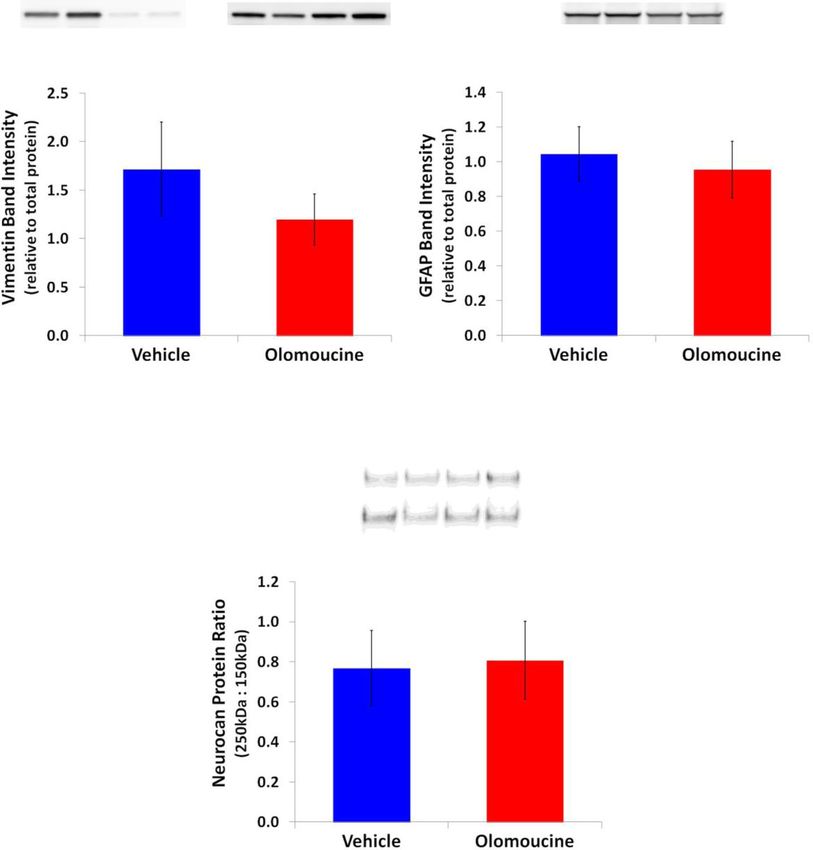
Yew et al. Journal of Neuroinflammation        (2021) 18:168                                                                               Page 4 of 19

 Fig. 1 Summary of experimental design. Olomoucine was injected at 1 h and 24 h after stroke induction. The effects on forelimb function were
 assessed up to 29 days and different aspects of cellular responses in brain tissue were assessed at 3, 7, and 29 days after the stroke. The numbers
 of rats used in different parts of the study are summarized in the figure. For the brains fixed at 29 days, coronal sections prepared from 6 rats per
 treatment group were subsequently analyzed following immunolabelling to detect GFAP and collagen IV

close as possible to the skull without contacting it. Rose                      Three variants of the test were used [11, 34]. The rat
bengal (10 mg/ml, 13 mg/kg) was injected at 200 μl/min                        was first moved towards the platform so that both sets
via the femoral vein cannula. The light source was then                       of vibrissae were stimulated. If a rat placed the forelimb
immediately activated and remained illuminated for 15                         being tested on the platform, the trial was scored as a
min.                                                                          success. Stimulation of the vibrissae ipsilateral and then
                                                                              contralateral to the limb being assessed was used in sub-
Olomoucine treatment                                                          sequent tests. For each forelimb, a score out of 30 was
Rats were treated with olomoucine using a minor modi-                         determined from the sum of successes with 10 trials of
fication of the protocol of Wang et al. [28]. A stock solu-                   each of the three variants of the test.
tion of 50 mg/ml olomoucine was prepared in DMSO                                A baseline value for this study was obtained within
and stored in aliquots at − 20 °C. Immediately before in-                     four days of stroke induction. Rats were tested again at 3
jection, the stock was diluted with water to 1 mg/ml.                         h and then at 1, 7, 14, 21 and 28 days after stroke. A
Rats were injected intraperitoneally with either olomou-                      score of 5 or less for the limb contralateral to the infarct
cine (5 mg/kg) or vehicle (DMSO in water) at 1 h and                          at 3 h after stroke was used as an inclusion criterion for
24 h after the conclusion of the light exposure to induce                     the study. This criterion was met by all 48 rats that sur-
photothrombosis.                                                              vived the procedure for stroke induction.

Assessment of forelimb function                                               Single pellet skilled reaching test
The forelimb function of rats was assessed up to 29 days                      Rats were trained to retrieve a pellet through a narrow
after stroke induction using a forelimb placing test and                      slit in a single pellet reaching box and tested at baseline
two components of a single pellet skilled reaching test.                      and up to 29 days after stroke for total success and first
                                                                              attempt success in retrievals using the forelimb contra-
Forelimb placing test                                                         lateral to the infarct as described previously [35, 36]. To
A minor modification [11] of the forelimb placing re-                         facilitate training, rats were food restricted so that their
sponse as described by Schallert and Woodlee [34] was                         body weight gradually reached 90 to 95% of that ex-
used as a criterion for inclusion in the study and as a                       pected if they had free access to food. Rats were trained
measure of recovery of forelimb motor function after                          in daily 10-min sessions over 2 to 3 weeks up to the day
stroke. No specific training was needed for this task.                        prior to stroke induction. Initial training allowed the
However, rats were familiarized with the procedure for                        paw preference for this task to be identified. Baseline
at least 10 min per day on three separate days leading                        scores are the average from two tests performed on sep-
up to stroke induction. In these sessions, rats were                          arate days within 4 days of stroke induction, including
placed on the testing platform for a few minutes and                          one test on the day preceding the induction. The average
were given approximately 5 trials of each variant of the                      baseline success for pellet retrieval exceeded 50% for all
forelimb placing test.                                                        rats.
Yew et al. Journal of Neuroinflammation (2021) 18:168 Page 5 of 19
In a previous study, recovery on this task was highly Immunohistochemistry
variable when rats were only tested at weekly intervals Two brain sections separated by at least 500 μm were
after stroke [11]. To improve reproducibility of the find- selected from each sample for immunolabelling with
ings and potentially promote recovery in this study, rats each marker. Brain sections from 3 days after stroke
were tested on day 1 and then in blocks of three con- were used to assess possible changes in the properties of
secutive days centred on days 7, 14, 21 and 28. The microglia and tissue macrophages by immunolabelling
values reported for days 7 to 28 are the average of the for Iba1 together with either CD68 or Ki67. Brain sec-
scores on the three consecutive days. On day 1, rats tions from 29 days after stroke were used in the investi-
showed reduced interest in pellet retrieval. Testing on gation of longer-term changes in astrogliosis and
this day was terminated if 20 trials were not completed angiogenesis using antibodies against GFAP and collagen
within 15 min. IV respectively. All sections were also co-labelled with
the NeuN antibody to visualize the infarct and assist in
identifying the peri-infarct region for analysis (see
Brain fixation and assessment of infarct volume
“Image acquisition and analysis” section).
Brain tissue from 12 rats was fixed for immunohisto-
The protocols for immunohistochemistry are essen-
chemistry and analysis of infarct volume at 3 days after
tially as described in detail in previous studies [11, 37].
stroke. The brains of the 20 rats that were used to test
Free-floating brain sections were blocked and perme-
recovery of forelimb function were fixed for additional
abilized in phosphate-buffered saline (PBS) containing
immunohistochemical analysis on day 29. Rats were
0.3% Triton X-100 and 5% donkey serum (blocking buf-
anesthetized with 100 mg/kg ketamine and 10 mg/kg
fer) for 2 h at room temperature, then incubated over-
xylazine injected intraperitoneally. The brains were per-
night at 4 °C in primary antibodies diluted in blocking
fusion fixed and prepared for sectioning as described
buffer (Iba1, 1/800; CD68 and collagen IV, 1/500; Ki67,
previously [11]. Coronal sections (20 μm) were prepared
GFAP and NeuN, 1/400). The sections were then
using a cryo-microtome (Leica Systems, NSW,
washed in PBS and incubated in secondary antibodies
Australia).
diluted (1/2000) in PBS for 2 h at room temperature on
Infarct volume was determined at 3 days after stroke
an orbital shaker. Finally, the sections were washed in
induction using cresyl-violet stained sections at 0.5 mm
PBS and mounted on glass slides in Prolong Gold anti-
intervals through the infarct, as described previously
fade mountant (Thermo Fisher Scientific). Sections to be
[11].
immunolabelled for collagen IV were incubated prior to
processing for immunohistochemistry in 10 mM citrate
Antibodies buffer (pH 8.5) for 10 min at 85 °C to promote antigen
Antibodies for immunohistochemistry to detect Iba1 retrieval.
(goat polyclonal; catalogue number ab5076), Ki67 (rabbit
monoclonal; clone SP6; catalogue number ab16667), Image acquisition and analysis
CD68 (rabbit polyclonal; catalogue number ab125212) The methods for image acquisition and analysis used in
and collagen IV (rabbit polyclonal; catalogue number the present study were largely as described in detail pre-
ab6586) were from Abcam (Cambridge, UK). The anti- viously [11, 37]. Minor modifications were made in the
body for detecting GFAP (rabbit polyclonal; catalogue image processing and analysis parameters to account for
number G4546) was from Sigma-Aldrich (St Louis, Mo) differences in image resolution and quality due to the
and for NeuN (mouse monoclonal; clone A60; catalogue different microscopy systems employed between the
number MAB377) was from Millipore-Chemicon studies.
(Merck, Darmstadt, Germany). Alexa-fluor-conjugated Whole immunolabelled sections, other than those
secondary antibodies were from Thermo Fisher Scien- immunolabelled for collagen IV, were batch-scanned
tific (Waltham, MA, USA). using the Pannoramic 250 Flash II slide scanner (3DHis-
For Western blot analysis, antibodies to detect GFAP tech; Budapest, Hungary). Images from the peri-infarct
(mouse monoclonal; clone G-A-5; catalogue number and equivalent contralateral regions were then extracted
G3893) were from Sigma-Aldrich, those for vimentin using the CaseViewer software (ver 2.1; 3DHistech) into
(rabbit monoclonal; clone EPR3776; catalogue number TIFF files for analysis in ImageJ (ver 1.51j8) [38].
ab92547) were from Abcam and for neurocan (mouse For sections immunolabelled for collagen IV, images
monoclonal; clone 650.24; catalogue number MAB5212) were acquired using an Olympus IX71 inverted micro-
were from Millipore-Chemicon. A peroxidase donkey scope with the 10× objective lens. Two adjacent images
anti-rabbit IgG (catalogue number: 711-035-712) from were captured and stitched together in ImageJ for each
Jackson ImmunoResearch was used as secondary hemisphere to create a field of view covering the entire
antibody. infarct and the equivalent contralateral region.Yew et al. Journal of Neuroinflammation (2021) 18:168 Page 6 of 19 Responses of microglia and macrophages within the tissue and equivalent contralateral regions were thre- peri-infarct tissue were assessed using the parameters of sholded using the “Li” method and the area fraction of circularity and area fraction of particles in Iba1- collagen IV immunolabelling within each ROI was deter- immunolabelled sections. Two rectangular regions of mined using the “Analyze Particle” function with the interest (ROIs) were analyzed. These encompassed the particle size set to a lower limit of 20 pixels (to exclude entire cortical layers extending 500 μm from the lateral debris). edge of the infarct beginning where NeuN immunoreac- tivity was preserved). Two corresponding ROIs in the Western blot analysis contralateral hemisphere were also analyzed (see Fig. Rats were decapitated under anesthesia induced by intra- 3A). Images were processed in ImageJ using the “sub- peritoneal injection of ketamine (100 mg/kg) and xyla- tract background” function with “rolling ball radius” set zine (10 mg/kg) at 7 days after stroke induction. For at 50 pixels and the “sliding paraboloid"” option selected. each rat, the brain was rapidly removed from the skull The two ROIs were then thresholded using the “Huang” and the infarct was identified from the paleness of the method and the circularity and area fraction of Iba1- tissue on the surface of the cortex. Cerebral cortex con- positive particles were determined using “Analyze parti- taining the infarct plus approximately 2 mm of sur- cles” with the particle size set at 2000-infinity pixels rounding tissue was dissected out for analysis. (equivalent to particles with diameters of approximately The tissue samples were homogenized in a buffer con- 8 μm and greater) and circularity at 0.00–1.00. taining 10 mM Tris, 1 mM EDTA, 0.32 M sucrose, pH Counts for CD68-positive and Ki67-positive particles 7.4 and a protease inhibitor cocktail (Sigma-Aldrich) were assessed using ROIs defined using the same ap- using a Retsch TissueLyser (Qiagen, Chadstone, Vic, proach as for Iba1 and background subtraction was per- Australia) at a frequency of 30 Hz for 2 min. Western formed using the same parameters. The images were blot analysis for the vimentin, GFAP and neurocan were thresholded using the “Otsu” method and the particles as described previously [11]. Total protein in the homog- analyzed with particle size set at 120-infinity pixels enates was determined using a Bradford protein assay (diameter 2 μm or greater) and circularity at 0.00–1.00. kit (Bio-Rad Laboratories, Gladesville, NSW, Australia). The results for CD68 and Ki67 from these analyses for Samples (20 μl) containing 20 μg protein were loaded each brain sample are the average of two sections. Iba1 for gel electrophoresis on 4–20% Criterion™ TGX Stain- was co-labelled with each of these two markers resulting Free™ Protein gels, (#5678094; Bio-Rad Laboratories). in four sections analyzed and averaged for each brain Samples for Western blot analysis were initially pre- sample for the circularity and area fraction analyses. pared from 6 rats in each of the two treatment groups. GFAP and collagen IV immunolabelling were analyzed Although all samples were handled identically, in initial using methods described previously [37]. The area frac- investigations to detect vimentin and GFAP, two sam- tion of GFAP in the peri-infarct tissue was assessed in ples from olomoucine-treated rats showed a substantially four adjoining 250-μm-wide rectangular ROIs on the lat- reduced content of these proteins and additional immu- eral edge of the infarct as well as in corresponding ROIs noreactive bands at lower molecular weights indicating in the contralateral hemisphere (see Fig. 5A). As re- protein degradation (as seen in the GFAP blot in Add- ported previously [37], the changes in collagen IV itional file 1: Fig. S1). Two further samples from each immunolabelling was limited to regions very close to the treatment were included in subsequent analyses. One of edge of the infarct and therefore analysis of this marker these from a vehicle-treated rat also showed degradation. was only performed in two adjacent rectangular ROIs The three degraded samples were not included in the extending 500 μm perpendicular to the lateral edge of subsequent analysis. Thus, results are presented for six the infarct. rats treated with olomoucine and seven with vehicle. Re- For analysis of GFAP immunolabelling, the raw unpro- sults for vimentin and neurocan were based on analysis cessed images were analyzed. The ROIs within the of all samples run in single blots. Two blots were used contralateral region were thresholded using the “Tri- for GFAP analysis with band intensities normalized angle” method in ImageJ and the same thresholding level based on the average values for the five samples that was applied to the ROIs within the peri-infarct region. were included in both blots. The area fraction of GFAP immunolabelling was then Stain-free images following protein transfer to the determined using the “Measure” function. polyvinylidene fluoride membrane were obtained using a For collagen IV labelling, background noise was first Gel Doc™ EZ Imager (Bio-Rad Laboratories) and the in- reduced by applying the “subtract background” function tensity of protein labelling in a full-length transect of with “rolling ball radius” set at 5 pixels and “sliding par- each lane was measured as described by Colella et al. aboloid” option selected, after which the “Despeckle” [39]. Chemiluminescence from the blots following anti- function was applied. The ROIs within the peri-infarct body treatment was captured digitally on a Gel Doc™ EZ
Yew et al. Journal of Neuroinflammation   (2021) 18:168                                                        Page 7 of 19

Imager and the band intensities analyzed using the Care-      as the untransformed data showed significant differences
stream Molecular Imaging Software (Version 5.0; Care-         in variance across the groups.
stream Health, NY, USA). The intensity of vimentin or           A two-sided type 1 error rate of alpha = 0.05 was used
GFAP bands were expressed relative to total protein in        for all hypothesis testing.
each lane as determined from the stain free images. For
neurocan, the intensity of the band for full-length pro-
                                                              Results
tein, which increases markedly in response to tissue
                                                              Infarct volume
damage [40–42], was determined as a ratio to a fragment
                                                              There was no significant difference in infarct volume (p
of this protein with a molecular weight of approximately
                                                              = 0.604; Student’s t test) between the brains of rats
150 kDa that is expressed in normal brain and is largely
                                                              treated with olomoucine (17.8 ± 2.3 mm3) and those
preserved during the response to damage [40–42].
                                                              treated with vehicle (16.8 ± 4.0 mm3) when assessed at 3
                                                              days after stroke.
Statistical analysis
Analysis was performed using SPSS (version 23.0; IBM
Corp., Armonk, NY, USA) and Stata (version 15.1; Stata-       Recovery of forepaw function
Corp College Station TX, USA).                                A forelimb placing test involving a motor response of
   Results from the tests of forelimb function are pre-       the forelimb to vibrissae stimulation was used as an in-
sented as box plots. The effect of olomoucine treatment       clusion criterion at 3 h after induction of the stroke and
on recovery of forelimb function was analyzed using lin-      to assess recovery over the subsequent 4 weeks. Prior to
ear mixed effects models. The models included both            surgery, all rats scored at or close to 30 (i.e. 30 success-
fixed effect and random effect terms. The random effects      ful placements from 30 trials on this test; Fig. 2A).
component consisted of a random intercept for each rat,       Within 3 h of surgery, this response was completely
thereby allowing variability around the overall fixed ef-     abolished (a score of 0) for the forelimb contralateral to
fect for each of the individual rats and accounting for       the infarct in 17 of the 20 rats and gave a score of 5 or
the correlation across time for each rat. The fixed effects   less in the other three rats. At 24 h, the performance on
terms consisted of the day, which was treated as categor-     this test for all rats from both treatment groups
ical variable and a day X treatment interaction term, in      remained almost completely impaired, with scores be-
order to test whether the recovery curves differed be-        tween 0 and 4. Median scores in the two groups im-
tween treatments across time. When testing for the sig-       proved over the next 4 weeks although most rats did not
nificance of the interaction, we first tested for global      fully regain their pre-stroke scores (Fig. 2A). Perform-
significance of the day X treatment interaction terms,        ance of the forelimb ipsilateral to the infarct was not sig-
and, when significant, then assessed at which of the time     nificantly affected by the stroke, with median scores for
points the difference in treatment effects occurred using     this forelimb between 28.5 and 30 on each day of
the separate day X treatment interaction terms from the       testing.
model. For analysis of the forelimb placing test, data for      Overall recovery on this task for the affected forelimb
all assessments from 3 h to 28 days after stroke were         was significantly worse for the rats treated with olomou-
used. For the skilled reaching test, initial post-stroke      cine compared with the vehicle-treated group (p < 0.02
performance was approximated by assigning a constant          for the overall day X treatment interaction term). Per-
value of 3 for total success and 1 for first attempt suc-     formance between the two groups was significantly dif-
cess, based on values obtained on day 1 (see “Results”        ferent on days 14, 21 and 28 (Fig. 2A).
section).                                                       The effect of olomoucine treatment on recovery of
   The results for infarct volume, parameters derived         forepaw function was also assessed using a single pellet
from analysis of the immunohistochemistry and band in-        skilled-reaching test. Both the total number of successes
tensity of the Western blots are shown as mean ± stand-       out of 20 (irrespective of the number of attempts re-
ard deviation. The effects of olomoucine treatment on         quired for each pellet) and the number of successes at
infarct volumes and band intensities from the Western         the first attempt out of 20 were evaluated (Fig. 2B, C).
blots were assessed using Student’s t tests. For the data     Performance on both aspects of this task was initially
obtained from analysis of images of immunolabelled            greatly affected by the stroke. When tested on day 1,
brain sections, the effects of olomoucine treatment and       only one rat in each group completed 20 trials within 15
distance from the infarct (or linear distance from an         min. The median value for the olomoucine-treated rats
equivalent starting point within the contralateral region)    was 9 total attempts with 0 total successes or first-
were analyzed using two-way analysis of variance for          attempt successes. For the vehicle-treated group, the
each brain hemisphere. Results for immunolabelling with       median values were 12 total attempts with 1.5 total suc-
CD68 and Ki67 were analyzed after log transformation          cesses and 0.5 first-attempt successes. Because of the
Yew et al. Journal of Neuroinflammation (2021) 18:168 Page 8 of 19
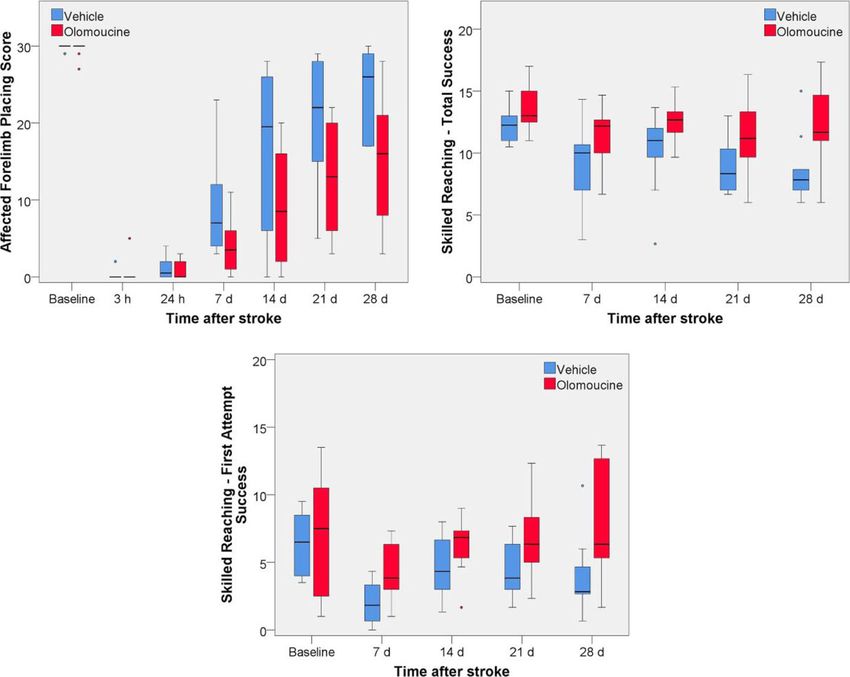
Fig. 2 Effect of olomoucine on forelimb function following photothrombotic stroke. Box plots of dysfunction and recovery up to 28 days after
photothrombotic stroke in A forelimb placing test, B single pellet skilled reaching test: total success from 20 trials, and C single pellet skilled
reaching test: first attempt success from 20 trials. Values are for 10 rats per group. Outliers are shown as circles. For each of the tests, there was
no statistically significant difference between the two treatment groups in the baseline values obtained prior to stroke induction (Mann-Whitney
test). There was a difference in recovery between the olomoucine-treated and vehicle-treated rats identified from a statistically significant overall
day X treatment interaction for the forelimb placing test (p < 0.02) and for both total success (p < 0.05) and first attempt success (p < 0.01) in the
skilled reaching test (Linear mixed effects models). *p < 0.05, **p < 0.01 identifies individual days in which there was a significant difference
between the two treatment groups
failure of most rats to complete 20 trials, the results for contralateral cerebral cortex (Fig. 3A, B). This increase
day 1 have not been included in Fig. 2B, C. was most obvious within the first 250 μm from the in-
The total number of successes showed more rapid re- farct but also extended into surrounding tissue. Treat-
covery than the number of first attempt successes during ment with olomoucine significantly reduced the area
the first 7 days. In contrast to the findings with the fore- fraction of Iba1 immunolabelling in peri-infarct tissue
limb placing test, olomoucine treatment led to improved (Fig. 3B). However, this change was small relative to the
recovery on both the total success (p < 0.05 for overall total change in area fraction. Furthermore, a similar re-
day X treatment interaction) and first-attempt success (p duction was seen in tissue from the contralateral hemi-
< 0.01 for overall day X treatment interaction) in the sphere. Thus, the increase attributable to the
skilled reaching test (Fig. 2B, C). Statistically significant development of the infarct was similar in the rats treated
differences between the treatment groups were seen at with olomoucine and those treated with vehicle.
days 7, 21 and 28 for both aspects of this test and also at The circularity of Iba1 immunolabelled cells provides
day 14 for total success. a more direct measure of the changes in microglial
morphology that occur during activation of these cells.
Cellular changes in peri-infarct tissue Values for this parameter increase in peri-infarct tissue
Key aspects of the response of microglia and blood- within the first day after stroke due to retraction of pro-
derived macrophages were assessed from the pattern of cesses and increases in the volume of the cell body [11].
Iba1 expression at 3 days when changes are near max- Increases in the circularity of Iba1-immunolabelled cells
imal following photothrombotic stroke [11]. Consistent in peri-infarct tissue compared with the contralateral
with previous findings [11], the area fraction of Iba1 cortex were again most prominent within 250 μm of the
immunolabelling was several-fold higher in peri-infarct infarct (Fig. 3C). Treatment with olomoucine signifi-
tissue compared with equivalent tissue in the cantly increased the circularity of Iba1 immunolabelledYew et al. Journal of Neuroinflammation (2021) 18:168 Page 9 of 19 Fig. 3 (See legend on next page.)
Yew et al. Journal of Neuroinflammation         (2021) 18:168                                                                                Page 10 of 19

 (See figure on previous page.)
 Fig. 3 Effect of olomoucine treatment on immunolabelling for Iba1, CD68, and Ki67 at 3 days after stroke. A The top panel shows Iba1
 immunolabelling in a coronal section including the infarct and surrounding cortex and equivalent tissue in the contralateral hemisphere. The
 rectangles indicate the location of the images in the lower panels, which have been processed for analysis of the area fraction of Iba1
 immunolabelling and circularity of Iba1-positive cells. The scale bar represents 200 μm. Representative unprocessed images are shown in
 Additional file1; Fig. S2. B, C The effect of treatment on the area fraction of Iba1 immunolabelling (B) and circularity of Iba1-immunolabelled cells
 (C). For both measures, there was a significant effect of treatment (p < 0.05) and distance from the lesion (p < 0.01) in the peri-infarct tissue with
 no significant interaction. There was also a significant effect of treatment but not location in the contralateral hemisphere. D. The effect of
 treatment on CD68-positive cells. In the peri-infarct tissue, there was a significant effect of distance (p < 0.05) but not treatment (p = 0.065),
 whereas the contralateral cortex showed a significant effect of treatment (p < 0.01) but not tissue location. E The effect of treatment on Ki67-
 positive cells. This marker differed significantly with distance from the lesion in peri-infarct tissue (p < 0.05) Olomoucine did not significantly affect
 the numbers of Ki67-positive cells in either hemisphere. Results in B to E are presented as mean ± SD (n = 6 rats per treatment group) with data
 for each hemisphere analyzed using two-way analysis of variance. *p < 0.05; **p < 0.01 for the effect of olomoucine treatment. #p < 0.05; ##p <
 0.01 for the effect of distance from the infarct or of location in the contralateral cortex

cells but again a similar change was seen in both peri-                         at 0 to 250 μm, 20 ± 13% at 250 to 500 μm; vehicle-
infarct tissue and equivalent tissue in the contralateral                       treated: 41 ± 5% at 0 to 250 μm, 24 ± 8% at 250 to 500
hemisphere (Fig. 3C).                                                           μm) or on the number of these double-labelled cells
   The effects of olmoucine on the number of cells in                           (olomoucine-treated: 158 ± 126 particles per mm2 at 0
peri-infarct tissue expressing CD68 was also investigated.                      to 250 μm, 69 ± 71 particles per mm2 at 250 to 500 μm;
CD68 is a protein produced by subpopulations of Iba1-                           vehicle-treated, 217 ± 110 particles per mm2 at 0 to 250
positive cells within the first few days of stroke induc-                       μm, 92 ± 71 particles per mm2 at 250 to 500 μm).
tion. Reductions in these cells have been seen previously                          The content of the intermediate filament protein,
following various treatments that led to improved func-                         vimentin, was measured using Western blots at 7 days in
tional recovery [11, 43–45]. The increases in CD68-                             samples of cerebral cortex that included the infarct and
positive cells were more restricted in distribution than                        adjacent peri-infarct tissue. Vimentin is not normally
those for Iba1 immunolabelling, resulting in relatively                         expressed in astrocytes in mature brain but expression
larger increases in the tissue immediately surrounding                          increases greatly in peri-infarct astrocytes during the
the infarct (Fig. 3D). Within the initial 250 μm of this                        first week after stroke [11, 46, 47]. Thus, changes in this
tissue, 84 ± 7% of the CD68-positive cells were also                            protein provide a sensitive indicator of reactive astroglio-
Iba1-positive. Very low numbers of CD68-positive cells                          sis. Preliminary analysis confirmed that samples contain-
were detected in the equivalent tissue from the contra-                         ing the infarct and the immediately surrounding tissue
lateral hemisphere but most (71 ± 17%) were again Iba1-                         contain much more vimentin than samples from the
positive. Olomoucine treatment did not result in statisti-                      equivalent region of the contralateral cortex (Fig. 4A).
cally significant differences in the number of CD68-                            Vimentin content was significantly decreased (to 70 ±
positive cells in peri-infarct tissue (Fig. 3D), although                       15%; p < 0.05) in tissue from olomoucine-treated rats
there was a trend towards an increase (p = 0.065). In                           compared with that from vehicle-treated rats (Fig. 4A;
equivalent tissue from the contralateral hemisphere, a                          Additional file 1: Fig. S3). The content of GFAP, a sec-
statistically significant increase (p < 0.01) in the numbers                    ond intermediate filament protein that also increases in
of these cells was seen in the olomoucine-treated rats.                         reactive astrocytes in peri-infarct tissue [46, 48, 49], was
   Immunolabelling for the cell-cycle protein, Ki67, was                        not significantly different between the two groups (Fig.
also measured at 3 days to assess possible downstream                           4B; Additional file 1: Fig. S1). Astrocytes are also an im-
changes in cell proliferation resulting from the olomou-                        portant contributor to increases in production of full-
cine treatment. There was substantial variability in Ki67                       length neurocan following stroke and other brain injury
immunolabelling between rats but cells containing Ki67                          [40, 41]. The content of full-length neurocan (expressed
were again most prominent in tissue adjacent to the in-                         relative to a truncated form of this protein) also was not
farct (Fig. 3E). Very few immunolabelled cells were seen                        significantly different in olomoucine-treated rats com-
in the contralateral hemisphere (Fig. 3E). There was no                         pared with the vehicle-treated rats (Fig. 4C; Additional
statistically    significant    difference    between    the                    file 1: Fig. S3).
olomoucine-treated and vehicle-treated groups. Cells la-                           The brains removed from rats at 29 days after comple-
belled with Iba1 were a substantial component of the                            tion of the assessment of forepaw function were ana-
Ki67-positive cells, particularly close to the infarct.                         lyzed for possible changes in immunolabelling for GFAP
There was again no significant effect of olomoucine                             and collagen IV. Increases in GFAP expression are still
treatment on the proportion of these cells relative to                          prominent at this time in the developing glial scar lo-
total Ki67-positive cells (olomoucine-treated: 35 ± 10%                         cated at the edge of the infarct and also remain elevated
Yew et al. Journal of Neuroinflammation         (2021) 18:168                                                                               Page 11 of 19

 Fig. 4 Effects of olomoucine treatment on markers of reactive astrogliosis at 7 days after stroke. A Vimentin: upper left image shows
 representative lanes from a blot comparing samples containing the infarct and peri-infarct tissue (I) and equivalent tissue from the contralateral
 hemisphere (C) from two rats; the upper right image shows representative lanes from a blot of infarct plus peri-infarct tissue from two rats
 treated with olomoucine (O) and two treated with vehicle (V). The bar graph shows the effect of olomoucine treatment on intensity of the
 vimentin band expressed relative to total protein in that sample. *p < 0.05 (Student’s t test). B GFAP: representative lanes from a blot of infarct
 plus peri-infarct tissue from two rats treated with olomoucine (O) and two treated with vehicle (V). The bar graph shows the effect of olomoucine
 treatment on the pooled data for intensity of the GFAP band expressed relative to total protein in that sample. The treatment did not produce a
 statistically significant difference in GFAP content (Student’s t test). C Neurocan: representative lanes from a blot of infarct plus peri-infarct tissue
 from two rats treated with olomoucine (O) or two treated with vehicle (V). The bar graph shows the effect of olomoucine treatment on the
 intensity of the bands for full-length neurocan (approx. 250 kDa) expressed relative to that of a proteolytic fragment of this protein (approx. 150
 kDa). The treatment did not produce a statistically significant difference in this ratio (Student’s t test). Group results in panels A to C are shown as
 mean ± SD (n = 6 to 7 per group). Additional file 1: Fig. S1 and S3 shows the full blots including the representative lanes for treatment effects in
 panels A to C

in the tissue exhibiting essentially normal neuronal via-                      extending 1 mm from the infarct (Fig. 5A, B), an ap-
bility that extends at least one millimetre into cortical                      proach that has previously detected effects of other
grey matter from the rim of the scar (Fig. 5A). The                            treatments on this cell population [37, 49]. As expected,
changes in GFAP expression in peri-infarct tissue be-                          GFAP immunolabelling was much higher in this tissue
yond the developing scar are indicative of an ongoing re-                      compared with equivalent tissue from the contralateral
activity in astrocytes that can more directly influence                        hemisphere. There was no effect of olomoucine treat-
adaptive responses in neighbouring neurons [18]. We in-                        ment on the GFAP response in either hemisphere. GFAP
vestigated possible effects of the olomoucine treatment                        labelling was significantly affected by distance from the
on this response by measuring the area fraction of GFAP                        lesion in peri-infarct tissue and location in the contralat-
immunolabelling in successive 250-μm-wide regions                              eral cortex. Post-hoc analysis (Tukey HSD test) revealed
Yew et al. Journal of Neuroinflammation   (2021) 18:168   Page 12 of 19

 Fig. 5 (See legend on next page.)
Yew et al. Journal of Neuroinflammation       (2021) 18:168                                                                             Page 13 of 19

 (See figure on previous page.)
 Fig. 5 Immunolabelling for GFAP and collagen IV at 29 days after stroke. A The low power image in the upper panel shows the typical pattern of
 GFAP labelling, with increases extending more than 1 mm into the grey matter surrounding the infarct. Prominent increases were seen on the
 rim of the infarct, in grey matter at the base of the infarct and in underlying white matter. The rectangles in this image identify the regions
 shown in the lower images. The rectangles in the lower images show the tissue that was analyzed. The narrow band of increased
 immunolabelling above the rectangles corresponds to the developing glial scar. B Effect of olomoucine treatment on the area fraction of GFAP
 immunolabelling. Olomoucine treatment did not significantly affect the area fraction of GFAP labelling in either hemisphere. There was a marked
 effect of distance from the lesion on immunolabelling in peri-infarct tissue (p < 0.01) and also a small but significant effect of sampling location
 in the contralateral cortex (p < 0.01). C Collagen IV immunolabelling. The rectangles delineate the regions in which area fraction of
 immunolabelling was assessed. D Effect of olomoucine treatment on the area fraction of collagen IV immunolabelling. There was a significant
 effect of distance from the lesion on immunolabelling in peri-infarct tissue (p < 0.01) but no significant differences in equivalent contralateral
 tissue. The area fraction of immunolabelling was not significantly affected by olomoucine treatment in either hemisphere. The scale bars in A and
 C = 200 μm. Results in B and D are shown as mean ± SD (n = 6 per group) and were analyzed using two-way analysis of variance. ##p < 0.01 for
 the effect of distance from the infarct or of location in the contralateral cortex

significant differences in pair-wise comparisons between                     olomoucine treatment on recovery. The forepaw placing
all regions in peri-infarct tissue except for that at 500 to                 test, which recovered more slowly following olomoucine
750 μm compared with 750 to 1000 μm. In the contra-                          treatment, requires a motor response to stimulation of
lateral hemisphere, a significant difference was seen be-                    the vibrissae. The effects of interventions on recovery of
tween tissue at 0 to 250 μm compared with 750 to 1000                        this task are often similar to that of other general mea-
μm.                                                                          sures of limb motor function or broader assessments of
  Collagen IV is expressed by endothelial cells in capil-                    neurological function (for examples see: [51–53]). In line
laries and has been shown to increase in peri-infarct tis-                   with our previous findings [11], this test was particularly
sue in association with angiogenesis, which can                              sensitive to the lesion induced by photothrombosis. In-
contribute to adaptive responses [8, 50]. The area frac-                     duction of stroke produced near complete loss of this re-
tion of collagen IV immunoreactivity was higher in peri-                     sponse in all rats. Rats exhibited substantial recovery
infarct tissue compared with the contralateral hemi-                         over the next 4 weeks, although most did not regain
sphere (Fig. 5C, D). Again, there was no significant dif-                    baseline performance at 28 days. No specific training
ference between treatment groups in either hemisphere.                       was required to elicit this response and progressive re-
                                                                             covery occurred without additional training beyond the
Discussion                                                                   weekly testing sessions.
The main finding from this study is that olomoucine                            The skilled reaching task, which showed a better re-
treatment initiated early after induction of stroke by                       covery following olomoucine treatment, is a complex
photothrombosis modified functional recovery. Interest-                      learned response that requires targeted movements and
ingly, the effects of the treatment on recovery were                         fine motor control in the forelimb. Rats in the present
markedly task dependent, with a worse outcome on per-                        study were tested 12 times between 6 and 29 days. The
formance in a forelimb placing test but improved recov-                      multiple sessions in this period probably contributed to
ery in both total success and first-trial success in a                       the extent of recovery as post-stroke training has been
skilled reaching test. The altered patterns of recovery                      found to promote spontaneous recovery [6] and improve
following olomoucine treatment occurred in the absence                       the response to other interventions [44, 54, 55].
of a significant change in infarct size. Thus, the effects of                  Previous investigations of the effects of several other
olomoucine were apparently not due to changes in gross                       interventions on functional recovery in rodent models of
tissue damage and probably arose from modifications in                       stroke have also identified marked differences in the ef-
the downstream cellular responses to this damage. Con-                       fects on general assessments of motor function or gross
sistent with this possibility, olomoucine treatment sig-                     neurological performance compared with skilled tasks.
nificantly reduced at least one major component of                           However, the responses to different tests of limb func-
reactive astrogliosis, as evidenced by reductions in tissue                  tion are generally not as markedly divergent as that fol-
vimentin content in peri-infarct tissue at 1 week after                      lowing olomoucine treatment. A pattern broadly
the stroke. In contrast, the treatment had little effect on                  consistent with the effects of olomoucine was seen in re-
key features of the responses of microglia and macro-                        sponse to a treatment involving forced use of the af-
phages in peri-infarct tissue.                                               fected forelimb following both ischemic and
   The two tests used to assess forelimb function in the                     hemorrhagic stroke [51, 55, 56]. Forced forelimb use im-
present study differ in several ways, including the com-                     proves recovery on skilled reaching tasks but does not
plexity of the task and a requirement for training, that                     usually improve more general measures of forelimb
could contribute to the differential influence of                            function or general motor performance. Indeed, an early
Yew et al. Journal of Neuroinflammation (2021) 18:168 Page 14 of 19 imposition of forced use of the affected forelimb in some blood flow is returned to the ischemic tissue within the studies impaired the recovery of measures of general first few hours following onset [62]. However, many pa- neurological function, including a forelimb placing test tients do not receive these interventions. Spontaneous [57, 58]. In contrast, environmental enrichment follow- reperfusion can develop without such treatments but ing stroke commonly promotes recovery in general as- this usually does not occur for many hours to days [63]. sessments of forelimb function but this intervention The permanent ischemia induced by photothrombotic alone is typically ineffective in improving outcomes in stroke provides a better model than short-term middle skilled reaching tests and similar tasks [52, 59–61]. cerebral artery occlusion for those patients in which Multiple changes in the brain are thought to contrib- local disruption to blood flow is permanent or restor- ute to recovery following stroke. This includes altered ation of flow is markedly delayed. neuronal activity, axonal sprouting, synaptogenesis, Proliferation of microglia is an important component neurogenesis and gliogenesis within the peri-infarct tis- of the response of these cells in peri-infarct tissue in the sue as well as modifications to the activity and connect- first few days after stroke [12, 19, 23]. Thus, these cells ivity of neurons at more distant locations [5–8]. The were considered a likely target for the olomoucine treat- specific elements of these responses underlying differen- ment that was given at 1 h and 24 h after the photo- tial outcomes between skilled tasks and general motor thrombosis. Features of Iba1 immunolabelling were used function following some treatments have not been well as the primary measure of microglial reactivity as this defined. A recent investigation found that improvements allowed us to reliably limit the analysis to cells in the in skilled reaching induced by forced limb use together peri-infarct tissue. Tissue of interest around the infarct with skilled forelimb training after induction of photo- was reproducibly defined on the basis of the presence of thrombotic stroke were associated with increased con- near-normal NeuN immunolabelling in the same sec- nectivity of preserved corticospinal neurons in tions. Iba1 immunohistochemistry has been widely used remaining motor cortex adjacent to the infarct [55]. to detect microglial reactivity in peri-infarct tissue fol- Thus, changes affecting the environment or activity of lowing stroke but most studies have relied on qualitative these cells in peri-infarct tissue could potentially modify comparisons of the extent or intensity of immunolabel- recovery of skilled tasks by altering axonal sprouting. ling. In only a few investigations have features of the re- Photothrombosis occludes vessels in the pia and cor- sponses of the Iba1-positive cells been quantified [10– tical parenchyma, producing a region of permanent is- 12]. chemia [31]. Under the conditions used in the present In the present study, both the circularity of Iba1- study, this approach generates a maximal infarct volume immunopositive cells and the area fraction of Iba1- within 24 h that is essentially maintained at 3 days and immunolabelling were analyzed at 3 days when these re- then slowly contracts [11]. The lack of a significant effect sponses are at or near the maximum. Circularity in- of olomoucine on infarct volume at 3 days strongly sup- creases primarily as a result of morphological changes in ports the conclusion that the differences in recovery the cell associated with activation of the microglia. It were not due to alterations in the severity of the initial reaches maximal values by 24 h in peri-infarct tissue and tissue damage but rather arose from modifications of the these increases are largely maintained at 3 days [11]. downstream cellular responses. This situation contrasts The area fraction of Iba1 immunoreactivity in peri- markedly with previous reports in which the cell cycle infarct tissue is markedly increased by 3 days, primarily inhibitor, roscovitine, improved recovery in general tests reflecting an increase in cell numbers [11]. The Iba1 area of motor function, a change that was associated with de- fraction remains similarly increased at 7 days. The local creases in infarct volume [23, 25, 26]. The use of a proliferation of the microglia is an important contributor model involving temporary middle cerebral artery occlu- to this increase [12, 19, 23], with migration of microglia sion and earlier initiation of treatment in these previous from surrounding tissue probably also involved. The studies are likely to have contributed to the different movement of microglia towards sites of tissue damage is outcomes compared with our investigation. a well characterized response of these cells following ac- Our use of photothrombotic stroke rather than a tivation [64]. This migration is commonly proposed to model involving temporary arterial occlusion also has contribute to increases in the numbers of these cells in implications for the potential relevance to stroke in the infarct and peri-infarct tissue [13, 14]. However, to humans. Early reperfusion results in major differences in our knowledge, the relative contribution of proliferation the progression to tissue damage when evaluated in ani- and migration to these changes following stroke has not mal models [2] and is also likely to alter downstream been determined. cellular responses. The increased clinical use of thromb- Olomoucine treatment produced a small increase in olysis and clot retrieval to treat acute stroke over the last the circularity of Iba1-immunolabelled cells and a small decade has increased the number of cases in which decrease in the Iba1 area fraction in peri-infarct tissue.
You can also read