Effect of Model Tear Film Lipid Layer on Water Evaporation
←
→
Page content transcription
If your browser does not render page correctly, please read the page content below

Cornea

              Effect of Model Tear Film Lipid Layer on Water
              Evaporation
              Xiaojie Xu,1 Guangle Li,1 and Yi Y. Zuo1,2
              1
                  Department of Mechanical Engineering, University of Hawaii at Manoa, Honolulu, Hawaii, United States
              2
                  Department of Pediatrics, John A. Burns School of Medicine, University of Hawaii, Honolulu, Hawaii, United States

              Correspondence: Yi Y. Zuo, 2540           PURPOSE. A majority of in vitro models were incapable of reproducing the evaporation
              Dole St, Holmes Hall 302, Honolulu,       resistance of tear film lipid layer (TFLL) in vivo. The purpose of this research is to develop
              HI 96822, USA;                            a novel in vitro model to study the effect of TFLL on water evaporation.
              yzuo@hawaii.edu.
                                                        METHODS. A ventilated, closed-chamber, droplet evaporimeter with a constant surface
              Received: November 19, 2022               area has been invented to study the evaporation resistance of TFLL. This evaporime-
              Accepted: January 3, 2023
                                                        ter ensures a rigorous control of environmental conditions, including the temperature,
              Published: January 19, 2023
                                                        relative humidity, airflow rate, surface area, and surface pressure, thus allowing for repro-
              Citation: Xu X, Li G, Zuo YY. Effect      ducible water evaporation measurements over a time period of only 5 minutes. The volu-
              of model tear film lipid layer on         metric evaporation rate of this droplet evaporimeter is less than 2.7 μL/min, comparable
              water evaporation. Invest                 to the basal tear production of healthy adults. Together with direct film imaging using
              Ophthalmol Vis Sci. 2023;64(1):13.
              https://doi.org/10.1167/iovs.64.1.13
                                                        atomic force microscopy (AFM), we have studied the effect of a model TFLL on water
                                                        evaporation, as a function of the lipid composition and surface pressure.
                                                        RESULTS. A model TFLL composed of 40% wax esters, 40% cholesteryl esters, and 20%
                                                        polar lipids was capable of reducing the water evaporation rate by 11% at surface pressure
                                                        47 mN/m. AFM revealed that the model TFLL at high surface pressures consists of discrete
                                                        droplets/aggregates of the nonpolar lipids residing atop a polar lipid monolayer with
                                                        phase separation.
                                                        CONCLUSIONS. The TFLL may resist water evaporation with a combined mechanism by
                                                        increasing film compactness of the polar lipid film at the air-water surface, and, to a
                                                        lesser extent, by increasing film thickness of the nonpolar lipid film.
                                                        Keywords: atomic force microscopy (AFM), constrained drop surfactometry, droplet, dry
                                                        eye disease, evaporimeter, surface tension, tear film lipid layer (TFLL), water evaporation

              T    ear film is a multilayered biological barrier covering
                   the ocular surface to protect and lubricate the cornea.1
              The tear film can be divided into three distinct layers: an
                                                                                      water evaporation leads to increased tear film instability
                                                                                      and premature breakup, which happens in evaporative dry
                                                                                      eye.13,17 In general, dysfunction of the TFLL results in dry eye
              inner mucus layer with sugar-rich glycosylated proteins,                disease that affects 10% to 30% of the world population.18–20
              an aqueous layer with dissolved proteins, metabolites, and                  Although it is generally accepted that the TFLL helps
              electrolytes, and an outmost lipid layer made up of vari-               reduce water evaporation in vivo,21–25 in vitro findings
              ous lipid species.2,3 This lipid layer, commonly known as               remain controversial. Most in vitro studies with meibomian
              the tear film lipid layer (TFLL), is approximately 100 nm               lipid films and model tear film lipids only demonstrated rela-
              thick.4 The current consensus is that the TFLL consists of              tively insignificant or nearly no retardation to water evap-
              two sublayers: a polar lipid layer at the air-water surface,            oration.26–32 These controversial results were likely related
              mainly consisting of phospholipids and (O-acyl)-ω-hydroxy               to the in vitro models used for studying water evaporation.
              fatty acids (OAHFAs), and a nonpolar lipid layer, composed              Quantitative study of monolayer retardation on water evap-
              of wax esters and cholesteryl esters, residing atop the polar           oration can be traced back to the seminal work by Victor
              lipid layer and directly exposing to the environment.5–7                La Mer in the 1950s for the interests of conserving water
              Nonpolar lipids in the TFLL are secreted by the meibomian               in reservoirs.33–35 To date, a vast majority of these in vitro
              glands, whereas the source of phospholipids in the TFLL                 studies relied on the classical Langmuir trough,26–32 which
              is still uncertain.8 The polar lipids may facilitate spread-            has a few limitations that prevent accurate evaluation of
              ing of the nonpolar lipids, rather than forming aggrega-                water evaporation. First, due to its large size, the Langmuir
              tions or droplets, over the aqueous surface of the tear                 trough generally lacks a rigorous control in environmen-
              film.9,10                                                               tal conditions, such as the temperature, relative humidity,
                  The TFLL has multiple physiological functions, such as              and airflow rate, all of which are essential factors that can
              host defense against ocular infection and retardation of                significantly affect the rate of water evaporation. Second, the
              water evaporation.11–15 Water evaporation is one of the                 evaporation rate is traditionally determined with gravimet-
              most important mechanisms for tear film thinning.16 Rapid               ric analysis (i.e. directly measuring the mass of water lost

              Copyright 2023 The Authors
              iovs.arvojournals.org | ISSN: 1552-5783                                                                                               1

                   This work is licensed under a Creative Commons Attribution-NonCommercial-NoDerivatives 4.0 International License.

Downloaded from iovs.arvojournals.org on 01/19/2023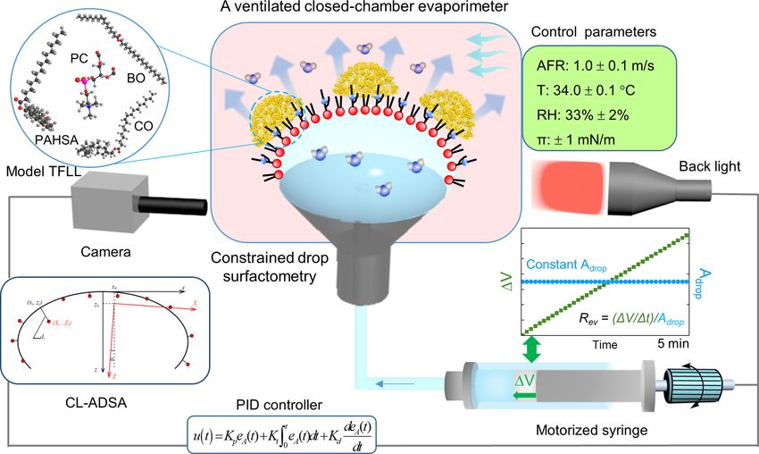
Evaporation Resistance of Tear Film Lipid Layer                                 IOVS | January 2023 | Vol. 64 | No. 1 | Article 13 | 2

              by evaporation from a Langmuir trough), which requires a             of the CDS is a carefully machined pedestal that uses its
              relatively long period of experiments (usually 0.5–2 hours)          knife-sharp edge to prevent film leakage even at very low
              to reduce system errors.27 This further increases the diffi-         surface tensions. System miniaturization of the CDS facili-
              culty of environmental control during such an extended               tates rigorous control of experimental conditions with an
              period of experiments. Third, Langmuir trough can hardly             environmental control chamber. The spread/adsorbed film
              reproduce the physiologically relevant high surface pres-            at the droplet surface can be compressed and expanded
              sure of the TFLL. The surface tension of whole tears                 by precisely controlling oscillation of the surface area of
              of healthy individuals was reported to be around 43 to               the droplet using a newly developed mechatronic system
              46 mN/m,36,37 which most likely represents the surface               called closed-loop axisymmetric drop shape analysis (CL-
              tension of major proteins in tears, such as lysozyme.38 Upon         ADSA).46 The CL-ADSA determines the surface tension of
              film compression during the blinking process, the TFLL can           the spread/adsorbed film by analyzing the shape of the
              reduce the surface tension to approximately 20 mN/m, corre-          film-covered droplet. The surface pressure (π ) can be deter-
              sponding to a surface pressure as high as 50 mN/m.38,39              mined from the surface tension (γ ) using π = γ 0 -γ , with
              However, most existing in vitro evaporation studies only             γ 0 being the surface tension of a clean, lipid-free air-water
              covered the surface pressure range between 5 and 30 mN/m,            surface.
              because the TFLL rapidly collapses at higher surface pres-               Specifically, a trace amount of the lipid sample was
              sures in a Langmuir trough. Hence, there is an urgent need           spread onto the air-water surface of a 15 μL droplet serv-
              for alternative biophysical models to evaluate the effect of         ing as the aqueous subphase to the spread lipid film. The
              TFLL on water evaporation under physiologically relevant             spread film was left undisturbed for 1 minute to allow evap-
              conditions.                                                          oration of the solvent and to reach equilibrium. The droplet
                  Here, we developed a novel droplet-based biophysical             was then slowly expanded to decrease the surface pressure
              model to study the effect of TFLL on water evaporation.              to around zero (i.e. increasing the surface tension to around
              Owing to system miniaturization, droplet-based evaporation           70 mN/m). Subsequently, the spread lipid film was
              models offer a more rigorous environmental control than the          compressed quasi-statically at a rate of 0.15 A%/s to a target
              classical Langmuir trough. Both pendant drop40 and sessile           surface pressure.
              drop41 methods have been attempted in previous studies.
              A key novelty of this work was the invention of a venti-
              lated, closed-chamber, droplet evaporimeter with a constant          Ventilated Closed-Chamber Droplet Evaporimeter
              surface area, analogous to the evaporimeter used for measur-         With a Constant Surface Area
              ing the tear evaporation rate in vivo.24,42,43 This in vitro evap-
              orimeter was realized with the combination of constrained            Figure 1 illustrates the schematic of the droplet-based evap-
              drop surfactometry and a novel feedback control system               orimeter. Lipid samples were spread onto the air-water
              called closed loop-axisymmetric drop shape analysis that             surface of a 5-mm droplet (27 μL in volume and 0.35 cm2
              decoupled surface area of the droplet from water evapora-            in surface area) to result in a target surface pressure. The
              tion. Using this novel biophysical model, together with direct       environmental temperature and relative humidity (RH) were
              film imaging using atomic force microscopy, we have stud-            controlled at 34.0 ± 0.1°C and 33.0 ± 2.0% with a closed
              ied the effect of a model TFLL on water evaporation. Our             environmental control chamber. The chamber was ventilated
              data suggest that the model TFLL is capable of reducing the          with a continuous airflow. The airflow rate was measured
              water evaporation rate by 11% at high surface pressures.             with a hot wire anemometer (TSI, Shoreview, MN, USA) and
              Our experimental results may provide novel implications              was controlled at 1.0 ± 0.1 m/s to simulate the ambient envi-
              into better understanding the biophysical and physiological          ronment.47
              function of the TFLL.                                                   A key feature of the droplet-based evaporimeter is its
                                                                                   capacity of maintaining a constant surface area during water
                                                                                   evaporation. As shown in Figure 1, CL-ADSA maintains the
              METHODS                                                              constant surface area of a droplet by determining its surface
                                                                                   area in real-time and feeding this information back to a
              Materials                                                            motorized syringe to automatically complete a proportional-
              Dipalmitoyl      phosphatidylcholine        (DPPC),     L-α-         integral-derivative (PID) control loop.46 The evaporation rate
              phosphatidylcholine (PC) from egg yolk, palmitic-acid-               (mm/min) was calculated as (V/t)/Adrop , where V was
              9-hydroxy-stearic-acid (PAHSA), and cholesteryl oleate (CO)          the volume of water replenished into the droplet, in order
              were purchased from Sigma-Aldrich (St. Louis, MO, USA).              to compensate for the evaporated water and thus to main-
              Behenyl oleate (BO) was purchased from Larodan (Monroe,              tain the constant surface area. The t is the time period of
              MI, USA). Physicochemical properties of these lipids can             the experiment (i.e. 5 minutes). The V/t was determined
              be found elsewhere.38 Individual lipids were dissolved in            from linear regression of the recorded V-t curve. The
              chloroform as 1 mM stock solutions. Water used was Milli-Q           Adrop was the surface area of the droplet, controlled at 0.35
              ultrapure water with a resistivity greater than 18 Mcm at          ± 0.01 cm2 . The volumetric evaporation rate (i.e. V/t) of
              room temperature.                                                    this droplet-based evaporimeter was determined to be less
                                                                                   than 2.7 μL/min, comparable to the basal tear production of
                                                                                   healthy adults (i.e. 0.8–2.0 μL/min).48,49
              Constrained Drop Surfactometry
              Constrained drop surfactometry (CDS) is a new genera-                Atomic Force Microscopy
              tion of droplet-based surface tensiometry technique devel-
              oped in our laboratory.44,45 It uses the air-water surface of        Lateral structure and topography of the tear lipid films were
              a millimeter-sized sessile drop to accommodate the spread            studied with the combination of in situ Langmuir-Blodgett
              or adsorbed film. As shown in Figure 1, a key design                 (LB) transfer from the CDS and atomic force microscopy

Downloaded from iovs.arvojournals.org on 01/19/2023
Evaporation Resistance of Tear Film Lipid Layer                                       IOVS | January 2023 | Vol. 64 | No. 1 | Article 13 | 3

              FIGURE 1. Schematic of a ventilated, closed-chamber, droplet evaporimeter with a constant surface area for studying evaporation resistance
              of the tear film lipid layer (TFLL). This droplet evaporimeter is constructed based on constrained drop surfactometry (CDS), in which a 5-mm
              water droplet (approximately 27 μL in volume and approximately 0.35 cm2 in surface area) is constrained on a carefully machined pedestal
              with knife-sharp edges. The water droplet is enclosed in an environmental control chamber that ensures a rigorous control of experimental
              conditions, including the temperature, relative humidity (RH), and airflow rate (AFR). The surface area of the droplet is maintained at a
              constant using closed-loop axisymmetric drop shape analysis (CL-ADSA) with a proportional-integral-derivative (PID) control loop. The
              measuring principle of ADSA is illustrated in a box: ADSA determines the surface tension by numerically fitting the experimental droplet
              profiles (indicated by red dots) to theoretical droplet profiles (indicated by black curves) obtained with numerical integration of the Laplace
              equation of capillarity. The PID controller is illustrated by a PID control function with the proportional, integral, and derivative terms. The
              evaporation rate (Rev , mm/min) is calculated as (V/t)/Adrop , where V is the volume of water replenished into the droplet, in order to
              compensate for the water lost by evaporation. The t is the time period of the experiment, usually 5 minutes. The Adrop is the surface
              area of the droplet, controlled at 0.35 cm2 . The model TFLL consists of 40 mol% behenyl oleate (BO) and 40 mol% cholesteryl oleate (CO)
              that represent two nonpolar lipid classes (i.e. wax ester and cholesteryl ester in the natural TFLL), and 15 mol% phosphatidylcholine (PC)
              and 5 mol% palmitic-acid-9-hydroxy-stearic-acid (PAHSA) that represent two polar lipid classes in the natural TFLL (i.e. phospholipids and
              OAHFAs).

              (AFM).39,50 The lipid film was first LB transferred from the              RESULTS     AND   DISCUSSION
              droplet by lifting a small piece of freshly peeled mica sheet
              at a speed of 1 mm/min. During the LB transfer process,                   Development of a Constant-Surface-Area Droplet
              the surface pressure of the lipid film was maintained at a                Evaporimeter for Studying Evaporation
              constant (± 1 mN/m). Topographical images of the lipid                    Retardation by Lipid Films
              film were obtained with an Innova AFM (Bruker, Santa
                                                                                        Figure 2 demonstrates the capacity of this new evaporimeter
              Barbara, CA, USA). Samples were scanned in air in contact
                                                                                        in determining the rate of evaporation from a water droplet
              mode and tapping mode. The contact mode used a silicon
                                                                                        while maintaining a constant surface area of the droplet.
              nitride cantilever with a spring constant of 0.12 N/m and
                                                                                        Within a 5-minute period, the RH of the environment was
              a tip radius of 2 nm, whereas the tapping mode used a
                                                                                        maintained at 33%, whereas the temperature and airflow
              silicon cantilever with the spring constant of 42 N/m and
                                                                                        rate were controlled at 34°C and 1 m/s, respectively. Surface
              a resonance frequency of 300 kHz. Relative height differ-
                                                                                        tension of the water droplet remained at a constant of
              ences between domains were determined with section anal-
                                                                                        71 mN/m, indicating no contamination of the water surface.
              ysis using Nanoscope Analysis (version 1.5).
                                                                                        It can be seen that during the 5-minute period, in spite
                                                                                        of water evaporation, the surface area and volume of
              Statistical Analysis
                                                                                        the droplet were maintained at 0.35 cm2 and 27 μL,
              All results were shown as mean ± standard deviation (n = 10               respectively, using CL-ADSA (see Movie S1 of the Supple-
              unless otherwise indicated). One-way ANOVA with Tukey’s                   mentary Data for this experiment). The volume of water
              means comparison test was used to determine group differ-                 replenished into the droplet, in order to maintain the
              ences (OriginPro, Northampton, MA, USA). A value P < 0.05                 constant surface area, increased linearly over the 5-minute
              was considered to be statistically significant.                           period, with a volumetric rate of 2.7 μL/min. The water

Downloaded from iovs.arvojournals.org on 01/19/2023
Evaporation Resistance of Tear Film Lipid Layer                                    IOVS | January 2023 | Vol. 64 | No. 1 | Article 13 | 4

                                                                                      oration rate by 4%. Third, at 40 and 50 mN/m, the DPPC
                                                                                      monolayer shows significant retardation effects on water
                                                                                      evaporation, with 9% and 17% reduction in the evaporation
                                                                                      rate, respectively. These findings are in qualitative agreement
                                                                                      with those reported by Miano et al. who determined the
                                                                                      effect of the DPPC monolayer on water evaporation up to
                                                                                      the surface pressure of 35 mN/m, at 36°C and 15% RH, using
                                                                                      the pendant drop method.40 These workers found that at
                                                                                      the surface pressure below 12 mN/m, the DPPC monolayer
                                                                                      showed no retardation effect on water evaporation. When
                                                                                      the surface pressure was increased to 20 mN/m, the DPPC
                                                                                      monolayer showed moderate effects on evaporation retar-
                                                                                      dation, whereas increasing the surface pressure to 35 mN/m
                                                                                      did not further increase the evaporation resistance.40
                                                                                         Figure 3 also shows the compression isotherm of the
                                                                                      DPPC monolayer at 34°C, superimposed on the water evap-
                                                                                      oration data. The DPPC monolayer undergoes a liquid-
                                                                                      expanded (LE) to tilted-condensed (TC) phase transition
                                                                                      within the surface pressure range between 20 and 30 mN/m,
                                                                                      indicated by a plateau region in the compression isotherm.
                                                                                      (Reproducibility of this compression isotherm can be found
                                                                                      in Supplementary Fig. S2.) This phase transition region of
                                                                                      the DPPC monolayer at 34°C is in good agreement with
                                                                                      our previous observations.45 The LE-TC phase transition, or
                                                                                      phase co-existence, can be visualized by the formation of TC
                                                                                      domains approximately 1 nm higher than the surrounding
                                                                                      LE phase, as demonstrated by the AFM image shown in the
                                                                                      inset of Figure 3.
                                                                                         These findings suggest that the evaporation resistance
                                                                                      of the DPPC monolayer is mainly determined by the phos-
                                                                                      pholipid polymorphism. The LE-TC phase co-existence in
                                                                                      the DPPC monolayer (i.e. at 20–30 mN/m), corresponds
              FIGURE 2. Typical experimental results for water evaporation deter-     to region II in which the DPPC monolayer starts to show
              mined within 5 min using the new droplet evaporimeter. Tempera-
              ture and relative humidity (RH) were maintained at 34°C and 33%,
                                                                                      moderate resistance to water evaporation (see Fig. 3). At
              respectively. Surface tension of water was relatively unchanged at 70   surface pressures lower than this phase transition pressure,
              mN/m, indicating no contamination. Surface area of the droplet was      the DPPC monolayer is in a disordered LE phase and hence
              actively controlled at a constant of 0.35 cm2 using CL-ADSA. Volume     does not significantly resist water evaporation (region I). At
              of the droplet was relatively unchanged at 27 μL. The volumetric rate   surface pressures higher than this phase transition pressure,
              of water replenished into the droplet (V/t) to complement the         the DPPC monolayer is compressed into a tightly packed,
              water lost by evaporation was determined by linear regression (red
              solid line), corresponding to a volumetric evaporation rate of 2.7
                                                                                      ordered TC phase, thus showing significant resistance to
              μL/min.                                                                 water evaporation (region III). These experimental data are
                                                                                      in line with the theory of an active energy barrier to water
                                                                                      evaporation through monolayers, originated from electro-
                                                                                      static and/or steric repulsions between lipid molecules upon
              evaporation rate (V/t)/Adrop , under the controlled exper-            monolayer compression.35
              imental conditions, was determined to be 0.078 mm/min. As
              shown in Supplementary Figure S1 of the Supplementary                   Effect of the Model TFLL on Water Evaporation
              Data, extending the experimental period to 15 minutes does
              not vary the water evaporation rate. Hence, the 5-minute                Figure 4a shows the quasi-static compression isotherms of
              experimental period is used thereafter.                                 three lipid films (i.e. egg PC, PAHSA, and a synthetic model
                  To demonstrate the effect of lipid monolayers on water              TFLL), at 34°C. This model TFLL consists of 40 mol% BO
              evaporation, we have studied the evaporation resistance of              and 40 mol% CO that represent two nonpolar lipid classes
              a DPPC monolayer at 34°C under various controlled surface               (i.e. wax ester and cholesteryl ester in the natural TFLL),
              pressures. DPPC (16:0, 16:0 PC) was selected as a model                 and 15 mol% PC and 5 mol% PAHSA that represent two
              lipid monolayer because C16 fatty acids are able to balance             polar lipid classes in the natural TFLL (i.e. phospholipids
              the rigidity needed for a sufficient resistance to water evapo-         and OAHFAs).51 It should be noted that human meibomian
              ration and the “self-healing” effect after rupture by waves.34          lipids are composed of a complex mixture of more than
                  As shown in Figure 3, effects of the DPPC monolayer on              200 lipid species, primarily including cholesterol esters, wax
              water evaporation can be roughly divided into three regions             esters, (O-acyl)-ω-hydroxy fatty acids, and triacylglycerols.1,6
              as a function of surface pressure. First, at 10 mN/m, the               Modern lipidomics data further suggested that the polar lipid
              DPPC monolayer shows no statistically significant effect on             content in healthy TFLL is generally less than 5 mol%.1,6
              water evaporation (P > 0.05 in comparison to the clean                  Hence, the model TFLL used here (i.e. BO:CO:PC:PAHSA
              air-water surface). Second, at 20 and 30 mN/m, the DPPC                 [40:40:15:5]), is not only overly simplified in its lipid compo-
              monolayer shows moderate, but statistically significant (P <            sition but also likely has an augmented abundance in polar
              0.05), effects on water evaporation, by reducing the evap-              lipids. Nevertheless, our previous studies have demonstrated

Downloaded from iovs.arvojournals.org on 01/19/2023
Evaporation Resistance of Tear Film Lipid Layer                                       IOVS | January 2023 | Vol. 64 | No. 1 | Article 13 | 5

              FIGURE 3. Superimposed compression isotherm of a DPPC monolayer at 34°C, and the corresponding evaporation resistance at various
              surface pressures. Surface pressure zero indicates a pure lipid-free air-water surface. The compression isotherm and evaporation resistance
              can be separated into three regions. Region I = No evaporation resistance for the DPPC monolayer in a disordered liquid-expanded (LE)
              phase; region II = Moderate evaporation resistance for the DPPC monolayer undergoing LE to tilted-condensed (TC) phase transitions; and
              region III = High evaporation resistance for the DPPC monolayer in a closely packed ordered TC phase. Insets are an AFM image showing
              LE-TC phase co-existence at 25 mN/m, and droplet images demonstrating the constant surface area. *P < 0.05 indicates statistically significant
              differences.

              that this model TFLL represents the biophysical and rheolog-             establish this finding.26–32 Using various synthetic models,
              ical properties of the natural TFLL to a certain degree.38,39            animal, or human meibomian lipids, numerous in vitro stud-
                  Figures 4b to d shows the retardation effects of the PC,             ies found no (
Evaporation Resistance of Tear Film Lipid Layer IOVS | January 2023 | Vol. 64 | No. 1 | Article 13 | 6
FIGURE 4. Effects of lipid films on water evaporation. (a) Compression isotherms of PC, PAHSA, and a model TFLL, BO:CO:PC:PAHSA
(40:40:15:5). (b-d) Evaporation rates of water (mm/min) through PC, PAHSA, and the model TFLL at various surface pressures. *P < 0.05,
**P < 0.01, ***P < 0.001.
differences between the environment and the surface of and packing density of the lipid molecules.33,34 La Mer and
the evaporating droplet. Supplementary Figure S4 shows coworkers found that the evaporation resistance of satu-
the surface temperature of the droplet under the controlled rated fatty acids was an exponential function of the chain
environmental temperature of 34°C. It can be seen that the length.33,34 Any addition of one carbon atom in the hydro-
airflow significantly affects the surface temperature of the carbon chain increases the evaporation resistance by a factor
droplet. Although without ventilation, there is only a 2°C of 1.65.15,33,34 However, the chain length effect of nonpolar
temperature difference between the droplet surface and the lipids on water evaporation is largely unknown. Nonpolar
environment, the temperature difference increases to 9°C lipids, such as wax esters and cholesteryl esters, account for
with a 1 m/s airflow. Therefore, the in vitro evaporation rate 80% of the TFLL.54 The model TFLL studied here contains
determined here might be underestimated in comparison to 40% behenyl oleate (C22:0-C18:1) and 40% cholesteryl oleate
in vivo conditions. (cholesterol-C18:1). Although these nonpolar lipids are inca-
Second, our study showcases the importance of lipid pable of directly spreading at the air-water surface, they
packing density in evaporation resistance. As shown somehow increase the evaporation resistance of the polar
in Figure 4d, when the surface pressure is increased from lipid monolayer (e.g. 4.2% for PC versus 6.1% for TFLL at
10 to 47 mN/m, the evaporation resistance of the TFLL 40 mN/m; see Figs. 4b vs. 4d).
increases by 3.2 times. Surface pressure 47 mN/m corre-
sponds to a surface tension approximately 23 mN/m, which
is significantly lower than the surface tension of whole tears Lateral Structure and Topography of the Model
but corresponds to the lowest surface tension of a highly TFLL
compressed TFLL.38
Third, our study indicates that the long-chain nonpolar Figure 5 shows the lateral structure and topography of three
lipids may play a role in evaporation resistance of the TFLL. lipid films (i.e. PC, PAHSA, and the model TFLL made up of
It has long been recognized that the evaporation resistance BO:CO:PC:PAHSA [40:40:15:5]), at 34°C. Reproducibility of
of polar lipid monolayers depends on both the chain length these AFM images can be found in Supplementary Figures S5
Downloaded from iovs.arvojournals.org on 01/19/2023Evaporation Resistance of Tear Film Lipid Layer IOVS | January 2023 | Vol. 64 | No. 1 | Article 13 | 7
FIGURE 5. AFM topography and lateral structure of lipid films at various surface pressures. (a, b) PC monolayer at 20 and 30 mN/m.
(c, d) PAHSA monolayer at 20 and 27 mN/m. (e-h) Model TFLL, BO:CO:PC:PAHSA (40:40:15:5) at 20, 30, 40, and 47 mN/m. (f1-h1) Three-
dimensional renderings of the TFLL corresponding to AFM images shown in panels f to h. All AFM images have the same scanning area of
20 × 20 μm. The z range for images in f to h is 100 nm, whereas it is 5 nm for all other images. Single-headed arrows indicate the heights
of the structures, whereas double-headed arrows indicate the lateral dimensions of the structures.
to S11. Films of the two polar lipids (i.e. PC; see Figs. 5a, 5b) thus forming nonpolar lipid multilayers/aggregates residing
and PAHSA (see Figs. 5c, 5d), assume a monolayer confor- atop the polar lipid monolayer.
mation with phase separation at surface pressures up to 30 Formation of nonpolar lipid droplets or aggregates by
mN/m. Lateral structures of the PC and PAHSA monolay- squeezing out from the TFLL at increasing surface pressure
ers show a network of ramified, fiber-like ordered domains is supported by multiple experimental evidence. First, film
approximately 1 nm higher than the surrounding disordered compressibility of the TFLL significantly decreases at surface
phase. The network of the ordered domains increases in pressures higher than 30 mN/m (see Fig. 4a). Second, AFM
density upon increasing surface pressure from 20 to 30 has detected a unique evaporation pattern closely analogous
mN/m, consistent with our previous observations.39 to the coffee-ring effect.55 These evaporation patterns have
At a low surface pressure of 20 mN/m (see Fig. 5e), heights of either 4 nm (see inset of Fig. 5h1) or 8 nm (see Fig.
the model TFLL also demonstrates a general monolayer 5f1), corresponding to 1 or 2 fully hydrated phospholipid
conformation with phase separation, similar to the polar bilayers. These “coffee-rings” are most likely formed by
lipids. However, at surface pressures equal to or higher than evaporation-driven self-assembly of reverse micelles of polar
30 mN/m (see Figs. 5f-h), the TFLL shows a completely lipids mixed with the nonpolar lipids.10,11 This also explains
different topography and lateral structure, compared to the various sizes of the oil “beads” found in the model TFLL,
those of the polar lipid monolayers. At all high surface pres- which could be a consequence of different degrees of oil
sures (i.e. 30, 40, and 47 mN/m), the TFLL shows discrete evaporation and oil droplet coalescence. Third, AFM has
bead-like structures ranging from approximately 150 to revealed structures similar to the polar lipid monolayers (PC
approximately 700 nm in height (see Figs. 5f1-h1 for three- or PAHSA) underneath the nonpolar multilayer, as shown in
dimensional renderings of the film topography). In compar- a high-resolution (1 × 1 μm) AFM image scanned through a
ison to lateral structures of the polar lipid films, the source “pore” on the surface layer. This AFM observation provides
of these high, bead-like structures must be the nonpolar direct evidence for layered structures of the TFLL.
lipids in the TFLL (i.e. BO and CO). Due to lack of affin- Formation of the nonpolar lipid multilayer/aggregates
ity to water, these nonpolar lipids are squeezed out from in the TFLL corresponds to a slight increase in the evap-
the surface when the surface pressure is increased above 30 oration resistance of the TFLL (see Figs. 4b, 4c vs. 4d).
mN/m (corresponding to the surface tension of whole tears), Therefore, the present study supports that the TFLL resists
Downloaded from iovs.arvojournals.org on 01/19/2023Evaporation Resistance of Tear Film Lipid Layer IOVS | January 2023 | Vol. 64 | No. 1 | Article 13 | 8
water evaporation with a combined mechanism by increas- 4. King-Smith E, Fink B, Hill R, Koelling K, Tiffany J. The thick-
ing film compactness of the polar lipid film at the air-water ness of the tear film. Curr Eye Res. 2004;29:357–368.
surface, and, to a lesser extent, by increasing film thick- 5. Chen JZ, Green-Church KB, Nichols KK. Shotgun lipidomic
ness of the nonpolar lipid film. It should be noted that analysis of human meibomian gland secretions with electro-
the model TFLL studied here only consists of four lipid spray ionization tandem mass spectrometry. Invest Ophthal-
components (i.e. BO:CO:PC:PAHSA [40:40:15:5]). Conse- mol Vis Sci. 2010;51:6220–6231.
quently, the squeezed-out nonpolar lipids formed discrete 6. Brown SHJ, Kunnen CME, Duchoslav E, et al. A compari-
droplets/aggregates due to inadequate lipid mixing. Human son of patient matched meibum and tear lipidomes. Invest
Ophthalmol Vis Sci. 2013;54:7417–7423.
meibomian lipids are composed of many different lipid
7. Butovich IA. The meibomian puzzle: Combining pieces
classes, and each of these lipid classes consists of many
together. Prog Retinal Eye Res. 2009;28:483–498.
homologous lipid species varying in lengths, degrees of
8. Dean AW, Glasgow BJ. Mass Spectrometric Identification of
unsaturation, and branching, which is essential for the natu- Phospholipids in Human Tears and Tear Lipocalin. Invest
ral meibomian lipids to have proper melting and lipid Ophthalmol Vis Sci. 2012;53:1773–1782.
mixing.1 Hence, the nonpolar lipid layer of natural TFLL is 9. Paananen RO, Rantamäki AH, Holopainen JM. Antievapora-
likely more continuous and more uniform than the model tive mechanism of wax esters: implications for the function
TFLL studied here, thus rendering more evaporation resis- of tear fluid. Langmuir. 2014;30:5897–5902.
tance.56 However, more recent studies, both in vitro57 and 10. Cwiklik L. Tear film lipid layer: A molecular level view.
in vivo,58,59 suggested that the thickness of the TFLL, and Biochimica et Biophysica Acta (BBA) - Biomembranes.
especially that of the nonpolar lipid layer of the TFLL, is 2016;1858:2421–2430.
not uniform but with regions of thicker lipid droplets or 11. Georgiev GA, Eftimov P, Yokoi N. Structure-function rela-
aggregates. Using in vitro surface rheological study of bovine tionship of tear film lipid layer: A contemporary perspective.
meibomian lipids, Bhamla et al. also inferred that the TFLL is Exp Eye Res. 2017;163:17–28.
likely not uniform in thickness, with the thicker area acting 12. Butovich IA. Lipidomics of human Meibomian gland secre-
as a more effective barrier to water evaporation.60 These tions: Chemistry, biophysics, and physiological role of
studies, including the present work, are consistent with the Meibomian lipids. Prog Lipid Res. 2011;50:278–301.
finding that tear film evaporation is not necessarily corre- 13. Bron AJ, Tiffany JM, Gouveia SM, Yokoi N, Voon LW. Func-
lated with a uniform thickness of the TFLL.61 tional aspects of the tear film lipid layer. Exp Eye Res.
2004;78:347–360.
14. Millar TJ, Schuett BS. The real reason for having a meibo-
CONCLUSIONS mian lipid layer covering the outer surface of the tear film -
A review. Exp Eye Res. 2015;137:125–138.
We have developed a novel ventilated, closed-chamber, 15. King-Smith PE, Bailey MD, Braun RJ. Four characteristics
droplet evaporimeter with a constant surface area for study- and a model of an effective tear film lipid layer (TFLL). Ocul
ing the effect of TFLL on water evaporation. This new evap- Surf. 2013;11:236–245.
orimeter is capable of a rigorous control of environmen- 16. Willcox MDP, Argueso P, Georgiev GA, et al. TFOS DEWS
tal conditions, including the temperature, relative humidity, II Tear Film Report. Ocul Surf. 2017;15:366–403.
airflow rate, surface area, and surface pressure, thus allow- 17. King-Smith PE, Begley CG, Braun RJ. Mechanisms, imag-
ing for reproducible water evaporation measurements over ing and structure of tear film breakup. Ocul Surf. 2018;16:
a time period of only 5 minutes. The volumetric evaporation 4–30.
rate of this droplet evaporimeter is less than 2.7 μL/min, 18. Nichols KK, Foulks GN, Bron AJ, et al. The Interna-
comparable to the basal tear production of healthy adults. tional Workshop on Meibomian Gland Dysfunction: Exec-
With this new evaporimeter, we have established the in vitro utive Summary. Invest Ophthalmol Vis Sci. 2011;52:1922–
evaporation resistance of a model TFLL that consists of 40% 1929.
wax esters, 40% cholesteryl esters, and 20% polar lipids. 19. Craig JP, Nichols KK, Akpek EK, et al. TFOS DEWS II Defini-
tion and Classification Report. Ocul Surf. 2017;15:276–283.
It was found that the TFLL resists water evaporation with
a combined mechanism by increasing film compactness of 20. Stapleton F, Alves M, Bunya VY, et al. TFOS DEWS II
Epidemiology Report. Ocul Surf. 2017;15:334–365.
the polar lipid film at the air-water surface, and, to a lesser
21. Craig JP, Tomlinson A. Importance of the lipid layer in
extent, by increasing film thickness of the nonpolar lipid
human tear film stability and evaporation. Optometry Vis
film. Sci. 1997;74:8–13.
22. Mathers W. Evaporation from the ocular surface. Exp Eye
Acknowledgments Res. 2004;78:389–394.
23. Wong S, Murphy PJ, Jones L. Tear evaporation rates: What
Supported by the National Science Foundation grant number does the literature tell us? Contact Lens Anterior Eye.
CBET-2011317 (to Y.Y.Z.) and the Mary & Paul Wagner Blind- 2018;41:297–306.
ness Prevention Fund of the Hawai’i Community Foundation 24. Peng CC, Cerretani C, Li Y, et al. Flow Evaporimeter To
grant number 20ADVC-102168 (to Y.Y.Z.). Assess Evaporative Resistance of Human Tear-Film Lipid
Layer. Industrial Engineer Chem Res. 2014;53:18130–18139.
Disclosure: X. Xu, None; G. Li, None; Y.Y. Zuo, None
25. Dursch TJ, Li W, Taraz B, Lin MC, Radke CJ. Tear-Film Evapo-
ration Rate from Simultaneous Ocular-Surface Temperature
References and Tear-Breakup Area. Optom Vis Sci. 2018;95:5–12.
26. Rantamäki AH, Javanainen M, Vattulainen I, Holopainen JM.
1. Butovich IA. Tear film lipids. Exp Eye Res. 2013;117:4–27. Do lipids retard the evaporation of the tear fluid? Invest
2. Davidson HJ, Kuonen VJ. The tear film and ocular mucins. Ophthalmol Vis Sci. 2012;53:6442–6447.
Veterinary Ophthalmol. 2004;7:71–77. 27. Borchman D, Foulks GN, Yappert MC, Mathews J, Leake K,
3. McCulley J, Shine W. A compositional based model for the Bell J. Factors affecting evaporation rates of tear film compo-
tear film lipid layer. Trans Am Ophthalmol Soc. 1997;95:79. nents measured in vitro. Eye Contact Lens. 2009;35:32–37.
Downloaded from iovs.arvojournals.org on 01/19/2023Evaporation Resistance of Tear Film Lipid Layer IOVS | January 2023 | Vol. 64 | No. 1 | Article 13 | 9
28. Herok GH, Mudgil P, Millar TJ. The effect of Meibo- 47. Cândido C, de Dear R, Lamberts R. Combined thermal
mian lipids and tear proteins on evaporation rate under acceptability and air movement assessments in a hot
controlled in vitro conditions. Curr Eye Res. 2009;34:589– humid climate. Building and Environment. 2011;46:379–
597. 385.
29. Cerretani CF, Ho NH, Radke CJ. Water-evaporation reduc- 48. Mishima S, Gasset A, Klyce SD, Baum JL. Determination
tion by duplex films: application to the human tear film. of Tear Volume and Tear Flow. Invest Ophthalmol Vis Sci.
Adv Colloid Interface Sci. 2013;197-198:33–57. 1966;5:264–276.
30. Sledge SM, Khimji H, Borchman D, et al. Evaporation and 49. Kim YH, Graham AD, Li W, Radke CJ, Lin MC. Human
Hydrocarbon Chain Conformation of Surface Lipid Films. Lacrimal Production Rate and Wetted Length of Modified
Ocul Surf. 2016;14:447–459. Schirmer’s Tear Test Strips. Transl Vis Sci Technol. 2019;
31. Kulovesi P, Rantamäki AH, Holopainen JM. Surface Proper- 8:40.
ties of Artificial Tear Film Lipid Layers: Effects of Wax Esters. 50. Xu L, Yang Y, Zuo YY. Atomic Force Microscopy Imag-
Invest Ophthalmol Vis Sci. 2014;55:4448–4454. ing of Adsorbed Pulmonary Surfactant Films. Biophysical
32. Borchman D, Yappert MC, Milliner SE, et al. 13C and 1H J. 2020;119:756–766.
NMR ester region resonance assignments and the compo- 51. Butovich IA, Millar TJ, Ham BM. Understanding and analyz-
sition of human infant and child meibum. Exp Eye Res. ing meibomian lipids–a review. Curr Eye Res. 2008;33:405–
2013;112:151–159. 420.
33. Archer RJ, La Mer VK. The Rate of Evaporation of Water 52. Mishima S, Maurice DM. The oily layer of the tear film
through Fatty Acid Monolayers. Journal Physic Chem. and evaporation from the corneal surface. Exp Eye Res.
1955;59:200–208. 1961;1:39–45.
34. Barnes GT, La Mer VK. The evaporation resistance of mono- 53. Goto E, Endo K, Suzuki A, Fujikura Y, Matsumoto Y, Tsub-
layers of long-chain acids and alcohols and their mixtures. ota K. Tear Evaporation Dynamics in Normal Subjects and
In: La Mer VK (ed), Retardation of Evaporation by Mono- Subjects with Obstructive Meibomian Gland Dysfunction.
layers: Transport Processes. New York, NY: Academic Press; Invest Ophthalmol Vis Sci. 2003;44:533–539.
1962:9–33. 54. Chen J, Green KB, Nichols KK. Quantitative profiling of
35. Blank M, La Mer VK. The energy barrier for monolayer pene- major neutral lipid classes in human meibum by direct
tration. In: La Mer VK (ed), Retardation of Evaporation by infusion electrospray ionization mass spectrometry. Invest
Monolayers: Transport Processes. New York, NY: Academic Ophthalmol Vis Sci. 2013;54:5730–5753.
Press; 1962:59–66. 55. Zhong X, Crivoi A, Duan F. Sessile nanofluid droplet drying.
36. Tiffany JM, Winter N, Bliss G. Tear film stability and tear Advances in Colloid Interface Sci. 2015;217:13–30.
surface tension. Curr Eye Res. 1989;8:507–515. 56. Rosenfeld L, Cerretani C, Leiske DL, Toney MF, Radke
37. Nagyová B, Tiffany JM. Components responsible for the CJ, Fuller GG. Structural and rheological properties of
surface tension of human tears. Curr Eye Res. 1999;19:4–11. meibomian lipid. Invest Ophthalmol Vis Sci. 2013;54:2720–
38. Xu X, Li G, Zuo YY. Biophysical properties of tear film lipid 2732.
layer I. Surface tension and surface rheology. Biophysical J. 57. Paananen RO, Viitaja T, Olżyńska A, Ekholm FS, Moilanen J,
2022;121:439–450. Cwiklik L. Interactions of polar lipids with cholesteryl ester
39. Xu X, Kang C, Sun R, Zuo YY. Biophysical properties of tear multilayers elucidate tear film lipid layer structure. Ocul
film lipid layer II. Polymorphism of FAHFA. Biophysical J. Surf. 2020;18:545–553.
2022;121:451–458. 58. Bai Y, Ngo W, Khanal S, Nichols JJ. Characterization of the
40. Miano F, Calcara M, Giuliano F, Millar T, Enea V. Effect thickness of the Tear Film Lipid Layer in Meibomian Gland
of meibomian lipid layer on evaporation of tears. J Phys: Dysfunction using high resolution optical microscopy. Ocul
Condensed Matter. 2004;16:S2461. Surf. 2022;24:34–39.
41. Svitova TF, Lin MC. Evaporation retardation by model tear- 59. Bai Y, Ngo W, Nichols JJ. Characterization of the thickness
lipid films: The roles of film aging, compositions and interfa- of the tear film lipid layer using high resolution microscopy.
cial rheological properties. Colloids and Surfaces B: Bioin- Ocul Surf. 2019;17:356–359.
terfaces. 2021;197:111392. 60. Bhamla MS, Chai C, Rabiah NI, Frostad JM, Fuller GG. Insta-
42. Mathers WD, Binarao G, Petroll M. Ocular water evapo- bility and Breakup of Model Tear Films. Invest Ophthalmol
ration and the dry eye. A new measuring device. Cornea. Vis Sci. 2016;57:949–958.
1993;12:335–340. 61. King-Smith PE, Reuter KS, Braun RJ, Nichols JJ, Nichols
43. Rohit A, Ehrmann K, Naduvilath T, Willcox M, Stapleton KK. Tear Film Breakup and Structure Studied by Simultane-
F. Validating a new device for measuring tear evaporation ous Video Recording of Fluorescence and Tear Film Lipid
rates. Ophthalmic Physiol Opt. 2014;34:53–62. Layer Images. Invest Ophthalmol Vis Sci. 2013;54:4900–
44. Valle RP, Wu T, Zuo YY. Biophysical influence of airborne 4909.
carbon nanomaterials on natural pulmonary surfactant. Acs
Nano. 2015;9:5413–5421.
SUPPLEMENTARY MATERIAL
45. Zuo YY, Chen R, Wang X, Yang J, Policova Z, Neumann AW.
Phase transitions in Dipalmitoylphosphatidylcholine mono- SUPPLEMENTARY MOVIE. Measurement of water eva-
layers. Langmuir. 2016;32:8501–8506.
poration from a water droplet for a time period
46. Yu K, Yang JL, Zuo YY. Automated droplet manipula-
tion using Closed-Loop Axisymmetric Drop Shape Analysis. of 5 minutes, while maintaining a constant surface
Langmuir. 2016;32:4820–4826. area of the droplet at 0.35 cm2 .
Downloaded from iovs.arvojournals.org on 01/19/2023You can also read