Effects of Peptide-Induced Immune Tolerance on Murine Lupus
←
→
Page content transcription
If your browser does not render page correctly, please read the page content below
ORIGINAL RESEARCH
                                                                                                                                               published: 19 May 2021
                                                                                                                                      doi: 10.3389/fimmu.2021.662901

                                            Effects of Peptide-Induced Immune
                                            Tolerance on Murine Lupus
                                            Ram P. Singh 1,2*†, Bevra H. Hahn 2 and David S. Bischoff 1,3
                                            1 Research Service, Veteran Administration Greater Los Angeles Healthcare System, Los Angeles, CA, United States,
                                            2 Division of Rheumatology, Department of Medicine, University of California, Los Angeles, Los Angeles, CA, United States,
                                            3 Department of Medicine, University of California, Los Angeles, Los Angeles, CA, United States

                                            The regulation of autoimmunity and the molecular mechanisms by which different immune
                                            cells, including T cells, polymorphonuclear leukocytes (PMN-granulocytes), and B cells
                                            suppress autoimmune diseases is complex. We have shown previously that BWF1 lupus
                                            mice are protected from autoimmunity after i.v. injection or oral administration of
                           Edited by:       tolerogenic doses of pCons, an artificial synthetic peptide based on sequences
                   Ciriaco A. Piccirillo,   containing MHC class I and MHC class II determinants in the VH region of a J558-
            McGill University, Canada
                                            encoded BWF1 anti-DNA Ab. Several T cell subsets can transfer this tolerance. In this
                      Reviewed by:
                   Bhagirath Singh,
                                            study, we determined the potential roles of granulocytes, B cells and regulatory T cells
          Western University, Canada        altered by pCons treatment in the BWF1 (NZB/NZW) mouse model of lupus.
                            Fan Pan,
                                            Immunophenotyping studies indicated that pCons treatment of BWF1 mice significantly
                  Chinese Academy
           of Sciences (CAS), China         increased CD4+FoxP3+ T cells, reduced the percent of B cells expressing CD19+CD5+
                 *Correspondence:           but increased the percent of CD19+CD1d+ regulatory B cells and increased the ability of
                       Ram P. Singh         the whole B cell population to suppress IgG anti-DNA production in vitro. pCons treatment
                  ram.singh@va.gov;
             rpsingh11@hotmail.com
                                            significantly decreased the expression of CTLA-4 (cytotoxic T-lymphocyte-associated
                  †
                 Present address:
                                            protein-4) in CD8+ T cells. In addition, peptide administration modified granulocytes so
                     Ram P. Singh,          they became suppressive. We co-cultured sorted naïve B cells from mice making anti-
           Research Service,Veteran
                                            DNA Ab (supported by addition of sorted naive CD4+ and CD8+ T cells from young auto-
 Administration Greater Los Angeles
Healthcare System, Los Angeles, CA,         antibody-negative BWF1 mice) with sorted B cells or granulocytes from tolerized mice.
                      United States         Both tolerized granulocytes and tolerized B cells significantly suppressed the production
                                            of anti-DNA in vitro. In granulocytes from tolerized mice compared to saline-treated
                   Specialty section:
         This article was submitted to
                                            littermate controls, real-time PCR analysis indicated that expression of interferon-induced
             Immunological Tolerance        TNFAIP2 increased more than 2-fold while Ptdss2 and GATA1 mRNA were up-regulated
                       and Regulation,
                                            more than 10-fold. In contrast, expression of these genes was significantly down-
               a section of the journal
              Frontiers in Immunology       regulated in tolerized B cells. Further, another IFN-induced protein, Bcl2, was reduced
        Received: 01 February 2021          in tolerized B cells as determined by Western blot analyses. In contrast, expression of
           Accepted: 28 April 2021          FoxP3 was significantly increased in tolerized B cells. Together, these data suggest that B
           Published: 19 May 2021
                                            cells and granulocytes are altered toward suppressive functions by in vivo tolerization of
                             Citation:
 Singh RP, Hahn BH and Bischoff DS          BWF1 mice with pCons and it is possible these cell types participate in the clinical benefits
    (2021) Effects of Peptide-Induced       seen in vivo.
 Immune Tolerance on Murine Lupus.
          Front. Immunol. 12:662901.        Keywords: regulatory B cells, immune tolerance and regulation, pConsensus peptide (pCons), systemic lupus
    doi: 10.3389/fimmu.2021.662901          erythematosus, Anti-DNA Ab, polymorphonuclear cells (PMNs), granulocytes

Frontiers in Immunology | www.frontiersin.org                                      1                                           May 2021 | Volume 12 | Article 662901
Singh et al.                                                                                            Peptide-Induced Immune Tolerance on Murine Lupus

INTRODUCTION                                                                       Polymorphonuclear neutrophils (PMN-granulocytes) have
                                                                               been shown to play a role in a variety of autoimmune diseases
Regulatory B cells and regulatory polymorphonuclear leukocytes                 including Rheumatoid Arthritis (18), Inflammatory Bowel
(PMNs-granulocytes) influence immunity but are not well                         Disease (19, 20), and Experimental Autoimmune Encephalomyelitis
understood in systemic autoimmunity. Lupus causes significant                   (EAE) (21). Studies have shown that PMNs-granulocytes are capable
morbidity, mortality, and economic loss. Systemic lupus                        of interacting with T cells by presenting class I and class II restricted
erythematosus (SLE) is probably initiated by autoantibodies                    antigens on their surface (22–26) as well as in a non-MHC restricted
(e.g. anti-DNA) and immune complexes that induced                              fashion (22). PMNs-granulocytes have also been shown to express the
inflammation and organ damage (1). One of the organs                            costimulatory molecules CD80 and CD86 (27), the regulation of
affected in some patients with SLE is the kidney and lupus                     which is important in autoimmunity and immune tolerance. In
nephritis is a leading cause of end stage kidney disease and                   patients with SLE, granulocytes undergoing NETosis are increased,
death. Approximately two million people suffer from this disease,              and the nets contain DNA/nucleosome/proteins that promote
with the majority of cases being women of childbearing age.                    autoreactivity and production of type 1 IFNs (28). The exact
Lupus is a gender-biased disease with a female to male ratio of                mechanisms of PMNs-granulocytes and Bregs interaction with other
9:1. African-American women are three times more likely to get                 regulatory cells and their cross-talk are currently poorly defined. In
lupus than Caucasian women. Lupus is also more common in                       this report, we provide novel evidence that pCons tolerance induces
Hispanic, Asian, and Native American women than in                             CD4+FoxP3+ T cells and potent regulatory B cells and granulocytes
Caucasians. In the last 50 years, there have been only two new                 capable of suppressing autoimmunity in vitro in a murine model of
lupus-specific therapy approved by the FDA, Benlysta (anti-                     SLE. Understanding the role of regulatory T cells, B cells and
BAFF, and voclosporin (a calcineurin inhibitor). Current                       granulocytes may provide novel mechanistic insight for SLE and
treatments, including the newer ones, rarely induce sustained                  expand our knowledge of immune tolerance and can identify
disease remission. Therefore, additional treatment strategies are              potential new targets for SLE.
urgently needed. The modulation of abnormal immune
regulation is an object of intense investigation in several
experimental autoimmune diseases with the goal to limit the
numbers of abnormal pathogenic cells and autoantibodies, and
                                                                               MATERIALS AND METHODS
to achieve restoration of immune system self-tolerance by the
administration of peptides that induce regulatory cells.                       Mice
    We have focused studies in a mouse model of SLE, the BWF1-                 NZB (H-2d/d), NZW (H-2z/z) and NZB/NZW F1 (H-2d/z) mice
New Zealand Black/New Zealand White (NZB/NZW) mouse,                           were purchased from the Jackson Laboratories (Bar Harbor, ME,
which has several characteristics in common with human SLE (2,                 USA) or bred at the University of California Los Angeles (UCLA).
3). These mice spontaneously develop fatal immune mediated                     All mice were treated in accordance with the guidelines of the
glomerulonephritis with high titers of anti-nuclear antibodies                 University of California Los Angeles Animal Research Committee,
including high affinity IgG antibodies to dsDNA and show                        an Institution accredited by the Association for Assessment and
female to male bias. In this model, we used a peptide, pCons,                  Accreditation of Laboratory Animal Care (AAALAC). Mice were
to induce regulatory cells which are protective in SLE. We                     housed in pathogen-free conditions. Female mice were used for
studied gene expression in splenocytes using Affymetrix                        all experiments.
microarray analysis (448 genes were differentially regulated one
week after tolerance induction), followed by validation studies
with quantitative real-time RT-PCR in CD4+ and CD8+ T cell                     Peptides
subsets. In the current study, we test for potential molecular                 The peptides used in this study and the MHC molecules they
mechanisms that govern the function of tolerized B cells                       bind have been described earlier (29, 30). pCons
(including Bregs) and PMNs-granulocytes in this model.                         (FIEWNKLRFRQGLEW), the artificial tolerizing peptide, contains
    Regulatory B cells (Bregs), a novel subpopulation of B cells, are          T-cell determinants based on the J558 VH regions of several murine
a significant area of research due to their therapeutic relevance,              anti-dsDNA Ab from BWF1 mice (29, 31–35). Peptides were
immune regulatory function, and ability to sustain self-tolerance              synthesized at Chiron Biochemicals (San Diego, CA, USA), purified
(4–6). Evidence suggesting a role for (Bregs) in the immune                    to a single peak on high-performance liquid chromatography, and
system has been described since the 1970s. These studies suggest               analyzed by mass spectroscopy for expected amino acid content.
that there is a potential role for Bregs in reducing T cell activity and
inducing immune tolerance (7–10). Over the past decade, Bregs have
been identified in many autoimmune diseases (11–17). Approaches                 Treatment of Mice
to manipulate B cells in a manner that is beneficial in attenuating             Ten- to twelve-week-old BWF1 mice received a single i.v. dose of
inflammatory and autoimmune conditions, including SLE, are not                  1 mg of pCons, dissolved in saline, as reported previously (29, 31,
clear. The mechanisms by which Bregs influence the functions of                 36) for tolerance induction. For immunophenotyping of
CD4+ and CD8+ Tregs are not known. Additionally, genes expressed               regulatory B cells, female 35-wk-old BWF1 mice were used and
in Breg cells (other than IL-10) that offer protective responses and           injected with pCons (1 mg i.v.). After 3 days, blood was obtained,
their molecular mechanisms of function remain to be defined.                    RBC lysed, and cells were stained with CD19, CD1d and CD5

Frontiers in Immunology | www.frontiersin.org                              2                                       May 2021 | Volume 12 | Article 662901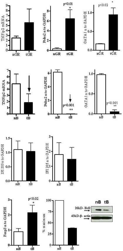
Singh et al.                                                                                       Peptide-Induced Immune Tolerance on Murine Lupus

antibodies and FACS performed. Control mice received either a              RNA Isolation and Real-Time PCR
similar amount of pNeg (negative control peptide) or saline.               Total cellular RNA was isolated from purified cell subsets or total
                                                                           splenocytes from saline-treated or pCons-tolerized BWF1 mice with
Cell Isolation, Preparation,                                               TRIzol (Invitrogen, Carlsbad, CA, USA) as per manufacturer’s
Immunophenotyping, and Flow cytometry                                      protocols. Real-time PCR was performed as described earlier (29,
Spleen cells were isolated ~1 week after administration of the             33–35). Each experimental group consists of the pooled spleen cells of
pCons peptide from tolerized, saline-treated, or naïve BWF1                3–4 mice from each group. 100 ng of total RNA was used with one-
mice. Single cell suspensions of splenocytes were prepared by              step RT-PCR reagents (Applied Biosystems, Foster City, CA, USA).
passing cells through cell strainers (Fisher). ACK lysing buffer,          Quantitative real-time reverse transcription was performed using
(Sigma, St Louis, MO, USA) was used to lyse red blood cells. Cells         TaqMan technology on an ABI Prism 7900 HT Sequence Detection
were washed and re-suspended in RPMI complete media. Cell                  System (Applied Biosystems). Primers and probes of IFI205, GATA1,
subsets were further enriched following incubation with anti-              Ptdss2, TNFip2, FoxP3 and GAPDH were obtained from Applied
CD4 (L3T4), anti-B (CD45R/B220), anti-CD8 (CD8a Ly-2), anti-               Biosystems, Foster City, CA, USA. The oligonucleotide sequences
NK1.1 (CD95b), anti-DX5, anti-CD11C, and anti-Gr-1 microbeads              used for the primers and TaqMan probes (Applied Biosystem, Foster
from Miltenyi Biotech (Auburn, CA, USA). Purity of cells was               City, CA) are described (29, 33–35). GAPDH was used as an
determined to be more than 90% pure as assessed by flow cytometry           endogenous control in each experimental set.
(FACS). For immunophenotyping, isolated cells were washed with
FACS buffer and 1–2 million cells were used for surface staining.          Cell Culture and Measurement of
Before staining, cells were incubated with rat anti-mouse CD16/            Anti-DNA Antibodies
CD32 (FC III/II receptor) Ab to block nonspecific binding.                  Assays were performed to measure anti-DNA Ab as described
    For regulatory B cell immunophenotyping, female 35 wk-old              earlier (29, 31, 34, 35, 37). For optimal Ab production, B cells
BWF1 mice were treated with pCons 1 mg i.v. and blood was                  (1x10⁵ cells) from keep old (40-50-wk-old) naïve BWF1 females
obtained after (3-5 days). Splenocytes were depleted of red blood          with 3+ proteinuria or higher, CD4+CD25- T cells (1x10⁶) from
cells (RBCs) and then stained with CD19, CD1d, and CD5                     young 10–12-wk-old naive BWF1 females without proteinuria,
antibodies for FACS analysis. Antibodies for cell surface                  naïve CD8+ T cells (1x10⁶), and irradiated APC (1x10⁵) cells were
staining and isotype controls were from BD Biosciences, BD                 isolated and cultured with granulocytes or B cells (1x10⁶) from
Pharmingen, eBiosciences, or BioLegend. CD4 (L3T4), CD25                   tolerized mice or controls. Cell cultures were performed in RPMI
(PC61.5) and CTLA-4 (UC10-4F10-11) staining was performed                  1640 supplemented with L-glutamine (2 mM), penicillin (100
with antibodies from BD Pharmingen. FoxP3 (FJK-16s) staining               units/ml), streptomycin (0.1 mg/ml), 2-mercaptoethanol (Gibco)
was performed with an eBiosciences intracellular kit. Data were            and 10% fetal bovine serum (FBS). For tolerized B cells analysis, we
collected using FACSCalibur (BD Biosciences) and analyzed by               cultured, as indicated above pCons-tolerized B cells (1x 10⁶) with
BD Cell Quest software (Becton-Dickinson, Mountain View,                   naïve CD4+CD25- T cells (1x 10⁶), naïve B (1x 10⁵) and/or naïve
CA) or FCS De Nova software (Thornhill, Ontario, Canada).                  CD8+ T cells (1x10⁶) cells. After 72-96 hours, culture supernatants
                                                                           were obtained and anti-DNA IgG was measured by ELISA.

Western Blot Analysis                                                      Statistical Analyses
Western blot analyses were performed as described earlier (37).            Data were analyzed using Prism 4.0 (GraphPad Software, San
In brief, cell lysates were prepared from the naïve and tolerized B        Diego, CA). Comparisons were performed using paired one- or
cells from the splenocytes of naïve and pCons-treated BWF1                 two-tailed test. Nonparametric testing among more than two
mice. Cells were lysed with RIPA buffer (150 nM NaCl, 1.0% NP-             groups was performed by one-way ANOVA. Results are
40, 0.5% sodium deoxycholate, 0.1% SDS, 10 mM Tris, pH 7.3)                expressed as mean ± SEM. p
Singh et al. Peptide-Induced Immune Tolerance on Murine Lupus
(from old BWF1 nephritic mice) and naïve CD8+ T cells to each
cultured subset either separately or in combination with tolerized
B cells (Bregs) and granulocytes. Anti-DNA Ab was analyzed as
described previously (29, 31, 34, 35, 37). Briefly, purified
populations of different spleen effector cell subsets including B
cells, naïve CD4+T cells (CD4+CD25-Tcells), naïve CD8+ T cells,
and tolerized granulocytes and B cells were harvested (using the
appropriate Miltenyi Biotec microbeads via AutoMACS) one
week after pCons treatment of BWF1 mice. Naïve CD4+ T cells
from young mice (10-12-weeks-old) and B cells from old
nephritic BWF1 mice (40-50-weeks-old with 3+ proteinuria)
were co-cultured in complete medium with tolerized/regulatory
B cells and granulocytes and other effector cell types such as
(naïve CD8+ T cells) from spleens of tolerized mice. Our previous
cell-dose response study with CD8+ Treg showed that 1×10⁶ cells
are optimum in mixed cell culture experiments (29) therefore, we
used the same number of cells in these experiments. In addition FIGURE 1 | Anti-DNA Ab was significantly decreased in the presence of
previously, we had found that a ratio of B cells to helper T cells tolerized B cells. Naïve CD4+ T cells, CD8+ T cells and CD45R/B220+B cells
(CD4+CD25-T cells) to regulatory/suppressor T cells of 1:10:10 were isolated from BWF1 mice spleen cells using microbeads from Miltenyi
are needed to observe optimal suppression of anti-DNA Biotech (Auburn, CA, USA). Cells were cultured in RPMI 1640 supplemented
with L-glutamine (2 mM), penicillin (100 units/ml), streptomycin (0.1 mg/ml),
antibodies (29, 32, 35). Therefore, we used this same ratio with
2-mercaptoethanol (Gibco) and 10% fetal bovine serum (FBS). Cells were co-
each effector cell type. We found that both tolerized B cells and cultured in the presence of tolerized B cells (1x10⁶ cells). Different immune cell
granulocytes suppressed the production of anti-DNA Ab subsets (naïve B cells, 1x10⁵ cells from old nephritic mice; CD4+CD25- T cells,
(Figures 1 and 2). Although we did not determine whether 1x10⁶; naïve CD8+ T cells, 1x10⁵) were isolated from splenocytes and cultured
this suppressive effect was direct or indirect on autoreactive B with tolerized B cells (1x10⁶ cells). After the 72-96 hours range, culture
supernatants were obtained. Anti-DNA Ab levels were measured from culture
cells through CD4+ or CD8+ T cells or by synergistic effect by
supernatants by ELISA. *p < 0.05.
those cells with tolerized B cells and granulocytes, this data
clearly suggests that pCons-induced regulatory B cells and
granulocytes suppress the anti-DNA Ab production and thus
play a significant role in autoimmunity.
Microarray Analysis Showed Altered
Regulation of Genes in Non-T Cells
Since our previous published microarray data showed that
pCons treatment induces major changes in white blood cells
(WBC) subsets of BWF1 spleen cells [448 genes differentially-
regulated in whole splenocytes of tolerized compared to control
mice (33)], we were interested to see the potential role of
regulatory B cells and granulocytes. Thus, in this report, we
characterized expression of selected genes (highly upregulated)
in different cell populations in non-T cells (tolerized B cells and
tolerized granulocytes cells) and further tested the ability of these
cell subsets to suppress production of anti-DNA Ab in lupus.
Our data suggests that cell types other than T cells may play
major roles in this model of immune tolerance.
B cells and Granulocytes Produced
Significantly Increased/Decreased
Amounts of mRNA for Several Genes of FIGURE 2 | Anti-DNA Ab was significantly decreased in the presence of
Interest Including Interferon Genes After tolerized PMNs-granulocytes. Different immune cell subsets (naïve B cells, 1x 10⁵
cells from old nephritic mice; CD4+CD25- T cells, 1x10⁶; naïve CD8+ T cells,
pCons Treatment in BWF1 Lupus Mice 1x10⁵) were isolated and cultured with tolerized granulocytes (GR, 1x10⁶). Cell
To address the role of tolerized non-T cell subsets after pCons subsets were isolated from total spleen cells of BWF1 mice. Cells were cultured in
treatment in BWF1 mice, B cells and granulocytes were obtained RPMI 1640 supplemented with L-glutamine (2 mM), penicillin (100 units/ml),
from the spleen of BWF1 mice 1 weeks after pCons treatment. streptomycin (0.1 mg/ml), 2-mercaptoethanol (Gibco) and 10% fetal bovine serum
RNA was isolated from these cell subsets and real-time PCR was (FBS). After the 72- 96 hours range, culture supernatants were obtained. Anti-
DNA Ab levels were measured from culture supernatants by ELISA. *p < 0.05.
performed as described earlier (33). Real-time PCR analyses
Frontiers in Immunology | www.frontiersin.org 4 May 2021 | Volume 12 | Article 662901Singh et al. Peptide-Induced Immune Tolerance on Murine Lupus
indicate that TNFAIP2 (Tumor necrosis factor alpha-induced CD19+CD1d+ regulatory B cells including median fluorescence
protein 2) was increased ~2-fold (Figure 3A), and Ptdss2 intensity (Figures 4B, G, D, E). This is an important finding
(Phosphatidylserine synthase 2 and GATA1 (GATA-binding because two previous studies have revealed similar phenotype of
factor 1) mRNA were upregulated more than 10-fold in Bregs in SLE patients (45, 46). Further, we found that pCons
tolerized granulocytes compared to naïve granulocytes (Figures treatment reduces the percent expression of CD19+CD5+ B cells
3B, C). In contrast, the mRNA of all the above genes (Figures (Figures 4C, H, I). The median fluorescence intensity (MFI) of
3D–F) and those of IFN-induced genes including IFI203 and CD19+ CD5+ cells were significantly decreased in pCons treated
IFI205 (Figures 3G, H) were down regulated in tolerized B cells. mice (Figure 4J). These data show that pCons treatment
Although, the decreased level of IFI203 and IFI205 did not reach modified the B cells expression markers CD1d and CD5, and
to the significance. However, other IFNs genes were significantly since we have also shown that CD4+ and CD8+ T cells from
decreased. Thus, this data displays dynamic interplay and tolerized mice suppress autoreactive B cells and could account
suggests that pCons has differential effects on different for their reduced numbers, this suggests that pCons treatment
interferon genes in our tolerance model. Collectively these data induces regulatory B cells.
demonstrate that pCons treatment modify the interferon’s gene
signature differentially in tolerized B cells and granulocytes. pCons Treatment Increased CD4+FoxP3+
Regulatory T Cells and Significantly
pCons-Tolerized B Cells From Lupus Mice Reduced Percent Expression and Median
Have Increased FoxP3 mRNA and Bcl2 Fluorescence Intensity of CTLA-4
Protein Levels (Cytotoxic T-Lymphocyte-Associated
The transcription factor forkhead box P3 (FoxP3), also known as Proten-4) in CD8+ T Cells of BWF1
scurfin, plays an important role in the maintenance of immunological Lupus Mice
homeostasis and restoration of self-tolerance. Dysfunction and We were interested to see whether pCons treatment induces
mutations of the FoxP3 gene causes immunodysregulation regulatory T cells and whether it affects CTLA-4 expression.
polyendocrinopathy enteropathy X-linked (or IPEX) syndrome. CTLA-4 plays an important role in immune tolerance and T-cell
FoxP3 also participates in maintaining the immune system activation. We found that pCons treatment significantly
response (38) and in the development and function of regulatory T increased the number of CD4+FoxP3+ T cells in BWF1 mice
cells (39–42). In the present study, we evaluated the expression of compared to naïve and/or saline-treated mice (Figures 5A–D,
FoxP3 in pCons-tolerized B cells of lupus (BWF1 mice) compared F–H). We also measured the CTLA-4 expression on T cells
with naïve B cells. Surprisingly, we found significantly increased (CD8+T cells) and found that percent expression of CTLA-4 was
expression of FoxP3 in tolerized B cells compared to naïve B cells significantly decreased in pCons treated mice (Figure 5E)
(Figure 3I). Next, we investigated the protein expression of bcl2 from Further, we found that pCons treatment significantly reduced
cell lysates of tolerized B cells and naïve B cells with Western blot the median fluorescence intensity (MFI) of CTLA-4 expression
assay. We found that tolerized B cells have decreased levels of Bcl2 in CD8+ T cells compared to naïve mice (Figures 5I, J). Thus, the
protein compared to naïve B cells (Figure 3J). Bcl-2 regulates cell data shows the immunomodulatory role of pCons in BWF1
death (apoptosis) by promoting or inhibiting apoptosis (43, 44). We mice. However, future study is warranted to pinpoint the exact
have shown previously that CD4+ and CD8+ T cells from tolerized mechanism of pCons activity in Lupus.
mice have significantly reduced apoptosis. Thus our data suggest that
pCons tolerance may also affect apoptosis of B cells in our tolerance
model and may play a significant role in survival of these cells by
DISCUSSION
regulating immune tolerance.
In the present study, we have added to previous work showing
pCons Treatment Induced and Modified that pCons induces CD8+ and CD4+ suppressive cells and shown
the Cell Surface Expression Markers for that B cells and granulocytes from tolerized mice suppress anti-
Regulatory B Cells DNA Ab production in vitro. Several suppressive mechanisms/
Our previous study showed that pCons treatment induces both factors may be involved including IL-10, TGFb, IL-35, and
CD4+ and CD8+ regulatory T cells (29, 32, 36). Based on these combinations of TLR9, CD40, and/or B cell receptor (BCR)
data and our gene expression study (33), we hypothesize that and engagement of CD80/CD86 on Bregs (45, 47). pCons
pCons treatment may induce suppressor/regulatory B cells treatment significantly increased the number of CD4+FoxP3+ T
(Bregs) and granulocyte cells with the potential to suppress the cells. In earlier studies, we showed that these FoxP3+ T cells (both
proliferation of naïve CD4+CD25- cells and naïve B cells as well CD4+ and CD8+ Treg) suppressed autoimmunity in vivo and anti-
as the production of anti-DNA Ab. To address this, we isolated DNA production in vitro (29, 32, 36). Immune tolerance induced
spleen cells from female BWF1 mice after one week of pCons by pCons prolonged survival of BWF1 lupus mice (NZB/NZW)
treatment (1 mg i.v) and performed immunophenotyping studies F1 and delayed the appearance of glomerulonephritis (29, 31,
with flow cytometry from naïve and pCons-treated mice 35). The pCons-induced regulatory T cells suppressed
(Figures 4A, F live gating scheme). We found that pCons proliferation of naïve CD4+ T cells and naïve CD19+/B220+ B
treatment of BWF1 mice increases percent expression of cells and the production of anti-DNA antibodies (29, 32, 34–36).
Frontiers in Immunology | www.frontiersin.org 5 May 2021 | Volume 12 | Article 662901Singh et al. Peptide-Induced Immune Tolerance on Murine Lupus
A B C
D E F
G H
I J
FIGURE 3 | Tolerized B cells have reduced IFNs gene mRNA and Bcl2 protein level and increased FoxP3 mRNA expression. RNA was isolated from naïve and
tolerized B cells and granulocytes. Real time PCR was performed with 100 ng of RNA with gene specific primers and probes. Data was normalized with GAPDH
mRNA levels. *p < 0.05. TNFAIP2, Ptdss2, and GATA1 mRNA was increased (A–C) in tolerized granulocytes (GR) but reduced in tolerized B cells (D–F). IFI203 and
IFI205 was decreased in tolerized B cells (G, H). (I) FoxP3 expression was increased in tolerized B cells. (J) Quantification of Western blot analysis of Bcl2 protein
levels in cell lysates from naïve and tolerized sorted B (CD45R/B220) cells.
T cell suppressive capacity correlated with modulation of significantly decreased in the CD8+ T cells of pCons-treated
Mitogen-Activated Protein kinase (p38 MAP kinase) activity BWF1 mice. This is a significant finding as CTLA-4 is involved in
and FoxP3 expression in CD4+ Tregs (48). In the current study negative signaling and plays a pivotal inhibitory role in T cell
we found that CTLA-4 median fluorescence intensity was anergy and prevention of autoimmunity. In addition, recent
Frontiers in Immunology | www.frontiersin.org 6 May 2021 | Volume 12 | Article 662901Singh et al. Peptide-Induced Immune Tolerance on Murine Lupus
A B C D E
F G H I J
FIGURE 4 | pCons treatment modified the cell surface expression markers for regulatory B cells. Female 35-wk old BWF1 mice were treated with pCons (1 mg i.v.).
After 3 days, blood was obtained, RBC lysed, and cells were stained with CD19, CD1d and CD5 antibodies and FACS performed. Representative live cell gating
strategy (A, F) and FACS analysis of CD1d (B, G) and CD5 (C, H) expression levels from representative naïve (A–C) and pCons treated (F–H) mice. Percent
expression of CD19+ CD1d+ cells in naïve vs pCons treated mice (D). Percent expression of CD19+CD5+cells in naïve vs pCons treated mice (I). Quantification of
Median Fluorescent Intensity of naïve and pCons-treated mice for CD1d (E) (from B and G Gate 3 and 4) and CD5 (J) (from C and H Gate 3 and 4). CD1d increased
and CD5 cells decreased with pCons treatment. *p < 0.05, **p < 0.001, ***p < 0.0001.
A B C D E
F G H I J
FIGURE 5 | pCons treatment increased CD4+FoxP3+ regulatory T cells and significantly reduced Median Fluorescence Intensity of CTLA-4 (Cytotoxic T-lymphocyte-
Associated Proten-4) in CD8+ T cells of BWF1 lupus mice. Female 12-20 wk-old BWF1 mice were treated with pCons (1mg i.v.). After 1-2 weeks, splenocytes were
obtained, RBC lysed, and cells were stained with CD4, CD8, CD25, CTLA-4 and FoxP3 antibodies and FACS performed. 10,000 minimum cells were gated.
Representative gating strategy (A, F) and FACS analyses of CD4+CD25+ (B, C), CD4+FoxP3+ (G, H) and cumulative two-three experiments data for CD4+FoxP3+
cells (D) are shown. Cumulative data of CD8+ CTLA-4+ T cells experiments (two-three) is shown (E). CTLA-4 staining (MFI) on CD8+ T cells is shown (I, J).
CD4+FoxP3+T cells are significantly increased. CTLA-4 MFI is significantly decreased. *p < 0.05, **p < 0.001, ***p < 0.0001.
studies show that CTLA-4 controls follicular helper T cells and and CD86 on antigen presenting cells (APC); thus, altering the
regulatory T cells, thereby controlling the B cells responses and level of CD28 engagement on follicular helper T cells (51).
humoral immunity (49–51). CTLA-4 also downregulates CD80 However, its precise mechanism of action has not been fully
Frontiers in Immunology | www.frontiersin.org 7 May 2021 | Volume 12 | Article 662901Singh et al. Peptide-Induced Immune Tolerance on Murine Lupus
resolved. Recently abnormal CTLA-4 gene polymorphisms and is thought to be central to induction of the type 1 IFN response
function has been reported in SLE patients (52, 53). (69, 70). Indeed, a recent therapeutic option in patients with
For the first time to our knowledge, in this system we found that lupus is through inhibition of type IFN–a and several recent
B cells and granulocytes also can be “tolerized” and subsequently clinical trial data suggest therapeutic benefit (71–73). Thus, our
function as regulatory/suppressor cells to prevent production of findings that pCons tolerance reduces the IFN genes in our lupus
autoantibodies. In the current experiments, we used whole tolerized model has direct clinical and translational significance.
B cells and granulocytes for the suppression assay. We acknowledge We have shown earlier that pCons peptide delayed the onset of
the next step is to test the specific Bregs subsets to determine cell autoimmunity in lupus mouse model by inducing immune
specificity and their mode of action and mechanism. Therefore, tolerance and up-regulating FoxP3 in T cells which are
detailed molecular and cellular mechanisms of regulatory B cells suppressive (31, 34, 74). Other studies have reported that peptides
and granulocytes are not completely clear and future study will be from CDRs of pathogenic anti-DNA Ab could also prevent
required to address this shortcoming. autoantibody production and down-regulate autoreactive T cell
Regulatory B cells and regulatory granulocytes are not well responses (75, 76). Similar to our results, these studies showed
characterized in this SLE tolerance model and this study provided that a peptide derived from the CDR1 of a human anti-DNA Ab
first novel mechanistic insight. We showed that 1) pCons tolerance (hCDR1) could ameliorate lupus by inducing Tregs and suppressing
altered expression of several candidate genes (see below) including the activation of autoreactive T cells through mechanisms including
interferon genes in tolerized B cells and granulocytes compared to downregulation of transcription factors responsible for negative
naïve cells; 2) pCons tolerance modified the cell surface expression of regulation of T-cell activation in lupus animal models (62, 76–79).
regulatory B cells (and/or deleted the CD19+CD5+ subset); 3) pCons Furthermore, clinical trial data has indicated safety and efficacy of
tolerance increased the percent expression of (CD19+CD1d+) cells; hCDR1 (edratide) in SLE patients (80).
and 4) pCons-tolerized B cells and granulocytes significantly reduced We demonstrated with flow cytometry (FACS) immunophenotyping
the production of anti-DNA antibody in cell culture experiments of that after pCons treatment the CD19+CD1d+ regulatory B cell
lupus mouse cells. subset was significantly increased in BWF1 lupus mice compared
pCons tolerance has been shown to affect various genes and to naïve mice (Figures 4B, G, D). However, the CD19+CD5+ B cell
markers, cell surface molecules, cytokines and different cell types subset was significantly decreased (Figures 4C, H, I). This is a
including regulatory T cells (both CD4 and CD8) (29–35, 37). In significant finding and the next step will be to decipher the
the present study, we showed that pCons induced B cells mechanisms with future functional studies including testing B
enriched in markers identifying suppressor B cells and these cells from anti-CD5 treated control mice. In agreement with our
cells have significantly reduced interferon-induced genes (IFN) experiments, a previous study showed that anti-CD5 therapy
such as, ptdss2, GATA1, and TNFip2 (Figure 3) compared to decreases severity of established disease in collagen-induced
naïve mice. In contrast, we also found that these genes were arthritis in DBA/1 mice (81). Thus, our data with pCons therapy
significantly increased in tolerized granulocytes, with the has clinical and therapeutic relevance in peptide induced immune
exception of TNFip2 which was upregulated but did not reach tolerance. We also found that tolerized B cells have significant
the significance level (Figure 3) demonstrating differential effect increased FoxP3 mRNA. Another study reported that the expansion
of pCons. Thus, our data indicate dynamic interplay of these of CD25+hiCD5+ and FoxP3+ regulatory B cells is associated with
genes or their gene products in different immune cell subsets in SLE disease activity in humans (82). Similarly, the presence of
our pCons-induced tolerance model. How this interplay affects FoxP3 + CD19 + CD5 + B cells in human peripheral blood
the overall immune response in lupus mice is not clear. However, mononuclear cells has also been reported (83). The diverse
recent studies have shown the importance of interferon genes in suppressive mechanisms of these regulatory B cells are through
lupus (54–58). Lupus is characterized by the dysregulation of IL-10, TGFb, and IL-35. Previously we have demonstrated that
both the innate and the adaptive immune systems. An increased pCons-induced splenocytes have significantly increased amount of
expression of type I IFN-regulated genes, termed IFN signature, TGFb, smad2, and smad3 expression and tolerized total CD8+ T
has been reported in the majority of patients with SLE (59–61). cells have increased amount of IL-10 (37). Although, we did not
In agreement with our findings, another study found that a measure the expression of IL-10, TGFb and IL-35 in the regulatory
tolerogenic peptide of the light chain complementarity- B cells in our model, it is tempting to speculate that these molecules
determining region 1 (hCDR1) down-regulates the expression will play important role in our system based on our previous data.
of interferon-alpha (IFN-a) in murine and human SLE (62). Thus, our findings may suggest that pCons tolerance promotes
IFN-a plays a major role in SLE pathogenesis and the levels of tolerized B cells that can suppress the autoimmune responses.
IFN-a were increased and correlated with SLE disease activity in Similar to our study, hCDR1 tolerance has effects on B cell
the sera of mice and humans (63–65). Administration of activating factor (BAFF) and B-cell CD74 macrophage inhibitory
exogenous IFN-a leads to worsening of disease in various factor in murine lupus (84, 85). The reduced levels of BAFF
mouse models (66). Type 1 IFN contributes to loss of tolerance correlated with reduced rate of maturation and differentiation of
and increases production of autoantibodies (67), induces B cells and decrease in integrin expression. Recent studies provided
differentiation of monocytes to myeloid-derived dendritic cells further evidence of targeting of BAFF/BLys and APRIL in the
(mDC) (56), and plays a vital role in the activation of management of lupus (86–88); and another study reported the effect
autoreactive T and B cells (68). Activation of TLR7 and TLR9 of hCDR1 on IL-7 and apoptosis (89) and showed the rate of
Frontiers in Immunology | www.frontiersin.org 8 May 2021 | Volume 12 | Article 662901Singh et al. Peptide-Induced Immune Tolerance on Murine Lupus
apoptosis is reduced with hCDR1 treatment in lupus mice. Bcl2 and AUTHOR CONTRIBUTIONS
Bcl-XL levels were further reduced, and this was associated with
reduced activation of T and B cells (90). We demonstrated earlier RPS contributed to the experimental design, obtaining funding,
that pCons-induced CD8+ Tregs are resistant to apoptosis (29, 34). In conducting experiments, analyzing data, preparing figures, and
the present study, we found that bcl2 protein level was significantly writing of the manuscript. BHH contributed to funding
decreased in tolerized B cells compared to naïve B cells thus affecting and editing of the manuscript. DSB contributed to figure and
the survival of these cells in BWF1 mice. This is in agreement with manuscript editing. All authors contributed to the article and
another study that revealed increased expression of Bcl2 leads to approved the submitted version.
development of SLE like symptoms in Bcl2 transgenic mice (91).
Thus, altogether, our data suggests that pCons’ effect on tolerized B
cells and down-regulation of IFNs and bcl2 may play overall FUNDING
therapeutic beneficial effects in our tolerance model.
This work was supported by the NIH grants AR54034, AI 083894,
AI65645 to RPS, UCLA Senate Core Grant to BHH and RPS, and
UCLA Oppenheimer Clinical Seed Grant and American
DATA AVAILABILITY STATEMENT
Autoimmune Related Disease Association grant to RPS.
The raw data supporting the conclusions of this article will be
made available by the authors, without undue reservation.
ACKNOWLEDGMENTS
ETHICS STATEMENT We thank the UCLA Flow Cytometry Core Facility for cell
sorting and FACS experiments, Dr. Desmond Smith’s Lab for
The animal study was reviewed and approved by UCLA Animal the real time PCR, Dr. Ravi Dinesh for technical help, and
Research Committee (ARC). Dr. Antonio La Cava for intellectual insights.
14. Mizoguchi A, Bhan AK. A Case for Regulatory B Cells. J Immunol (2006)
REFERENCES 176:705–10. doi: 10.4049/jimmunol.176.2.705
1. Kotzin BL. Systemic Lupus Erythematosus. Cell (1996) 85:303–6. doi: 15. Serra P, Santamaria P. To ‘B’ Regulated: B Cells as Members of the Regulatory
10.1016/S0092-8674(00)81108-3 Workforce. Trends Immunol (2006) 27:7–10. doi: 10.1016/j.it.2005.11.003
2. Wakeland EK, Liu K, Graham RR, Behrens TW. Delineating the Genetic Basis 16. Yanaba K, Bouaziz JD, Haas KM, Poe JC, Fujimoto M, Tedder TF. A
of Systemic Lupus Erythematosus. Immunity (2001) 15:397–408. doi: Regulatory B Cell Subset With a Unique CD1dhiCD5+ Phenotype Controls
10.1016/S1074-7613(01)00201-1 T Cell-Dependent Inflammatory Responses. Immunity (2008) 28:639–50. doi:
3. Theofilopoulos AN, Dixon FJ. Murine Models of Systemic Lupus Erythematosus. 10.1016/j.immuni.2008.03.017
Adv Immunol (1985) 37:269–390. doi: 10.1016/S0065-2776(08)60342-9 17. Yanaba K, Bouaziz JD, Matsushita T, Tsubata T, Tedder TF. The
4. Karim MR, Wang YF. Phenotypic Identification of CD19(+)CD5(+)CD1d(+) Development and Function of Regulatory B Cells Expressing IL-10 (B10
Regulatory B Cells That Produce Interleukin 10 and Transforming Growth Cells) Requires Antigen Receptor Diversity and TLR Signals. J Immunol
Factor Beta1 in Human Peripheral Blood. Arch Med Sci (2019) 15:1176–83. (2009) 182:7459–72. doi: 10.4049/jimmunol.0900270
doi: 10.5114/aoms.2018.77772 18. Iking-Konert C, Ostendorf B, Sander O, Jost M, Wagner C, Joosten L, et al.
5. Li W, Tian X, Lu X, Peng Q, Shu X, Yang H, et al. Significant Decrease in Transdifferentiation of Polymorphonuclear Neutrophils to Dendritic-Like Cells
Peripheral Regulatory B Cells is an Immunopathogenic Feature of at the Site of Inflammation in Rheumatoid Arthritis: Evidence for Activation by
Dermatomyositis. Sci Rep (2016) 6:27479. doi: 10.1038/srep27479 T Cells. Ann Rheum Dis (2005) 64:1436–42. doi: 10.1136/ard.2004.034132
6. Mauri C, Menon M. Human Regulatory B Cells in Health and Disease: 19. Premchand P, Takeuchi K, Bjarnason I. Granulocyte, Macrophage, Monocyte
Therapeutic Potential. J Clin Invest (2017) 127:772–9. doi: 10.1172/JCI85113 Apheresis for Refractory Ulcerative Proctitis. Eur J Gastroenterol Hepatol
7. Morris A, Moller G. Regulation of Cellular Antibody Synthesis Effect of (2004) 16:943–5. doi: 10.1097/00042737-200409000-00023
Adoptively Transferred Antibody-Producing Spleen Cells on Cellular 20. Suzuki Y, Yoshimura N, Saniabadi AR, Saito Y. Selective Granulocyte
Antibody Synthesis. J Immunol (1968) 101:439–45. and Monocyte Adsorptive Apheresis as a First-Line Treatment for Steroid
8. L’Age-Stehr J, Teichmann H, Gershon RK, Cantor H. Stimulation of Naive Patients With Active Ulcerative Colitis: A Prospective Uncontrolled
Regulatory T Cell Circuits by Immunoglobulin-Dependent Structures on Study. Dig Dis Sci (2004) 49:565–71. doi: 10.1023/B:DDAS.00000
Activated B Cells. Eur J Immunol (1980) 10:21–6. doi: 10.1002/eji.1830100105 26299.43792.ae
9. Kennedy MW, Thomas DB. A Regulatory Role for the Memory B Cell as 21. Zehntner SP, Brickman C, Bourbonniere L, Remington L, Caruso M,
Suppressor-Inducer of Feedback Control. J Exp Med (1983) 157:547–58. doi: Owens T. Neutrophils That Infiltrate the Central Nervous System Regulate
10.1084/jem.157.2.547 T Cell Responses. J Immunol (2005) 174:5124–31. doi: 10.4049/jimmunol.
10. Evans DE, Munks MW, Purkerson JM, Parker DC. Resting B Lymphocytes as 174.8.5124
APC for Naive T Lymphocytes: Dependence on CD40 Ligand/CD40. 22. Prior C, Townsend PJ, Hughes DA, Haslam PL. Induction of Lymphocyte
J Immunol (2000) 164:688–97. doi: 10.4049/jimmunol.164.2.688 Proliferation by Antigen-Pulsed Human Neutrophils. Clin Exp Immunol
11. Bouaziz JD, Yanaba K, Tedder TF. Regulatory B Cells as Inhibitors of Immune (1992) 87:485–92. doi: 10.1111/j.1365-2249.1992.tb03024.x
Responses and Inflammation. Immunol Rev (2008) 224:201–14. doi: 10.1111/ 23. Potter NS, Harding CV. Neutrophils Process Exogenous Bacteria Via an Alternate
j.1600-065X.2008.00661.x Class I MHC Processing Pathway for Presentation of Peptides to T Lymphocytes.
12. Lund FE. Cytokine-Producing B Lymphocytes-Key Regulators of Immunity. J Immunol (2001) 167:2538–46. doi: 10.4049/jimmunol.167.5.2538
Curr Opin Immunol (2008) 20:332–8. doi: 10.1016/j.coi.2008.03.003 24. Tvinnereim AR, Hamilton SE, Harty JT. Neutrophil Involvement in Cross-
13. Mauri C, Ehrenstein MR. The ‘Short’ History of Regulatory B Cells. Trends Priming CD8+ T Cell Responses to Bacterial Antigens. J Immunol (2004)
Immunol (2008) 29:34–40. doi: 10.1016/j.it.2007.10.004 173:1994–2002. doi: 10.4049/jimmunol.173.3.1994
Frontiers in Immunology | www.frontiersin.org 9 May 2021 | Volume 12 | Article 662901Singh et al. Peptide-Induced Immune Tolerance on Murine Lupus
25. Beauvillain C, Delneste Y, Scotet M, Peres A, Gascan H, Guermonprez P, et al. (14;18) Translocation. Cell (1986) 47:19–28. doi: 10.1016/0092-8674(86)
Neutrophils Efficiently Cross-Prime Naive T Cells In Vivo. Blood (2007) 90362-4
110:2965–73. doi: 10.1182/blood-2006-12-063826 45. Blair PA, Norena LY, Flores-Borja F, Rawlings DJ, Isenberg DA, Ehrenstein
26. Culshaw S, Millington OR, Brewer JM, McInnes IB. Murine Neutrophils MR, et al. CD19(+)CD24(Hi)CD38(Hi) B Cells Exhibit Regulatory Capacity
Present Class II Restricted Antigen. Immunol Lett (2008) 118:49–54. doi: in Healthy Individuals But are Functionally Impaired in Systemic Lupus
10.1016/j.imlet.2008.02.008 Erythematosus Patients. Immunity (2010) 32:129–40. doi: 10.1016/
27. Muller I, Munder M, Kropf P, Hansch GM. Polymorphonuclear Neutrophils j.immuni.2009.11.009
and T Lymphocytes: Strange Bedfellows or Brothers in Arms? Trends 46. Yang X, Yang J, Chu Y, Xue Y, Xuan D, Zheng S, et al. T Follicular Helper
Immunol (2009) 30:522–30. doi: 10.1016/j.it.2009.07.007 Cells and Regulatory B Cells Dynamics in Systemic Lupus Erythematosus.
28. Gupta S, Kaplan MJ. The Role of Neutrophils and NETosis in Autoimmune and PloS One (2014) 9:e88441. doi: 10.1371/journal.pone.0088441
Renal Diseases. Nat Rev Nephrol (2016) 12:402–13. doi: 10.1038/nrneph.2016.71 47. Flores-Borja F, Bosma A, Ng D, Reddy V, Ehrenstein MR, Isenberg DA, et al.
29. Hahn BH, Singh RP, La Cava A, Ebling FM. Tolerogenic Treatment of Lupus Cd19+Cd24hicd38hi B Cells Maintain Regulatory T Cells While Limiting
Mice With Consensus Peptide Induces FoxP3-expressing, Apoptosis- TH1 and TH17 Differentiation. Sci Trans Med (2013) 5:173ra23. doi: 10.1126/
Resistant, TGFbeta-secreting Cd8+ T Cell Suppressors. J Immunol (2005) scitranslmed.3005407
175:7728–37. doi: 10.4049/jimmunol.175.11.7728 48. Lourenco EV, Procaccini C, Ferrera F, Iikuni N, Singh RP, Filaci G, et al.
30. Hahn BHA, La Cava A. The Anti-DNA Ig Consensus Peptide Pcons Modulation of P38 MAPK Activity in Regulatory T Cells After Tolerance
Facilitates Regulatory T Cell Activity in SLE Patients. Arthritis Rheum With Anti-DNA Ig Peptide in (NZB X NZW)F1 Lupus Mice. J Immunol
(2007) 56:S546–547. (2009) 182:7415–21. doi: 10.4049/jimmunol.0804214
31. Hahn BH, Singh RR, Wong WK, Tsao BP, Bulpitt K, Ebling FM. Treatment 49. Sage PT, Paterson AM, Lovitch SB, Sharpe AH. The Coinhibitory Receptor
With a Consensus Peptide Based on Amino Acid Sequences in Autoantibodies CTLA-4 Controls B Cell Responses by Modulating T Follicular Helper, T
Prevents T Cell Activation by Autoantigens and Delays Disease Onset in Follicular Regulatory, and T Regulatory Cells. Immunity (2014) 41:1026–39.
Murine Lupus. Arthritis Rheum (2001) 44:432–41. doi: 10.1002/1529-0131 doi: 10.1016/j.immuni.2014.12.005
(200102)44:23.0.CO;2-S 50. Wing K, Onishi Y, Prieto-Martin P, Yamaguchi T, Miyara M, Fehervari Z,
32. La Cava A, Ebling FM, Hahn BH. Ig-Reactive CD4+CD25+ T Cells From et al. CTLA-4 Control Over FoxP3+ Regulatory T Cell Function. Science
Tolerized (New Zealand Black X New Zealand White)F1 Mice Suppress In (2008) 322:271–5. doi: 10.1126/science.1160062
Vitro Production of Antibodies to DNA. J Immunol (2004) 173:3542–8. doi: 51. Wang CJ, Heuts F, Ovcinnikovs V, Wardzinski L, Bowers C, Schmidt EM,
10.4049/jimmunol.173.5.3542 et al. CTLA-4 Controls Follicular Helper T-cell Differentiation by Regulating
33. Singh RP, Dinesh R, Elashoff D, de Vos S, Rooney RJ, Patel D, et al. Distinct the Strength of CD28 Engagement. Proc Natl Acad Sci USA (2015) 112:524–9.
Gene Signature Revealed in White Blood Cells, CD4(+) and CD8(+) T Cells in doi: 10.1073/pnas.1414576112
(Nzbx NZW) F1 Lupus Mice After Tolerization With Anti-DNA Ig Peptide. 52. Jury EC, Flores-Borja F, Kalsi HS, Lazarus M, Isenberg DA, Mauri C, et al.
Genes Immun (2010) 11:294–309. doi: 10.1038/gene.2010.6 Abnormal CTLA-4 Function in T Cells From Patients With Systemic Lupus
34. Singh RP, La Cava A, Hahn BH. Pconsensus Peptide Induces Tolerogenic Cd8+ Erythematosus. Eur J Immunol (2010) 40:569–78. doi: 10.1002/eji.200939781
T Cells in Lupus-Prone (NZB X NZW)F1 Mice by Differentially Regulating 53. Liu J, Zhang HX. CTLA-4 Polymorphisms and Systemic Lupus
FoxP3 and PD1 Molecules. J Immunol (2008) 180:2069–80. doi: 10.4049/ Erythematosus: A Comprehensive Meta-Analysis. Genet Test Mol
jimmunol.180.4.2069 Biomarkers (2013) 17:226–31. doi: 10.1089/gtmb.2012.0302
35. Singh RP, La Cava A, Wong M, Ebling F, Hahn BH. Cd8+ T Cell-Mediated 54. Choubey D, Panchanathan R. Interferon-Inducible Ifi200-family Genes in
Suppression of Autoimmunity in a Murine Lupus Model of Peptide-Induced Systemic Lupus Erythematosus. Immunol Lett (2008) 119:32–41. doi: 10.1016/
Immune Tolerance Depends on FoxP3 Expression. J Immunol (2007) j.imlet.2008.06.001
178:7649–57. doi: 10.4049/jimmunol.178.12.7649 55. Crow MK. Type I Interferon in the Pathogenesis of Lupus. J Immunol (2014)
36. Hahn BH, Anderson M, Le E, La Cava A. Anti-Dna Ig Peptides Promote Treg 192:5459–68. doi: 10.4049/jimmunol.1002795
Cell Activity in Systemic Lupus Erythematosus Patients. Arthritis Rheum 56. Ronnblom L, Pascual V. The Innate Immune System in SLE: Type I
(2008) 58:2488–97. doi: 10.1002/art.23609 Interferons and Dendritic Cells. Lupus (2008) 17:394–9. doi: 10.1177/
37. Dinesh R, Hahn BH, La Cava A, Singh RP. Interferon-Inducible Gene 202b 0961203308090020
Controls CD8(+) T Cell-Mediated Suppression in Anti-DNA Ig Peptide- 57. Luo S, Wang Y, Zhao M, Lu Q. The Important Roles of Type I Interferon and
Treated (NZB X NZW) F1 Lupus Mice. Genes Immun (2011) 12:360–9. doi: Interferon-Inducible Genes in Systemic Lupus Erythematosus. Int
10.1038/gene.2011.4 Immunopharmacol (2016) 40:542–9. doi: 10.1016/j.intimp.2016.10.012
38. Brunkow ME, Jeffery EW, Hjerrild KA, Paeper B, Clark LB, Yasayko SA, et al. 58. Bezalel S, Guri KM, Elbirt D, Asher I, Sthoeger ZM. Type I Interferon
Disruption of a New Forkhead/Winged-Helix Protein, Scurfin, Results in the Signature in Systemic Lupus Erythematosus. Isr Med Assoc J (2014) 16:246–9.
Fatal Lymphoproliferative Disorder of the Scurfy Mouse. Nat Genet (2001) 59. Crow MK. Type I Interferon in Systemic Lupus Erythematosus. Curr Top
27:68–73. doi: 10.1038/83784 Microbiol Immunol (2007) 316:359–86. doi: 10.1007/978-3-540-71329-6_17
39. Fontenot JD, Gavin MA, Rudensky AY. FoxP3 Programs the Development 60. Crow MK. Type I Interferon in Organ-Targeted Autoimmune and Inflammatory
and Function of CD4+CD25+ Regulatory T Cells. Nat Immunol (2003) 4:330– Diseases. Arthritis Res Ther (2010) 12(Suppl 1):S5. doi: 10.1186/ar2886
6. doi: 10.1038/ni904 61. Crow MK. Advances in Understanding the Role of Type I Interferons in
40. Fontenot JD, Rasmussen JP, Williams LM, Dooley JL, Farr AG, Rudensky AY. Systemic Lupus Erythematosus. Curr Opin Rheumatol (2014) 26:467–74. doi:
Regulatory T Cell Lineage Specification by the Forkhead Transcription Factor 10.1097/BOR.0000000000000087
FoxP3. Immunity (2005) 22:329–41. doi: 10.1016/j.immuni.2005.01.016 62. Sthoeger Z, Zinger H, Sharabi A, Asher I, Mozes E. The Tolerogenic Peptide,
41. Hori S, Nomura T, Sakaguchi S. Control of Regulatory T Cell Development by hCDR1, Down-Regulates the Expression of Interferon-Alpha in Murine and
the Transcription Factor FoxP3. Science (2003) 299:1057–61. doi: 10.1126/ Human Systemic Lupus Erythematosus. PloS One (2013) 8:e60394. doi:
science.1079490 10.1371/journal.pone.0060394
42. Marson A, Kretschmer K, Frampton GM, Jacobsen ES, Polansky JK, MacIsaac 63. Baechler EC, Batliwalla FM, Karypis G, Gaffney PM, Ortmann WA, Espe KJ,
KD, et al. FoxP3 Occupancy and Regulation of Key Target Genes During T- et al. Interferon-Inducible Gene Expression Signature in Peripheral Blood
cell Stimulation. Nature (2007) 445:931–5. doi: 10.1038/nature05478 Cells of Patients With Severe Lupus. Proc Natl Acad Sci USA (2003) 100:2610–
43. Tsujimoto Y, Finger LR, Yunis J, Nowell PC, Croce CM. Cloning of the 5. doi: 10.1073/pnas.0337679100
Chromosome Breakpoint of Neoplastic B Cells With the T(14;18) 64. Crow MK, Kirou KA. Interferon-Alpha in Systemic Lupus Erythematosus. Curr
Chromosome Translocation. Science (1984) 226:1097–9. doi: 10.1126/ Opin Rheumatol (2004) 16:541–7. doi: 10.1097/01.bor.0000135453.70424.1b
science.6093263 65. Ytterberg SR, Schnitzer TJ. Serum Interferon Levels in Patients With Systemic
44. Cleary ML, Smith SD, Sklar J. Cloning and Structural Analysis of cDNAs for Lupus Erythematosus. Arthritis Rheum (1982) 25:401–6. doi: 10.1002/
Bcl-2 and a Hybrid bcl-2/immunoglobulin Transcript Resulting From the T art.1780250407
Frontiers in Immunology | www.frontiersin.org 10 May 2021 | Volume 12 | Article 662901Singh et al. Peptide-Induced Immune Tolerance on Murine Lupus
66. Liu Z, Davidson A. Ifnalpha Inducible Models of Murine Sle. Front Immunol 81. Plater-Zyberk C, Taylor PC, Blaylock MG, Maini RN. Anti-CD5 Therapy
(2013) 4:306. doi: 10.3389/fimmu.2013.00306 Decreases Severity of Established Disease in Collagen Type II-induced
67. Domeier PP, Chodisetti SB, Schell SL, Kawasawa YI, Fasnacht MJ, Soni C, Arthritis in DBA/1 Mice. Clin Exp Immunol (1994) 98:442–7. doi: 10.1111/
et al. B-Cell-Intrinsic Type 1 Interferon Signaling Is Crucial for Loss of j.1365-2249.1994.tb05510.x
Tolerance and the Development of Autoreactive B Cells. Cell Rep (2018) 82. Vadasz Z, Peri R, Eiza N, Slobodin G, Balbir-Gurman A, Toubi E. The
24:406–18. doi: 10.1016/j.celrep.2018.06.046 Expansion of CD25 High IL-10 High FoxP3 High B Regulatory Cells is in
68. Kiefer K, Oropallo MA, Cancro MP, Marshak-Rothstein A. Role of Type I Association With SLE Disease Activity. J Immunol Res (2015) 2015:254245.
Interferons in the Activation of Autoreactive B Cells. Immunol Cell Biol (2012) doi: 10.1155/2015/254245
90:498–504. doi: 10.1038/icb.2012.10 83. Noh J, Choi WS, Noh G, Lee JH. Presence of FoxP3-expressing Cd19(+)Cd5
69. Celhar T, Fairhurst AM. Toll-Like Receptors in Systemic Lupus (+) B Cells in Human Peripheral Blood Mononuclear Cells: Human CD19(+)
Erythematosus: Potential for Personalized Treatment. Front Pharmacol CD5(+)FoxP3(+) Regulatory B Cell (Breg). Immune Netw (2010) 10:247–9.
(2014) 5:265. doi: 10.3389/fphar.2014.00265 doi: 10.4110/in.2010.10.6.247
70. Fillatreau S, Manfroi B, Dorner T. Toll-Like Receptor Signalling in B Cells 84. Parameswaran R, Ben David H, Sharabi A, Zinger H, Mozes E. B-Cell
During Systemic Lupus Erythematosus. Nature Reviews. Rheumatology Activating Factor (BAFF) Plays a Role in the Mechanism of Action of a
(2020) 17, 98-108. doi: 10.1038/s41584-020-00544-4 Tolerogenic Peptide That Ameliorates Lupus. Clin Immunol (2009) 131:223–
71. Khamashta M, Merrill JT, Werth VP, Furie R, Kalunian K, Illei GG, et al. 32. doi: 10.1016/j.clim.2008.12.009
Sifalimumab, an Anti-Interferon-Alpha Monoclonal Antibody, in Moderate 85. Lapter S, Ben-David H, Sharabi A, Zinger H, Telerman A, Gordin M, et al. A
to Severe Systemic Lupus Erythematosus: A Randomised, Double-Blind, Role for the B-cell CD74/Macrophage Migration Inhibitory Factor Pathway in
Placebo-Controlled Study. Ann Rheum Dis (2016) 75:1909–16. doi: 10.1136/ the Immunomodulation of Systemic Lupus Erythematosus by a Therapeutic
annrheumdis-2015-208562 Tolerogenic Peptide. Immunology (2011) 132:87–95. doi: 10.1111/j.1365-
72. Kalunian KC, Merrill JT, Maciuca R, McBride JM, Townsend MJ, Wei X, et al. 2567.2010.03342.x
A Phase II Study of the Efficacy and Safety of Rontalizumab (rhuMAb 86. La Cava A. Targeting the BLyS-APRIL Signaling Pathway in SLE. Clin
Interferon-Alpha) in Patients With Systemic Lupus Erythematosus (ROSE). Immunol (2013) 148:322–7. doi: 10.1016/j.clim.2012.11.010
Ann Rheum Dis (2016) 75:196–202. doi: 10.1136/annrheumdis-2014-206090 87. Stohl W. Therapeutic Targeting of the BAFF/APRIL Axis in Systemic Lupus
73. Chaichian Y, Wallace DJ, Weisman MH. A Promising Approach to Targeting Erythematosus. Expert Opin Ther Targets (2014) 18:473–89. doi: 10.1517/
Type 1 IFN in Systemic Lupus Erythematosus. J Clin Invest (2019) 129:958– 14728222.2014.888415
61. doi: 10.1172/JCI127101 88. Stohl W. Inhibition of B Cell Activating Factor (BAFF) in the Management of
74. Singh RR, Ebling FM, Sercarz EE, Hahn BH. Immune Tolerance to Systemic Lupus Erythematosus (SLE). Expert Rev Clin Immunol (2017)
Autoantibody-Derived Peptides Delays Development of Autoimmunity in 13:623–33. doi: 10.1080/1744666X.2017.1291343
Murine Lupus. J Clin Invest (1995) 96:2990–6. doi: 10.1172/JCI118371 89. Ben-David H, Sharabi A, Parameswaran R, Zinger H, Mozes E. A Tolerogenic
75. Waisman A, Ruiz PJ, Israeli E, Eilat E, Konen-Waisman S, Zinger H, et al. Peptide Down-Regulates Mature B Cells in Bone Marrow of Lupus-Afflicted
Modulation of Murine Systemic Lupus Erythematosus With Peptides Based Mice by Inhibition of interleukin-7, Leading to Apoptosis. Immunology (2009)
on Complementarity Determining Regions of a Pathogenic anti-DNA 128:245–52. doi: 10.1111/j.1365-2567.2009.03109.x
Monoclonal Antibody. Proc Natl Acad Sci USA (1997) 94:4620–5. doi: 90. Sharabi A, Luger D, Ben-David H, Dayan M, Zinger H, Mozes E. The Role of
10.1073/pnas.94.9.4620 Apoptosis in the Ameliorating Effects of a CDR1-based Peptide on Lupus
76. Sthoeger ZM, Dayan M, Tcherniack A, Green L, Toledo S, Segal R, et al. Manifestations in a Mouse Model. J Immunol (2007) 179:4979–87. doi:
Modulation of Autoreactive Responses of Peripheral Blood Lymphocytes of 10.4049/jimmunol.179.8.4979
Patients With Systemic Lupus Erythematosus by Peptides Based on Human 91. Strasser A, Whittingham S, Vaux DL, Bath ML, Adams JM, Cory S, et al.
and Murine anti-DNA Autoantibodies. Clin Exp Immunol (2003) 131:385–92. Enforced BCL2 Expression in B-lymphoid Cells Prolongs Antibody Responses
doi: 10.1046/j.1365-2249.2003.02058.x and Elicits Autoimmune Disease. Proc Natl Acad Sci USA (1991) 88:8661–5.
77. Sharabi A, Haviv A, Zinger H, Dayan M, Mozes E. Amelioration of Murine Lupus doi: 10.1073/pnas.88.19.8661
by a Peptide, Based on the Complementarity Determining Region-1 of an
Autoantibody as Compared to Dexamethasone: Different Effects on Cytokines Conflict of Interest: BHH and RPS have a patent through the University of
and Apoptosis. Clin Immunol (2006) 119:146–55. doi: 10.1016/j.clim.2006.01.007 California, Los Angeles for the use of pCons as an immune modulator in systemic
78. Sharabi A, Zinger H, Zborowsky M, Sthoeger ZM, Mozes E. A Peptide Based lupus erythematosus.
on the Complementarity-Determining Region 1 of an Autoantibody
Ameliorates Lupus by Up-Regulating CD4+CD25+ Cells and TGF-Beta. The remaining author declares that the research was conducted in the absence of
Proc Natl Acad Sci USA (2006) 103:8810–5. doi: 10.1073/pnas.0603201103 any commercial or financial relationships that could be construed as a potential
79. Sela U, Mauermann N, Hershkoviz R, Zinger H, Dayan M, Cahalon L, et al. conflict of interest.
The Inhibition of Autoreactive T Cell Functions by a Peptide Based on the
CDR1 of an anti-DNA Autoantibody is Via TGF-beta-mediated Suppression Copyright © 2021 Singh, Hahn and Bischoff. This is an open-access article distributed
of LFA-1 and CD44 Expression and Function. J Immunol (2005) 175:7255–63. under the terms of the Creative Commons Attribution License (CC BY). The use,
doi: 10.4049/jimmunol.175.11.7255 distribution or reproduction in other forums is permitted, provided the original author(s)
80. Urowitz MB, Isenberg DA, Wallace DJ. Safety and Efficacy of hCDR1 (Edratide) in and the copyright owner(s) are credited and that the original publication in this journal is
Patients With Active Systemic Lupus Erythematosus: Results of Phase II Study. cited, in accordance with accepted academic practice. No use, distribution or
Lupus Sci Med (2015) 2:e000104. doi: 10.1136/lupus-2015-000104 reproduction is permitted which does not comply with these terms.
Frontiers in Immunology | www.frontiersin.org 11 May 2021 | Volume 12 | Article 662901You can also read